Is Extracellular Vesicle-Based Therapy the Next Answer for Cartilage Regeneration?
←
→
Page content transcription
If your browser does not render page correctly, please read the page content below
MINI REVIEW
                                                                                                                                             published: 23 April 2021
                                                                                                                                     doi: 10.3389/fbioe.2021.645039

                                            Is Extracellular Vesicle-Based
                                            Therapy the Next Answer for
                                            Cartilage Regeneration?
                                            Émilie Velot 1,2* , Henning Madry 3 , Jagadeesh K. Venkatesan 3 , Arnaud Bianchi 2 and
                                            Magali Cucchiarini 3
                                            1
                                             Faculté de Médecine, Biopôle de l’Université de Lorraine, Campus Brabois-Santé, Laboratoire UMR 7365 CNRS-Université
                                            de Lorraine, Ingénierie Moléculaire et Physiopathologie Articulaire (IMoPA), Université de Lorraine, Vandoeuvre-Lès-Nancy,
                                            France, 2 Campus Brabois-Santé, Laboratoire de Travaux Pratiques de Physiologie, Faculté de Pharmacie, Université
                                            de Lorraine, Vandoeuvre-Lès-Nancy, France, 3 Center of Experimental Orthopaedics, Saarland University, Homburg,
                                            Germany

                                            “Extracellular vesicles” (EVs) is a term gathering biological particles released from cells
                                            that act as messengers for cell-to-cell communication. Like cells, EVs have a membrane
                                            with a lipid bilayer, but unlike these latter, they have no nucleus and consequently cannot
                                            replicate. Several EV subtypes (e.g., exosomes, microvesicles) are described in the
                                            literature. However, the remaining lack of consensus on their specific markers prevents
                                            sometimes the full knowledge of their biogenesis pathway, causing the authors to focus
                           Edited by:       on their biological effects and not their origins. EV signals depend on their cargo, which
                       Roberta Tasso,       can be naturally sourced or altered (e.g., cell engineering). The ability for regeneration
             University of Genoa, Italy
                                            of adult articular cartilage is limited because this avascular tissue is partly made of
                       Reviewed by:
                  Ryan Michael Porter,
                                            chondrocytes with a poor proliferation rate and migration capacity. Mesenchymal stem
    University of Arkansas for Medical      cells (MSCs) had been extensively used in numerous in vitro and preclinical animal
              Sciences, United States       models for cartilage regeneration, and it has been demonstrated that their therapeutic
                  Guus van den Akker,
    Maastricht University, Netherlands      effects are due to paracrine mechanisms involving EVs. Hence, using MSC-derived
                   *Correspondence:         EVs as cell-free therapy tools has become a new therapeutic approach to improve
                           Émilie Velot     regenerative medicine. EV-based therapy seems to show similar cartilage regenerative
           emilie.velot@univ-lorraine.fr
                                            potential compared with stem cell transplantation without the associated hindrances
                    Specialty section:      (e.g., chromosomal aberrations, immunogenicity). The aim of this short review is to take
          This article was submitted to     stock of occurring EV-based treatments for cartilage regeneration according to their
    Preclinical Cell and Gene Therapy,
                a section of the journal
                                            healing effects. The article focuses on cartilage regeneration through various sources
       Frontiers in Bioengineering and      used to isolate EVs (mature or stem cells among others) and beneficial effects depending
                         Biotechnology
                                            on cargos produced from natural or tuned EVs.
       Received: 22 December 2020
          Accepted: 15 March 2021           Keywords: extracellular vesicles, cell-to-cell communication, stem cells, regenerative medicine, cartilage
           Published: 23 April 2021         regeneration, cell-free therapy

                            Citation:
    Velot É, Madry H, Venkatesan JK,
Bianchi A and Cucchiarini M (2021) Is
                                           INTRODUCTION
  Extracellular Vesicle-Based Therapy
        the Next Answer for Cartilage
                                           With an aging population, musculoskeletal diseases remain a worldwide challenge, both economical
                        Regeneration?      and therapeutic, for public health (Woolf and Pfleger, 2003; Sanchez-Adams et al., 2014; Hunter
Front. Bioeng. Biotechnol. 9:645039.       and Bierma-Zeinstra, 2019). Among these diseases, those including articular cartilage degeneration
     doi: 10.3389/fbioe.2021.645039        are very deleterious by contributing to the societal burden through disability and morbidity

Frontiers in Bioengineering and Biotechnology | www.frontiersin.org               1                                             April 2021 | Volume 9 | Article 645039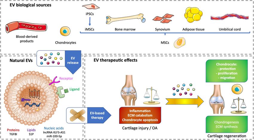
Velot et al.                                                                                              Extracellular Vesicles for Cartilage Regeneration

(Woolf and Pfleger, 2003; Hunter and Bierma-Zeinstra, 2019;                  often lead to the production of fibrocartilage instead of hyaline
Komarraju et al., 2020). Cicuttini et al. (2005) examined                    cartilage. It is important to integrate that repair is different from
a population of healthy, middle-aged subjects. The authors                   regeneration. Repair means healing joint through fibrocartilage
demonstrated that individuals with cartilage defects are affected            synthesis, resulting in a tissue with poorer functional quality
by a loss of articular cartilage compared with their initial                 that is not mechanically equivalent to native hyaline cartilage
cartilage volume. This loss of cartilage represents a 2.5%                   and cannot restore full original function. Besides, end-stage
annual rate of loss in terms of cartilage volume. However, a                 OA leads to invasive surgery with total joint replacement
pathological early degeneration of articular cartilage can ensue             by limited life prostheses (Chard et al., 2005; Lindahl, 2015;
following osteoarthritis (OA), trauma, or other causes (infections,          Kolasinski et al., 2020).
hereditary conditions, etc.) (Komarraju et al., 2020). OA is                     Regenerative medicine strategies have been initiated as a new
estimated to be the most common affliction triggering cartilage              opportunity to treat cartilage defects and bring regeneration
degeneration by affecting more than 300 million patients all                 along. Numerous tissue engineering approaches have been
around the world and more than 40% of the elderly older than                 developed by using biomaterials as ECM derivatives or
70 years (Hunter and Bierma-Zeinstra, 2019; Magnusson et al.,                mesenchymal stem cells (MSCs) as chondrogenic precursors
2019; Kolasinski et al., 2020). The mechanisms causing OA are                to alleviate the lack of accessibility for primary chondrocytes
still not fully understood, and the global therapeutic strategy is to        (Bakhshayesh et al., 2020; Tsiapalis and O’Driscoll, 2020).
maintain the patient quality of life mainly by treating symptoms             Because of their promising results in vitro or on animal models,
(i.e., the consequences of cartilage degeneration) (Hunter and               these approaches are currently investigated to be transferred to
Bierma-Zeinstra, 2019; Hsu and Siwiec, 2020; Kolasinski et al.,              clinic practice (Borić et al., 2019; Kim et al., 2020; Kyriakidis
2020; Oliveira Silva et al., 2020).                                          et al., 2020; Lehoczky et al., 2020; Mendes et al., 2020). Cell
    Articular cartilage is a hyaline cartilage covering the surface of       therapy approaches involving patients are also encouraging, and
bones in diarthrodial joints. This specialized connective tissue is          many clinical trials are ongoing. Larger sample sizes and long-
aneuronal and avascular, meaning it does not have its own blood              term follow-ups would be necessary for a validation in the
supply. Therefore, its nutrition depends on synovium through the             clinics. In addition, the proof of effectiveness for intra-articular
imbibition from synovial fluid or subchondral bone, which blood              cell injection remains limited in terms of repair as much as
supply is brought to cartilage bounds (Oliveira Silva et al., 2020).         regeneration (Ha et al., 2019; Monckeberg et al., 2019; Lee et al.,
The main cell type constituting cartilage is called chondrocyte.             2019; Zhao et al., 2019).
Chondrocytes are surrounded by an extracellular matrix (ECM)                     Other regenerative medicine strategies involved cell-free
they synthetize. Articular cartilage viscoelastic properties provide         therapies by delivering exogenous active biomolecules (growth
wear-resistant surfaces to the joint by improving the distribution           factors, drugs, etc.) as therapeutic agents directly through intra-
of mechanical loads and moderating the friction between its                  articular injection. The flaws of this route are that most mediators
surfaces (Sanchez-Adams et al., 2014; Lindahl, 2015; Bianchi                 have a short half-life and are promptly removed from the joint
et al., 2020; Masson and Krawetz, 2020). When injured or                     space and that mediators cannot reach chondrocytes suitably
degenerating, articular cartilage is not able to restore its original        because these cells are embedded into a thick avascular ECM (Yan
organization because it has a very weak regenerative capacity.               et al., 2019; Eckstein et al., 2020). The first flaw can be moderated
The lost hyaline cartilage is replaced by fibrocartilage, but                by encapsulating the mediator in an appropriate scaffold to
this reparative process is mechanically inadequate with a new                ensure a gradual diffusion inside the joint and a more long-
cartilage lacking inherent functionality (Goldring et al., 2017;             lasting effect, but the ECM penetration to get to chondrocytes still
Lepage et al., 2019; Masson and Krawetz, 2020).                              remains challenging (Szychlinska et al., 2018).
    OA is a disease related to cartilage degradation, osteophyte                 The cells, among which MSCs are the most solicited, used in
formation, and subchondral bone alteration. It leads gradually               tissue engineering or cell therapy are supposed to have intrinsic
to a joint destruction, which generates severe impairment                    capabilities to mediate tissue repair. For example, numerous
of mobility for the patient (Lindahl, 2015; Hsu and Siwiec,                  studies have now demonstrated that MSC healing effect is a
2020). The management of OA patients mostly starts with                      paracrine effect due to cell secretome (Julianto and Rindastuti,
non-surgical treatments that are replaced by surgical ones if                2016; Khatab et al., 2018; Chen et al., 2019b; Niada et al.,
losing their efficacy. Usually, pain management is prioritized               2019; Mancuso et al., 2019; Brennan et al., 2020; Parate et al.,
over other treatments for early stages OA, whereas invasive                  2020; Ragni et al., 2020b; Kadir et al., 2021). The secretome
surgery is preferred for late stages (Hsu and Siwiec, 2020;                  includes all cell secretions, i.e., soluble mediators (growth factors,
Yu and Hunter, 2020). There is still no pharmacological                      cytokines, etc.) (Maumus et al., 2017) and also very unique
treatment that would be able to repair or regenerate cartilage               entities called extracellular vesicles (EVs) (Dostert et al., 2017;
(Chevalier et al., 2013). Cartilage repair can be enhanced                   Tsiapalis and O’Driscoll, 2020).
by surgery as described hereinafter. Conventional surgical                       Since the first decade of the twenty-first century, literature
approaches include methods such as subchondral bone                          about EVs has been growing exponentially. But in parallel, a
microfracture, soft tissue transplantation with an autograft of              lack of accuracy for certain publications was noticed by experts
periosteum/perichondrium, osteochondral allograft/autograft                  in the field. A new scientific community in need of dogma
transplantation, and autologous chondrocyte implantation                     developed around EV research leaders resulting in the foundation
(Bakhshayesh et al., 2020). When effective, these methods                    of the International Society for Extracellular Vesicles (ISEV) in

Frontiers in Bioengineering and Biotechnology | www.frontiersin.org      2                                         April 2021 | Volume 9 | Article 645039
Velot et al. Extracellular Vesicles for Cartilage Regeneration 2012 (Witwer and Théry, 2019). EVs are commonly described as to produce EVs and the positive effects of the therapeutic cargos membranous entities released by every cell type into extracellular from natural or modified EVs. space. They are not able to replicate and are involved in cell-to- cell communication. Most publications depict EVs as particles enriched with components from the releasing cell and bordered BIOLOGICAL SOURCE-DERIVED EVs by a lipid bilayer comparable to the plasma membrane (Yáñez- FOR CARTILAGE HEALING Mó et al., 2015). Many subtypes of cell-released structures are encompassed in the term “EV” [e.g., small/medium/large EVs participate in the modulation of numerous cell regulatory vesicles, exosomes, ectosomes, oncosomes, microvesicles (MVs), processes (e.g., proliferation, differentiation, or inflammation), microparticles, apoptotic bodies (ABs), matrix vesicles, etc.]. making these membranous entities perfect stakeholders for tissue Consequently, the whole EVs constitute hard-to-characterize regeneration according to their various sources (Dostert et al., heterogeneous populations. The worldwide harmonization on 2017; Tsiapalis and O’Driscoll, 2020). EVs are secreted by diverse “how to term” the various EV subtypes has still not been set, cell types and found in solid tissues (e.g., ECM) or in body and their designation could depend on the size, biogenesis, fluids (blood, saliva, urine, milk, etc.). They can be isolated from specific markers, location or origin (ECM, cell/tissue, tumor, etc.), these biological samples, but determining their exact cell origin and/or conditioning (identified culture conditions). This lack of is not always possible because of their heterogeneity. According consensus can sometimes make the designation of EV subtypes to the use of EVs, particularly for therapeutic purpose, their ambiguous (Théry et al., 2018; Witwer and Théry, 2019). availability in sufficient quantity must be demonstrated to prevent Three categories of EVs are frequently found in publications precious sample scarcity and to ensure the results obtained and are described according to biogenesis. ABs are large from their exploitation. Despite the restrictions linked to good EVs (±1–5 µm) released after dismantling of apoptotic manufacturing/clinical practice (GMP/GCP), cell culture is also a cells and made of subcellular fragments. MVs, also termed reasonable answer to overcome the lack of biological sources and ectosomes or microparticles, are medium/large EVs (±100 nm to standardize an in vitro EV production from a well-defined cell to 1 µm) materialized from the budding of the plasma type. It allows EV isolation from cell-conditioned medium (Théry membrane. Exosomes are small EVs (±30–150 nm depending et al., 2006, 2018; Grant et al., 2011; Chen et al., 2013; Wolf et al., on the authors) derived from endocytic pathway (Dostert 2015; Iwai et al., 2016; Palamà et al., 2020; Ragni et al., 2020b). et al., 2017; Witwer and Théry, 2019; Cocozza et al., 2020; Blood products, such as serum- or plasma-based whole- Tsiapalis and O’Driscoll, 2020). blood derivatives, have a good tolerance and can reduce pain In 2014, ISEV set up guidelines called minimal information and inflammation. Intra-articular injection of blood products for studies of EVs (MISEV), which were updated in 2018. over other intra-articular treatments has been investigated MISEV were a necessary step to support scientists interested and improves pain scales in knee OA. The benefits become in this complex and still evolving field and to clarify/prevent statistically and clinically significant starting from 6 months the confusion about EV designation. MISEV2018 define “EV” and increase up to 12-month follow-up (Filardo et al., 2020). as a generic term. The authors are free to choose the required Whole blood non-invasively drawn from patients allows a direct approaches to characterize EV subtypes. However, if they cannot conditioning to obtain autologous blood-derived products with match the guidelines to validate EV identity, they are requested a higher tolerance (Gato-Calvo et al., 2019). Among cell-free to use the term extracellular particle or to find their own strategies to regenerate cartilage, EVs from autologous blood term after setting a clear definition (Théry et al., 2018). For products have a stronger effect in OA chondrocytes than full example, extracellular mitochondria alone or encapsulated in blood products by influencing cartilage ECM metabolism and vesicles constitute a noticeable subset of EVs (Puhm et al., inflammation. Acute OA could be improved by the positive anti- 2019; Al Amir Dache et al., 2020). Likewise, Yefimova et al. inflammatory effect of citrate-anticoagulated platelet-rich plasma (2020), have recently designated a new kind of EVs found in EVs. Chronic OA could benefit the chondrogenesis elicited by seminal plasma called myelinosome. Myelinosomes are secretory hyperacute serum EVs. These promising in vitro results have to organelles entirely released by Sertoli cells through a process be tested in vivo to demonstrate cartilage regeneration (Otahal close to endocytic pathway. These newly discovered EVs have a et al., 2020; Figures 1A,C). multilamellar demarcation very different from the lipid bilayer Current regenerative strategies for cartilage regeneration typically encountered in EVs (Yefimova et al., 2020). rely on the use of MSCs. These cells are multipotent with EVs have many interesting assets that could improve cartilage a chondrogenic potential. They also have self-renewal and defects. They have the innate capacity to target difficult-to- immunomodulatory properties (Dostert et al., 2017; Tsiapalis and reach places by crossing the blood–brain barrier or ECM. O’Driscoll, 2020). However, using growth factors to differentiate Their membranous structure allows protecting their cargo MSCs toward an articular phenotype remains difficult and from the environment. According to their sources, they have can lead to hypertrophy associated with unwanted ossification immunomodulatory properties and are biocompatible (Tsiapalis (Deng et al., 2019). Lately, primary articular chondrocytes and O’Driscoll, 2020). All these benefits offer new insights to use harvested from patients who underwent polydactyly surgery EVs as regenerative medicine tools to heal cartilage. were used to produce EVs. The aim was to use chondrocyte- This mini review summarizes current EV-based treatments to derived EVs (C-EVs) instead of growth factors to mediate improve cartilage degeneration by focusing on various sources MSC differentiation into chondrocytes. Human umbilical Frontiers in Bioengineering and Biotechnology | www.frontiersin.org 3 April 2021 | Volume 9 | Article 645039
Velot et al.                                                                                                             Extracellular Vesicles for Cartilage Regeneration

  FIGURE 1 | Therapeutic effects of natural EV-based therapy for cartilage healing. (A) Various cell sources for generating non-modified EVs. (B) Example of
  therapeutic cargos for natural EVs conveying proteins (TGFBI), nucleic acids (lnc KLF3-AS1, miR-100-5p), or lipids (SP1). (C) EV therapeutic effects on cartilage
  injury or OA. ECM, extracellular matrix; EV, extracellular vesicle; iPSC, induced pluripotent stem cell; iMSC, induced pluripotent stem cell-derived mesenchymal stem
  cell; lncRNA, long non-coding RNA; miR, microRNA; MSC, mesenchymal stem cell; OA, osteoarthritis; S1P, sphingosine-1-phosphate; TGFBI, transforming growth
  factor β–induced protein.

cord–derived MSCs (UCMSCs) were treated with C-EVs.                                    (e.g., cord blood or umbilical cord connective tissue, also known
In vitro, C-EVs promoted chondrogenic differentiation of MSCs                          as Wharton jelly) or MSCs derived from embryonic stem
and decreased fibrotic and hypertrophic markers. In vivo, MSCs                         cells. Perinatal MSCs are easily harvested, whereas embryonic
conditioned by C-EVs were successfully used to repair knee                             MSCs can cause ethical issues. Induced pluripotent stem cells
cartilage defect in a rabbit model. This approach needs further                        (iPSCs) have limitless self-renewal and can differentiate into
developments to be tested for human therapeutic purpose. Its                           MSCs to generate iPSC-derived MSCs (iMSCs). Nevertheless,
main flaws reside in the accessibility of mature chondrocytes                          the use of iPSCs or iMSCs in clinics is questionable because
and their tolerance in an allogeneic context (Ma et al., 2020;                         of their possible genomic instability, immunogenicity, and
Figures 1A,C).                                                                         tumorigenicity (Zhang et al., 2016; Dostert et al., 2017; Zhu et al.,
   MSCs have a unique capacity to build a regenerative                                 2017; Tsiapalis and O’Driscoll, 2020). Whether it is from an
microenvironment, which is considered to promote                                       allogeneic or an autologous origin, the best source of MSC-EVs
chondrogenic differentiation in the case of cartilage injury                           has not been yet highlighted. Several examples of MSC-derived
through soluble mediators and EVs. In vitro and in vivo studies                        EV therapy will be illustrated in the following sections of this
have demonstrated the immunomodulatory, chondroprotective,                             article. To date, there is only one clinical trial involving EVs to
and regenerative effects of MSC-derived EVs (MSC-EVs), which                           treat cartilage injury. It is based on the promising results from
is equivalent or superior to MSCs alone. Harvesting human                              Niada et al. (2019), where MSCs derived from adipose tissue of
adult MSCs (e.g., bone marrow, adipose tissue, or dental pulp)                         healthy donors undergoing aesthetic or prosthetic surgery. This
often requires invasive procedures, and these cells are not                            observational study has been posted in January 2020, and there is
systematically available, particularly in sufficient amount for                        still no recruitment (National Clinical Trial no. NCT04223622)1 .
cell therapy or tissue engineering. Consequently, adult MSCs                           One of its purposes is to validate a cell-free approach based
need to be expanded in vitro, which can lead to chromosomal                            on the use of EVs produced by adipose tissue–derived MSCs
aberrations. EV-based therapy is an answer to alleviate the                            (ATMSCs) to improve an ex vivo OA model. Besides, one of the
previous hindrances and potential cell immunogenicity.                                 possible sources of autologous ATMSCs could come from OA
The stemness of adult MSCs decreases with the increase of                              patients’ infrapatellar fat pad (IFP) obtained after arthroscopy,
the donor age. Adult MSCs have limited self-renewal and
immunomodulation properties compared with perinatal MSCs                               1
                                                                                           https://clinicaltrials.gov

Frontiers in Bioengineering and Biotechnology | www.frontiersin.org                4                                               April 2021 | Volume 9 | Article 645039
Velot et al. Extracellular Vesicles for Cartilage Regeneration
although such cells may be compromised in their regenerative and repress ECM catabolism in an in vitro OA model made
potential as the IFP has been involved in the pathogenesis of of OA patients’ chondrocytes treated with IL-1β. Intra-articular
OA. Autologous MSCs from OA patients’ joint tissues such injections of these ATMSC-EVs in a mouse model based on knee
as the synovium or IFP could originate from an inflammatory joint instability induced by surgery enhance articular cartilage
environment characterized by progressive OA causing their protection from damage and improve gait abnormality due to OA
potential priming by proinflammatory factors. This priming does pain and disturbance (Wu et al., 2019; Figures 1B,C).
not seem to compromise the healing aptitudes of MSCs. Instead, One of the limitations to have information on EV cargos
Kouroupis et al. (2019) showed that primed MSCs derived from is that numerous studies characterize EV effects and not their
the IFP exhibit enhanced immunomodulatory properties in vitro content or membrane composition. Moreover, the differences
and in vivo (Figures 1A,C). between cell type and cell donor have to be taken into account
to ensure the potential of the therapeutic message conveyed by
EVs (Ragni et al., 2019). The following examples illustrate the
THERAPEUTIC EFFECTS OF REGULAR positive use of natural EVs on cartilage without deciphering
EVs FOR CARTILAGE HEALING their cargos. It was previously mentioned that blood product–
derived EVs beneficially influence cartilage ECM metabolism and
The therapeutic effects of EVs depend on the cargo they inflammation in vitro (Otahal et al., 2020) and that articular
conveyed. Indeed, tissue regeneration relies on healing factors C-EVs favor chondrogenic differentiation of human UCMSCs
carried by EVs to generate a trophic microenvironment suitable in vitro and in vivo (Ma et al., 2020). Besides, human embryonic
for chondrogenesis and hyaline ECM upkeep (Liu et al., MSC-derived EVs promote cartilage regeneration in an adult rat
2017; Tao et al., 2017; Jing et al., 2020; Zhao et al., 2020). cartilage defect model (Zhang et al., 2016). In an OA model based
EVs can be untouched and used as therapeutic agents in on rabbit chondrocytes treated with IL-1β, EVs secreted by rabbit
their regular or natural state after isolation from biological BMMSCs prevent chondrocyte mitochondrial-induced apoptosis
samples or conditioned medium in standard cell culture in response to IL-1β in vitro (Qi et al., 2019). Human BMMSCs
conditions (Figure 1). produce chondroprotective and anti-inflammatory EVs in vitro
The following studies highlight the molecules of interest that prevent mice to develop collagenase-induced OA (Cosenza
conveyed by therapeutic EVs that could become part of the et al., 2017; Vonk et al., 2018). EVs secreted by human synovial
future strategies used for cartilage healing. Transforming growth membrane–derived MSCs (SMMSCs) and human iMSCs are able
factor β (TGF-β) is a major regulator of cartilage homeostasis, to attenuate OA in the same animal model. However, human
and the deregulation of its pathway is involved in OA. iMSC-EVs show a superior therapeutic effect associated with an
A member of the TGF-β family, the TGF-β–induced protein improvement of chondrocyte migration and proliferation (Zhu
(TGF-BI), is upregulated in bone and cartilage of OA patients, et al., 2017). Intra-articular injections of human iMSCs-derived
but is downregulated in human bone marrow–derived MSCs EVs also allow ECM restoration and cartilage regeneration
(BMMSCs) (Ruiz et al., 2019). Recently, Ruiz et al. (2020), showed in a rat temporomandibular joint OA model induced by
that TGBI silencing inhibits murine BMMSCs’ chondroinductive monosodium iodoacetate (Zhang et al., 2019). In addition, EVs
effect in vitro and healing effect in a collagenase-induced OA secreted by human iMSCs alleviate OA in the mouse, in vitro in
mouse model. These positive effects are due to the presence of chondrocytes treated with IL-1β and in vivo by limiting cartilage
TGF-BI mRNA and protein in BMMSC-derived EVs, suggesting destruction and matrix degradation according to Osteoarthritis
that TGF-BI is a new key factor released by MSCs to protect Research Society International (OARSI) scores in a model based
cartilage and favor its anabolism (Ruiz et al., 2020). The long non- on knee joint instability induced by surgery (Wang et al.,
coding RNA (lncRNA) KLF3-AS1 (see Ref seq NR_026804.1) 2017). Human ATMSCs derived from abdominoplasty release
is found in human MSCs and their EVs. When used in an EVs that decrease the inflammation caused in vitro by IL-1β
in vitro OA model based on rat articular chondrocytes treated in patient-derived OA chondrocytes (Tofiño-Vian et al., 2018;
with the proinflammatory cytokine interleukin 1β (IL-1β), EVs Figures 1A,C).
carrying lncRNA KLF3-AS1 suppress IL-1β–induced apoptosis Furthermore, EVs can also be tuned to carry selected
of chondrocytes. Additionally, these EVs engender cartilage mediators favoring a specific enrichment to elicit cartilage
repair and chondrocyte proliferation in a knee collagenase– regeneration. On the one end, engineered or primed tissues/cells
induced OA rat model (Liu et al., 2018b). EV subtypes secreted can lead to the production of enriched EVs. On the other hand,
by human BMMSCs contain a high level of sphingosine-1- EVs can also be loaded after their isolation.
phosphate (S1P) compared with MSCs alone. This enrichment
is due to the cell enzyme sphingosine kinase 1. S1P-enriched
EVs enhance the proliferation of human chondrocytes and THERAPEUTIC EFFECTS OF TUNED EVs
inhibit IL-1β–induced apoptosis in vitro. In a rabbit model of FOR CARTILAGE HEALING
knee articular cartilage injury made by drilling, the injection
of these EVs into the knee capsule promotes the recovery of Tuning EVs to increase their therapeutic potential require EV
the cartilage defect (Xiang et al., 2018). OA patients’ ATMSCs drug-loading strategies. Priming or transfecting cells involved
derived from IFP released EVs conveying high levels of miR-100- an EV loading performed by the donor cell, which secrete EVs.
5p. These EVs inhibit cell apoptosis, promote ECM anabolism, Postisolation drug loading occurs after EV secretion by the
Frontiers in Bioengineering and Biotechnology | www.frontiersin.org 5 April 2021 | Volume 9 | Article 645039Velot et al. Extracellular Vesicles for Cartilage Regeneration
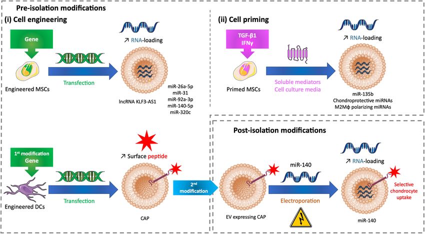
FIGURE 2 | Tuning methods to improve the drug loading of therapeutic EVs for cartilage healing. (A) Methods of modifications used before EV isolation to tune EVs
with (i) cell engineering by transfecting cells or (ii) cell priming by treating cells with soluble mediators added to cell culture medium. (B) Methods of modifications
used after EV isolation to tune EVs with miRNA electroporation. DC, dendritic cell; EV, extracellular vesicle; CAP, chondrocyte affinity peptide; IFN-γ, interferon γ;
lncRNA, long non-coding RNA; M2Mϕ, M2 macrophage; miR, microRNA; miRNA, microRNA; MSC, mesenchymal stem cell; TGF-β1, transforming growth factor β1.
donor cell. EV-based therapy for cartilage regeneration with the synthesis of chondrocyte ECM (Tao et al., 2017). The
tuned EVs depends mainly on RNA loading from transfected microRNA (miR)-140-5p is known to play a role in chondrogenic
MSCs (Figure 2). differentiation and OA prevention (Tardif et al., 2009; Barter
As previously described, lncRNA KLF3-AS1 conveyed by et al., 2015). SMMSC-derived EVs contain this miRNA weakly.
MSC-EVs improves OA in vitro and in vivo by respectively, When modified to overexpress miR-140-5p, SMMSCs secrete
suppressing IL-1β–induced apoptosis of chondrocytes and EVs enriched in miR-140-5p. This packaging enhanced the
eliciting cartilage repair and chondrocyte proliferation in a knee healing properties of human SMMSC-EVs by delaying the
collagenase-induced OA model (Liu et al., 2018b). The role of progression of early stage OA and cartilage damage (Tao et al.,
lncRNAs is to act as competitive endogenous RNA (ceRNAs) 2017). Likewise, miR-26a-5p and miR-31 promote chondrocyte
to separate microRNAs (miRNAs) from target mRNAs, and proliferation and improve OA. Respectively, human BMMSCs
thus restoring mRNA expression. Among miRNAs, miR-206 and human SMMSCs overexpressing miR-26a-5p and miR-31
is known to be highly expressed in OA. When transfected secrete EVs enriched in these miRNAs. These EVs alleviate
to overexpress lncRNA KLF3-AS1, MSCs release EVs with a cartilage destruction, matrix degradation, and inflammation in
high load of this lncRNA. These enriched EVs used in similar OA models based on knee joint instability induced by surgery,
in vitro and in vivo models showed a significant improve of in the rat for EVs carrying miR-26a-5p according to the authors’
IL-1β–induced chondrocyte apoptosis and cartilage injury. The subjective scores based on synovitis inflammation, synovial
authors demonstrated that lncRNA KLF3-AS1 conveyed by thickening, and subchondral bone erosion (Jin et al., 2020) and
MSC-EVs acts as a ceRNA to prevent miR-206 to downregulate in the mouse for EVs carrying miR-31 according to OARSI
G-protein-coupled receptor kinase interacting protein-1 (GIT1) scores (Wang et al., 2020). EVs derived from rat BMMSCs
expression, which induces EV therapeutic effects. Indeed, GIT1 transfected to overexpress miR-135b favor rat chondrocyte
inhibits chondrocytes apoptosis and promotes chondrocytes proliferation in vitro (Wang et al., 2018). Human BMMSCs
proliferation. Delivering this lncRNA by EVs into cartilage could overexpressing miR-92a-3p, a chondrogenesis promoter and
be a future strategy for cartilage regeneration (Liu et al., 2018a; cartilage degradation inhibitor (Mao et al., 2017), produce
Figure 2Ai upper panel). EVs sustaining its properties in vitro. Human BMMSC-EVs
In an OA rat model based on knee joint instability induced enriched in miR-92a-3p interfere with the progression of early-
by surgery, human SMMSCs promote to a lesser extent the stage OA and prevent cartilage damage in a knee collagenase–
same positive effects -as described previously- but inhibit induced OA mouse model (Mao et al., 2018). Human BMMSCs
Frontiers in Bioengineering and Biotechnology | www.frontiersin.org 6 April 2021 | Volume 9 | Article 645039Velot et al. Extracellular Vesicles for Cartilage Regeneration
undergoing chondrogenic differentiation upregulate the EV EVs can penetrate deep down into tissue, and their faintly
loading of specific miRNAs, like miR-320c. EVs derived from negative net charge allows long circulation in the body.
human BMMSCs transfected to overexpress miR-320c promote Moreover, they are able to escape immune cells by avoiding
OA patients’ chondrocyte proliferation and migration and clearance or degradation (Malhotra et al., 2016; Vader et al.,
increase human BMSC chondrogenesis (Sun et al., 2019). The 2016). These assets made them ideal biocompatible carriers to
previous studies demonstrate the potential of miRNA-loaded convey molecules of interest for cartilage healing. Whatever EV
EVs to develop novel therapeutic strategies for cartilage healing enrichment, all of these molecules have not yet been discovered,
(Figure 2Ai upper panel). and there is still an in-depth screening ongoing to bring them
Palamà et al., showed that human BMMSCs grown in a xeno- up to light by the scientific community (Endisha et al., 2018; Sun
free culture system produce chondroprotective EVs in vitro. et al., 2019; Ragni et al., 2020a).
In addition, these EVs were produced in a higher amount
and inhibit the adverse effects of IL-1α-induced inflammation
in an in vitro OA model compared with cells grown in a DISCUSSION
conventional culture system with fetal bovine serum (Palamà
et al., 2020). This demonstrates that a xeno-free environment Although a lack of consensus concerning EV isolation methods
can prime cells and influence EV composition and consequently and EV characterization/designation remains, EV therapeutic
EV effects. Similarly, cell priming with biochemical factors potential cannot be denied to improve cartilage injury as
influences EV cargos. EVs derived from rat BMMSCs primed shown in the preclinical studies presented in this article. Cell-
with TGF-β1 are enriched in miR-135b and favor rat chondrocyte free therapy based on EVs appears to demonstrate similar
proliferation by upregulating S1P in vitro (Wang et al., 2018). or sometimes even better regenerative properties of cartilage
Moreover, these EVs engender cartilage repair in a rat OA model compared with cell therapy. For example, phosphate-buffered
based on knee joint instability induced by surgery. Interferon saline (PBS), EVs derived from human BMMSCs, or human
γ (IFN-γ) is a versatile proinflammatory cytokine involved in BMMSCs were injected in the knee capsule in a rabbit model
tissue regeneration. In vitro IFN-γ–primed human ATMSCs of knee articular cartilage injury performed by drilling. The
secrete EVs enriched in chondroprotective and M2 (i.e., anti- control group treated with PBS exhibited defect filling with
inflammatory) macrophage polarizing miRNAs (Ragni et al., adipose and fibrotic cells but without ECM, suggesting a poor
2020b). This cargo improvement makes a suitable association to cartilage repair. The EV-treated group or the MSC-treated group
promote cartilage regeneration (Figure 2Aii). displayed contrasting results, with defects progressively filled
The delivery of EVs after intra-articular administration in by a hyaline cartilage–like tissue. There was no significant
chondrocytes remains challenging as these cells are surrounded difference between the EV-treated and MSC-treated groups,
by an avascular, dense ECM. The density of hyaline ECM is indicating that EVs are as efficient as MSCs to promote the
due to proteoglycans with high negative charges entangled in recovery of cartilage defects (Xiang et al., 2018). Similarly,
a collagen fibril network. Only solutes less than or equal to intra-articular injection of human BMMSC full secretome
10 nm have been shown to cross this thick barrier and penetrate (containing EVs), equally to injection of human BMMSCs,
the cartilage (Bajpayee et al., 2014). Although EVs are greater was shown to reduce pain and have protective effects on
than 10 nm and have a slight negative charge that could push the development of cartilage damage in a knee collagenase–
them away from cartilage, the previous references showed their induced OA mouse model (Khatab et al., 2018). Cosenza et al.
healing effects on damaged cartilage whether they are regular (2017) reported the superiority of human BMMSC-EVs over
or tuned. However, their penetration in the hyaline ECM has human BMMSCs to protect the joint from OA in the same
not always been tested. Recently, Liang et al. (2020), transfected in vivo preclinical model. Moreover, EVs are easier to handle
dendritic cells to produce engineered EVs that are tagged with and display minor regulatory concerns compared with cells,
a chondrocyte affinity peptide (CAP) at their surface. CAP-EVs mainly because they are less immunogenic, and because they
have the same size range (40–200 nm) as EVs without CAP. have no nucleus, they do not replicate. To translate EV-based
CAP-EVs target and enter selectively chondrocytes in vitro and therapy into clinics, several hurdles would have to be overcome
in vivo. They can more efficiently and deeply penetrate healthy rat to guarantee the procurement of safe therapy products and
cartilage than non-tagged EVs that mainly reside in the surface prevent side effects, such as EV cell sources/donor related to
layer. After their isolation, CAP-EVs and non-tagged EVs were potential immunogenicity and EV dose and route related to
loaded with miR-140 mimic by electroporation. Loaded CAP- pharmacokinetics, pharmacodynamics, and toxicity (Théry et al.,
EVs were able to improve an in vitro OA model based on human 2018; Tieu et al., 2020).
articular chondrocytes treated with IL-1β by a targeted delivery of Alongside the GMP/GCP, the future of patients’ cartilage
miR-140. Intra-articular injection of EVs was made in an OA rat regeneration appears to have a great potential within cell-free
model based on knee joint instability induced by surgery. CAP- therapies based on EVs. EV aptitude to cross ECM allows them
EVs remained preferentially into cartilage with minimal diffusion to reach and heal cartilage. However, to penetrate the cartilage,
compared with non-tagged EVs. Loaded CAP-EVs delivered EVs seemed to need a special tag allowing to cross the hyaline
miR-140 to deep cartilage areas and prevent cartilage degradation ECM. While tuned EVs can be enhanced to carry an established
and OA progression (Liang et al., 2020; Figure 2Ai lower panel, tag (Liang et al., 2020), regular EV healing cartilage could be
Figure 2B). enriched and carry inherently those kinds of tags such as S1P
Frontiers in Bioengineering and Biotechnology | www.frontiersin.org 7 April 2021 | Volume 9 | Article 645039Velot et al. Extracellular Vesicles for Cartilage Regeneration
(Xiang et al., 2018). The selective mechanisms to favor EVs and engineered constructs (Malda et al., 2016; Szychlinska et al.,
ECM interaction could rely on a chemotactic response (Headland 2018). After being implanted subcutaneously in nude mice,
et al., 2015). According to the disease state of OA and to the depth constructs made of rabbit cartilage progenitor cells and alginate
of the defects, EVs could also have an easier access to exposed have developed into ectopic cartilage once injected with rabbit
chondrocytes without being hindered by the hyaline ECM, which C-EVs (Chen et al., 2018). An acellular tissue patch made of
is removed from cartilage by erosion. hydrogel glue and human iMSC-EVs was tested in a rabbit model
EVs are used in their natural state or tuned to be drug- of patellar groove defect. The hydrogel glue could retain and
loaded in order to be enriched with cartilage regenerative then release EVs sustainably. The patch was easily integrated
RNAs (e.g., miRNAs or lncRNAs). The tuning can also apply to cartilage ECM and improved the articular cartilage injury
to EV membrane to improve chondrocyte targeting (e.g., through cell deposition (Liu et al., 2017). The same animal model
surface modification with CAP). To date, these modifications was used to test a three-dimensional (3D) construct printed
are the only existing for cartilage, despite a broad range of with a bioink made of cartilage ECM, gelatin methacrylate,
possibilities that would probably inspire forthcoming research and BMMSC-EVs. The 3D construct stimulated chondrocyte
(Liao et al., 2019). Strategies using cotherapy by tuning EVs migration and M2 macrophage polarization and also elicited
with drug encapsulation can also be considered to boost cartilage cartilage regeneration (Chen et al., 2019a). Like cell-free therapies
regeneration. For example, loading EVs with pharmacological based on EVs, few EV-based tissue engineering strategies have
agents such as glucosamine, chondroitin sulfate, or non-steroidal been investigated in the field of cartilage regeneration, and the
anti-inflammatory drugs could improve ECM degradation and course to new discoveries remains.
reduce pain and cartilage degeneration (Artuzi et al., 2020; Sun All the presented preclinical studies show that EVs embody a
et al., 2020). Although liposomes can be used as synthetic vesicles great hope to become part of the next-generation treatments in
to convey drugs to cells, it has been shown that they could be regenerative medicine for articular cartilage regeneration.
silenced through phagocytosis by monocytes and macrophages.
Consequently, they require surface adjustments to obtain smart
targeting abilities. EVs carry specific membrane proteins that AUTHOR CONTRIBUTIONS
protect them from phagocytosis and facilitate their delivery
(Kamerkar et al., 2017). A strategy for cotherapy could also be ÉV and MC drafted the manuscript and gave the final approval
to engineer “smarter” delivery systems by creating hybrid EV- of the version to be published. AB designed the figures. ÉV,
liposome carriers with membrane fusion (Elkhoury et al., 2020). HM, JV, AB, and MC helped to write, discuss, and edit the
Apart from cell-free therapy, EVs can also be used for cartilage manuscript. All authors contributed to the article and approved
tissue engineering strategies by being associated with implantable the submitted version.
REFERENCES Brennan, M. Á, Layrolle, P., and Mooney, D. J. (2020). Biomaterials functionalized
with MSC secreted extracellular vesicles and soluble factors for tissue
Al Amir Dache, Z., Otandault, A., Tanos, R., Pastor, B., Meddeb, R., et al. (2020). regeneration. Adv. Funct. Mater. 30:1909125. doi: 10.1002/adfm.20190
Blood contains circulating cell-free respiratory competent mitochondria. 9125
FASEB J. 34, 3616–3630. doi: 10.1096/fj.201901917RR Chard, J., Lohmander, S., Smith, C., and Scott, D. (2005). Osteoarthritis of the knee.
Artuzi, F. E., Puricelli, E., Baraldi, C. E., Quevedo, A. S., and Ponzoni, D. (2020). Clin. Evid. 14, 1506–1522.
Reduction of osteoarthritis severity in the temporomandibular joint of rabbits Chen, C. Y., Hogan, M. C., and Ward, C. J. (2013). Purification of exosome-like
treated with chondroitin sulfate and glucosamine. PLoS One 15:e0231734. doi: vesicles from urine. Methods Enzymol. 524, 225–241. doi: 10.1016/b978-0-12-
10.1371/journal.pone.0231734 397945-2.00013-5
Bajpayee, A. G., Wong, C. R., Bawendi, M. G., Frank, E. H., and Grodzinsky, A. J. Chen, P., Zheng, L., Wang, Y., Tao, M., Xie, Z., Xia, C., et al. (2019a).
(2014). Avidin as a model for charge driven transport into cartilage and drug Desktop-stereolithography 3D printing of a radially oriented extracellular
delivery for treating early stage post-traumatic osteoarthritis. Biomaterials 35, matrix/mesenchymal stem cell exosome bioink for osteochondral defect
538–549. doi: 10.1016/j.biomaterials.2013.09.091 regeneration. Theranostics 9, 2439–2459. doi: 10.7150/thno.31017
Bakhshayesh, A. R. D., Babaie, S., Nasrabadi, H. T., Asadi, N., Akbarzadeh, A., and Chen, W., Sun, Y., Gu, X., Hao, Y., Liu, X., Lin, J., et al. (2019b). Conditioned
Abedelahi, A. (2020). An overview of various treatment strategies, especially medium of mesenchymal stem cells delays osteoarthritis progression in a rat
tissue engineering for damaged articular cartilage. Artif. Cells Nanomed. model by protecting subchondral bone, maintaining matrix homeostasis, and
Biotechnol. 48, 1089–1104. doi: 10.1080/21691401.2020.1809439 enhancing autophagy. J. Tissue Eng. Regen. Med. 13, 1618–1628. doi: 10.1002/
Barter, M. J., Tselepi, M., Gómez, R., Woods, S., Hui, W., Smith, G. R., et al. term.2916
(2015). Genome-Wide MicroRNA and gene analysis of mesenchymal stem cell Chen, Y., Xue, K., Zhang, X., Zheng, Z., and Liu, K. (2018). Exosomes derived from
chondrogenesis identifies an essential role and multiple targets for miR-140- mature chondrocytes facilitate subcutaneous stable ectopic chondrogenesis of
5p: miR-140-5p targets in MSC chondrogenesis. Stem Cells 33, 3266–3280. cartilage progenitor cells. Stem Cell Res. Ther. 9:318. doi: 10.1186/s13287-018-
doi: 10.1002/stem.2093 1047-1042
Bianchi, A., Velot, É, Kempf, H., Elkhoury, K., Sanchez-Gonzalez, L., Linder, M., Chevalier, X., Eymard, F., and Richette, P. (2013). Biologic agents in osteoarthritis:
et al. (2020). Nanoliposomes from agro-resources as promising delivery systems hopes and disappointments. Nat. Rev. Rheumatol. 9, 400–410. doi: 10.1038/
for chondrocytes. Int. J. Mol. Sci. 21:3436. doi: 10.3390/ijms21103436 nrrheum.2013.44
Borić, I., Hudetz, D., Rod, E., Jeleč, Ž, Vrdoljak, T., Skelin, A., et al. (2019). A 24- Cicuttini, F., Ding, C., Wluka, A., Davis, S., Ebeling, P. R., and Jones, G. (2005).
Month follow-up study of the effect of intra-articular injection of autologous Association of cartilage defects with loss of knee cartilage in healthy, middle-
microfragmented fat tissue on proteoglycan synthesis in patients with knee age adults: a prospective study. Arthritis Rheum. 52, 2033–2039. doi: 10.1002/
osteoarthritis. Genes 10:1051. doi: 10.3390/genes10121051 art.21148
Frontiers in Bioengineering and Biotechnology | www.frontiersin.org 8 April 2021 | Volume 9 | Article 645039Velot et al. Extracellular Vesicles for Cartilage Regeneration
Cocozza, F., Grisard, E., Martin-Jaular, L., Mathieu, M., and Théry, C. (2020). Kadir, N. D., Yang, Z., Hassan, A., Denslin, V., and Lee, E. H. (2021). Electrospun
SnapShot: extracellular vesicles. Cell 182, 262–262.e1. doi: 10.1016/j.cell.2020. fibers enhanced the paracrine signaling of mesenchymal stem cells for cartilage
04.054 regeneration. Stem Cell Res. Ther. 12:100. doi: 10.1186/s13287-021-02137-2138
Cosenza, S., Ruiz, M., Toupet, K., Jorgensen, C., and Noël, D. (2017). Mesenchymal Kamerkar, S., LeBleu, V. S., Sugimoto, H., Yang, S., Ruivo, C. F., Melo, S. A.,
stem cells derived exosomes and microparticles protect cartilage and bone et al. (2017). Exosomes facilitate therapeutic targeting of oncogenic KRAS in
from degradation in osteoarthritis. Sci. Rep. 7:16214. doi: 10.1038/s41598-017- pancreatic cancer. Nature 546, 498–503. doi: 10.1038/nature22341
15376-15378 Khatab, S., van Osch, G., Kops, N., Bastiaansen-Jenniskens, Y., Bos, P., Verhaar,
Deng, Y., Lei, G., Lin, Z., Yang, Y., Lin, H., and Tuan, R. S. (2019). Engineering J., et al. (2018). Mesenchymal stem cell secretome reduces pain and prevents
hyaline cartilage from mesenchymal stem cells with low hypertrophy potential cartilage damage in a murine osteoarthritis model. Eur. Cell. Mater. 36, 218–
via modulation of culture conditions and Wnt/β-catenin pathway. Biomaterials 230. doi: 10.22203/eCM.v036a16
192, 569–578. doi: 10.1016/j.biomaterials.2018.11.036 Kim, J.-K., Bae, H. C., Ro, D. H., Lee, S., Lee, M. C., and Han, H.-S. (2020).
Dostert, G., Mesure, B., Menu, P., and Velot, É (2017). How do mesenchymal stem Enhancement of cartilage regeneration of synovial stem cells /hydrogel by using
cells influence or are influenced by microenvironment through extracellular transglutaminase-4. Tissue Eng. Part A doi: 10.1089/ten.TEA.2020.0271 Online
vesicles communication? Front. Cell Dev. Biol. 5:6. doi: 10.3389/fcell.2017.00006 ahead of print.
Eckstein, F., Kraines, J. L., Aydemir, A., Wirth, W., Maschek, S., and Hochberg, Kolasinski, S. L., Neogi, T., Hochberg, M. C., Oatis, C., Guyatt, G., Block, J., et al.
M. C. (2020). Intra-articular sprifermin reduces cartilage loss in addition to (2020). 2019 american college of rheumatology/arthritis foundation guideline
increasing cartilage gain independent of location in the femorotibial joint: post- for the management of osteoarthritis of the hand. hip, and knee. Arthritis Care
hoc analysis of a randomised, placebo-controlled phase II clinical trial. Ann. Res. 72, 149–162. doi: 10.1002/acr.24131
Rheum. Dis. 79, 525–528. doi: 10.1136/annrheumdis-2019-216453 Komarraju, A., Goldberg-Stein, S., Pederson, R., McCrum, C., and Chhabra,
Elkhoury, K., Koçak, P., Kang, A., Arab-Tehrany, E., Ellis Ward, J., and Shin, A. (2020). Spectrum of common and uncommon causes of knee joint
S. R. (2020). Engineering smart targeting nanovesicles and their combination hyaline cartilage degeneration and their key imaging features. Eur. J. Radiol.
with hydrogels for controlled drug delivery. Pharmaceutics 12:849. doi: 10.3390/ 129:109097. doi: 10.1016/j.ejrad.2020.109097
pharmaceutics12090849 Kouroupis, D., Bowles, A. C., Willman, M. A., Perucca Orfei, C., Colombini,
Endisha, H., Rockel, J., Jurisica, I., and Kapoor, M. (2018). The complex landscape A., Best, T. M., et al. (2019). Infrapatellar fat pad-derived MSC response to
of microRNAs in articular cartilage: biology, pathology, and therapeutic targets. inflammation and fibrosis induces an immunomodulatory phenotype involving
JCI Insight 3:e121630. doi: 10.1172/jci.insight.121630 CD10-mediated Substance P degradation. Sci. Rep. 9:10864. doi: 10.1038/
Filardo, G., Previtali, D., Napoli, F., Candrian, C., Zaffagnini, S., and Grassi, s41598-019-47391-47392
A. (2020). PRP injections for the treatment of knee osteoarthritis: a meta- Kyriakidis, T., Iosifidis, M., Michalopoulos, E., Melas, I., Stavropoulos-Giokas,
analysis of randomized controlled trials. CARTILAGE 19:1947603520931170. C., and Verdonk, R. (2020). Good mid-term outcomes after adipose-derived
doi: 10.1177/1947603520931170 culture-expanded mesenchymal stem cells implantation in knee focal cartilage
Gato-Calvo, L., Magalhaes, J., Ruiz-Romero, C., Blanco, F. J., and Burguera, defects. Knee Surg. Sports Traumatol. Arthrosc. 28, 502–508. doi: 10.1007/
E. F. (2019). Platelet-rich plasma in osteoarthritis treatment: review of s00167-019-05688-5689
current evidence. Ther. Adv. Chronic Dis. 10:204062231982556. doi: 10.1177/ Lee, W.-S., Kim, H. J., Kim, K.-I., Kim, G. B., and Jin, W. (2019). Intra-Articular
2040622319825567 injection of autologous adipose tissue-derived mesenchymal stem cells for the
Goldring, M. B., Culley, K. L., and Otero, M. (2017). “Pathogenesis of osteoarthritis treatment of knee osteoarthritis: a phase IIb, randomized, placebo-controlled
in general,” in Cartilage, eds S. Grässel and A. Aszódi (Cham: Springer clinical trial. Stem Cells Transl. Med. 8, 504–511. doi: 10.1002/sctm.18-0122
International Publishing), 1–25. doi: 10.1007/978-3-319-45803-8_1 Lehoczky, G., Wolf, F., Mumme, M., Gehmert, S., Miot, S., Haug, M., et al. (2020).
Grant, R., Ansa-Addo, E., Stratton, D., Antwi-Baffour, S., Jorfi, S., Kholia, S., et al. Intra-individual comparison of human nasal chondrocytes and debrided knee
(2011). A filtration-based protocol to isolate human plasma membrane-derived chondrocytes: relevance for engineering autologous cartilage grafts. Clin.
vesicles and exosomes from blood plasma. J. Immunol. Methods 371, 143–151. Hemorheol. Microcirc. 74, 67–78. doi: 10.3233/CH-199236
doi: 10.1016/j.jim.2011.06.024 Lepage, S. I. M., Robson, N., Gilmore, H., Davis, O., Hooper, A., St. John, S., et al.
Ha, C.-W., Park, Y.-B., Kim, S. H., and Lee, H.-J. (2019). Intra-articular (2019). Beyond cartilage repair: the role of the osteochondral unit in joint health
mesenchymal stem cells in osteoarthritis of the knee: a systematic review of and disease. Tissue Eng. Part B Rev. 25, 114–125. doi: 10.1089/ten.teb.2018.0122
clinical outcomes and evidence of cartilage repair. Arthrosc. J. Arthrosc. Relat. Liang, Y., Xu, X., Li, X., Xiong, J., Li, B., Duan, L., et al. (2020). Chondrocyte-
Surg. 35, 277–288.e2. doi: 10.1016/j.arthro.2018.07.028 Targeted MicroRNA delivery by engineered exosomes toward a cell-free
Headland, S. E., Jones, H. R., Norling, L. V., Kim, A., Souza, P. R., Corsiero, E., et al. osteoarthritis therapy. ACS Appl. Mater. Interfaces 12, 36938–36947. doi: 10.
(2015). Neutrophil-derived microvesicles enter cartilage and protect the joint in 1021/acsami.0c10458
inflammatory arthritis. Sci. Transl. Med. 7:315ra190. doi: 10.1126/scitranslmed. Liao, W., Du, Y., Zhang, C., Pan, F., Yao, Y., Zhang, T., et al. (2019). Exosomes: the
aac5608 next generation of endogenous nanomaterials for advanced drug delivery and
Hsu, H., and Siwiec, R. M. (2020). “Knee osteoarthritis,” in StatPearls. Treasure therapy. Acta Biomater. 86, 1–14. doi: 10.1016/j.actbio.2018.12.045
Island, FL: StatPearls Publishing. Lindahl, A. (2015). From gristle to chondrocyte transplantation: treatment of
Hunter, D. J., and Bierma-Zeinstra, S. (2019). Osteoarthritis. Lancet 393, 1745– cartilage injuries. Philos. Trans. R. Soc. B Biol. Sci. 370:20140369. doi: 10.1098/
1759. doi: 10.1016/S0140-6736(19)30417-30419 rstb.2014.0369
Iwai, K., Minamisawa, T., Suga, K., Yajima, Y., and Shiba, K. (2016). Liu, X., Yang, Y., Li, Y., Niu, X., Zhao, B., Wang, Y., et al. (2017). Integration of
Isolation of human salivary extracellular vesicles by iodixanol density stem cell-derived exosomes with in situ hydrogel glue as a promising tissue
gradient ultracentrifugation and their characterizations. J. Extracell. Vesicles patch for articular cartilage regeneration. Nanoscale 9, 4430–4438. doi: 10.1039/
5:10.3402/jev.v5.30829. doi: 10.3402/jev.v5.30829 C7NR00352H
Jin, Z., Ren, J., and Qi, S. (2020). Human bone mesenchymal stem cells-derived Liu, Y., Lin, L., Zou, R., Wen, C., Wang, Z., and Lin, F. (2018a). MSC-derived
exosomes overexpressing microRNA-26a-5p alleviate osteoarthritis via down- exosomes promote proliferation and inhibit apoptosis of chondrocytes via
regulation of PTGS2. Int. Immunopharmacol. 78:105946. doi: 10.1016/j.intimp. lncRNA-KLF3-AS1/miR-206/GIT1 axis in osteoarthritis. Cell Cycle 17, 2411–
2019.105946 2422. doi: 10.1080/15384101.2018.1526603
Jing, H., Zhang, X., Luo, K., Luo, Q., Yin, M., Wang, W., et al. (2020). miR-381- Liu, Y., Zou, R., Wang, Z., Wen, C., Zhang, F., and Lin, F. (2018b). Exosomal KLF3-
abundant small extracellular vesicles derived from kartogenin-preconditioned AS1 from hMSCs promoted cartilage repair and chondrocyte proliferation in
mesenchymal stem cells promote chondrogenesis of MSCs by targeting TAOK1. osteoarthritis. Biochem. J. 475, 3629–3638. doi: 10.1042/BCJ20180675
Biomaterials 231:119682. doi: 10.1016/j.biomaterials.2019.119682 Ma, K., Zhu, B., Wang, Z., Cai, P., He, M., Ye, D., et al. (2020). Articular
Julianto, I., and Rindastuti, Y. (2016). Topical delivery of mesenchymal chondrocyte-derived extracellular vesicles promote cartilage differentiation of
stem cells “Secretomes” in wound repair. Acta Medica Indones. 48, human umbilical cord mesenchymal stem cells by activation of autophagy.
217–220. J. Nanobiotechnol. 18:163. doi: 10.1186/s12951-020-00708-700
Frontiers in Bioengineering and Biotechnology | www.frontiersin.org 9 April 2021 | Volume 9 | Article 645039Velot et al. Extracellular Vesicles for Cartilage Regeneration Magnusson, K., Turkiewicz, A., and Englund, M. (2019). Nature vs nurture in knee reference genes as a crucial step for donor selection. Cells 8:369. doi: 10.3390/ osteoarthritis - the importance of age, sex and body mass index. Osteoarthritis cells8040369 Cartilage 27, 586–592. doi: 10.1016/j.joca.2018.12.018 Ragni, E., Perucca Orfei, C., De Luca, P., Colombini, A., Viganò, M., and Malda, J., Boere, J., van de Lest, C. H. A., van Weeren, P. R., and Wauben, M. H. M. de Girolamo, L. (2020a). Secreted factors and EV-miRNAs orchestrate the (2016). Extracellular vesicles — new tool for joint repair and regeneration. Nat. healing capacity of adipose mesenchymal stem cells for the treatment of knee Rev. Rheumatol. 12, 243–249. doi: 10.1038/nrrheum.2015.170 osteoarthritis. Int. J. Mol. Sci. 21:1582. doi: 10.3390/ijms21051582 Malhotra, H., Sheokand, N., Kumar, S., Chauhan, A. S., Kumar, M., Jakhar, P., Ragni, E., Perucca Orfei, C., De Luca, P., Mondadori, C., Viganò, M., Colombini, et al. (2016). Exosomes: tunable nano vehicles for macromolecular delivery A., et al. (2020b). Inflammatory priming enhances mesenchymal stromal cell of transferrin and lactoferrin to specific intracellular compartment. J. Biomed. secretome potential as a clinical product for regenerative medicine approaches Nanotechnol. 12, 1101–1114. doi: 10.1166/jbn.2016.2229 through secreted factors and EV-miRNAs: the example of joint disease. Stem Mancuso, P., Raman, S., Glynn, A., Barry, F., and Murphy, J. M. (2019). Cell Res. Ther. 11:165. doi: 10.1186/s13287-020-01677-1679 Mesenchymal stem cell therapy for osteoarthritis: the critical role of the cell Ruiz, M., Maumus, M., Fonteneau, G., Pers, Y.-M., Ferreira, R., Dagneaux, L., et al. secretome. Front. Bioeng. Biotechnol. 7:9. doi: 10.3389/fbioe.2019.00009 (2019). TGFβi is involved in the chondrogenic differentiation of mesenchymal Mao, G., Zhang, Z., Hu, S., Zhang, Z., Chang, Z., Huang, Z., et al. (2018). stem cells and is dysregulated in osteoarthritis. Osteoarthritis Cartilage 27, Exosomes derived from miR-92a-3p-overexpressing human mesenchymal stem 493–503. doi: 10.1016/j.joca.2018.11.005 cells enhance chondrogenesis and suppress cartilage degradation via targeting Ruiz, M., Toupet, K., Maumus, M., Rozier, P., Jorgensen, C., and Noël, D. (2020). WNT5A. Stem Cell Res. Ther. 9:247. doi: 10.1186/s13287-018-1004-1000 TGFBI secreted by mesenchymal stromal cells ameliorates osteoarthritis and Mao, G., Zhang, Z., Huang, Z., Chen, W., Huang, G., Meng, F., et al. (2017). is detected in extracellular vesicles. Biomaterials 226:119544. doi: 10.1016/j. MicroRNA-92a-3p regulates the expression of cartilage-specific genes by biomaterials.2019.119544 directly targeting histone deacetylase 2 in chondrogenesis and degradation. Sanchez-Adams, J., Leddy, H. A., McNulty, A. L., O’Conor, C. J., and Guilak, Osteoarthritis Cartilage 25, 521–532. doi: 10.1016/j.joca.2016.11.006 F. (2014). The mechanobiology of articular cartilage: bearing the burden Masson, A. O., and Krawetz, R. J. (2020). Understanding cartilage protection in of osteoarthritis. Curr. Rheumatol. Rep. 16:451. doi: 10.1007/s11926-014- OA and injury: a spectrum of possibilities. BMC Musculoskelet. Disord. 21:432. 0451-456 doi: 10.1186/s12891-020-03363-3366 Sun, H., Hu, S., Zhang, Z., Lun, J., Liao, W., and Zhang, Z. (2019). Expression Maumus, M., Manferdini, C., Toupet, K., Chuchana, P., Casteilla, L., Gachet, M., of exosomal microRNAs during chondrogenic differentiation of human bone et al. (2017). Thrombospondin-1 partly mediates the cartilage protective effect mesenchymal stem cells. J. Cell. Biochem. 120, 171–181. doi: 10.1002/jcb.27289 of adipose-derived mesenchymal stem cells in osteoarthritis. Front. Immunol. Sun, Y., Wang, C., and Gong, C. (2020). Repairing effects of glucosamine sulfate 8:1638. doi: 10.3389/fimmu.2017.01638 in combination with etoricoxib on articular cartilages of patients with knee Mendes, L. F., Bosmans, K., Van Hoven, I., Viseu, S. R., Maréchal, M., and osteoarthritis. J. Orthop. Surg. 15:150. doi: 10.1186/s13018-020-01648-z Luyten, F. P. (2020). Developmental engineering of living implants for deep Szychlinska, M. A., D’Amora, U., Ravalli, S., Ambrosio, L., Di Rosa, M., osteochondral joint surface defects. Bone 139:115520. doi: 10.1016/j.bone.2020. and Musumeci, G. (2018). Functional biomolecule delivery systems and 115520 bioengineering in cartilage regeneration. Curr. Pharm. Biotechnol. 20, 32–46. Monckeberg, J. E., Rafols, C., Apablaza, F., Gerhard, P., and Rosales, J. (2019). doi: 10.2174/1389201020666190206202048 Intra-articular administration of peripheral blood stem cells with platelet- Tao, S.-C., Yuan, T., Zhang, Y.-L., Yin, W.-J., Guo, S.-C., and Zhang, C.-Q. rich plasma regenerated articular cartilage and improved clinical outcomes (2017). Exosomes derived from miR-140-5p-overexpressing human synovial for knee chondral lesions. Knee 26, 824–831. doi: 10.1016/j.knee.2019.05. mesenchymal stem cells enhance cartilage tissue regeneration and prevent 008 osteoarthritis of the knee in a rat model. Theranostics 7, 180–195. doi: 10.7150/ Niada, S., Giannasi, C., Gomarasca, M., Stanco, D., Casati, S., and Brini, thno.17133 A. T. (2019). Adipose-derived stromal cell secretome reduces TNFα-induced Tardif, G., Hum, D., Pelletier, J.-P., Duval, N., and Martel-Pelletier, J. (2009). hypertrophy and catabolic markers in primary human articular chondrocytes. Regulation of the IGFBP-5 and MMP-13 genes by the microRNAs miR-140 and Stem Cell Res. 38:101463. doi: 10.1016/j.scr.2019.101463 miR-27a in human osteoarthritic chondrocytes. BMC Musculoskelet. Disord. Oliveira Silva, M., Gregory, J. L., Ansari, N., and Stok, K. S. (2020). Molecular 10:148. doi: 10.1186/1471-2474-10-148 signaling interactions and transport at the osteochondral interface: a review. Théry, C., Amigorena, S., Raposo, G., and Clayton, A. (2006). “Isolation and Front. Cell Dev. Biol. 8:750. doi: 10.3389/fcell.2020.00750 characterization of exosomes from cell culture supernatants and biological Otahal, A., Kramer, K., Kuten-Pella, O., Weiss, R., Stotter, C., Lacza, Z., et al. (2020). fluids,” in Current Protocols in Cell Biology, eds J. S. Bonifacino, M. Dasso, J. B. Characterization and chondroprotective effects of extracellular vesicles from Harford, J. Lippincott-Schwartz, and K. M. Yamada (Hoboken, NJ: John Wiley plasma- and serum-based autologous blood-derived products for osteoarthritis & Sons, Inc). therapy. Front. Bioeng. Biotechnol. 8:584050. doi: 10.3389/fbioe.2020.584050 Théry, C., Witwer, K. W., Aikawa, E., Alcaraz, M. J., Anderson, J. D., Palamà, M. E. F., Shaw, G. M., Carluccio, S., Reverberi, D., Sercia, L., Persano, L., Andriantsitohaina, R., et al. (2018). Minimal information for studies et al. (2020). The secretome derived from mesenchymal stromal cells cultured in of extracellular vesicles 2018 (MISEV2018): a position statement of the a xeno-free medium promotes human cartilage recovery in vitro. Front. Bioeng. international society for extracellular vesicles and update of the MISEV2014 Biotechnol. 8:90. doi: 10.3389/fbioe.2020.00090 guidelines. J. Extracell. Vesicles 7:1535750. doi: 10.1080/20013078.2018. Parate, D., Kadir, N. D., Celik, C., Lee, E. H., Hui, J. H. P., Franco-Obregón, A., 1535750 et al. (2020). Pulsed electromagnetic fields potentiate the paracrine function of Tieu, A., Lalu, M. M., Slobodian, M., Gnyra, C., Fergusson, D. A., Montroy, J., et al. mesenchymal stem cells for cartilage regeneration. Stem Cell Res. Ther. 11:46. (2020). An analysis of mesenchymal stem cell-derived extracellular vesicles for doi: 10.1186/s13287-020-1566-1565 preclinical use. ACS Nano 14, 9728–9743. doi: 10.1021/acsnano.0c01363 Puhm, F., Afonyushkin, T., Resch, U., Obermayer, G., Rohde, M., Penz, T., et al. Tofiño-Vian, M., Guillén, M. I., Pérez, del Caz, M. D., Silvestre, A., and Alcaraz, (2019). Mitochondria are a subset of extracellular vesicles released by activated M. J. (2018). Microvesicles from human adipose tissue-derived mesenchymal monocytes and induce type i IFN and tnf responses in endothelial cells. Circ. stem cells as a new protective strategy in osteoarthritic chondrocytes. Cell. Res. 125, 43–52. doi: 10.1161/CIRCRESAHA.118.314601 Physiol. Biochem. 47, 11–25. doi: 10.1159/000489739 Qi, H., Liu, D.-P., Xiao, D.-W., Tian, D.-C., Su, Y.-W., and Jin, S.-F. (2019). Tsiapalis, D., and O’Driscoll, L. (2020). Mesenchymal stem cell derived extracellular Exosomes derived from mesenchymal stem cells inhibit mitochondrial vesicles for tissue engineering and regenerative medicine applications. Cells dysfunction-induced apoptosis of chondrocytes via p38, ERK, and Akt 9:991. doi: 10.3390/cells9040991 pathways. Vitro Cell. Dev. Biol. - Anim. 55, 203–210. doi: 10.1007/s11626-019- Vader, P., Mol, E. A., Pasterkamp, G., and Schiffelers, R. M. (2016). Extracellular 00330-x vesicles for drug delivery. Adv. Drug Deliv. Rev. 106, 148–156. doi: 10.1016/j. Ragni, E., De Luca, P., Perucca Orfei, C., Colombini, A., Viganò, M., Lugano, addr.2016.02.006 G., et al. (2019). Insights into inflammatory priming of adipose-derived Vonk, L. A., van Dooremalen, S. F. J., Liv, N., Klumperman, J., Coffer, P. J., Saris, mesenchymal stem cells: validation of extracellular vesicles-embedded miRna D. B. F., et al. (2018). Mesenchymal stromal/stem cell-derived extracellular Frontiers in Bioengineering and Biotechnology | www.frontiersin.org 10 April 2021 | Volume 9 | Article 645039
You can also read