DIAPH3 deficiency links microtubules to mitotic errors, defective neurogenesis, and brain dysfunction - eLife
←
→
Page content transcription
If your browser does not render page correctly, please read the page content below
RESEARCH ARTICLE

                                     DIAPH3 deficiency links microtubules to
                                     mitotic errors, defective neurogenesis,
                                     and brain dysfunction
                                     Eva On-Chai Lau1‡, Devid Damiani1†§, Georges Chehade1†, Nuria Ruiz-Reig1,
                                     Rana Saade1, Yves Jossin2, Mohamed Aittaleb3, Olivier Schakman4,
                                     Nicolas Tajeddine4, Philippe Gailly4, Fadel Tissir1,3*
                                     1
                                       Université catholique de Louvain, Institute of Neuroscience, Developmental
                                     Neurobiology, Brussels, Belgium; 2Université catholique de Louvain, Institute of
                                     Neuroscience, Mammalian Development and Cell Biology, Brussels, Belgium;
                                     3
                                       College of Health and Life Sciences, HBKU, Doha, Qatar; 4Université catholique de
                                     Louvain, Institute of Neuroscience, Cell Physiology, Brussels, Belgium

                                     Abstract Diaphanous (DIAPH) three (DIAPH3) is a member of the formin proteins that have the
                                     capacity to nucleate and elongate actin filaments and, therefore, to remodel the cytoskeleton.
*For correspondence:                 DIAPH3 is essential for cytokinesis as its dysfunction impairs the contractile ring and produces
fadel.tissir@uclouvain.be            multinucleated cells. Here, we report that DIAPH3 localizes at the centrosome during mitosis and
†                                    regulates the assembly and bipolarity of the mitotic spindle. DIAPH3-deficient cells display
 These authors contributed
equally to this work                 disorganized cytoskeleton and multipolar spindles. DIAPH3 deficiency disrupts the expression and/
                                     or stability of several proteins including the kinetochore-associated protein SPAG5. DIAPH3 and
Present address: ‡Division of        SPAG5 have similar expression patterns in the developing brain and overlapping subcellular
Life Science, The Hong Kong          localization during mitosis. Knockdown of SPAG5 phenocopies DIAPH3 deficiency, whereas its
University of Science and
                                     overexpression rescues the DIAHP3 knockdown phenotype. Conditional inactivation of Diaph3 in
Technology, Hong Kong, China;
§                                    mouse cerebral cortex profoundly disrupts neurogenesis, depleting cortical progenitors and
  Center for RNA Technologies,
RNA Lab, Center for Human            neurons, leading to cortical malformation and autistic-like behavior. Our data uncover the
Technologies, IstitutoItaliano di    uncharacterized functions of DIAPH3 and provide evidence that this protein belongs to a molecular
Tecnologia, Genova, Italy            toolbox that links microtubule dynamics during mitosis to aneuploidy, cell death, fate
                                     determination defects, and cortical malformation.
Competing interest: See
page 17
Funding: See page 17

Received: 10 August 2020             Introduction
Accepted: 23 April 2021              Development of the cerebral cortex requires the production and positioning of the right number of
Published: 26 April 2021             neurons. At initial stages of cortical development, the dorsal telencephalon is organized in a pseu-
                                     dostratified epithelium consisting of neural stem cells (NSCs, also known as neuroepithelial cells)
Reviewing editor: Joseph G
                                     that undergo multiple rounds of proliferative division to expand the initial pool of progenitors. Once
Gleeson, Howard Hughes
Medical Institute, The Rockefeller
                                     neurogenesis begins, the neocortex comprises two germinal zones: the ventricular zone (VZ), which
University, United States            forms the lining of lateral ventricles and contains radial glial cells (RG), also known as apical neural
                                     progenitor cells (aNPCs), and the adjacent subventricular zone (SVZ), which is located dorsally to the
   Copyright Lau et al. This
                                     VZ and contains basal progenitors (BP). In the VZ, aNPCs undergo several rounds of divisions to self-
article is distributed under the
                                     renew and generate glutamatergic neurons. aNPCs can also give rise to BP cells, which delaminate
terms of the Creative Commons
Attribution License, which           from the VZ and translocate to the SVZ, where they divide a limited number of times to increase the
permits unrestricted use and         final output of neurons (Florio and Huttner, 2014; Noctor et al., 2008). A delicate balance between
redistribution provided that the     proliferation and differentiation of aNPC must be preserved during neurogenesis. This balance is
original author and source are       regulated by intrinsic and extrinsic factors, and involves rearrangements of the cytoskeleton to sup-
credited.                            port a rigorous sequence of fate decisions. During cell division, filamentous actin rearranges at the

Lau et al. eLife 2021;10:e61974. DOI: https://doi.org/10.7554/eLife.61974                                                             1 of 21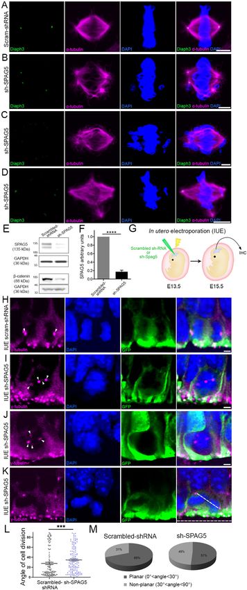
Research article                                                                                                    Neuroscience

                                    cell cortex to enhance cell membrane rigidity for the anchorage of astral microtubule (MT)
                                    (Heng and Koh, 2010). In the meantime, the centrosome, also known as the microtubule organising
                                    centre (MTOC), duplicates, and the two nascent centrosomes migrate toward the poles of the cell.
                                    Astral and spindle MT nucleate from the centrosomes and extend to the cortex and equator of the
                                    cell, respectively. Polarity proteins G-protein signaling modulator 2 (GPSM2, aka PINS/LGN) and
                                    nuclear mitotic apparatus (NUMA) are distributed underneath the cell cortex and interact with corti-
                                    cal actin to connect microtubule plus-end motor proteins dynein/dynactin, and pull on astral MT
                                    (Morin and Bellaı̈che, 2011). On the other hand, spindle MT grow inwardly and attach to chromo-
                                    somes at the metaphase plate. Once the chromosomes are properly aligned and each chromosome
                                    is bilaterally connected to two spindle MT, cohesin is degraded and sister chromatids migrate to
                                    opposing poles of the cell, thus enabling nuclear division (aka karyokinesis). Thereafter, actin redis-
                                    tributes to the contractile ring, which constricts, creating the cleavage furrow. The cell cycle is com-
                                    pleted by cytokinesis that splits the cytoplasm. Hence, the coordinated action of actin and MT is key
                                    to cell division. Errors in centrosome duplication, actin or microtubule polymerization, spindle assem-
                                    bly, or chromosome segregation lead to aneuploidy and/or mitotic catastrophe.
                                        Formins are key regulators of actin dynamics. There are fifteen mammalian formins and they all
                                    possess two formin homolog (FH) domains (Breitsprecher and Goode, 2013). FH1 delivers profilin-
                                    bound actin monomers to the actin filament-barbed ends accelerating elongation, whereas the FH2
                                    dimerizes, enabling the bundling of actin filaments (Kovar, 2006). Diaphanous (DIAPH) formins form
                                    a subgroup of three members that have three regulatory domains: a GTPase-binding domain (GBD)
                                    at the N terminus, a diaphanous autoregulatory domain (DAD), and a diaphanous inhibitory domain
                                    (DID). DAD binds to DID, maintaining the DIAPH proteins in an inactive state. Activation occurs
                                    through the binding of Rho-GTPases, which releases the DAD/DID interaction. The best-character-
                                    ized functions of diaphanous three (DIAPH3, also referred to as mDia2) are actin-related. In prolifer-
                                    ating cells, DIAPH3 is required for the formation of the contractile ring and the cleavage furrow to
                                    enable cytokinesis (Chen et al., 2017; DeWard and Alberts, 2009; Watanabe et al., 2013). DIAPH3
                                    is also involved in filopodia assembly, supporting cell migration (Stastna et al., 2012), and mesen-
                                    chymal-amoeboid transition (Hager et al., 2012; Morley et al., 2015). In addition to the above-
                                    mentioned roles, evidence for DIAPH3 implication in actin/cytokinesis-independent functions has
                                    emerged. Early in vitro studies have suggested that DIAPH3 interacts with EB1 and APC, which
                                    localizes to plus-end, thereby stabilizing MT (Wen et al., 2004; Bartolini et al., 2008). Analysis of
                                    the full Diaph3 knockout mice has revealed an important role of DIAPH3 in the biology of NSCs. The
                                    lack of DIAPH3 severely compromises chromosome segregation, leading to aneuploidy, mitotic
                                    catastrophe, and loss of these cells (Damiani et al., 2016). Yet, the precise function of DIAPH3 in
                                    nuclear division, especially in cytoskeletal rearrangements, remains elusive. Furthermore, the impact
                                    of DIAPH3 deficiency on brain development and function was not assessed due to early embryonic
                                    lethality.
                                        Here, we report that DIAPH3 localizes at the centrosome during mitosis and regulates the assem-
                                    bly of MT as well as the bipolar shape and orientation of the mitotic spindles. DIAPH3 deficiency dis-
                                    rupts the expression and/or stability of several proteins, leading to multipolar spindles and
                                    disorganized cytoskeleton. One of the affected proteins is SPAG5 (also known as the mitotic spin-
                                    dle-associated protein 126 or Astrin), which localizes at the centrosome and kinetochore, and has a
                                    documented function in cell division. SPAG5 displays a similar expression pattern as DIAPH3 in the
                                    developing cortex. Downregulation of SPAG5 phenocopies DIAPH3 deficiency, whereas its overex-
                                    pression rescues the DIAPH3 knockdown phenotype. We also used a conditional approach to delete
                                    Diaph3 specifically in the cerebral cortex and report that this causes a marked depletion of cortical
                                    neurons, microcephaly, locomotor impairment, and social interaction defects.

                                    Results
                                    DIAPH3 localizes to centrosome and is required for assembly and
                                    function of mitotic spindle
                                    We studied the subcellular distribution of DIAPH3 by immunofluorescence in U2OS cells. The protein
                                    co-localized with the centrosomal marker g-tubulin during the whole mitosis (Figure 1A–D). In telo-
                                    phase, it was also seen at the midbody (Figure 1D), which is consistent with its documented role in

Lau et al. eLife 2021;10:e61974. DOI: https://doi.org/10.7554/eLife.61974                                                            2 of 21
Research article                                                                                                                 Neuroscience

Figure 1. Loss of DIAPH3 disrupts the expression/stability of SPAG5 and causes mitotic defects. (A–D) Immunostaining of U2OS cells at prophase (A),
metaphase (B), anaphase (C), and telophase (D), with anti-DIAPH3 (green) and anti-g tubulin (magenta). The chromosomes were counterstained with
DAPI (blue). Diaphanous three (DIAPH3) localizes at the centrosome at all mitotic stages. Insets are zooms in the centrosomal region, showing that the
DIAPH3 signal is pericentrosomal. Scale bar, 10 mm. n = 20 cells for each phase from three distinct experiments. (E, F) Western blot analysis of DIAPH3
and SPAG5 levels upon shRNA downregulation in U2OS cells. shRNA against DIAPH3 (sh-DIAPH3) significantly reduced the levels of DIAPH3 and
SPAG5 proteins (p=0.000044 and 0.000046, respectively). Reciprocally, shRNA against SPAG5 (sh-SPAG5) reduced SPAG5 and DIAPH3 protein levels
(p=0.000001 and 0.000097, respectively). Scrambled shRNA were used as control for the transfection and GAPDH as loading control. n = 3 independent
experiments, Student’s t-test; error bars represent s.e.m. (G–J) Representative images of mitotic cells transfected with scrambled shRNA (G) and shRNA
against DIAPH3 (sh-DIAPH3) (H–J), immunostained with anti-a tubulin (magenta). Chromosomes were counterstained with DAPI (blue). Yellow arrows in
(H) point to lagging chromosomes. Arrowheads in (I) depict poles of mitotic spindle. (J) Representative image of a mitotic cell with disorganized/shrunk
microtubule (MT), single centrosome/pole, and asymmetric metaphase plate. Scale bar, 5 mm. (K) Quantification of mitotic errors. 82% of DIAPH3
knockdown cells exhibit mitotic abnormalities (4% in control cells). n = 118 and 115 cells from five individual experiments of scrambled shRNA and sh-
DIAPH3, respectively. (L–N) Diminished cortical actin in DIAPH3 knockdown cells. (L, M) Illustration of mitotic cells transfected with scrambled shRNA (L)
or sh-DIAPH3 (M), and immunostained with phalloidin (magenta) and a-tubulin (green). Chromosomes were counterstained with DAPI; scale bar, 5 mm.
(N) Quantification of cortical actin (fluorescence intensity) showing a reduction of 48% of cortical actin in DIAPH3 knockdown cells. n = 113 cells per
condition from three distinct experiments. Student’s t-test, p=1.07  10 32. Error bars represent s.e.m.
The online version of this article includes the following source data and figure supplement(s) for figure 1:
Source data 1. Downregulation of DIAPH3 and SPAG5.
Figure supplement 1. DIAPH3 knockdown reduced cell survival and overexpression of SPAG5 rescued the phenotype.
Figure supplement 1—source data 1. Downregulation of DIAPH 3 and SPAG5 in U2OS cells.

                                     the ingression of the cleavage furrow during cytokinesis (Watanabe et al., 2013; DeWard and
                                     Alberts, 2009). We downregulated the expression of DIAPH3 by shRNA (Figure 1E,F) and found
                                     that this disrupted cell division (82% of DIAPH3 knockdown cells showed abnormal division versus
                                     4% in control cells; Figure 1G–K, Supplementary file 1). Notably, DIAPH3 knockdown compro-
                                     mised the integrity of the centrosome and bipolar shape of the spindle (40% of cells exhibited
                                     an abnormal number of centrosomes), as well as the organization of MT, leading to mis-segregation
                                     of chromosomes (Supplementary file 1). Knockdown of DIAPH3 also reduced cell survival to 59.1 ±
                                     0.73% of control cells (Figure 1—figure supplement 1A), likely by inducing apoptosis (percentage
                                     of aCas3+ cells: 1.1 ± 0.12% for scrambled shRNA versus 3.2 ± 0.16% for Diaph3 shRNA; Figure 1—
                                     figure supplement 1B). However, it did not affect cell proliferation as the ratio of Ki67+ cells was
                                     preserved (Figure 1—figure supplement 1C).

Lau et al. eLife 2021;10:e61974. DOI: https://doi.org/10.7554/eLife.61974                                                                          3 of 21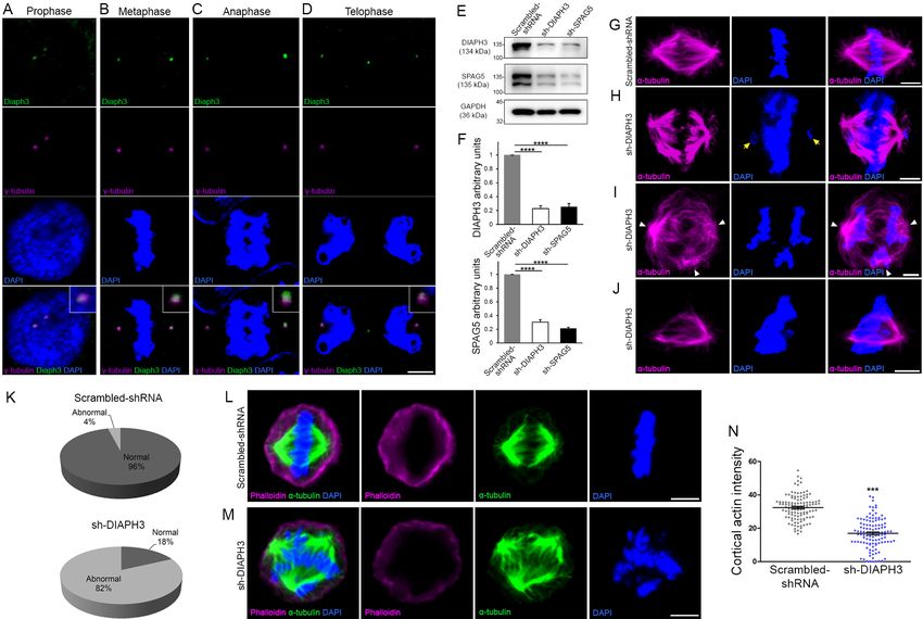
Research article Neuroscience
Downregulation of DIAPH3 alters SPAG5 expression
Spindle positioning during mitosis is governed by multiple mechanisms. Key among those is the ‘cor-
tical pulling’, which refers to the capacity of specific sites on the cell cortex to capture and exert
forces on astral MT to orient the mitotic spindle. Cortical pulling is steered by cytocortical
proteins, which involve NUMA and GPSM2 (also known as LGN or partner of inscuteable [PINS]);
polarity proteins PAR3 and NUMB; and the adapter protein Inscuteable (INSC). The assembly of
NUMA-GPSM2 beneath cortical actin recruits the dynein/dynactin motor protein complex to haul
astral MT (Morin and Bellaı¨che, 2011). Downregulation of DIAPH3 dramatically reduced the amount
of cortical actin (Figure 1L–N). We used Western blotting on dorsal telencephalon lysates from cor-
tex-specific Diaph3 knockout mice (hereafter referred to as Diaph3 cKO, obtained by crossing a
floxed allele with mice expressing the recombinase Cre under the control of Emx1 promoter; Fig-
ure 2—figure supplement 1) to assess the level of core cortical proteins NUMA, GPSM2, PAR3,
NUMB, INSC, dynein, and dynactin; MT plus-end-associated proteins sperm-associated antigen
(SPAG 5) and Kinastrin (KNSTRN, aka small kinetochore-associated protein/SKAP); and the cyto-
plasmic linker-associated protein (CLASP)1. We also used CENPA, a protein downregulated in
DIAPH3-depleted cells (Liu and Mao, 2016) as the positive control for Western blotting and normal-
ized the results to GAPDH levels. We did not detect significant changes in the level of endogenous
NUMA, dynein, dynactin, INSC, or CLASP1 between Diaph3 cKO and control mice. In contrast,
polarity proteins GPSM2, NUMB, and PAR3, and microtubule-associated proteins SPAG5 and
KNSTRN were downregulated (Figure 2A,B, Supplementary file 2). We studied the subcellular dis-
tribution of differentially expressed proteins in Diaph3 cKO (GPSM2 and PAR3) or knockdown cells
(NUMB, SPAG5, and KNSTRN). All displayed mild to important alterations of their partition during
mitosis (Figure 2C–H, Figure 2—figure supplement 2). The distribution of GPSM2, NUMB, SPAG5,
and KNSTRN in the pericentrosomal region was compromised as was that of PAR3 in the apical sur-
face, even though we cannot exclude that some of these changes are due to the reduction in protein
levels. The most evident change was observed in SPAG5. In control cells, the protein localized to the
spindle pole and kinetochore during metaphase and progressively concentrated in the pericentroso-
mal region as mitosis proceeded to anaphase (Figure 2C,D). In DIAPH3-depleted cells, SPAG5
expression declined sharply (Figure 1E,F; Figure 2C–H), and its distribution was disrupted
(Figure 2E–H, Supplementary file 1). The bipolar organization of the spindle was altered, and astral
MT were shrunk. The chromosomes failed to congress at the metaphase plate and did not migrate
to the cell poles at anaphase (Figure 2E–H). Importantly, the expression pattern of the Spag5 tran-
script resembled that of Diaph3 with a strong expression in cortical progenitor cells (http://www.eur-
express.org/ee/databases/assay.jsp?assayID=euxassay_004999&image=01; Damiani et al., 2016).
To explore further the relationship between DIAPH3 and SPAG5, we conducted real-time RT-PCR
(Figure 2I) and RNAscope in situ hybridization (Figure 2J), both of which showed a reduction of
Spag5 mRNA in Diaph3 cKO.
Downregulation of SPAG5 phenocopies the DIAPH3 phenotype
SPAG5 is an essential component of the mitotic spindle. It is required for chromosome alignment,
sister chromatid segregation, and progression to anaphase (Mack and Compton, 2001;
Gruber et al., 2002; Thein et al., 2007). SPAG5 promotes microtubule-kinetochore attachments
and regulates the localization of several centrosomal proteins (e.g. CDK2, CDK5RAP2, CEP152,
WDR62, and CEP63; Kodani et al., 2015). Silencing SPAG5 in U2OS cells triggered multipolar spin-
dles and chromosome mis-segregation, a phenotype reminiscent of DIAPH3 knockdown
(Figure 1G–J and Figure 3A–D; Thein et al., 2007). It also compromised the expression and/or sta-
bility of DIAPH3 (Figure 1E,F and Figure 3A–D). Remarkably, overexpression of SPAG5 significantly
rescued the DIAPH3 knockdown phenotype and partially restored cell viability (survival: 87.1 ±
1.71% in SPAG5 rescue versus 59.1 ± 0.73% in DIAPH3 knockdown; aCas3+ cells: 1.1 ± 0.15% in
SPAG5 rescue versus 3.2 ± 0.16% in DIAPH3 knockdown; Figure 1—figure supplement 1A,B) and
spindle bipolarity (Figure 1—figure supplement 1D). These results, along with the expression of
SPAG5 and its downregulation in the Diaph3 cKO mice, incited us to investigate its function in vivo.
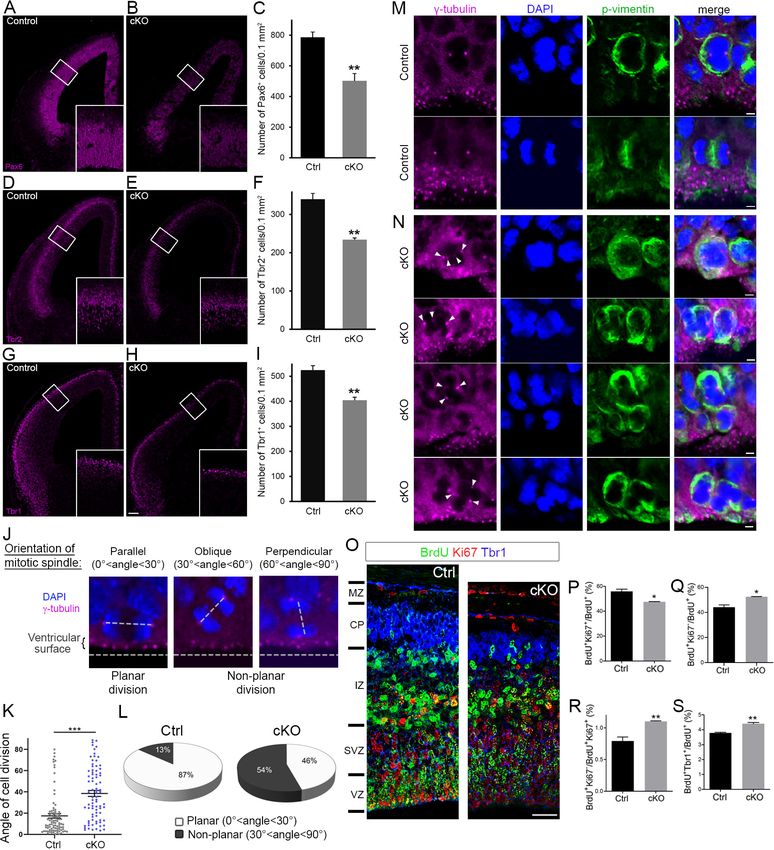
After testing the efficiency of SPAG5 shRNA in NIH3T3 cells (Figure 3E,F), we electroporated e13.5
embryos in utero with scrambled shRNA or sh-SPAG5 cDNA constructs and analyzed cell division of
aNPC at e15.5. We used g-tubulin (centrosome) and DAPI (chromosomes) to envision the orientation
Lau et al. eLife 2021;10:e61974. DOI: https://doi.org/10.7554/eLife.61974 4 of 21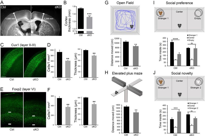
Research article                                                                                                                Neuroscience

Figure 2. Diaph3 deficiency impairs the expression and stability of mitotic spindle polarity proteins. (A, B) Assessment of nuclear mitotic
apparatus (NUMA), G-protein signaling modulator 2 (GPSM2), INSC, NUMB, PAR3, Dynein, Dynactin, SPAG5, KNSTRN, and CLASP1 levels in
telencephalon extracts of Diaph3 cKO embryos by western blotting. CENPA was used as positive control (Liu and Mao, 2016) and GAPDH as loading
control for quantification. Both the higher and lower bands of SPAG5 were quantified. n = 4 embryos for each genotype. *p
Research article                                                                                                         Neuroscience

                                    Figure 3. Downregulation of SPAG5 phenocopies DIAPH3 deficiency. (A–D) Representative images of mitotic cells
                                    transfected with scrambled shRNA (A) or SPAG5 shRNA (sh-SPAG5) (B–D) and immunostained with anti-DIAPH3
                                    (green) and anti-a tubulin (magenta) antibodies. Chromosomes were counterstained with DAPI (blue). SPAG5
                                    Figure 3 continued on next page

Lau et al. eLife 2021;10:e61974. DOI: https://doi.org/10.7554/eLife.61974                                                                 6 of 21
Research article Neuroscience
Figure 3 continued
deficiency downregulated diaphanous three (DIAPH3) and caused mitotic errors. Note the mis-localization of
DIAPH3 and spindle abnormalities in sh-SPAG5-transfected cells. Scale bar, 5 mm. n = 100 cells per condition from
five distinct experiments. (E, F) Western blot analysis of SPAG5 levels upon shRNA-mediated downregulation in
NIH3T3 cells. Transfection of shRNA against SPAG5 (sh-SPAG5) significantly reduced the protein levels ( 82.8%,
p=0.00001, n = 3 independent experiments, Student’s t-test; error bars represent s.e.m.). Beta-catenin, a protein
downregulated by knockdown of SPAG5 (Liu et al., 2019), was used as control of knockdown efficiency. (G)
Schematic representation of in utero electroporation of sh-SPAG5 in cortical progenitors at e13.5 and
immunohistochemistry (IHC) analysis at e15.5. (H–K) Ventricular zone of telencephalic sections of e15.5 embryos
electroporated in utero with scrambled shRNA and GFP (H) or SPAG5 shRNA and GFP (I–K) at e13.5, and
immunostained with anti-g tubulin (magenta) at e15.5. White arrowheads depict normal centrosomes in (H) and
numerical abnormalities of the centrosome in (I–K). (K) Illustration of a neural progenitor undergoing a non-planar
division. The horizontal and oblique dashed lines delineate the ventricular surface and mitotic spindle orientation,
respectively. When the mitotic spindle is parallel to the ventricular surface (0˚Research article Neuroscience Figure 4. Cortex-specific inactivation of Diaph3 disrupts cortical neurogenesis. (A, B, D, E, G, and H) Forebrain coronal sections from e13.5 stained with Pax6 (apical neural progenitor cells [aNPCs]; A, B), Tbr2 (basal progenitors; D, E), and Tbr1 (neurons; G, H) antibodies. Insets are enlargements of the boxed areas. Quantifications shown in (C, F, and I) emphasize reduction in the number of apical radial glia (aNPC, Pax6+, p=0.00853), basal progenitors (Tbr2+, p=0.00287), and neurons (Tbr1+, p=0.00535), respectively. Cells were counted in 0.1 mm2 cortical areas. n = 3 embryos per genotype. Student’s t-test; error bars represent s.e.m. Scale bar, 100 mm. (J) e12.5 cortical sections stained with anti-g tubulin antibodies (magenta) and DAPI (blue) to label centrosomes and chromosomes, respectively, and ‘foresee’ the mitotic spindle. The orientation of the mitotic spindle with respect to the ventricular surface is categorized into three types: parallel (left), oblique (middle), or perpendicular (right). When the mitotic spindle is parallel to the ventricular surface (0˚
Research article                                                                                                                    Neuroscience

Figure 4 continued
increased in Diaph3 cKO. n = 3 embryos for each genotype; Student’s t test, p=0.011 for (P), p=0.011 for (Q), p=0.0078 for (R), p=0.0031 for (S); error
bars represent s.e.m. Scale bar, 30 mm. CP, cortical plate; IZ, intermediate zone; MZ, marginal zone; SVZ, subventricular zone; VZ, ventricular zone.
The online version of this article includes the following source data and figure supplement(s) for figure 4:
Source data 1. Quantification of cell division modalities.
Figure supplement 1. Cell apoptosis in Diaph3 cKO mice.
Figure supplement 1—source data 1. Quantification of cell death in the Diaph3 cKO cortex.

                                      Diaph3 cKO mice exhibit cortical hypoplasia and impaired behavior
                                      The cerebral cortex of adult Diaph3 cKO mice was markedly thin (Figure 5A,B). Immunostaining
                                      with markers of different cortical layers (Cux1: layer II-III, Foxp2: layer VI) revealed a diminished num-
                                      ber of cells along with a reduced thickness of cortex (Figure 5C–F). We then assessed the conse-
                                      quences of cortical hypoplasia in terms of behavior. Diaph3 cKO mice displayed lower spontaneous

Figure 5. Diaph3 cKO mice display cortical hypoplasia and behavioral defects. (A) Dark field micrograph of coronal sections of the forebrain from
control (left) and Diaph3 cKO (right) mice, depicting a marked reduction in cortex thickness in Diaph3 cKO, quantified in (B) (1.3 ± 0.028 mm in Diaph3
cKO versus 1.5 ± 0.014 mm in control, p=0.00038). (C) Coronal sections stained with the upper layer marker Cux1. (D) Quantification of the number of
Cux1+ cells (left) and thickness (right) in layers II-III (Cux1+ cells: 614 ± 20 cells in Diaph3 cKO versus 933 ± 73 in control, p=0.0056; thickness: 385 ± 15
mm in Diaph3 cKO versus 482 ± 13 mm in control, p=0.0025). (E) Coronal sections stained with deep layer marker Foxp2. (F) Quantification of the
number of Foxp2+ cells (left) and thickness (right) in layer VI (Foxp2+ cells: 375 ± 22 cells in Diaph3 cKO versus 558 ± 32 in control, p=0.0033; thickness:
383 ± 20 mm in Diaph3 cKO versus 505 ± 8 mm in control, p=0.0012). Cux1+ and Foxp2+ cells were counted in 1 mm2 cortical area. n = 4 mice for each
genotype, Student’s t-test; error bars represent s.e.m. Scale bar, 200 mm. (G) Distance moved by Diaph3 cKO and control mice in the open field.
Student’s t test, p=0.0015. (H) Distance travelled by mice in elevated plus maze. Student’s t test, p=0.0043. (I) Social behavior in the ‘three-chamber’
test. One-way ANOVA test, p=8.4  10 7 for control, p=0.99 for Diaph3 cKO (stranger versus empty chamber). (J) Social novelty behavior in the ‘three-
chamber’ test. One-way ANOVA test, p=0.00050 for control, p=0.16 for Diaph3 cKO (stranger 1 versus stranger 2 chamber). Compared with control,
Diaph3 cKO mice have reduced locomotor activity and defective social interactions. n = 10 per genotype. Error bars represent s.e.m.
The online version of this article includes the following source data and figure supplement(s) for figure 5:
Source data 1. Cortical histogenesis and behaviour.
Figure supplement 1. Behavior assessment of Diaph3 cKO mice.
Figure supplement 1—source data 1. Assessement of learning and memory, and olfactory behaviour.

Lau et al. eLife 2021;10:e61974. DOI: https://doi.org/10.7554/eLife.61974                                                                              9 of 21
Research article Neuroscience
locomotor activity in open-field test and elevated plus maze (Figure 5G,H), but no difference was
detected when we analyzed the time spent in the periphery of the open field or in the closed arms
of the elevated plus maze (Figure 5—figure supplement 1A,B). These results suggest that DIAPH3
deficiency impairs locomotor activity, but has little effect, if any, on anxiety and/or attention. In addi-
tion, Diaph3 cKO mice exhibited a defective social behavior in the ‘three-chamber’ test. They spent
more time in the empty chamber than with a stranger mouse (stranger 1), emphasizing the lower
sociability of Diaph3 cKO mice, as compared to control littermates (Figure 5I). When another
stranger mouse was introduced, Diaph3 cKO mice spent less time with the new stranger, indicating
impairment in social novelty behavior (Figure 5J). Diaph3 cKO mice did not show any olfactory
defect (Figure 5—figure supplement 1C). They performed as well as controls in the Morris water
maze (Figure 5—figure supplement 1D) and modified Y-maze tests (Figure 5—figure supplement
1E), implying that long-term and short-term memories were preserved. Finally, they did not display
any repetitive behavior, as assayed by self-grooming (Figure 5—figure supplement 1F) or marble
burying (Figure 5—figure supplement 1G). These results show that Diaph3 cKO mice have specific
deficits in motor activity and social behavior, which is in line with previous studies that have associ-
ated DIAPH3 mutations with autism spectrum disorder in humans (Vorstman et al., 2011; Xie et al.,
2016).
Discussion
DIAPH3 is an effector of Rho GTPases that has been classically associated with actin dynamics in
vitro (Watanabe et al., 2010; Watanabe et al., 2013). Functional analysis of knockout mice showed
that it regulates cytokinesis in erythroid cells by promoting the accumulation of actin in the cleavage
furrow at telophase (Watanabe et al., 2013). In addition to this well-documented role in actin cyto-
skeleton and cytokinesis, we have reported that DIAPH3 plays a role during karyokinesis and that its
loss causes chromosome mis-segregation, aneuploidy, and cell death (Damiani et al., 2016). Here
we provide evidence that DIAPH3 localizes to the centrosome during mitosis. Its depletion compro-
mises the integrity of centrosome, spindle and astral MT, as well as the expression or stability of sev-
eral proteins involved in spindle-cell cortex and spindle-kinetochore interactions (Figure 6). These
results are consistent with previous in vitro data, suggesting that DIAPH3 may serve as a scaffold
protein that binds to and stabilizes MT (Palazzo et al., 2001; Bartolini et al., 2008; Wen et al.,
2004). By regulating centrosome integrity, microtubule stability, and spindle orientation, DIAPH3
sits at the heart of cell division accuracy and fate determination (Figure 6). All these processes are
requisites for the production and maintenance of neural progenitors and neurons. The centrosome
acts as a seed and anchoring point for the minus end of MT as they grow toward the equatorial
region (spindle MT) or the cell cortex (astral MT). Consistent with this, we found that depletion of
DIAPH3 disrupts the centrosome and results in disorganized spindle and astral MT, abnormal align-
ment, and segregation of chromosomes. DIAPH3 depletion affects the expression level and/or distri-
bution of several proteins. Among those, SPAG5, which localizes at the centrosome and
kinetochore, is essential to the recruitment of other centrosomal proteins such as CDK5RAP2
(MCPH3) (Kodani et al., 2015). SPAG5 controls sister chromatid cohesion by regulating separase
activity (Thein et al., 2007). Knockdown of SPAG5 induces similar mitotic errors to those triggered
by knockdown of DIAPH3 (Kodani et al., 2015; Thein et al., 2007 and this work), and its overex-
pression rescues DIAPH3 knockdown phenotype. DIAPH3 depletion also affects the expression level
or the stability of GPSM2, NUMB, and PAR3, all of which localize at the region of the cell cortex
where astral MT are anchored.
Microcephaly is a reduction of more than three standard deviations of the head circumference
compared to matching gender, age, and ethnicity controls (Kaindl et al., 2010; Jayaraman et al.,
2018). Primary microcephaly is an inherited neurodevelopmental defect that appears mostly during
pregnancy or at the first postnatal year. Eighteen ‘microcephaly’ genes named MCPH 1–18 (for
microcephaly primary hereditary 1–18) have been identified (Jayaraman et al., 2018). All these
genes are expressed in the germinal zones, wherein the neural progenitors reside, proliferate, and
differentiate into neurons during the second trimester of pregnancy (Verloes et al., 1993;
Gilmore and Walsh, 2013). A key determinant of brain size is the production and maintenance of
the right numbers of neural progenitors and neurons. To attain the initial pool of progenitors and
right number of neurons, NSCs must reconcile two imperatives: the high speed and fidelity of cell
Lau et al. eLife 2021;10:e61974. DOI: https://doi.org/10.7554/eLife.61974 10 of 21Research article Neuroscience
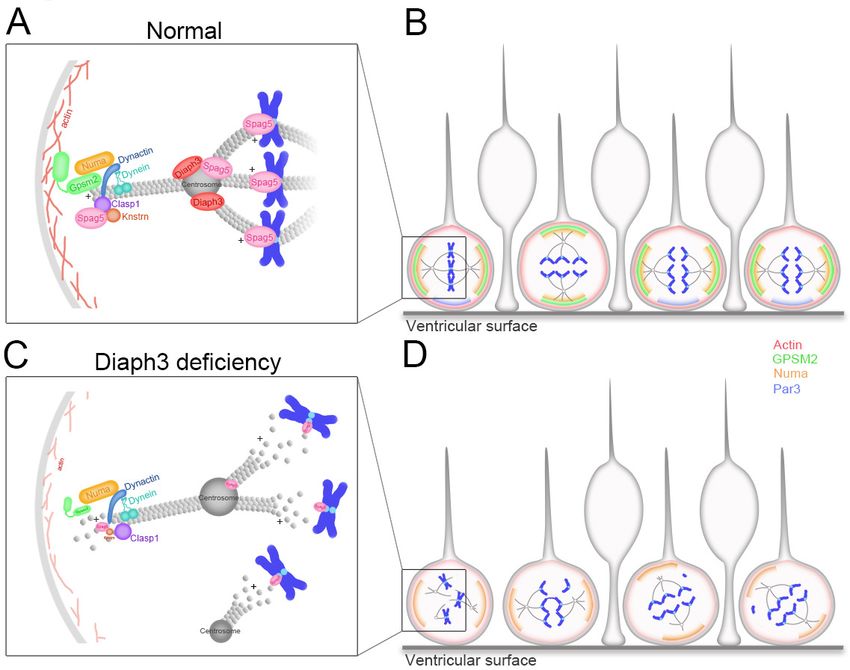
Figure 6. Working model of DIAPH3 function in aNPC. (A) During mitosis, diaphanous three (DIAPH3) maintains
the dynamics of cytoskeleton. Polarity proteins nuclear mitotic apparatus (NUMA) and G-protein signaling
modulator 2 (GPSM2) assemble underneath cortical F-actin and recruit the dynein/dynactin motor protein
complex. Dynein and dynactin interact with SPAG5/KNSTRN/CLASP1 located at the microtubule plus-end and
attach astral microtubule (MT) to cell cortex, thus providing the pulling force for chromosome bipolar segregation
(Okumura et al., 2018; Dunsch et al., 2011; Kern et al., 2016). (B) The position of polarity proteins NUMA/
GPSM2 directs the orientation of the mitotic spindle and determines the type of division (proliferative versus
neurogenic) of apical neural progenitor cells (aNPCs). (C, D) Absence of DIAPH3 destabilizes actin and MT and
disrupts the expression of SPAG5, KNSTRN, GPSM2, PAR3, and NUMB, therefore weakening astral MT-cell cortex
and spindle MT-kinetochore interactions. This causes spindle abnormalities, chromosome mis-alignment, and mis-
segregation, and alters the fate decision of aNPCs.
division. This relies on the interplay between cell cycle regulators, mitotic spindle assembly, mitotic
checkpoints, and cytoskeleton. Hence, the division of NSCs and the balance between proliferation
and differentiation of apical progenitors is a critical factor of brain size (Gilmore and Walsh, 2013).
Defects in the balance between proliferative and neurogenic division, or in the timing of neurogenic
switch, affect cell fate and production of neurons. Loss of function of DIAPH3 disrupts the modality
of cell division and fate determination of apical progenitors, enhancing the number of neurogenic
divisions at the expense of proliferative division, prematurely exhausting the pool of progenitors.
Even though the cytocortical proteins help to orientate the mitotic spindle, the loss of GPSM2 does
not lead to microcephaly despite the randomized cleavage planes in dividing neural progenitors
(Konno et al., 2008; Blumer et al., 2008). Therefore, the role of DIAPH3 in fidelity of nuclear divi-
sion and the cell loss observed in Diaph3 cKO mice may be more instrumental to the emergence of
microcephaly in Diaph3 cKO mice than its role in oriented cell division. It will be interesting to test
whether the blockade of apoptosis induces neoplastic transformation due to accumulation of aneu-
ploid cells. Finally, DIAPH3 is expressed in several epithelia and may govern cell proliferation,
thereby influencing the size of different organs and/or organisms. In support of this, patients with
microcephalic osteodysplastic primordial dwarfism type 1 (MOPD 1) syndrome display a 50% reduc-
tion in DIAPH3 expression (Edery et al., 2011). Furthermore, the Diaph3 gene has been associated
with body size in equines, as copy number variants were reported in donkeys, ponies, and horses
with short stature (Metzger et al., 2018).
In humans, two clinical studies point to DIAPH3 as an autism susceptibility gene (Vorstman et al.,
2011; Xie et al., 2016). The first study identified a double-hit mutation with one amino acid substitu-
tion (Pro614Thr) in one allele and a deletion in the second allele. The second is a de novo point
mutation in the 18th exon (c.2156T > C, pI719T). Several chromosomal deletions spanning the
Lau et al. eLife 2021;10:e61974. DOI: https://doi.org/10.7554/eLife.61974 11 of 21Research article Neuroscience
DIAPH3 locus have also been associated with language impairment, autism, and intellectual disability
(https://decipher.sanger.ac.uk/ and Nathalie Sans et al., 2016). Importantly, among autistic
patients, a significant fraction exhibits microcephaly (Fombonne et al., 1999). In this work, we report
that the conditional deletion of Diaph3 in the mouse cerebral cortex dramatically affects neurogene-
sis and leads to microcephaly and autistic-like behavior, with typical altered motor activity and social
interactions. Our finding that the deletion of Diaph3 affects the accuracy and modalities of division
of neural progenitor cells, ultimately regulating the final number of cortical neurons, may provide a
molecular and cellular basis to the brain malformations and neurological disorders that have been
associated with DIAPH3 dysfunction in humans.
Combined together, our results suggest a novel role of DIAPH3 as a centrosomal protein. Its defi-
ciency induces centrosome abnormalities and disrupts spindle and astral MT. These defects cause
inaccuracies in nuclear division and fate decision of neural progenitors, leading to the loss of cortical
progenitors, microcephaly, and autistic-like behavior.
Materials and methods
Key resources table
Reagent type
(species) or Source or Additional
resource Designation reference Identifiers information
Antibody Anti-CDP/Cux1 (rabbit polyclonal) Santa Cruz Cat# SC-13024, IF (1:200)
RRID:AB_2261231
Antibody Anti-Foxp2 (rabbit polyclonal) Abcam Cat# ab16046, IF (1:500)
RRID:AB_2107107
Antibody Anti-Tbr1 (rabbit polyclonal) Abcam Cat# ab31940, IF (1:500)
RRID:AB_2200219
Antibody Anti-Tbr2 (rabbit polyclonal) Abcam Cat# ab23345, IF (1:500)
RRID:AB_778267
Antibody Anti-Pax6 (rabbit polyclonal) Covance Cat# PRB-278P, IF (1:500)
RRID:AB_291612
Antibody Anti-g-tubulin (rabbit polyclonal) Abcam Cat# ab11317, IF (1:500)
RRID:AB_297921
Antibody Anti-Diaph3 (rabbit polyclonal) Tominaga et al., 2000 IF (1:500)
WB (1:500)
Antibody Anti-Spag5 (rabbit polyclonal) Proteintech Cat# 14726–1-AP, IF (1:200)
RRID:AB_2194787
Antibody Anti-a-tubulin (mouse monoclonal) Sigma Cat# T6199, IF (1:500)
RRID:AB_477583
Antibody Anti-cleaved caspase-3 Cell Signaling Cat# 9603, IF (1:100)
(Alexa RRID:AB_11179205
Fluor 488
conjugate) (rabbit
monoclonal)
Antibody Anti-Ki67 (mouse BD phamingen Cat# 556003, IF (1:500)
monoclonal) RRID:AB_396287
Antibody Anti-SPAG5 (rabbit Sigma Cat# HPA022008, WB (1:750)
polyclonal) RRID:AB_1853519
Antibody Anti-GAPDH (chicken Millipore Cat# AB2302, WB (1:2000)
polyclonal) RRID:AB_10615768
Antibody Anti-NUMA (mouse BD Biosciences Cat# 610561, WB (1:500)
monoclonal) RRID:AB_397913
Antibody Anti-PAR3 (rabbit Millipore Cat# 07–330, IF (1:500)
polyclonal) RRID:AB_2101325 WB (1:500)
Antibody Anti-GPSM2 (rabbit Ezan et al., 2013 IF (1 :200)
polyclonal) WB (1 :200)
Continued on next page
Lau et al. eLife 2021;10:e61974. DOI: https://doi.org/10.7554/eLife.61974 12 of 21Research article Neuroscience
Continued
Reagent type
(species) or Source or Additional
resource Designation reference Identifiers information
Antibody Anti-CENPA (rabbit Cell Signaling Cat# 2048, WB (1 :500)
monoclonal) RRID:AB_1147629
Antibody Anti-Dynein IC1/2 Santa Cruz Cat# sc-13524, WB (1:500)
(mouse monoclonal) RRID:AB_668849
Antibody Anti-KNSTRN (rabbit Sigma Cat# HPA042027, IF (1:1000)
polyclonal) RRID:AB_10797378 WB (1:1000)
Antibody Anti-INSC (rabbit Abcam Cat# ab102953, WB (1:1000)
polyclonal) RRID:AB_10711784
Antibody Anti-NUMB (goat Abcam Cat# ab4147, IF (1:100)
polyclonal) RRID:AB_304320 WB (1:100)
Antibody Anti-p150Glued BD Biosciences Cat# 610474, WB (1:500)
/dynactin RRID:AB_397846
(mouse polyclonal)
Antibody Anti-CLASP1 (rabbit monoclonal) Abcam Cat# ab108620, WB (1:5000)
RRID:AB_10864427
Antibody Anti-BrdU Serotec Cat# MCA2060GA, IF (1:200)
(rat monoclonal) RRID:AB_10545551
Antibody Anti-Ki67 (rabbit Abcam Cat# ab15580, IF (1:250)
polyclonal) RRID:AB_443209
Antibody Anti-Tbr1 (chicken Millipore Cat# AB2261, IF (1:100)
polyclonal) RRID:AB_10615497
Antibody Anti-SPAG5 (rabbit polyclonal) NovusBio Cat# NB100-74638, WB (1:1000)
RRID:AB_2239831
Antibody Alexa Fluor Invitrogen Cat# A11017, IF (1:800)
488 anti-mouse RRID:AB_143160
IgG (goat
polyclonal)
Antibody Alexa Fluor Invitrogen Cat# A11034, IF (1:1000)
488 anti- RRID:AB_2576217
rabbit
IgG (goat
polyclonal)
Antibody Alexa Fluor Jackson Cat# 103-545-155, IF (1:1000)
488 anti- ImmunoResearch RRID:AB_2337390
chicken
IgY (IgG)
(goat polyclonal)
Antibody Alexa Fluor Invitrogen Cat# A21124, IF (1:1000)
568 anti-mouse RRID:AB_141611
IgG (goat
polyclonal)
Antibody Alexa Fluor Invitrogen Cat# A11036, IF (1:1000)
568 anti-rabbit RRID:AB_10563566
IgG (goat
polyclonal)
Antibody Anti-chicken Upstate Biotech Cat# 12–341, WB (1:1000)
IgY/HRP-linked RRID:AB_390189
(rabbit
polyclonal)
Antibody Anti-mouse Dako Cat# P0447, WB (1:1000)
IgG/HRP- RRID:AB_2617137
linked
(goat polyclonal)
Continued on next page
Lau et al. eLife 2021;10:e61974. DOI: https://doi.org/10.7554/eLife.61974 13 of 21Research article Neuroscience
Continued
Reagent type
(species) or Source or Additional
resource Designation reference Identifiers information
Antibody Anti-rabbit Cell Signaling Cat# 7074, WB (1:1000)
IgG/HRP- RRID:AB_2099233
linked
(goat polyclonal)
Cell line U2OS cell ATCC Cat# HTB-96,
(Homo-sapiens) (osteosarcoma) RRID:CVCL_0042
Cell line NIH3T3 ATCC Cat# CRL-1658
(Mus musculus) immortalized MEFs RRID:CVCL_KS54
Strain, strain Mus musculus Jackson Lab Emx1tm1(cre)Krj/J
background RRID:IMSR_JAX:005628
Strain, strain Mus musculus This paper Diaph3Emx1-Cre cKO
background (Diaph3f/f;
Emx1-Cre)
Transfected Scrambled shRNA Origene Cat# TR30012 Transfected construct
construct (human)
Transfected sh-Diaph3 Origene Cat# TR304992 Transfected construct
construct (human)
Transfected sh-Spag5 Origene Cat# TR309161 Transfected construct
construct (human)
Transfected sh-Spag5 Origene Cat# TR509034 Transfected construct
construct
(Mus musculus)
Commercial Mycoplasma PCR detection kit Sigma Cat#, MP0035
assay or kit
Commercial RNeasy Kit Qiagen Cat# 74004
assay or kit
Commercial RT cDNA synthesis Kit Promega Cat# A5003
assay or kit
Commercial SYBR Green SuperMix Biorad Cat# 170–8882
assay or kit
Commercial Lipofectamine LTX Thermofisher Cat# 15338030
assay or kit Scientific
Commercial RNAscope Probe-Mm-Spag5 ACD Cat# 505691
assay or kit
Commercial Pierce BCA protein assay kit Thermofisher Cat# 23225
assay or kit Scientific
Commercial Bolt 4–12% Bis-Tris Plus Gels Thermofisher Cat# NW04125 BOX
assay or kit Scientific
Commercial StartingBlock T20 (TBS) blocking buffer Thermofisher Cat# 37543
assay or kit Scientific
Commercial 20x Bolt MOPS SDS running buffer Thermofisher Cat# B0001
assay or kit Scientific
Software, Prism GraphPad, USA RRID:SCR_002798
algorithm
Software, Zen lite Zeiss
algorithm
Software, Ethovision 6.1, Noldus Wageningen,
algorithm The Netherlands
Mutant mice
All animal procedures were carried out in accordance with European guidelines and approved by
the animal ethics committee of the Université Catholique de Louvain. Mouse lines used in this study
were Emx1tm1(cre)Krj/J; (Jackson Lab) (Gorski et al., 2002; Damiani et al., 2016), Diaph3f/f; Emx1-Cre
Lau et al. eLife 2021;10:e61974. DOI: https://doi.org/10.7554/eLife.61974 14 of 21Research article Neuroscience
(Diaph3 cKO), and Diaph3f/f (control for Diaph3 cKO). All mice were maintained in a mix
background.
Immunostaining and antibodies
For immunohistochemistry, embryos were fixed in 4% paraformaldehyde (PFA), cryoprotected by
gradients of sucrose solution. Cryosections were blocked in PBST (0.1% Triton X-100 in phosphate-
buffered saline [PBS]) supplemented with 5% normal goat serum and 1% bovine serum albumin
(BSA) for 30 min. Slides were incubated in primary antibodies diluted in blocking buffer at 4˚C over-
night. Slides were washed and incubated with secondary antibodies (Alexa, Thermofisher) diluted in
the same blocking buffer. Cultured cells were fixed with chilled methanol for 5 min, followed by
washing in PBST. Cells were incubated in blocking buffer for 30 min, primary antibodies for 1.5 hr,
and secondary antibodies for 1.5 hr at room temperature. Primary antibodies used were as follows:
rabbit anti-Cux1 (Santa Cruz, SC-13024, 1:200), rabbit anti-Foxp2 (Abcam, ab16046, 1:500), rabbit
anti-Tbr1 (Abcam, ab31940, 1:500), rabbit anti-Tbr2 (Abcam, ab23345, 1:500), rabbit anti-Pax6
(Covance, PRB-278P, 1:500), rabbit anti-g-tubulin (Abcam, ab11317, 1:500), Diaph3 (1:500)
(Tominaga et al., 2000), rabbit anti-Spag5 (Proteintech, 14726–1-AP, 1:200), mouse anti-a-tubulin
(Sigma, T6199, 1:500), rabbit anti-aCas3 (Cell Signaling, 9603, 1:100), and mouse anti-Ki67 (BD Pha-
mingen, 556003, 1:500). Nuclei/chromosomes were counterstained with DAPI. Images were acquired
with an Olympus FV1000 confocal microscope. Mitotic cells were imaged with Z-stack, which
focused on the levels of centrosomes.
Cell fate assessment
For cell fate and neurogenesis study, pregnant females were intraperitoneally injected with BrdU
(Sigma B5002) (50 mg/kg body weight) at E13.5. Embryos were collected 24 hr later (E14.5) and
brains were fixed in PFA 4% and processed for cryosectioning (20 mm). For BrdU staining, sections
were pretreated with HCl 1 N for 20 min at room temperature, followed by a 10 min incubation with
0.1 M sodium borate buffer (pH 8.5). After that, antigen retrieval was performed by heating sections
for 20 min in 0.01 M sodium citrate buffer (pH 6). Immunodetection was done using primary antibod-
ies rat anti-BrdU (1:200, Serotec MCA2060GA), rabbit anti-Ki67 (1:250, Abcam ab15580),
and chicken anti-Tbr1 (1:100, Millipore AB2261) and secondary antibodies as described above.
Western blotting
Tissues were homogenized in lysis buffer containing 50 mM Tris HCl (pH 7.5), 150 mM NaCl, 1%
NP40, and protease inhibitors (Roche). Cell lysates were incubated on ice for 30 min prior to centri-
fugation at 2000 g for 10 min at 4˚C. Protein quantification was performed with BCA Protein Assay
kit (Pierce). Supernatants were mixed with SDS-loading buffer and heated at 85˚C for 10 min. Equal
amounts of proteins were loaded on 4–12% gel (Invitrogen) and transferred to PVDF membrane
(Merck Millipore). Membranes were blocked with StartingBlock buffer (ThermoScientific) and incu-
bated overnight at 4˚C with rabbit anti-DIAPH3 (1:5000) (Tominaga et al., 2000), rabbit anti-SPAG5
(Sigma, HPA022008, 1:750), chicken anti-GAPDH (Millipore, AB2302, 1:2000), mouse anti-NUMA
(Becton Dickinson, 610561, 1:500), rabbit anti-PAR3 (Millipore, 07–330, 1:500), rabbit anti-GPSM2
(1:200) (Ezan et al., 2013), rabbit anti-CENPA (Cell Signaling, 2048, 1:500), mouse anti-dynein (Santa
Cruz, sc-13524, 1:500), rabbit anti-KNSTRN (Sigma, HPA042027, 1:1000), rabbit anti-INSC (Abcam,
ab102953, 1:1000), goat anti-NUMB (Abcam, ab4147, 1:100), mouse anti-dynactin (BD Biosciences,
610474, 1:500), and rabbit anti-CLASP1 (Abcam, ab108620, 1:5000). Proteins were detected with
SuperSignal West Pico PLUS solution (ThermoScientific) or SuperSignal West Femto Maximum Sensi-
tivity Substrate kit (ThermoScientific). Band intensity was imaged by fusion pulse and quantified with
ImageJ Gel analyzer tool. Values were normalized with GAPDH. The amount of protein in control
was set to one.
qPCR
Total mRNA was isolated from control or Diaph3 cKO E11.5 telecephalon using the RNeasy mini kit
(Qiagen) according to the supplier’s instructions. Reverse transcription was performed with an RT
cDNA Synthesis Kit (Promega). Real-time PCR was performed with SYBR green SuperMix using an
iCycler real-time PCR detection system (Bio-Rad).
Lau et al. eLife 2021;10:e61974. DOI: https://doi.org/10.7554/eLife.61974 15 of 21Research article Neuroscience
Cell lines and knockdown experiments
U2OS and NIH/3T3 cell lines were purchased from ATCC. Their identity has been authenticated by
STR profiling (ATCC). They were tested negative for mycoplasma using the ‘LookOut Mycoplasma
PCR Detection Kit’ (Sigma, MP0035). Cells were cultured in Dulbecco’s modified eagle’s
medium (DMEM) supplemented with penicillin-streptomycin and 10% fetal bovine serum (FBS; Invi-
trogen). Cells were seeded in 12-well plates (day 0) and were transfected with Lipofectamine LTX
(Invitrogen) (day 1) using scrambled shRNA (Origene, TR30012) or pool of sh-DIAPH3 (Origene,
TR304992) with sequences ATTTATGCGTTGTGGATTGAAAGAGATAT, AATCAGCATGAGAAGA
TTGAATTGGTTAA, CACGGCTCAGTGCTATTCTCTTTAAGCTT, and AAGAGCAGGTGAACAACA
TCAAACCTGAC or pool of sh-Spag5 (Origene, TR309161) with sequences CCTCAAGGACACTG
TAGAGAACCTAACGG, GGTAGGATTCTTGGCTCTGATACAGAGTC, CTCCAAGGAAAGCCTGAG-
CAGTAGAACTG, and AGATGAAGAGCCAGAATCAACTCCTGTGC. Cells were harvested 72 hr
after transfection for western blotting. For immunostaining, cells were serum-deprived at day 3 (48 h
after transfection) and seeded onto glass coverslips pre-coated with gelatin at day 4 (72 h after
transfection). Cell cycle was synchronized by serum add back, and cells were fixed after 16 hr. For
cell survival assay, cells were seeded at the same density (50% confluency, 2 105 cells per well)
onto glass coverslips pre-coated with gelatin in 12-well plates at day 0. Cell number was counted at
day 1 (just before transfection) and day 4 (72 h after transfection). Cells were then immunostained
with apoptotic marker (aCas3), proliferation marker (Ki67), and DAPI. Cell density was imaged and
counted by Zen lite with Zeiss AXIO light microscope.
Behavioral tests
All the behavioral tests were performed using adult Diaph3 cKO (Diaph3f/f; Emx1-Cre) and control
littermate (Diaph3f/f) males. The ‘open-field’ test was performed to assess locomotor activity and
anxiety. Mice were allowed to move freely in a square arena (60 60 cm) and video tracked (Ethovi-
sion 6.1, Noldus; Wageningen, The Netherlands) for 20 min (Mallon et al., 2008; Mignion et al.,
2013). The total distance covered by test mice (locomotor activity) and the time spent in the periph-
ery (anxiety) were measured.
The ‘elevated plus maze’ test was performed to assess locomotor activity and anxiety. Mice were
placed in a setup consisting of two opposing open arms (exposed place) and two opposing closed
arms (safer place). Total distance travelled and time spent in the closed arm were recorded by a
video tracking system (Ethovision 6.1, Noldus; Wageningen, The Netherlands) for 5 min.
The ‘three-chamber’ test was performed to assess sociability. Test box was divided into three
equal compartments of 20 cm each, and dividing walls had retractable doorways allowing access
into each chamber. Mice were habituated in the middle chamber for 5 min with the doorways
closed. To evaluate social interactions, mice were enclosed in the center compartment, and an unfa-
miliar mouse (called stranger 1) was restricted in a wire cage placed in one side compartment, while
the other side compartment contained an empty wire cage. Mice were video tracked (Ethovision
6.1, Noldus; Wageningen, The Netherlands) for 10 min. The time spent in each chamber was mea-
sured. To evaluate the preference for social novelty, a second unfamiliar mouse (called stranger 2)
was introduced into the empty wire cage in the other side compartment of the sociability test box.
Mice were video tracked for 10 min, and the time spent in each chamber was measured (Moy et al.,
2007).
The ‘food localization’ test was performed to evaluate the olfactory function. Test mice were
fasted for 16 hr with water supply ad libitum. They were then transferred into a clean cage with 3-
cm-thick wood-chip bedding and lightly tamped down to make a flat surface. 1.0 g of food pellet
was placed at the same location under the bedding and mice were introduced into the cage at a
constant position. The time from which the mouse was placed into the cage until it retrieved the
food pellet with its front paws was counted up to a maximum of 300 s.
The ‘Morris water maze’ test was performed to assess long-term memory. Water maze was made
of a round pool with a diameter of 113 cm, virtually divided into four quadrants (North, South, West,
and East) and filled with water (26˚C). Several visual cues were placed around the pool. The platform
was placed at the center of the North-East quadrant of the pool and maintained in this position
throughout the four days. Mice were video-tracked (Ethovision 6.1, Noldus; Wageningen, The
Lau et al. eLife 2021;10:e61974. DOI: https://doi.org/10.7554/eLife.61974 16 of 21Research article Neuroscience
Netherlands). The time latency to reach the platform was measured (Rzem et al., 2015;
Boucherie et al., 2018).
The ‘Y-maze’ test was performed to assess short-term memory (Rzem et al., 2015;
Lepannetier et al., 2018). The Y-maze was made of three identical opaque arms. The test mouse
was freely accessible to only two arms for 10 min. After a 30 min inter-trail interval, the third arm
was opened and the mouse was put back into the maze. The mouse was video tracked, and the time
spent in the novel arm was assessed.
‘Self-grooming’ and ‘marble burying’ tests were performed to assess repetitive behavior. For self-
grooming assessment, mice were individually placed in a clear plastic cage for 20 min. The first 10
min served as a habituation period, and during the second 10 min of testing, time spent grooming
was recorded. For the marble burying test, test cages were filled with wood-chip bedding and lightly
tamped down to make a flat surface. Twenty glass marbles were placed on the surface in a regular
pattern and mice were then placed in the cage. After 30 min, the number of bedding-buried marbles
was counted (Amodeo et al., 2012).
In utero electroporation
CD1 pregnant females were used for in utero electroporation (IUE). e13.5 embryos were electropo-
rated with 1 mg/ml of scrambled shRNA or sh-Spag5, and 0.5 mg/ml of CAG-GFP in 10 mM Tris buffer
(pH 8.0) and 0.01% fast green as described in Jossin and Cooper, 2011. Needles for injection were
pulled from Wiretrol II glass capillaries (Drummond Scientific). Forcep-type electrodes (Nepagene)
with 5 mm pads were used for electroporation using the ECM830 electroporation system (Harvard
Apparatus). Embryos were collected and fixed at e15.5. shRNA were purchased from Origene;
scrambled shRNA (Origene, TR30012) and pool of sh-Spag5 (Origene, TR509034) with sequences
TAGTCTCTGGAGACCTGTTGTCCTTGCTT, GGAGGAAGCAATAGAAACAGTGGATGACT,
GACAAGTATCTGAGCCATAGGCACATCCT, and GCAACAAGGAGCAGGCTACTCAATGGCAA
were used.
In-situ hybridization
RNase-free coronal cryosections from e11.5 Diaph3 cKO and control embryos were hybridized with
Spag5 fast red-labelled RNAscope probe (Cat No. 505691) as described by the manufacturer.
Acknowledgements
We thank Dr. Ulrike Gruneberg (University of Oxford, UK) for the Spag5 plasmid, Dr. Mireille Mon-
tcouquiol (INSERM, France) for the GPSM2/PINS antibody, and Valérie Bonte, Rachid El Kaddouri,
Isabelle Lambermont, and Younes Massaoudi for technical support. This study makes use of data
generated by the DECIPHER community. A full list of centers that contributed to the generation of
the data is available from http://decipher.sanger.ac.uk. Funding for the DECIPHER project was pro-
vided by the Wellcome Trust. This work was supported by the following grants: FNRS PDR
T00075.15, FNRS PDR T0236.20, FNRS-FWO EOS 30913351, Fondation Médicale Reine Elisabeth,
and Fondation JED-Belgique. EOL was supported by ‘Move-In Louvain’ postdoctoral fellowship
funded by Marie Skłodowska-Curie Actions of European Commission and Université Catholique de
Louvain. RS is supported by an FNRS-Télévie Grant. GC, NRR, and YJ are research fellow, postdoc-
toral researcher, and research associate at the Belgian Fund for Scientific Research (FNRS),
respectively.
Additional information
Competing interests
Fadel Tissir: Reviewing editor, eLife. The other authors declare that no competing interests exist.
Funding
Funder Grant reference number Author
Fonds De La Recherche Scien- Fadel Tissir
Lau et al. eLife 2021;10:e61974. DOI: https://doi.org/10.7554/eLife.61974 17 of 21Research article Neuroscience
tifique - FNRS
Fonds De La Recherche Scien- FNRS PDR T0236.20 Fadel Tissir
tifique - FNRS
Fonds De La Recherche Scien- EOS 30913351 Fadel Tissir
tifique - FNRS
H2020 Marie Skłodowska-Curie Eva On-Chai Lau
Actions
Université Catholique de Lou- Eva On-Chai Lau
vain
Fonds De La Recherche Scien- Rana Saade
tifique - FNRS
Fonds De La Recherche Scien- Georges Chehade
tifique - FNRS Nuria Ruiz-Reig
Yves Jossin
The funders had no role in study design, data collection and interpretation, or the
decision to submit the work for publication.
Author contributions
Eva On-Chai Lau, Investigation, Visualization, Methodology, Writing - original draft, Writing - review
and editing; Devid Damiani, Conceptualization, Investigation, Methodology, Writing - review and
editing, Contributed equally with Georges Chehade; Georges Chehade, Supervision, Validation,
Investigation, Visualization, Writing - review and editing, Contributed equally with Devid Damiani;
Nuria Ruiz-Reig, Validation, Investigation, Methodology, Writing - review and editing; Rana Saade,
Mohamed Aittaleb, Formal analysis, Investigation; Yves Jossin, Validation, Investigation, Visualiza-
tion; Olivier Schakman, Validation, Investigation, Writing - original draft; Nicolas Tajeddine, Supervi-
sion, Validation, Methodology, Writing - review and editing; Philippe Gailly, Conceptualization,
Validation, Investigation, Writing - review and editing; Fadel Tissir, Conceptualization, Resources,
Formal analysis, Supervision, Funding acquisition, Validation, Visualization, Methodology, Writing -
original draft, Writing - review and editing
Author ORCIDs
Nuria Ruiz-Reig https://orcid.org/0000-0001-7008-7920
Yves Jossin http://orcid.org/0000-0001-8466-7432
Fadel Tissir https://orcid.org/0000-0002-9292-6622
Ethics
Animal experimentation: All animal procedures were carried out in accordance with European guide-
lines and approved by the animal ethics committee of the Université Catholique de Louvain (permit
number 2019/UCL/MD/006).
Decision letter and Author response
Decision letter https://doi.org/10.7554/eLife.61974.sa1
Author response https://doi.org/10.7554/eLife.61974.sa2
Additional files
Supplementary files
. Supplementary file 1. Cell division abnormalities in DIAPH3 knockdown cells. Over 81% of
DIAPH3 knockdown cells exhibited mitotic errors (4.2% in control cells). These errors were divided
into five ‘non-exclusive’ categories: (1) defects in chromosome alignment (57.4%); (2) abnormal astral
microtubule (MT) (52.2%); (3) abnormal number of centrosomes (40%); (4) abnormal mitotic spindle
(53%); and (5) mis-localization of SPAG5 (60%). n = 118 cells transfected with scrambled shRNA and
115 with sh-DIAPH3 from five individual experiments.
Lau et al. eLife 2021;10:e61974. DOI: https://doi.org/10.7554/eLife.61974 18 of 21You can also read