Tinnitus and hyperacusis involve hyperactivity and enhanced connectivity in auditory-limbic-arousal-cerebellar network
←
→
Page content transcription
If your browser does not render page correctly, please read the page content below
RESEARCH ARTICLE
           elifesciences.org

                                     Tinnitus and hyperacusis involve
                                     hyperactivity and enhanced connectivity in
                                     auditory-limbic-arousal-cerebellar network
                                     Yu-Chen Chen1, Xiaowei Li2, Lijie Liu2, Jian Wang2,3, Chun-Qiang Lu1, Ming Yang1,
                                     Yun Jiao1, Feng-Chao Zang1, Kelly Radziwon4, Guang-Di Chen4, Wei Sun4,
                                     Vijaya Prakash Krishnan Muthaiah4, Richard Salvi4*, Gao-Jun Teng1*
                                     1
                                      Jiangsu Key Laboratory of Molecular Imaging and Functional Imaging, Department
                                     of Radiology, Zhongda Hospital, Medical School, Southeast University, Nanjing,
                                     China; 2Department of Physiology, Southeast University, Nanjing, China; 3School of
                                     Human Communication Disorders, Dalhousie University, Halifax, Canada; 4Center for
                                     Hearing and Deafness, University at Buffalo, The State University of New York,
                                     Buffalo, United States

                                     Abstract Hearing loss often triggers an inescapable buzz (tinnitus) and causes everyday sounds to
                                     become intolerably loud (hyperacusis), but exactly where and how this occurs in the brain is
                                     unknown. To identify the neural substrate for these debilitating disorders, we induced both tinnitus
                                     and hyperacusis with an ototoxic drug (salicylate) and used behavioral, electrophysiological, and
                                     functional magnetic resonance imaging (fMRI) techniques to identify the tinnitus–hyperacusis
                                     network. Salicylate depressed the neural output of the cochlea, but vigorously amplified sound-
                                     evoked neural responses in the amygdala, medial geniculate, and auditory cortex. Resting-state fMRI
                                     revealed hyperactivity in an auditory network composed of inferior colliculus, medial geniculate, and
                                     auditory cortex with side branches to cerebellum, amygdala, and reticular formation. Functional
                                     connectivity revealed enhanced coupling within the auditory network and segments of the auditory
*For correspondence: salvi@          network and cerebellum, reticular formation, amygdala, and hippocampus. A testable model
buffalo.edu (RS); gjteng@vip.sina.   accounting for distress, arousal, and gating of tinnitus and hyperacusis is proposed.
com (GT)                             DOI: 10.7554/eLife.06576.001

Competing interests: The
authors declare that no
competing interests exist.
                                     Introduction
Funding: See page 15                 A third of adults over the age of 65 suffer from significant hearing loss, a condition exacerbated by
Received: 21 January 2015            two debilitating condition, subjective tinnitus, a phantom ringing or buzzing sensation, and
Accepted: 13 April 2015              hyperacusis, normal sounds perceived as intolerably loud or even painful. Roughly 12% of adults
Published: 12 May 2015               experience tinnitus, but the prevalence skyrockets to 50% in young combat personnel (Leske, 1981;
                                     Andersson et al., 2002; Cave et al., 2007; Michikawa et al., 2010; Hebert et al., 2013). Tinnitus is
Reviewing editor: Heidi
Johansen-Berg, University of         costly with more than $2 billion paid annually in veteran disability payments. Hyperacusis affects
Oxford, United Kingdom               roughly 9% of adults (Andersson et al., 2002), but its prevalence is likely higher because of the
                                     difficulty of self-diagnosis (Gu et al., 2010). Remarkably, among those whose primary complaint is
    Copyright Chen et al. This
                                     hyperacusis, 90% also suffer from tinnitus (Baguley, 2003). Since tinnitus and hyperacusis are often
article is distributed under the
                                     triggered by cochlear hearing loss, it was long assumed that these auditory distortions resulted from
terms of the Creative Commons
                                     hyperactivity disorders in the peripheral auditory nerve. This hypothesis, however, is contradicted by
Attribution License, which
permits unrestricted use and         studies showing that auditory nerve spontaneous and sound-evoked firing rates are depressed in
redistribution provided that the     subjects with cochlear damage (Kiang et al., 1970; Wang et al., 1997). Moreover, surgical section of
original author and source are       the auditory nerve fails to eliminate tinnitus (Baguley et al., 1992; Lockwood et al., 2001). These
credited.                            negative results plus recent imaging studies now suggest that tinnitus and hyperacusis arise from

Chen et al. eLife 2015;4:e06576. DOI: 10.7554/eLife.06576                                                                           1 of 19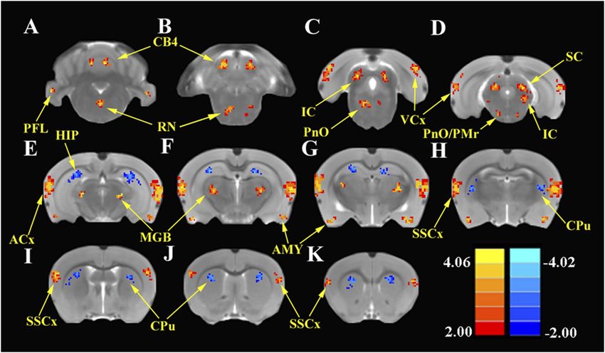
Research article                                                                                                   Neuroscience

                                   eLife digest One in three adults over the age of 65 will experience a significant loss of hearing.
                                   This is often worsened by related conditions, such as: tinnitus, an unexplained constant buzzing or
                                   ringing sound; and hyperacusis, whereby everyday sounds are perceived as too loud or painful.
                                      Most hearing loss is caused by damage to the sound-sensitive cells within a structure in the inner
                                   ear called the cochlea. Some studies have also identified regions of the brain that show abnormal
                                   activity in people with tinnitus and hyperacusis. However, the results from different patients have
                                   often been inconsistent and sometimes contradictory, and so it remains unclear what exactly causes
                                   these conditions.
                                      To overcome this problem, Chen et al. made use of the fact that tinnitus and hyperacusis are
                                   common short-term side effects of certain drugs and measured the brain activity in rats before and
                                   after they were given one such drug. Before receiving the drug, the rats had first been trained to
                                   expect to receive a food pellet from the left side of their cage when they heard a steady buzzing
                                   sound. The rats were also trained to expect a food pellet from their right if they heard nothing at all.
                                   Shortly after receiving the drug, the rats often failed to respond correctly in the ‘quiet tests’ and
                                   behaved like they were already experiencing a constant buzzing sound, as would be expected if they
                                   had tinnitus. Further tests confirmed that the drug also triggered behavior in the rats that is typical of
                                   people with hyperacusis.
                                      Chen et al. then discovered that the drug treatment reduced the nerve signals that are sent from
                                   a rat’s cochlea. Moreover, the drug treatment greatly increased the activity in response to sound
                                   within parts of the rat’s brain; these and other parts of the brain also became overactive in drug-
                                   treated rats in the absence of sound. Finally, further experiments revealed that drug-treated rats had
                                   stronger connections between these brain regions than in normal rats.
                                      Chen et al. used these results to propose a model to explain the underlying causes of tinnitus and
                                   hyperacusis. However, because the drug treatment only induces short-term hearing impairment,
                                   further studies are needed to see if this model also applies when these conditions are long-term.
                                   DOI: 10.7554/eLife.06576.002

                                   maladaptive neuroplastic change in the central nervous system (CNS) provoked by cochlear pathology
                                   (Lockwood et al., 1998; Husain et al., 2011; Sereda et al., 2011).
                                       Several models of tinnitus and hyperacusis have been proposed that involve increased central gain,
                                   altered functional connectivity (FC), and aberrant neural oscillations in neural networks (Weisz et al.,
                                   2007; Sereda et al., 2011; Henry et al., 2014). Most of these conceptual models have emerged from
                                   human imaging studies using magnetoencephalography, electroencephalography, magnetic
                                   resonance imaging (MRI), and functional MRI (fMRI) of the blood oxygen level-dependent (BOLD)
                                   response (Llinas et al., 1999; Weisz et al., 2005; Auer, 2008; Gu et al., 2010; Moazami-Goudarzi
                                   et al., 2010; Leaver et al., 2012; Maudoux et al., 2012; Husain and Schmidt, 2014). In the context
                                   of central gain models, some human imaging data indicate that hyperacusis is associated with
                                   enhanced sound-evoked activity in multiple-auditory processing centers, namely auditory cortex
                                   (ACx), medial geniculate body (MGB), and inferior colliculus (IC), whereas tinnitus can be triggered
                                   solely by enhanced central gain in the ACx (Gu et al., 2010). On the other hand, active loudness
                                   models suggest that tinnitus arises entirely from increased central noise independent of gain, whereas
                                   hyperacusis results exclusively from increased nonlinear gain that results in loudness intolerance
                                   (Zeng, 2013).
                                       While cross-sectional human brain imaging studies have identified many different sites of aberrant
                                   neural activity, published results from patients have often produced diverse, inconsistent, or
                                   contradictory findings. Some discrepancies are likely due to confounding factors such as patient
                                   heterogeneity, unknown etiology, genetic diversity, social and environmental factors, and duration or
                                   severity of tinnitus and hyperacusis. Animal models could potentially overcome many of these
                                   limitations provided that tinnitus and hyperacusis can be reliably induced, behaviorally measured, and
                                   functionally imaged. While tinnitus can develop in some individuals after intense noise exposure, the
                                   percentage of affected individuals is highly variable and its duration is unpredictable (Heffner and
                                   Harrington, 2002; Lobarinas et al., 2006; Heffner, 2011). High doses of aspirin, an anti-
                                   inflammatory drug used to treat rheumatoid arthritis, have long been known to consistently induce

Chen et al. eLife 2015;4:e06576. DOI: 10.7554/eLife.06576                                                                             2 of 19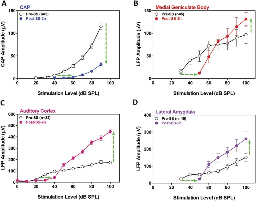
Research article                                                                                                   Neuroscience

                                   acute tinnitus in humans and animals (Myers and Bernstein, 1965; Myers et al., 1965; Mongan et al.,
                                   1973). Moreover, high-dose sodium salicylate (SS), the active ingredient in aspirin, not only consistently
                                   induces tinnitus (Jastreboff et al., 1988; Lobarinas et al., 2004; Stolzberg et al., 2013), but also
                                   hyperacusis (Chen et al., 2014; Hayes et al., 2014); these perceptual disorders disappear a day or two
                                   post-treatment. The highly predictable time course of SS-induced tinnitus and hyperacusis makes it an
                                   extremely powerful tool for studying the neural correlates of these perceptual disturbances. Therefore,
                                   we took advantage of our unique behavioral techniques for assessing SS-induced tinnitus and
                                   hyperacusis in rats and combined this with focused electrophysiological measurements plus global fMRI
                                   assessment techniques to map out the regions of neural hyperactivity and enhanced FC that
                                   characterize the tinnitus–hyperacusis network. To identify regions of heightened or depressed
                                   spontaneous neural activity, we measured the amplitude of low-frequency fluctuations (ALFF) in
                                   resting-state fMRI (Zang et al., 2007; Zhang et al., 2010; Yao et al., 2012; Wen et al., 2013) and
                                   combined this with resting-state FC to identify regions of increased or decreased functional coupling
                                   between regions of the auditory pathway and other parts of the CNS. This is the first animal study to use
                                   ALFF and FC combined with detailed electrophysiological measures to provide a comprehensive
                                   neurological map of the tinnitus–hyperacusis network.

                                   Results
                                   Three complimentary experiments involving behavioral, electrophysiological, and functional imaging
                                   were conducted in separate groups of rats. In Experiment 1, three behavioral studies were performed
                                   on separate groups of rats to assess SS-induced tinnitus, hyperacusis, and startle reflex hyperactivity.
                                   In Experiment 2, electrophysiological measurements were carried out on a separate group of rats to
                                   determine how SS altered the neural input/output functions in cochlea, as reflected in the compound
                                   action potential (CAP) from the auditory nerve, and the local field potentials (LFP) recorded in the
                                   MGB, ACx, and lateral amygdala (LA). In Experiment 3, resting-state fMRI studies were conducted in
                                   another group of rats to determine how SS altered the ALFF and FC patterns obtained with seeds
                                   placed in ACx, MGB, and IC.

                                   Experiment 1
                                   Tinnitus
                                   To determine if SS-induced tinnitus, we tested three rats using our 2AFC-tinnitus paradigm. All three
                                   rats developed tinnitus-like behavior; data from two representative animals are shown in Figure 1A,B.
                                   During baseline testing (B1–B4; B6–B9), rats correctly identified Quiet (no sound stimulus) trials at
                                   greater than 70% correct and AM and NBN trials >80% correct. The saline-control treatment had no
                                   noticeable effect on performance during Quiet, NBN, or AM trials. However, when the rats were
                                   treated with SS, performance dropped to 50% or less only on Quiet trials, that is, rats shifted their
                                   response preference from the feeder associated with Quiet to the feeder associated with a continuous
                                   NBN, behavior indicative of tinnitus. When SS treatment was discontinued (P1–P4), performance on
                                   Quiet trials returned to baseline indicating that tinnitus had disappeared. Performance on NBN and
                                   AM trials was unaffected indicating that behavior was under sound stimulus control.

                                   Hyperacusis
                                   To test for SS-induced hyperacusis, we measured reaction time-intensity functions to broadband
                                   noise bursts before and after Saline or SS-treatment (Figure 1C). Reaction time–time intensity
                                   functions obtained with Saline were nearly identical to those obtained during baseline indicating
                                   that the injection had no effect on behavior. In contrast, reaction times obtained after SS were
                                   significantly different from baseline at low and high intensities [Two-way, repeated measures
                                   ANOVA; significant effect of treatment (p < 0.0001), intensity(p < 0.0001), interaction of treatment
                                   × sound intensity (p < 0.001); Bonferroni post-hoc analysis between baseline and SS significant at 30
                                   dB (p < 0.001), 50 dB (p < 0.05), 60 dB (p < 0.01), 70 and 80 dB (p < 0.001), and 90 dB (p < 0.05)].
                                   Reaction times after SS were significantly shorter than baseline at moderate to high intensities
                                   behavioral evidence indicative of hyperacusis, that is, these intensities were perceived as louder
                                   than normal (Lauer and Dooling, 2007; Chen et al., 2014; Hayes et al., 2014). However, at 30 dB
                                   SPL (sound pressure level) reaction times were longer than normal due to hearing loss, which
                                   reduces the loudness of sounds near threshold. Reaction time–intensity functions returned to
                                   normal after SS treatment was discontinued (data not shown).

Chen et al. eLife 2015;4:e06576. DOI: 10.7554/eLife.06576                                                                             3 of 19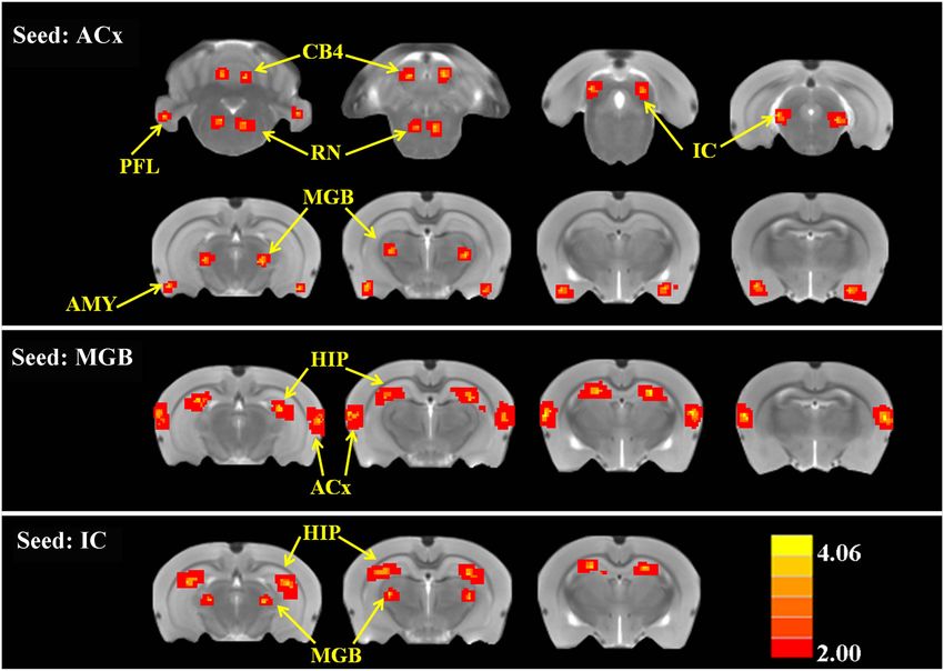
Research article                                                                                                        Neuroscience

Figure 1. SS-induced tinnitus, hyperacusis, and startle reflex hyperactivity. (A and B) 2AFC-tinnitus task for two representative rats during
baseline days B1–B4, during Saline (SAL) treatment, during baseline days B6–B9, 2 hr post-sodium salicylate (SS), and days P1–P4 post-SS
treatment. Percent correct performance shown for NBN, AM, and Quiet trials. Purple-shaded region is the 99% confidence interval for baseline
measurements (B1–B4; B6–B9). (C) Mean (+SEM, n = 7) reaction time-intensity functions measured at baseline, after Saline-treatment and after
SS-treatment. Reaction times during SS treatment were significantly longer than baseline at 30 dB SPL and significantly shorter than baseline at
50–90 dB SPL (*p < 0.05; **p < 0.01; and ***p < 0.001). (D) Mean (+SEM, n = 6) startle amplitude-intensify functions after treatment with Saline or
SS. Startle amplitudes after SS treatment were significantly larger than after Saline at 95 and 105 dB SPL (p < 0.001).
DOI: 10.7554/eLife.06576.003

                                   Startle reflex hyperactivity
                                   Startle reflex hyperactivity has been linked to hyperacusis (Sun et al., 2009; Lu et al., 2011).
                                   Therefore, acoustic reflex amplitude-intensity functions were compared in the same group of rats after
                                   Saline and SS treatments (Figure 1D). SS treatment caused a significant increase in startle amplitude
                                   at 95 and 105 dB SPL [Two-way, repeated measure, ANOVA, significant main effect of intensity
                                   (p < 0.001), treatment (p < 0.001), intensity × treatment interaction (p < 0.001); Bonferroni post-test
                                   significant at 95 dB and 105 dB (p < 0.001)]. Startle amplitudes returned to normal when SS was
                                   discontinued (data not shown).

                                   Experiment 2
                                   Electrophysiology
                                   SS is known to cause temporary hearing loss and reduce the neural output of the cochlea. To
                                   quantify the effects, CAP amplitude-intensity functions were measured before and 2 hr post-SS. The
                                   mean (+SEM) CAP amplitude-intensity function (average of 6, 8, 12, 16, 20, 24, 30, and 40 kHz)
                                   measured 2 hr after SS treatment was shifted to the right at low intensities due to a threshold shift of
                                   approximately 20 dB (horizontal arrow, Figure 2A). In addition, the amplitude of the CAP was
                                   greatly reduced (70–80%) at suprathreshold intensities (down arrow, Figure 2A) indicating
                                   a profound reduction in the neural output of the cochlea. LFP amplitude-intensity functions were
                                   also recorded from the MGB, ACx, and LA before and 2 hr after SS treatment. The LFP amplitude-
                                   intensity functions from all three structures were shifted to the right approximately 20 dB

Chen et al. eLife 2015;4:e06576. DOI: 10.7554/eLife.06576                                                                                  4 of 19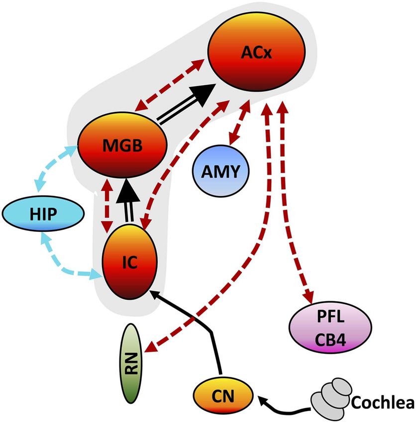
Research article Neuroscience
Figure 2. SS depresses cochlear potentials but enhances central auditory evoked responses. Effects of 300 mg/kg SS on peripheral and central
electrophysiological measures. (A) Mean (+SEM, n = 5) compound action potential (CAP) input/output function (average of 6, 8, 12, 16, 20, 24, 30, and 40
kHz; 10-ms tone burst) recorded from the round window pre- and 2 hr post-SS. Note, 20 dB threshold shift of the function to the right at low intensities
(horizontal arrow) and large reduction in CAP amplitude at high intensities (down arrow). (B, C, D) Local field potential input/output functions (50-ms noise
bursts) from medial geniculate body, auditory cortex, and lateral amygdala (AMY), respectively, before and 2 hr post-SS. Note, 20 dB threshold shift of the
functions to the right at low intensities (horizontal arrows) and large increase in response amplitude (up arrow) at suprathreshold levels (>60 dB SPL).
DOI: 10.7554/eLife.06576.004
(Figure 2B–D) 2 hr post-SS consistent with the CAP. These results indicate that the threshold shift
measured in central structures is largely determined by the loss of sensitivity at the cochlea. LFP
amplitudes in the MGB, ACx, and LA increased rapidly with increasing intensity, and response
amplitudes became substantially larger than pre-treatment values (Figure 2B–D) in contrast to the
large CAP amplitude reductions (∼70% decrease) (Figure 2A). The SS-induced enhancement of
suprathreshold LFP amplitudes at 100 dB SPL was approximately 50% in the MGB and 140% in the
ACx, results indicative of a progressive increase in gain from peripheral to more central auditory loci
(Noreña, 2010; Lu et al., 2011).
Experiment 3
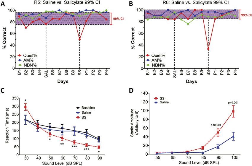
ALFF
To identify the global effects of SS on brain activity, we compared the ALFF in the SS group with the
Saline group 2 hr post-treatment using two-sample t-tests corrected for multiple comparisons.
Figure 3 shows the regions where significant increases or decreases in ALFF were observed due to SS;
Table 1 shows the cluster sizes and t-values in left and right hemispheres for each region. Within the
cerebellum, SS produced significant bilateral increases in ALFF in the parafloccular lobes (PFL, 38–37
voxels) and cerebellar lobules 4 (CB4, 38–37 voxels) (Figure 3A,B). Significant bilateral increases in
Chen et al. eLife 2015;4:e06576. DOI: 10.7554/eLife.06576 5 of 19Research article Neuroscience
Figure 3. SS enhances and depresses amplitude of low-frequency fluctuations (ALFF) in specific CNS regions. Panels
A (most caudal) through K (most rostral) show MR images of rat brain. Significant differences in ALFF between the SS
group vs Saline group 2 hr post-treatment. Thresholds set at a corrected p value ofResearch article Neuroscience
Table 1. SS-induced changes in amplitude of low-frequency fluctuations (ALFF); SS group vs Saline
group; p < 0.001 corrected for multiple comparisons
Left Right
Brain region Cluster size t-value Cluster size t-value
ALFF increased
ACx 167 3.812 178 3.746
IC 72 3.383 68 3.473
MGB 52 3.432 48 3.339
SSCx 37 3.383 40 3.402
VCx 32 3.342 39 3.312
SC 40 4.123 43 4.094
AMY 61 4.192 60 3.923
RN/PnO 35 3.249 28 3.290
PnO/PMr 12 3.498 10 3.313
PFL 38 3.349 37 3.292
CB4 38 3.349 37 3.292
ALFF decreased
HIP 108 −4.087 92 −4.002
CPu 72 −3.772 71 −3.741
Abbreviations: auditory cortex (ACx), inferior colliculus (IC), medial geniculate body (MGB), somatosensory cortex
(SSCx), visual cortex (VCx), superior colliculi (SC), amygdala (AMY), gigantocellular reticular nucleus (RN), pontine
reticular nucleus oral (PnO), paramedian raphe nucleus (PMr), parafloccular lobe of cerebellum (PFL), cerebellum
lobule 4 (CB4), hippocampus (HIP), caudate-putamen (CPu), sodium salicylate (SS).
DOI: 10.7554/eLife.06576.006
Discussion
Brain gain
SS induced a peripheral threshold shift of approximately 20 dB for the CAP (Figure 2A) (Chen et al.,
2013, 2014). The same amount of threshold shift occurred at higher levels of the auditory pathway
indicating that the SS-induced hearing loss originates in the cochlea and is relayed centrally. SS also
reduced the CAP neural output by ∼70% at suprathreshold intensities. Paradoxically, suprathreshold
LFP amplitudes in the MGB, ACx, and LA were larger than normal despite the massive reduction in the
output of the cochlea (Figure 2B–D). These provocative findings provide compelling evidence for an
increase in central gain, a form of homeostatic plasticity implicated in tinnitus and hyperacusis (Salvi
et al., 1990; Auerbach et al., 2014). The enhanced LFPs seen in ACx are consistent with the
enhanced fMRI response observed in the ACx of tinnitus patients, whereas the enhanced LFPs seen in
both ACx and MGB are consistent with the enhanced fMRI responses observed these regions in
hyperacusis patients (Gu et al., 2010). These results are consistent with previous models and data
linking tinnitus and loudness intolerance to increased central gain in the central auditory pathway in
particular regions from the IC to ACx (Salvi et al., 1990; Qiu et al., 2000; Auerbach et al., 2014). In
some models, enhanced central gain amplifies central neural noise resulting in tinnitus (Noreña,
2010). However, in other models, central neural noise increases independent of central gain (Zeng,
2013); this could potentially explain why some patients only experience tinnitus, but not hyperacusis
(Baguley, 2003). However, this distinction is clouded by the fact that many tinnitus patients are
unaware of their mild hyperacusis, that is, hyperacusis may be more prevalent in tinnitus patients than
currently believed because many patients are unaware of their hyperacusis (Gu et al., 2010).
Many cellular mechanisms could enhance central gain, but one likely candidate is reduced
inhibition (disinhibition). Considerable evidence exists for dysregulated inhibition in central gain-
control models (Auerbach et al., 2014). First, SS can suppress GABA-mediated inhibition and
enhance excitability (Xu et al., 2005; Gong et al., 2008). Second, SS enhances sound evoked activity
Chen et al. eLife 2015;4:e06576. DOI: 10.7554/eLife.06576 7 of 19Research article Neuroscience
Figure 4. SS alters functional connectivity (FC) in specific brain resions. ROI FC heat maps showing the regions of
the brain where SS induced a statistically significant increase in FC with the ROI placed in the ACx (top row), MGB (middle
row), or inferior colliculus (IC) (bottom row). Scale bar shown in lower right; corrected t-values ranged from +4.06 to −2.00.
CB4, lobules 4 of cerebellum; PFL, parafloccular lobe of cerebellum; RN, gigantocellular reticular nucleus; IC, inferior
colliculius; MGB, medial geniculate body; ACx, auditory cortex; HIP, hippocampus; AMY, amygdala.
DOI: 10.7554/eLife.06576.007
The following figure supplement is available for figure 4:
Figure supplement 1. The ALFF and FC data (seeds: ACx, MGB, and IC) for baseline and after SS application
separately.
DOI: 10.7554/eLife.06576.008
in ACx when given systemically or applied locally to the LA or ACx, whereas it depresses ACx
responses when only applied to the cochlea (Sun et al., 2009; Chen et al., 2012). Third, drugs that
enhance GABA-mediated inhibition, prevent SS-induced gain enhancement (Sun et al., 2009;
Lu et al., 2011), and suppress tinnitus (Brozoski et al., 2007b).
Behaviorally, hyperacusis was initially observed at 50 dB SPL; the same low intensity at which
sound-evoked neural hyperactivity occurred in the ACx. In contrast, sound-evoked hyperactivity
occurred at noticeably higher intensities for the LA (∼60 dB SPL), MGB (∼70 dB SPL), and acoustic-
startle reflex amplitude (∼95 dB SPL). These results suggest that neural responses from the ACx may
be one of the most sensitive biomarkers of hyperacusis (Juckel et al., 2004; Gu et al., 2010).
However, since neural responses increased in magnitude from cochlea to cortex, loudness intolerance
issues likely result from multiple stages of neural amplification as signals are relayed rostrally from the
cochlea to the ACx (Auerbach et al., 2014). Indeed, there is growing evidence that the neural
amplification gradually develops in the auditory brainstem and serially accumulates to supernormal
levels after reaching the MGB and ACx consistent with previous electrophysiological results
(Qiu et al., 2000; Schaette and McAlpine, 2011).
Tinnitus
Some models of tinnitus are based on changes in spontaneous spiking patterns such as increased
firing rate or increased neural synchrony (Eggermont, 2015). SS either decreased or had no effect on
Chen et al. eLife 2015;4:e06576. DOI: 10.7554/eLife.06576 8 of 19Research article Neuroscience
Table 2. SS-induced increases in functional connectivity (FC); p < 0.001 corrected for multiple
comparisons
Left Right
Seed region Brain region Cluster size t-value Cluster size t-value
ACx MGB 62 3.900 70 3.859
IC 82 3.912 88 3.632
AMY 67 3.897 68 3.839
RN 52 3.983 48 3.902
PFL 39 3.992 35 3.954
CB4 51 4.066 53 4.070
MGB ACx 195 4.074 210 4.084
HIP 162 4.032 178 4.109
IC MGB 72 4.098 60 4.013
HIP 140 4.064 131 3.904
Abbreviations: medial geniculate body (MGB), inferior colliculus (IC), amygdala (AMY), reticular nucleus (RN),
parafloccular lobe of cerebellum (PFL), cerebellum lobule 4 (CB4), auditory cortex (ACx), hippocampus (HIP), sodium
salicylate (SS).
DOI: 10.7554/eLife.06576.009
spontaneous spike rate in primary ACx (Ochi and Eggermont, 1996; Yang et al., 2007) and
reportedly no effect on synchrony between neuron pairs (Eggermont, 2015). Since the BOLD and LFP
responses mainly represent presynaptic activity, it is difficult to directly relate our results to these
spiking models. However, the increase in very low-frequency BOLD oscillations (0.01 Hz) represented
by ALFF could be interpreted as evidence for increased presynaptic synchrony, which would likely
enhance spike synchrony albeit at much longer time intervals than previously studied or over much
larger neuronal populations than that reflected by spike correlations between neuron pairs. SS has
also been found to increase gamma-band (50–100 Hz) oscillatory activity in ACx (Stolzberg et al.,
2013); oscillations substantially higher than in ALFF. An alternative view is that the tinnitus percept is
derived from coordinated activity among several auditory and nonauditory regions (Horwitz and
Braun, 2004; Husain et al., 2006). Enhanced FC between the HIP and auditory areas provides
a substrate for assigning a spatial location to a phantom sound, while coordinated activity between
specific auditory areas and the reticular formation and AMY may draw attention to and add emotional
significance to neural activity in the auditory pathway. Thus, functionally coordinated activity within
the network may be essential for bringing tinnitus into consciousness.
Reallocating network resources
Tinnitus and hyperacusis, like phantom limb pain and cutaneous allodynia, are triggered by peripheral
damage presumably leading to widespread changes in the CNS that involve altered connections in
networks that include portions of the central auditory pathway and other regions linked to emotion,
memory, attention, and arousal (Llinas et al., 1999; Leaver et al., 2012; Husain and Schmidt, 2014).
In the resting state, SS increased ALFF and FC in a broad-neural network that included core auditory
structures extending from the IC to the ACx consistent with previous studies implicating these central
auditory structures in tinnitus (Paul et al., 2009). SS also enhanced sound-evoked LFP in the MGB and
ACx suggesting a key role for these auditory structures in amplifying auditory information that could
manifest as loudness intolerance (Gu et al., 2010).
Cerebellar gating and gain
Although the cerebellum is mainly involved in motor planning and control, some cerebellar regions
such as the PFL and vermis receive inputs from auditory centers (Petacchi et al., 2005) and respond to
sound (Lockwood et al., 1999). Interestingly, the perception of tinnitus has been linked to activation
of the PFL and vermis (Brozoski et al., 2007a) consistent with our results. Since ablation or
Chen et al. eLife 2015;4:e06576. DOI: 10.7554/eLife.06576 9 of 19Research article Neuroscience
inactivation of the PFL eliminates the perception of noise-induced tinnitus (Bauer et al., 2013), some
have suggested that the PFL acts as a gain control mechanism comparing the afferent input from the
cochlea with descending signals from the ACx (Bauer et al., 2013). Consistent with this view, our
results show that SS leads to hyperactivity in the ACx and increases the FC between the ACx and PFL
and CB4. If this cerebellar-tinnitus gating hypothesis is correct, then ablating or inactivating the PFL
should suppress behavioral measures of SS-induced tinnitus and possibly hyperacusis, providing
a clear test of this model. The functional role of the PFL in tinnitus–hyperacusis network could be
further elucidated by inactivating the PFL and determining the effects this has on SS-induced changes
we observed in our electrophysiological and fMRI measures.
Negative valence
The AMY, which assigns emotions such as fear or anxiety to sensory events, lies outside the classical
auditory pathway; however, it is linked to several auditory areas and responds robustly to sound
(Romanski and LeDoux, 1993; Stutzmann et al., 1998; Chen et al., 2014). In the resting state, SS
enhanced the ALFF in the AMY and increased FC between ACx and AMY consistent with prior results
showing increased coupling between ACx and AMY in tinnitus patients (Kim et al., 2012) and
increased c-fos immunolabeling in the AMY following SS treatment (Wallhäusser-Franke et al.,
2003). SS also enhanced sound-evoked activity in the AMY consistent with the increased activation
seen in the AMY of hyperacusis patients (Levitin et al., 2003). Importantly, infusion of SS directly into
AMY increases sound-evoked activity in the ACx, effects that illustrate the potent independent role
that the AMY can exert on central auditory function and aural perception (Chen et al., 2012).
Collectively, these results reinforce the view that the AMY contributes to the fear and anxiety
experienced by many patients with tinnitus and hyperacusis (van Veen et al., 1998; Juris et al., 2013;
Aazh et al., 2014). Sound and cognitive therapies aimed at reducing the emotional distress of tinnitus
and hyperacusis would be expected to reduce the level of activity in the AMY and/or the functional
coupling between the AMY and ACx without necessarily eliminating aberrant auditory percepts
(Hazell and Jastreboff, 1990). Human imaging studies employing ALFF and FC could be used to test
this hypothesis and provide an objective and independent assessment of how these therapies work
and their effects on the tinnitus–hyperacusis neural network.
Arousal
A novel finding observed during resting-state fMRI was the SS-induced enhancement of ALFF in the
reticular formation (RN, PnO, and PMr) together with increased FC between the reticular formation
and ACx. The reticular formation is an important arousal center with numerous inputs from the
cochlear nucleus and IC (Kandler and Herbert, 1991). Giant neurons in pontine reticular formation
control the amplitude of the acoustic startle reflex (Koch et al., 1992), and stimulation of the AMY
enhances the response of these giant neurons (Koch et al., 1992). Thus, the SS-induced increases of
ALFF observed in the AMY and reticular formation likely contribute to the enhancement of the
acoustic startle reflex.
Feeling and seeing
SS unexpectedly increased ALFF in SSCx, VCx, and SC raising the question of whether this might be
linked to phantom visual or somatosensory perceptions. However, after an extensive search, we were
unable to find evidence of such aberrant somatosensory or visual phenomena. While the heightened
ALFF response in these areas is novel, such changes seem reasonable, given the multisensory
interactions known to exist between auditory, somatosensory, and visual areas. One possibility is that
heightened ALFF activity in ACx, MGB, and IC could spill over and enhance activity in visual and
somatosensory areas (Murray et al., 2005). However, these increases may not lead to altered
perception because FC was not enhanced in visual or somatosensory areas. Alternatively, the SS-
induced cochlear loss could unmask pre-existing multisensory circuits in visual and somatosensory
areas leading to increased activation (Barone et al., 2013).
Tinnitus–hyperacusis network
Although SS has long been known to cause tinnitus (Mongan et al., 1973; Jastreboff et al., 1988), it is
now clear that it also induces strong hyperacusis-like behavior (Chen et al., 2014; Hayes et al., 2014).
Chen et al. eLife 2015;4:e06576. DOI: 10.7554/eLife.06576 10 of 19Research article Neuroscience
Although tinnitus and hyperacusis could conceivably arise from different mechanisms (Zeng, 2013), they
frequently co-occur more frequently than previously believed (Gu et al., 2010). Tinnitus and hyperacusis
do not exist in isolation but are linked to other brain regions associated with emotions, arousal,
memories, spatial location, and motor activity as schematized in the tinnitus–hyperacusis network model
defined by our imaging results (Figure 5). With the seed region in IC, a significant increase in FC
occurred in the MGB; this increase is likely due to the SS-induced enhancement of ALFF in the IC, which
is relayed rostrally to the MGB (Figure 5, thick black line) resulting in a larger and more coherent MGB
response. Similarly, with the seed in the MGB, increased FC occurred in the ACx; this increase is likely
due to the increased ALFF and FC occurring in MGB, which is relayed rostrally to the ACx (Figure 5,
thick black line). With the seed in the ACx, increased FC was seen in the same two lower auditory
centers, the MGB and IC, raising the possibility of a recurrent feedback loop in this auditory subnetwork
(Figure 5, shaded area, bidirectional dashed red lines). These data combined with our electrophys-
iological results suggest that SS enhances the FC and response magnitude in a central auditory
subnetwork that extends from the IC through the MGB to the ACx.
The FC data suggest that the ACx is a major hub in the tinnitus–hyperacusis network with side
branches that extend caudally to the AMY, RN, and cerebellum (PFL, CB4); these subdivisions all show
large SS-induced increases in ALFF, as well as increased FC with the ACx. The side branch connecting
the ACx to the AMY provides a pathway through which emotional significance can be attached to
tinnitus or hyperacusis (Chen et al., 2014)
consistent with earlier studies linking anxiety,
annoyance, and fear to tinnitus and hyperacusis
(Moller, 2007). The ACx-RN network provides
a conduit by which increased arousal can increase
awareness or attention, enhance motivation, or
amplify motor responses to tinnitus or supra-
threshold sounds (Carlson and Willott, 1998;
Paus, 2000). The ACx-cerebellar branch could
serve as a gating path for tinnitus (Boyen et al.,
2014) or modulate the motor responses to or
perceptual salience of suprathreshold sounds
thereby contributing to hyperacusis (Mobbs
et al., 2007). Increased FC between HIP-MGB
and HIP-IC could facilitate the formation or
stabilization of a memory trace for tinnitus, assign
a spatial location to the phantom sound within or
outside the head, or promote the retrieval of
previously stored auditory memories (De Ridder
Figure 5. Tinnitus–hyperacusis network model. Sche- et al., 2006; Ulanovsky and Moss, 2008). Our
matic showing some of the major centers in the auditory
network model can be explicitly tested by
pathway and areas in the CNS showing increased FC
administering a high-dose of SS while activating
with the auditory cortex (ACx). Black lines show the
neural transmission path from the cochlea through the
or inactivating part of the network, such as the
cochlear nucleus (CN), inferior colliculus (IC), medial PFL, and determining if the manipulation abol-
geniculate body (MGB), and ACx; black double-line ishes tinnitus or hyperacusis or alternatively de-
reflects SS-induced increases in ALFF and/or increased termining how such manipulations affects the
FC. SS increased ALFF response magnitudes and FC in electrophysiological and fMRI properties of the
a central auditory subnetwork (gray shaded area) network. Since high-dose SS is one of the most
comprised of the IC, MGB, and AC. The AC serves as predictable and reliable inducers of tinnitus and
a major hub with side branches to the amygdala (AMY), hyperacusis, SS provides researchers with a pow-
reticular nuclei (RN), and parafloccular lobe (PFL) and erful tool to gain mechanistic insights into the
cerebellar lobules 4 (CB4); these side branch regions
neurophysiological conditions needed to induce
contribute to the emotional, motoric, and conscious
these debilitating auditory perceptions. Since
awareness of tinnitus and/or hyperacusis. Enhanced
activity in the MGB and IC combined with the increased SS-induced tinnitus and hyperacusis are transient
FC of these auditory structures with the hippocampus phenomena that begin shortly after drug treat-
(HIP) could facilitate tinnitus memory storage or retrieval ment, some of the neuro-pathophysiological
or assign spatial location to phantom or real sounds. changes we observed are likely to be similar to
DOI: 10.7554/eLife.06576.010 those that occur during the early stages of
Chen et al. eLife 2015;4:e06576. DOI: 10.7554/eLife.06576 11 of 19Research article Neuroscience
tinnitus or hyperacusis induced by acoustic trauma, Meniere’s disease, or sudden hearing loss. In cases
of acoustic overstimulation, most individuals develop transient tinnitus immediately post-exposure,
which gradually disappears; interestingly only a small percentage develops permanent tinnitus
(Gilles et al., 2012; Degeest et al., 2014). Thus, an important question that remains to be answered is
whether the neural correlates of chronic tinnitus and hyperacusis are similar or different from the
immediate and acute condition.
Summary
SS-induced tinnitus and hyperacusis appear to arise from enhanced central gain and increased FC in
an auditory network with four side branches. The core of the network is composed of auditory
structures extending from the IC, through the MGB to the ACx plus branches to the AMY, RN,
cerebellum, and HIP. These side branches presumably contribute to the emotional significance,
arousal, motor response, gating, and memories associated with tinnitus and hyperacusis.
Materials and methods
Behavioral Experiment 1 and electrophysiological Experiment 2 were approved by the Institutional
Animal Care and Use Committee at the University at Buffalo, Buffalo, NY, USA in accordance with NIH
guidelines. The fMRI studies in Experiment 3 were conducted in accordance with the National
Institutes of Health Guide for the Care and Use of Laboratory Animals and approved by the Animal
Care Committee of Southeast University, Nanjing, China.
Experiment 1
SS-induced tinnitus, hyperacusis, and startle reflex hyperactivity
Subjects
Sprague Dawley rats were used to obtain behavioral measures of tinnitus, hyperacusis, and acoustic
startle reflex amplitude-intensity functions.
Acoustic startle reflex
Our procedures for measuring acoustic startle reflex amplitude-intensity functions have been
described previously (Chen et al., 2014). Six rats were acclimated for 2 days to testing chambers.
Acoustic reflex amplitude-intensity functions (50-ms broadband noise bursts, 55–105 dB, 10 dB steps,
18–22 s inter-stimulus interval, 10 presentations/intensity, random order) were measured 2 hr after
treatment with Saline (1 ml, i.p.) or SS (300 mg/kg, 1 ml, i.p.). Saline control and SS measurements
were separated by 3 days or more, and the order of treatments was randomized across subjects.
Hyperacusis assessment
Our procedures for assessing hyperacusis using reaction time-intensity functions are described in
detail in recent publications (Chen et al., 2014; Hayes et al., 2014). Seven food-restricted rats were
trained to detect a broadband-noise burst (300 ms, 5 ms rise/fall) presented at intensities from 30 to
90 dB SPL. The rat initiated a trial by holding its nose in a nose-poke hole for 1–4 s until a noise-burst
stimulus was presented. If the rat withdrew its nose from the hole during a 2-s response interval that
began at noise-burst onset, it received a food pellet, and the trial was scored as a ‘hit’. Failure to
respond during the 2-s response interval was scored as a ‘miss’ and not rewarded. Approximately,
30% of trials were catch trials (no stimulus). If the rat removed its nose on a catch trial (‘false alarm’),
the light in the test chamber was turned off and the rat received a 4-s timeout. However, if it
continued to nose-poke through the response interval during a catch trial the trial was scored as
a ‘correct rejection’, and no reinforcement was given. Reaction time measures were only recorded on
trials scored as a ‘hit’. After obtaining stable baseline reaction time measurements, rats were treated
either with SS (300 mg/kg, i.p.; Sigma–Aldrich, St. Louis, MO, USA) or an equivalent volume of Saline
and tested on the go/no-go paradigm for approximately 60 min beginning 2 hr post-treatment.
Tinnitus assessment
Details of our two-alternative forced choice (2AFC) behavioral paradigm to test for tinnitus are
described in previous publications (Stolzberg et al., 2013; Chen et al., 2014). Three food-restricted
Chen et al. eLife 2015;4:e06576. DOI: 10.7554/eLife.06576 12 of 19Research article Neuroscience
rats (85–90% free feeding weight) were trained to nose-poke a center-hole to start a trial and then
wait 4–8 s for a cue light to come on before responding in a bidirectional manner to one of three
randomly presented ongoing stimuli. If an unmodulated narrow-band noise (NBN, 50% of trials) was
present (4, 5, 6, 7, or 11 kHz center frequency, ∼70 dB SPL), the rat was trained to nose-poke the left
feeder to obtain a food pellet. In contrast, if an amplitude-modulated (AM, 30% of trials) narrow-band
noise (100% modulation depth, 4, 5, 6, 7, or 11 kHz center frequency, ∼70 dB SPL) or if no sound
(Quiet, 20% of trials) was present, the rat was trained to nose-poke the right feeder to obtain a food
pellet. During baseline testing, performance was typically greater than 80% correct. After reaching
criterion, rats were treated either with SS or an equivalent volume of Saline and tested daily on the
2AFC paradigm for approximately 75 min beginning 2 hr post-treatment. Since the rats were trained
to respond left to a steady NBN vs right to fluctuating AM noise or Quiet, we expected that if a rat
developed SS-induced tinnitus, it would only shift its response on Quiet trials from the right side
(associated with Quiet) to the left side (associated with a steady NBN) as previously discussed
(Stolzberg et al., 2013). Behavioral evidence of tinnitus was defined as a significant shift in behavior
only on Quiet trials (percent correct below the 99% confidence interval established during baseline
testing).
Experiment 2
SS-induced changes in auditory electrophysiology
Subjects
Sprague Dawley rats were used to obtain electrophysiological measures from the cochlea, MGB, ACx,
and LA before and after SS treatment (300 mg/kg, i.p.).
CAP
Rats (n = 5) were anesthetized with ketamine/xylazine (50/6 mg/kg, i.m.), and the CAP recorded
before and 2 hr after SS treatment using procedures described in our earlier publications (Chen et al.,
2010; Lu et al., 2011). Tone bursts (6, 8, 12, 16, 20, 24, 30, and 40 kHz, 10-ms duration, 1 ms rise/fall
time, cosine gated) were presented at levels ranging from approximately 0 to 90 dB SPL. The
response was amplified (1000×), filtered (0.1 Hz–5 kHz), and averaged (50 repetitions). The amplitude
of the CAP N1 response was measured and used to construct mean CAP amplitude-intensity
functions.
ACx, MGB, LA
Rats were anesthetized with ketamine/xylazine (50/6 mg/kg, i.m.). LFPs were recorded from the MGB (n =
5), ACx (n = 32), and LA (n = 19) using procedures described in our earlier publications (Stolzberg et al.,
2011; Chen et al., 2013). LFP were recorded before and 2 hr after SS treatment (300 mg/kg, i.p.) using 16-
channel silicon electrodes (A-1 × 16–10 mm 100–177, NeuroNexus Technologies). The LFP were filtered
(2–300 Hz), sampled at 608 Hz, and averaged over 100 stimulus presentations (50 ms noise-burst, 1 ms
rise/fall time, cosine gated). For each intensity, the root mean square amplitude of the LFP was computed
over a 50 ms time window for the MGB and ACx and a 100 ms time window for the LA; the data were used
to construct LFP amplitude-intensity functions before and 2 hr after SS treatment.
Experiment 3
ALFF and FC
Subjects
Sprague Dawley rats weighing between 180–230 g were used as subjects. Animals were divided into
two groups, a Saline-control group (n = 15) and a SS-treated group (n = 15). Rats were anesthetized
with urethane (1 mg/kg body weight, i.p.) in order to maintain a stable long-term plane of anesthesia
during data acquisition.
Salicylate
Prior to positioning the rat in the scanner, a catheter (25-gage needle) was inserted into the
intraperitoneal space. The catheter was attached to a syringe (5 ml), which contained either normal
Chen et al. eLife 2015;4:e06576. DOI: 10.7554/eLife.06576 13 of 19Research article Neuroscience
Saline or SS dissolved in normal Saline. After collecting all the baseline MRI data, the rats were treated
either with normal Saline (3 ml) or 300 mg/kg of SS dissolved in normal Saline (3 ml) (Lobarinas et al.,
2004; Stolzberg et al., 2013; Chen et al., 2014).
MRI acquisition and analysis
Each rat was positioned in the scanner in a prone position. Rectal temperature was maintained at 37.5˚C
with a temperature-controlled water blanket beneath the rat. The respiratory rate of the rat was monitored
continuously during the entire experiment using an MRI-compatible pulse oximeter. Head position was
stabilized with a bite bar and two rods located on opposite sides of the temporal surface of the head.
MRI data were acquired with a 7.0 T animal MRI scanner (PharmaScan, Bruker Biospin GmbH,
Germany) using a quadrature surface RF coil. Anatomical images were acquired with a turbo-rapid
acquisition relaxation enhancement (RARE) T2-weighted sequence (repetition time (TR)/echo
time (TE) = 3200/36 ms, slices = 27, field of view (FOV) = 2.5 × 2.5 cm, number of averages = 1,
matrix = 384 × 384, slice thickness/gap = 1/0 mm, flip angle = 90˚). The 27 contiguous anatomical
images extended anteriorly from the cerebral-olfactory bulb to the caudal region of the cerebellum
posteriorly. The BOLD measurements were acquired with a single-shot gradient-echo echo-planar-
imaging (GE-EPI) sequence to acquire multiple slices of images. The parameters were:
TR/TE = 2000/19 ms, slices = 27, FOV = 2.5 × 2.5 cm, number of averages = 1, matrix = 96 × 96, slice
thickness/gap = 1/0 mm, flip angle = 90˚, 100 volumes. Baseline and salicylate/Saline data acquisition
occurred over a period of approximately 2.5 hr. Anatomical and functional scans were obtained from each
rat before and 2 hr after administering salicylate or Saline.
Data processing and statistical analysis
The first 10 time points were eliminated to allow for scanner calibration and adaptation of the subject
to the environment. Processing of the fMRI data was carried out with statistical Parametric Mapping
software (SPM8, http://www.fil.ion.ucl.ac.uk/spm/) and Resting State fMRI Data Analysis Toolkit V1.8
software (REST, http://www.restfmri.net). Sequential data processing steps included: slice-timing
adjustment, realignment and correction for head-motion, spatial normalization to the standard rat
brain atlas (Paxinos and Watson, 2004), smoothing with an isotropic Gaussian kernel (FWHM = 1 mm),
detrending and filtering (0.01–0.1 Hz). Data were excluded if head movements exceeded 1.0 mm of
maximum translation in the x, y, or z directions or 2.0 of maximum rotation about the three axes. Images
for the ALFF analysis were band-pass filtered (0.01–0.1 Hz). Afterward, a fast Fourier transform was
performed on the corrected time series data to obtain the power spectrum in each voxel within the
brain; the square root of the power spectrum was calculated to obtain the amplitude at each frequency.
To identify significant differences in ALFF values between the SS experimental group and the Saline
control group, a between-group, two-sample t-test was calculated. Thresholds were set at a corrected p
value of p < 0.001 (cluster size > 10 voxels) using the multiple comparison correction obtained with the
AlphaSim method employing Monte Carlo simulation. Region of interest (ROI)-based FC analysis was
performed for three auditory regions consisting of 9 voxels in the ACx, MGB, and IC. These regions
were chosen based on the increased ALFF values observed in these areas during SS and because of their
putative roles in tinnitus and hyperacusis. The mean time series for each of these three ROIs was
computed for the reference time course. Cross-correlation analysis was then carried out between
the mean signal change in each ROI and the time series of every voxel in the whole rat brain. Finally,
a Fisher z-transform was applied to improve the normality of the correlation coefficients. Both
motion parameters resulting from the realignment and the global signal time course were regressed
out during this analysis to improve the specificity of the FC (Büchel et al., 1996). For each ROI, two-
sample t-tests were performed to identify significant changes in FC between the SS experimental
group and the Saline control group. Thresholds were set at a corrected p value of p < 0.001
(cluster size > 10 voxels) using the multiple comparison correction obtained with the AlphaSim
method employing Monte Carlo simulation.
Acknowledgements
The fMRI studies were supported by a grant from the National Key Basic Research Program (973
Program) (NOs. 2013CB733800, 2013CB733803), National Natural Science Foundation of China
(NOs. 81230034, 81271739), Jiangsu Provincial Special Program of Medical Science (NO. BL2013029),
Key Project of Jiangsu Province Natural Science Foundation of China (NO. BK20130577),
Chen et al. eLife 2015;4:e06576. DOI: 10.7554/eLife.06576 14 of 19Research article Neuroscience
Fundamental Research Funds for the Central Universities, and Jiangsu Graduate Student Innovation
Grant (NO. KYZZ_0076). The electrophysiological and behavioral studies were supported in part by
grants from ONR (N000141210731) and NIH (5R01DC011808). YCC acknowledged the financial
support from the China Scholarship Council for his joint PhD scholarship (NO. 201406090139). RS
acknowledges support from Overseas Master Project Grant, Chinese Educational Ministry, 2012–17.
Additional information
Funding
Funder Grant reference Author
Ministry of Science and 2013CB733800 Gao-Jun Teng
Technology of the People’s
Republic of China
Ministry of Science and 2013CB733803 Gao-Jun Teng
Technology of the People’s
Republic of China
National Natural Science 81230034 Gao-Jun Teng
Foundation of China
National Natural Science 81271739 Gao-Jun Teng
Foundation of China
Natural Science Foundation of BK20130577 Gao-Jun Teng
Jiangsu Province
Fundamental Research Funds for KYZZ_0076 Gao-Jun Teng
the Central Universities and
Jiangsu Graduate Student
Innovation Grant
National Institutes of Health (NIH) 5R01DC011808 Richard Salvi
China Scholarship Council PhD scholarship 201406090139 Yu-Chen Chen
Ministry of Education of the Overseas Master Project Grant Richard Salvi
People’s Republic of China
Natural Science Foundation of BK20130577 Gao-Jun Teng
Jiangsu Province
Office of Naval Research N000141210731 Richard Salvi
The funders had no role in study design, data collection and interpretation, or the
decision to submit the work for publication.
Author contributions
Y-CC, Collected the fMRI data, performed the analysis, and wrote and revised the manuscript; XL,
LL, Assisted with fMRI data collected and analysis; JW, Helped design and execute parts of the MRI
experiment and revise the manuscript; C-QL, MY, YJ, F-CZ, Contributed to the discussion and
manuscript revision; KR, Collected, analyzed and helped write and revise the tinnitus and hyperacusis
behavioral data; G-DC, Collected the electrophysiological data; WS, VPKM, Collected and analyzed
the startle reflex results and write and revise this portion of the manuscript; RS, G-JT, Helped design
the MRI experiment and write and revise the manuscript
Ethics
Animal experimentation: All of the animals were handled according to approved Institutional Animal
Care and Use Committee of the University at Buffalo and the Southeast University (Permit Number:
HER05080Y) in accordance with the National Institutes of Health Guide. All surgery was performed
under ketamine/xylazine anesthesia, and every effort was made to minimize suffering.
References
Aazh H, McFerran D, Salvi R, Prasher D, Jastreboff M, Jastreboff P. 2014. Insights from the first international
conference on hyperacusis: causes, evaluation, diagnosis and treatment. Noise & Health 16:123–126.
doi: 10.4103/1463-1741.132100.
Chen et al. eLife 2015;4:e06576. DOI: 10.7554/eLife.06576 15 of 19Research article Neuroscience
Andersson G, Lindvall N, Hursti T, Carlbring P. 2002. Hypersensitivity to sound (hyperacusis): a prevalence
study conducted via the internet and post. International Journal of Audiology 41:545–554. doi: 10.3109/
14992020209056075.
Auer DP. 2008. Spontaneous low-frequency blood oxygenation level-dependent fluctuations and functional
connectivity analysis of the ‘resting’ brain. Magnetic Resonance Imaging 26:1055–1064. doi: 10.1016/j.mri.2008.
05.008.
Auerbach BD, Rodrigues PV, Salvi RJ. 2014. Central gain control in tinnitus and hyperacusis. Frontiers in Neurology
5:206. doi: 10.3389/fneur.2014.00206.
Baguley DM. 2003. Hyperacusis. Journal of the Royal Society of Medicine 96:582–585. doi: 10.1258/jrsm.96.12.582.
Baguley DM, Moffat DA, Hardy DG. 1992. What is the effect of translabyrinthine acoustic schwannoma removal
upon tinnitus? The Journal of Laryngology and Otology 106:329–331. doi: 10.1017/S0022215100119413.
Barone P, Lacassagne L, Kral A. 2013. Reorganization of the connectivity of cortical field DZ in congenitally deaf
cat. PLOS ONE 8:e60093. doi: 10.1371/journal.pone.0060093.
Bauer CA, Kurt W, Sybert LT, Brozoski TJ. 2013. The cerebellum as a novel tinnitus generator. Hearing Research
295:130–139. doi: 10.1016/j.heares.2012.03.009.
Boyen K, de Kleine E, van Dijk P, Langers DR. 2014. Tinnitus-related dissociation between cortical and subcortical
neural activity in humans with mild to moderate sensorineural hearing loss. Hearing Research 312:48–59. doi: 10.
1016/j.heares.2014.03.001.
Brozoski TJ, Ciobanu L, Bauer CA. 2007a. Central neural activity in rats with tinnitus evaluated with manganese-
enhanced magnetic resonance imaging (MEMRI). Hearing Research 228:168–179. doi: 10.1016/j.heares.2007.02.003.
Brozoski TJ, Spires TJ, Bauer CA. 2007b. Vigabatrin, a GABA transaminase inhibitor, reversibly eliminates tinnitus
in an animal model. Journal of the Association for Research in Otolaryngology 8:105–118. doi: 10.1007/s10162-
006-0067-2.
Büchel C, Wise RJ, Mummery CJ, Poline JB, Friston KJ. 1996. Nonlinear regression in parametric activation studies.
Neuroimage 4:60–66. doi: 10.1006/nimg.1996.0029.
Carlson S, Willott JF. 1998. Caudal pontine reticular formation of C57BL/6J mice: responses to startle stimuli,
inhibition by tones, and plasticity. Journal of Neurophysiology 79:2603–2614.
Cave KM, Cornish EM, Chandler DW. 2007. Blast injury of the ear: clinical update from the global war on terror.
Military Medicine 172:726–730. doi: 10.7205/MILMED.172.7.726.
Chen GD, Kermany MH, D’Elia A, Ralli M, Tanaka C, Bielefeld EC, Ding D, Henderson D, Salvi R. 2010. Too much of
a good thing: long-term treatment with salicylate strengthens outer hair cell function but impairs auditory neural
activity. Hearing Research 265:63–69. doi: 10.1016/j.heares.2010.02.010.
Chen GD, Manohar S, Salvi R. 2012. Amygdala hyperactivity and tonotopic shift after salicylate exposure. Brain
Research 1485:63–76. doi: 10.1016/j.brainres.2012.03.016.
Chen GD, Radziwon KE, Kashanian N, Manohar S, Salvi R. 2014. Salicylate-induced auditory perceptual disorders and
plastic changes in nonclassical auditory centers in rats. Neural Plasticity 2014:658741. doi: 10.1155/2014/658741.
Chen GD, Stolzberg D, Lobarinas E, Sun W, Ding D, Salvi R. 2013. Salicylate-induced cochlear impairments, cortical
hyperactivity and re-tuning, and tinnitus. Hearing Research 295:100–113. doi: 10.1016/j.heares.2012.11.016.
De Ridder D, Fransen H, Francois O, Sunaert S, Kovacs S, Van De Heyning P. 2006. Amygdalohippocampal
involvement in tinnitus and auditory memory. Acta Oto-Laryngologica Supplementum 50–53. doi: 10.1080/
03655230600895580.
Degeest S, Corthals P, Vinck B, Keppler H. 2014. Prevalence and characteristics of tinnitus after leisure noise
exposure in young adults. Noise & Health 16:26–33. doi: 10.4103/1463-1741.127850.
Eggermont JJ. 2015. Tinnitus and neural plasticity (Tonndorf lecture at XIth international tinnitus Seminar, Berlin,
2014). Hearing Research 319:1–11. doi: 10.1016/j.heares.2014.10.002.
Gilles A, De Ridder D, Van Hal G, Wouters K, Kleine Punte A, Van de Heyning P. 2012. Prevalence of leisure noise-
induced tinnitus and the attitude toward noise in university students. Otology & Neurotology 33:899–906.
doi: 10.1097/MAO.0b013e31825d640a.
Gong N, Zhang M, Zhang X-B, Chen L, Sun G-C, Xu T-L. 2008. The aspirin metabolite salicylate enhances neuronal
excitation in rat hippocampal CA1 area through reducing GABAergic inhibition. Neuropharmacology 54:
454–463. doi: 10.1016/j.neuropharm.2007.10.017.
Gu JW, Halpin CF, Nam EC, Levine RA, Melcher JR. 2010. Tinnitus, diminished sound-level tolerance, and elevated
auditory activity in humans with clinically normal hearing sensitivity. Journal of Neurophysiology 104:3361–3370.
doi: 10.1152/jn.00226.2010.
Hayes SH, Radziwon KE, Stolzberg DJ, Salvi RJ. 2014. Behavioral models of tinnitus and hyperacusis in animals.
Frontiers in Neurology 5:179. doi: 10.3389/fneur.2014.00179.
Hazell JW, Jastreboff PJ. 1990. Tinnitus. I: auditory mechanisms: a model for tinnitus and hearing impairment. The
Journal of Otolaryngology 19:1–5.
Hebert S, Fournier P, Noreña A. 2013. The auditory sensitivity is increased in tinnitus ears. The Journal of
Neuroscience 33:2356–2364. doi: 10.1523/JNEUROSCI.3461-12.2013.
Heffner HE. 2011. A two-choice sound localization procedure for detecting lateralized tinnitus in animals. Behavior
Research Methods 43:577–589. doi: 10.3758/s13428-011-0061-4.
Heffner HE, Harrington IA. 2002. Tinnitus in hamsters following exposure to intense sound. Hearing Research 170:
83–95. doi: 10.1016/S0378-5955(02)00343-X.
Henry JA, Roberts LE, Caspary DM, Theodoroff SM, Salvi RJ. 2014. Underlying mechanisms of tinnitus: review and
clinical implications. Journal of the American Academy of Audiology 25:5–22. quiz 126. doi: 10.3766/jaaa.25.1.2.
Chen et al. eLife 2015;4:e06576. DOI: 10.7554/eLife.06576 16 of 19You can also read