Spontaneous Switching among Conformational Ensembles in Intrinsically Disordered Proteins - MDPI
←
→
Page content transcription
If your browser does not render page correctly, please read the page content below
Review

Spontaneous Switching among Conformational
Ensembles in Intrinsically Disordered Proteins
Ucheor B. Choi 1, Hugo Sanabria 2, Tatyana Smirnova 3, Mark E. Bowen 4,*
and Keith R. Weninger 5,*
 1 Department of Molecular and Cellular Physiology, Department of Neurology and Neurological Sciences,
   Department of Structural Biology, Department of Photon Science, Howard Hughes Medical Institute,
   Stanford University, Stanford, CA 94305, USA; ucheor@stanford.edu
 2 Department of Physics and Astronomy, Clemson University, Clemson, SC, 29634, USA;

   hsanabr@clemson.edu
 3 Department of Chemistry, North Carolina State University, Raleigh, NC, 27695, USA; tismirno@ncsu.edu

 4 Department of Physiology and Biophysics, Stony Brook University, Stony Brook, NY, 11794, USA

 5 Department of Physics, North Carolina State University, Raleigh, NC, 27695, USA

 * Correspondence: mark.bowen@stonybrook.edu (M.E.B.); krwening@ncsu.edu (K.R.W.);
   Tel.: +1-919-513-3696 (M.E.B.); +1-631-444-2536 (K.R.W.)

 Received: 17 January 2019; Accepted: 15 March 2019; Published: 22 March 2019

 Abstract: The common conception of intrinsically disordered proteins (IDPs) is that they
 stochastically sample all possible configurations driven by thermal fluctuations. This is certainly
 true for many IDPs, which behave as swollen random coils that can be described using polymer
 models developed for homopolymers. However, the variability in interaction energy between
 different amino acid sequences provides the possibility that some configurations may be strongly
 preferred while others are forbidden. In compact globular IDPs, core hydration and packing density
 can vary between segments of the polypeptide chain leading to complex conformational dynamics.
 Here, we describe a growing number of proteins that appear intrinsically disordered by biochemical
 and bioinformatic characterization but switch between restricted regions of conformational space.
 In some cases, spontaneous switching between conformational ensembles was directly observed,
 but few methods can identify when an IDP is acting as a restricted chain. Such switching between
 disparate corners of conformational space could bias ligand binding and regulate the volume of
 IDPs acting as structural or entropic elements. Thus, mapping the accessible energy landscape and
 capturing dynamics across a wide range of timescales are essential to recognize when an IDP is
 acting as such a switch.

 Keywords: dynamic configuration; free energy landscape; intrinsically disordered protein; IDP

1. Introduction
     In general, proteins do not fold into static structures. Rather, most proteins fluctuate within a
pseudocontinuum of accessible configurations about their lowest energy folded state. Often, these
fluctuations are coupled to the protein’s functional cycle such as catalytic activity or molecular
recognition. However, many proteins lack a predominant low-energy state, and instead sample a
broad and disparate ensemble of configurations. Such intrinsically disordered proteins (IDPs) lack
the stable folded state, which is a central element in the classical structure–function paradigm.
Nonetheless, IDPs, and proteins containing intrinsically disordered regions (IDRs), are now
understood to play essential roles in cell signaling pathways and regulatory networks [1–6].
     Models for how function arises from an ensemble of rapidly interconverting IDP conformers
generally fall into two categories: those directly involved in molecular recognition and those acting

Biomolecules 2019, 9, 114; doi:10.3390/biom9030114                        www.mdpi.com/journal/biomolecules
Biomolecules 2019, 9, 114                                                                      2 of 16

as linker or structural elements. Unlike molecular recognition by folded domains, wherein the
interaction residues are spread across the polypeptide chain, IDP interactions tend to involve short
linear motifs (SLiMs), which are contiguous within the chain. Disordered SLiMs adopt an ensemble
of conformations and these different conformations of the same sequence may be recognized by
distinct binding partners [7]. The timescale of IDP conformational dynamics is an important
determinant of the binding mode for IDPs. Rapid conformational dynamics supports the classic
induced fit model, where the IDP can reconfigure the binding site before the initial encounter
complex dissociates. Many IDPs couple the energy from order transitions to the recognition event,
which provides a powerful mechanism for allostery. In other cases, the disorder persists even in the
bound state [8,9]. If the timescale for IDP dynamics slows, then the binding mode is limited to
conformational selection wherein the interaction is only possible during those windows when the
binding competent configuration is present. In this way, the timescale of IDP conformational
dynamics plays a key role in differentiating modes of molecular recognition by IDPs.
     The dynamic nature of IDPs and IDRs makes them well suited to function as linkers between
functional elements such as binding sites or folded domains. For example, the fly-casting model
revealed how conformational exchange allows IDPs to explore a large volume in the search for
binding partners [10–12]. The entropic clock model showed how the degree of extension of the IDP
linker between a pore and its blocking domain controlled the open time of an ion channel [13]. The
entropic bristle model suggested that IDPs can fill large volumes of three-dimensional (3D) space
during their conformational searching, which can regulate protein interactions [14]. Thus, the
timescale and the range of conformational sampling within the ensemble governs the structural
properties of IDPs acting as linkers or bristles.
     These functional roles for IDPs rely on their dynamic properties to control the energetics of
binding reactions as well as for regulating hydrodynamic volume and spacing. Understanding
protein structure is the key to describing its function. With IDPs this necessitates extending our
understanding beyond minimum energy states to further characterize the ensemble both in terms of
the accessible landscape as well as the timescales of conformational dynamics.
     The rise of polymer science led to a great foundation of Nobel prize-winning theory to describe
the behavior of homopolymers composed of many repeated subunits [15]. Based on different starting
assumptions about the polymer, a continuum ensemble of end-to-end distances can be estimated by
well-established approximations such as a Gaussian chain, a self-avoiding random coil, or a worm-
like chain model [16,17]. While fully appropriate for polymers, in application to IDPs, such models
lack molecular detail and rely on assumptions about polymer behavior that are not strictly applicable
to polypeptides. Nonetheless, such polymer models still retain great predictive power, particularly
for proteins in near ideal solvent conditions. As such, polymer models have shown great utility in
describing the ensemble properties of chemically denatured proteins and coil-like IDPs.
     Due to the intrinsic flexibility of IDPs, models from polymer physics have proven useful to
describe their ensemble properties in many specific cases. A common extrapolation from polymer
models is the assumption that the conformational landscape is generally featureless and that the
entire landscape is accessible. For proteins selected through evolution to fold, neither of these
assumptions is true. The extent to which these assumptions hold true for IDPs depends on their class.
Simple swollen coils often follow polymer scaling, but now it is recognized that many IDPs, even
polar polypeptides like Huntingtin [18], undergo collapse to form more compact, disordered globules
[19,20]. At these higher packing densities, self-interaction becomes more prevalent and
conformational dynamics become more complex. In addition, while specific long-range
intramolecular interactions are generally absent in IDPs, molecular dynamics simulations have
shown that charge patterning does govern IDP conformation through intramolecular electrostatic
effects [19,21,22]. Thus, sequence evolution can select for IDPs with limited or biased conformational
ensembles such that a continuum of conformations is not possible.
     Conformational switching is well accepted in folded proteins, and IDPs may also be able to
switch between discrete conformational ensembles while remaining disordered in both states. Here,
we will focus on ensemble switching behavior identified in an increasing number of IDPs (Figure 1).
Biomolecules 2019, 9, 114 3 of 16
In these cases, IDPs were observed to stochastically switch between distinct states within the entirety
of conformational space or showed evidence of dynamics on slow timescales (Figure 1B). Both
phenomena are suggestive of large energy barriers between states or the existence of metastable
intermediates. A combination of methods from ensemble to single molecule is required to elucidate
this individual molecular behavior. Regulating the access to distinct regions of IDP conformational
space provides mechanisms to govern IDP activity. Understanding the origins of switch-like
transitions will provide new insights into the mechanisms by which IDP conformational dynamics
can modulate cell signaling.
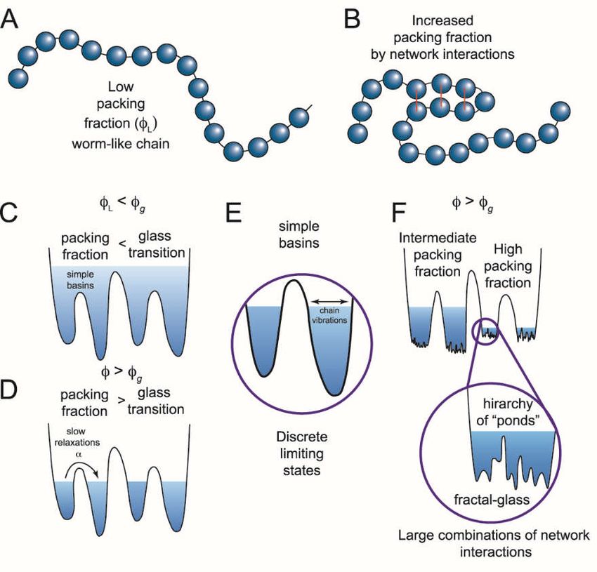
Figure 1. Single molecule observation of state switching in an intrinsically disordered protein (IDP)
with single molecule Förster resonance energy transfer (smFRET). The soluble N-ethylmaleimide-
sensitive factor activating protein receptor (SNARE) protein synaptosomal nerve-associated protein
25A (SNAP-25A) and the C-terminal tail of the GluN2B subunit of the N-methyl-D-aspartate (NMDA)
receptor (NMDAR) (C-term-N2B) are intrinsically disordered in their native states. Residues 20 and
197 of SNAP-25A and residues 1273 and 1394 of C-term-N2B were randomly labeled with donor and
acceptor fluorophores for smFRET measurements [23]. Labeled single molecules were encapsulated
in liposomes (100 nm in diameter) that were then surface-tethered through biotin–streptavidinBiomolecules 2019, 9, 114 4 of 16
linkage on a surface passivated with biotinylated bovine serum albumin (BSA). Fluorescence emission
was recorded using an emCCD (electron multiplying charge coupled device) camera at a frame rate
of 10 Hz. At this time resolution, rapid conformational dynamics are time-averaged, but surface
attachment allows extended observations of the same molecule for seconds to minutes. (A)
Representative single molecule fluorescence intensity time trace of SNAP-25A (top panel) does not
show spontaneous switching. The fluorescence intensities were converted to Förster resonance energy
transfer (FRET) efficiency (bottom panel, black line). SNAP-25A molecules showed a stable FRET
efficiency with no switching between different FRET values. (B) Representative single molecule
fluorescence intensity time trace of C-term-N2B (top panel) and FRET efficiency (bottom panel, black
line) fit by hidden Markov modeling (HMM) to obtain the dwell times in each state (bottom panel,
red line). Donor and acceptor signals for C-term-N2B molecules show step-wise, anticorrelated
changes in intensity, yielding steady FRET for seconds before spontaneously switching to different
FRET values, which would correspond to distinct disordered states with a different average size. (C)
FRET efficiency histogram for C-term-N2B molecules show a broad distribution across the entire
range of FRET values (black line) in contrast to SNAP-25A molecules (red lines). (D) Histogram of
state dwell times for C-term-N2B obtained from HMM. The mean state dwell time is on the order of
seconds and showed no correlation with FRET efficiency. (E) Transition density plot for C-term-N2B
shows the FRET efficiency before a transition (y-axis) plotted against the FRET efficiency after that
transition (x-axis) for all observed transitions. These transitions proved too variable to resolve or
assign to specific conformations. A.U.: Arbitrary units.
2. Evidence of Configuration Switching in IDP Studies
One of the best examples of state switching in polypeptides is protein folding. There are two
states: the unfolded state, which is characterized by a broad ensemble of disparate, interconverting
conformations, and the folded state, which is characterized by local fluctuation within a narrow range
of conformational space. In most cases, such folding transitions are unidirectional under
physiological conditions. However, some proteins, such as the ankyrin repeat (AR) domain from the
IκB transcription inhibitor, undergo reversible order to disorder transitions at room temperature [24].
Only single molecule fluorescence resonance energy transfer (smFRET) of surface attached molecules
could detect the “nanospring dynamics” that occurred on the seconds timescale as individual akyrin
repeats came “unglued” [25]. Ensemble nuclear magnetic resonance (NMR) measurements on the
same protein showed well-resolved NMR cross-peaks and high-order parameters but no signs of
dynamic behavior [24]. Such a slow timescale for state switching suggests a large energy barrier
separating these regions of conformational space.
A similar conformational state switching has been reported in α-synuclein, an IDP linked to
Parkinson’s disease that undergoes a disorder-to-order transition upon interaction with amphiphilic
small molecules or membranes. However, in this case, the transition is not spontaneous but regulated
by functional interactions [26–28]. Other IDPs are known to change their form of disorder in response
to physiological signals such as ion influx or posttranslational modifications like phosphorylation
[29,30]. A key aspect of state switching in these IDPs is that the entire conformational landscape is
not always accessible or there are sharp energy barriers, which separate discrete subregions of
conformational space. The conformation is not “continuously tunable” [26].
Given the broad ensemble of conformations an IDP may adopt, it is not always possible to know
how much of the conformational landscape is being explored and when such switching is occurring.
One hallmark of deeper energy basins within the conformational landscape is the presence of slow
timescale dynamics. However, most structural methods are not well suited for detecting slow
timescale conformational dynamics.
A comparison of synaptic IDPs and IDRs revealed stochastic conformational switching in two
out of five proteins in a test set despite the fact that all proteins appeared similarly intrinsically
disordered by other measures [23]. The cytoplasmic domains from both neuroligin and the GluN2B
subunit of the N-methyl-D-aspartate (NMDA) receptor (NMDAR) showed continuous hop-like
conformational diffusion with Förster resonance energy transfer (FRET) shifts equivalent to
nanometer scale motions in a single 100 millisecond time bin (Figure 1B). The sensitivity toBiomolecules 2019, 9, 114 5 of 16
stoichiometry provided by single molecule detection allows IDP clustering or aggregation to be
distinguished from single molecule conformational switching. These IDRs adopted a compact
globular form of disorder, yet another IDP (synaptobrevin) with a similar form of disorder failed to
show transitions [23].
The transitions in GluN2B were detected with seven different FRET labeling combinations
representing dye separations from 83 to 172 residues [31]. Only the shortest separation of 15 residues
failed to show any transitions, which is expected because a short polypeptide segment should not be
capable of large conformational changes. This important control confirms that photophysical effects
on dye environment and orientation are not the origin of the transitioning phenomena. Because of
their complexity, the transitions observed in synaptic IDRs proved uninterpretable in terms of
structural intermediates [23].
Similar stochastic transitions between FRET efficiency levels were observed in the smFRET
traces for protein 4.1, a cytoskeletal adaptor protein that stabilizes spectrin–actin crosslinks [32]. The
protein appeared to switch between an unresolved number of discrete conformational states.
Interestingly, while binding of protein 4.1 to the nuclear mitotic apparatus (NuMA) protein changed
the pattern of transitions, it did not eliminate the transitions, indicating that the complex retains
switch-like dynamics. Similarly, binding partner interactions with the synaptic scaffold protein
postsynaptic density protein 95 (PSD-95) also showed no effect on conformational switching in the
IDR from neuroligin [23]. Conformational switching may play a functional role even after these IDPs
interact with downstream factors.
State switching in IDPs is also possible on faster timescales. The yeast prion protein Sup35 is a
translation termination factor that forms amyloid fibrils. At low concentrations, smFRET showed that
the protein formed a compact disordered globule [33]. However, fluorescence correlation
spectroscopy (FCS) analysis of fluorescence quenching revealed at least two well separated
components to the dynamics, including a slower component that originated from long-range
contacts.
3. Theoretical Framework for Understanding IDP Conformational Switching
Swollen random coils can fully sample a “flat” energy landscape. Fast sampling is possible
because little energy is needed to overcome small barriers between different configurations. Such
IDPs have been described using equilibrium statistical mechanics as freely joint Gaussian chains, self-
avoiding coils, and worm-like chains [17,23,34]. The conformational switching, described above in
Section 2, challenges such approaches because these IDPs are sequestered off from the entirety of the
conformational space. Thus, in the absence of the simplifying assumption that all states are present
and equally probable, one cannot construct a statistical ensemble that represents all the possible
states.
Intrinsically disordered protein conformational switching has been likened to the Lorenz
attractor in nonlinear chaotic systems [35]. Like the Lorenz attractor, this switching behavior in IDPs
has been shown to be sensitive to initial conditions and is also restricted to be near the attractor points
in conformational space. Chaos, in dynamical mechanical systems, is dependent on underlying
nonlinear relationships in the governing interactions. Intrinsically disordered proteins certainly have
interactions within their peptide chain, with other components in solution, and with the host fluid
that are likely to involve nonlinearities.
All conformational transitions are noise-assisted reactions of the type described by Kramers’
transition state theory [36–38]. The energy for excursions between basins must come from
fluctuations where the polypeptide gains enough energy from the thermal milieu. Therefore, the
continuum of timescales relates to the continuum of barriers between different regions of
conformational space. Polypeptides are not uniform chemical polymers. Within the core of a compact,
globular IDP there exists a nonlinear potential as alternate long-range interactions (favorable and
unfavorable) are transiently sampled as distinct regions of the polypeptide chain are brought into
close proximity. Given such a fluctuating force, thermally activated transitions could occur across a
range of timescales. Even rare transitions are possible within some finite time.Biomolecules 2019, 9, 114 6 of 16
Within Kramers’ theory, the time to escape a basin is related to the damping factor associated
with internal friction. Studies of IDPs have pointed to the important role of internal friction in
modulating conformational dynamics and folding [39,40]. Friction within expanded IDPs depends
on the quality of solvent interactions. However, in collapsed, globular IDPs, shielding of residues in
the interior removes solvent interactions and can create a complex network of coupled and decoupled
chain segments [34,41]. Thus, nonlinear dynamics arising from feedback among these different
couplings should not be surprising. Large transitions (e.g., folding–unfolding) involve a complex set
of possible pathways within the energy landscape [42].
Conformational switching occurs over a wide range of timescales, ranging from very slow
processes lasting for seconds down to microsecond timescales [43]. To explain this, a scale-free
approach is needed. The energy landscape we have postulated for these IDPs, containing multiple
minima with high energy barriers, is also characteristic of glasses (Figure 2). Mean-field theories of
disordered glasses can describe the existence of stable and metastable states [44–46]. At low density
or low packing fraction (ϕ), glasses remain liquid and can sample all states (Figure 2C). As the
packing fraction increases, the individual particles are subject to jamming and undergo a glass
transition where they become trapped within individual basins (Figure 2D). In this jammed state,
excitations can extend over a wide range of timescales [47].
Figure 2. Switching in IDPs shows similarity to structural glasses. (A) IDPs can be modeled as worm-
like chains when the packing fraction (ϕ of the constituent particles is low (ϕL). Glasses can flow
when the packing fraction is low but undergo a phase transition at high packing fractions when
particles are caged by interactions with their neighbors. Applying this analogy of glasses to describe
IDPs, the amino acids are the constituent particles. (B) If amino acids interact, the packing fraction
increases, giving rise to a more complex energy landscape with states at different energy levels. (C)
These states (simple basins) represent the minima of the energy landscape. When the packing fraction
is lower than the glass transition (ϕg), or collapsed state, the polypeptide can sample a continuum of
states following polymer models. Transitions between states would be described by Kramers’
transition state theory. (D) When there are many network interactions, the packing fraction increases
and not all conformations are accessible. The limiting states are separated by deep wells and discrete
states can be identified. Such conformational switching could result from a number of different
mechanisms including internal friction, long rage potentials, ion mediated interactions, or other forces
that modulate the network interactions. (E) Discrete limiting states produce conformationalBiomolecules 2019, 9, 114 7 of 16
switching, yet there is still a subensemble of conformations inside each basin. (F) At high packing
fractions, the subensemble of states can also become discontinuous, generating potential transitions
across widely separated temporal domains. (Figure adapted from reference [47]).
Deep within the glass phase, each individual basin becomes a metabasin composed of a fractal
hierarchy of sub-basins (Figure 2F) [48]. This fractal free-energy landscape was recently proposed to
explain the roughness transition in structural glasses [47]. A similar fractal hierarchy or scale free-
energy landscape could arise from packing of chain segments within an IDP. The packing particles
are collapsed chain segments within the polymer along with coordinated ions and bound solvent
(Figure 2B). A high packing fraction could lead to jamming and produce metastable configurations,
which might switch to other equally stable configurations as intrachain interaction forces evolve with
the conformational search. In this model, such local phase transitions would extend to other packed
chain segments, leading to the observed IDP conformational switching across a wide temporal
regime.
4. Possible Mechanisms to Sequester regions of Conformational Space in IDPs
The origin of continuous slow timescale dynamics or state switching is not clear. Proline
isomerization is one of the conformational changes in polypeptides known to operate on this
timescale and can result in state switching. Conformational exchange rates linked to proline
isomerization were detected in the IDR of the transcriptional regulator E2 from human
papillomavirus (HPV) by collecting NMR nuclear overhauser spectroscopy (NOESY) spectra at
different mixing times [20]. This discrete conformational transition in E2 is the rate-limiting step for
antibody recognition of this viral antigen. Interestingly, proline depletion of GluN2B reduced the
number of molecules that showed any stochastic FRET transitions, but proline-depleted constructs
showed similar transition rates to the wild type [49]. Thus, for GluN2B, proline appeared to facilitate
switching but not govern the timescale.
Aside from proline isomerization, the major determinant of IDP conformation are self-
interactions (i.e., between amino acids in the chain) and solvent interactions. Most intrachain
interactions in IDPs are local, yet chain segments can partition into both packed and extended forms
of disorder. These chain segments are continuously exposed to one another during the
conformational search, with favorable interactions restricting chain motions and unfavorable
interactions limiting access to some conformations. Within the disordered globule, the quality and
quantity of the solvent is evolving as water and ions interact with local chain segments and are drawn
into the “core” of the IDP. For example, ions from solution could neutralize local charge densities
stabilizing distinct configuration space from those visited in the absence of the ions. Compacted chain
segments of IDPs can temporarily exclude water [34,41,50], which would result in stable local minima
that would limit the conformational search. As noted above, the complexity of conformational space
within these fractal basins would mean that available thermal energy could be dissipated by small,
local rearrangements rather than long-range motions.
Chain reconfiguration could also transiently trap unfavorable interactions within the globular
interior. This unfavorable interaction would destabilize any metastable local conformations and drive
the system to a new region of conformational space. Reintroduction of water to a temporarily
dehydrated chain segment would upset the balance of the chain interactions. Chain reconfiguration
will inevitably bring charged amino acids within the globular interior with a finite probability of
unfavorable interactions being trapped by approach jamming. These sorts of trapped interactions
represent high energy metastable intermediates that could suddenly be released when a random
fluctuation exposes a pathway that permits access to alternate regions of conformational space. Such
an event would manifest as sudden stochastic changes in the sampled conformational space, or what
we have termed IDP conformational switching.
It remains to be established that effects as subtle as sequestering an additional ion or water
molecule could generate the spontaneous IDP ensemble switching that is the focus of this discussion.
There are some relevant examples where such small perturbations can affect molecular properties.Biomolecules 2019, 9, 114 8 of 16
Deoxyribonucleic acid (DNA) certainly can transiently recruit or bind ions from solution changing
its polymeric properties [51], bearing in mind that DNA can easily be modeled as a worm-like chain
[52,53]. Certainly, changes in proteins at the level of a single amino acid can impact disordered states
[54–63]. Changes of the amino acid sequence in IDPs and IDRs have been linked to human diseases.
Aggregation of α-synuclein has been linked to sequence composition in familial forms of Parkinson’s
disease. Familial mutations that enhanced aggregation slowed conformational dynamics, while
mutations that sped up intramolecular diffusion inhibited aggregation [64]. De novo missense
mutations in the cytoplasmic IDR of the NMDAR, discussed above, are linked familial forms of
epilepsy [65,66]. That changes at the level of individual amino acids can reshape the energy landscape
of IDPs suggests that transient interactions with ions or water could generate the observed ensemble
switching phenomenon.
5. State Switching Can Occur Close to the Boundary of a Folding Transition
Most protein folding studies to date have focused on single domain proteins displaying simple
two state behavior. The folding of large multidomain proteins can be more complex with metastable
intermediate states in their energy landscapes. At low denaturant concentrations (0.65 M GdmCl),
the three-domain, 214 amino acid protein adenylate kinase (AK) began to show stochastic FRET
transitions between six states [42]. The transitions were too variable to resolve nor could they all be
assigned to specific denatured intermediates. Furthermore, the “situation [became] even more
complex at higher concentrations of denaturant.” [42]. This shows that a folded protein slightly
destabilized by denaturants shows similarly complex transitioning to that observed in switching
IDPs.
Thus, transitioning molecules arise when the impetus for a polypeptide to fold is lowered
slightly and becomes more complex as more of the conformational landscape becomes available.
Ultimately, transitions become faster until they are unresolved and become continuous dynamics of
the type described by polymer models. Thus, a similar transition should be possible if an expanded
coil is brought close to a folded state. Interestingly, smFRET measured in the presynaptic fusion
protein SNAP-25 did show scaling that fit to polymer models [23]. However, stochastic FRET
switching was induced in SNAP-25 upon binding the SNARE protein syntaxin, which is a binary
intermediate in the formation of the tripartite SNARE complex [67]. This confirms that an expanded
coil-like IDP can be converted to state switching as protein interactions restrict access to portions of
the conformational landscape.
6. Functional Relevance of IDP Ensemble Switching through Phosphorylation
Aside from transitions involving an ordered state, few biological functions have been directly
connected to the modulation of IDP conformational ensembles. Post-translational modifications can
change the interaction potential of existing amino acids [29,30,68–71]. Phosphorylation is among the
best-documented mechanism for dynamically affecting biological function of proteins. In particular,
phosphorylation has been connected with modulating disorder in IDPs [29,68–75].
For example, the ribonucleic acid (RNA) binding protein fused in sarcoma (FUS) forms
aggregated protein deposits in neurodegenerative disorders, which are modulated by
phosphorylation. Nuclear magnetic resonance studies found that phosphorylation of FUS, or
phosphomimetic mutations, did not “alter the disordered structure of FUS” [50]. However,
phosphorylation of FUS did decrease transient polypeptide collapse and increase the radius of
gyration. These changes in the IDP ensemble were associated with reduced intermolecular interaction
and aggregation such that a phosphomimetic variant reduced the toxicity of FUS to live cells [50].
Phosphorylation of an IDR was also linked to biological function of the NMDAR, which uses the
energy of neurotransmitter binding to open its ion channel. Allosteric inhibition of channel gating by
extracellular zinc can be alleviated by Src kinase phosphorylation of the C-terminal IDR of the
GluN2B subunit (C-term-N2B) [76], which switches conformation as noted above (Figure 3A) [23].
The effect of Src phosphorylation is mediated by expansion of this globular IDR without affecting
conformational transitions (Figure 3B,C) [31]. Deleting prolines near the phosphorylation sites hadBiomolecules 2019, 9, 114 9 of 16
the opposite effect and compacted the IDR while reducing the probability of transitions. Both of these
modifications, which change the size of the disordered states in opposite directions, eliminated the
ability of zinc to allosterically regulate channel gating without disrupting the underlying gating
mechanisms [49]. Thus, this IDR appears to have an optimal packing density that supports allosteric
coupling between domains of the receptor.
Figure 3. Effect of Src kinase phosphorylation on the conformational ensemble of the NMDA receptor.
The C-terminal intrinsically disordered region (IDR) of the GluN2B subunit of the NMDA receptor
(C-term-N2B) was randomly labeled with donor and acceptor fluorophores at residues 1323 and 1453
[31]. Src kinase phosphorylates C-term-N2B on tyrosine residues 1336 and 1472. (A) Representative
single molecule fluorescence intensity (top panels) and fluorescence resonance energy transfer (FRET)
efficiency fit by hidden Markov modeling (HMM) (bottom panels) for unphosphorylated (left) and
phosphorylated (right) C-term-N2B. Phosphorylation did not induce structure in C-term-N2B as the
dynamic transitions continued. (B) FRET efficiency histogram of C-term-N2B before (black line) and
after (red line) Src phosphorylation. Phosphorylation led to shift towards lower FRET indicating a
general expansion of the polypeptide, which was confirmed with hydrodynamic measurements. (C)
Dwell time histograms for transitions in C-term-N2B, obtained from HMM, before (black line) and
after (red line) Src phosphorylation. Although phosphorylation shifted the ensemble FRET efficiency,
stochastic transitions were unaffected. A.U.: Arbitrary units.
Phosphorylation-induced modulation of an IDP ensemble was also connected to regulation of
cellular signaling processes in prostate cancer. Prostate-associated gene 4 (PAGE4) is an IDP that is
expressed exclusively in adult males who have prostate cancer. Interactions between PAGE4 and
transcription factors have been suggested to control androgen sensitivity [77–83]. Both
homeodomain-interacting protein kinase 1 (HIPK1) and CDC-Like Kinase 2 (CLK2) phosphorylate
PAGE4, with CLK2 modifying many more sites. Experiments combining NMR, paramagnetic
relaxation enhancement (PRE), small angle X-ray scattering (SAXS) and smFRET have determined
that HIPK1 phosphorylation compacts the ensemble, while CLK2 phosphorylation leads to
expansion. Molecular dynamics (MD) simulations linked changes in PAGE4 ensembles to distinct
phosphorylation patterns [84]. These different phosphorylation patterns influenced transcription
factor binding. HIPK1-treated PAGE4 binds to AP-1, whereas CLK2 treatment of PAGE4 decreases
its affinity for AP-1. Phosphorylation by HIPK1 effectively disrupts the PAGE4 interaction with c-JunBiomolecules 2019, 9, 114 10 of 16
and consequently stimulated c-Jun dependent transcription in prostate cancer cell models [85,86]. In
contrast, CLK2 phosphorylation of PAGE4 inhibited c-Jun dependent transcription. Importantly,
HIPK1 is expressed in both androgen-dependent and androgen-independent prostate cancer cells,
whereas CLK2 and PAGE4 are expressed only in androgen-dependent cells. A model for the PAGE4–
Jun-Fos (AP-1)–AR regulatory circuit suggests phosphorylation patterns in prostate cancer cells can
oscillate. Thus, it was proposed that androgen dependence may vary in time [78] with switching
between androgen-dependent and androgen-independent phenotypes being a result of details of
PAGE4 phosphorylation [77,79,80,84]. This differential phosphorylation is associated with opposing
shifts in the conformational ensemble of PAGE4.
7. Conclusions and Prospects for Understanding IDP Switching
Intrinsically disordered proteins are essential components of cellular signaling pathways
because of their adaptability to the local environment. Cell signaling events can lead to changes in
ionic composition or pH that affect solvent quality while posttranslational modifications affect net
charge and hydropathy. Intrinsically disordered proteins can respond instantly to such signals by
shifting to alternate conformational ensembles. Their unique capabilities can allow a single IDP to
interact with multiple binding partners. Intrinsically disordered proteins also play structural roles as
linkers or entropic elements. Additionally, IDPs are linked to the formation of membraneless
organelles (i.e., liquid phase separation) [87,88]. These important functions allow for more nuanced
coordination of signal transduction.
Here, we have highlighted a recently identified conformational switching phenomenon
observed in a small but growing number of IDPs. Intrinsically disordered proteins in this class appear
disordered by standard measures of secondary structure or hydrodynamic mobility but also appear
to fluctuate between well-separated regions of conformational space. If the conformational ensembles
are functionally distinct, then mechanisms that biases conformational sampling will govern protein
activity. We suggest such control over IDP switching may be an important regulator of cellular
signaling networks.
Few experimental methods are sensitive to conformational switching in IDPs [89]. Integrative
structural biology approaches where several methods are applied to a single protein may help inform
our understanding of the molecular mechanisms controlling intrinsic disorder in proteins. Several
methods including smFRET, NMR, electron paramagnetic resonance double electron–electron
resonance (EPR-DEER), paramagnetic relaxation enhancement (PRE), and small-angle neutron
scattering (SANS)/small-angle X-ray scattering (SAXS) have all been applied to IDP studies. These
different methods are able to provide complementary information about local chain contacts and
global disordered structure. Comparing and cross-validating such results with multiple methods
may help reveal specific interactions that are critical for determining details of the disordered state.
To date, switching has only been observed in vitro, so it remains to be determined if such switching
transitions are present within live cells [3], where smFRET may enable direct observation [90–92].
Similarly, switching has been primarily studied under dilute conditions so it remains unknown
whether the phenomenon persists in the condensed phase [93,94].
In addition to experimental approaches, MD simulation is a critical tool to understand the
mechanisms that govern access to the full conformational ensemble. Molecular dynamics simulations
of IDPs are particularly challenging. Details of the force fields are critically important and are a topic
of continued development [95,96]. The long timescales required to observe ensemble switching are
difficult to achieve for standard MD simulation and require more sophisticated ensemble sampling
approaches. Despite these difficulties progress applying MD simulation to IDPs is an area of active
work [84,97–103]. Critically, experimental studies and simulations provide essential feedback to each
other [103–107]. The molecular mechanisms controlling rapid dynamics in IDPs are becoming clearer
through MD simulation [83,101]. However, spontaneous ensemble switching of an IDP has yet to be
reported so molecular details remain unknown.
If IDP ensemble switching does regulate signaling, then these mechanisms could be used for
therapeutic intervention in those pathways. Efforts are already underway to identify small moleculesBiomolecules 2019, 9, 114 11 of 16
that can specifically bind IDPs [2,108,109]. An exciting functional connection has been found for the
transcription factor TFIID where a drug-like molecule affected the DNA interaction to prevent
transcription initiation by RNA polymerase [2,109]. Additional strategies for intervening in signaling
pathways within the diseased state will likely emerge as our understanding grows as to how IDP
conformational dynamic leads to function within cellular networks.
Author Contributions: Conceptualization, M.E.B., H.S., T.S. and K.R.W.; writing—review and editing, U.B.C.,
M.E.B., H.S., T.S. and K.R.W.
Funding: This research has thus far been unable to secure any external funding.
Conflicts of Interest: The authors declare no conflict of interest.
References
1. Uversky, V.N.; Dunker, A.K. Understanding protein non-folding. Biochim. Biophys. Acta - Proteins Proteomics
2010, 1804, 1231–1264, doi:10.1016/j.bbapap.2010.01.017.
2. Shammas, S.L. Mechanistic roles of protein disorder within transcription. Curr. Opin. Struct. Biol. 2017, 42,
155–161, doi:10.1016/j.sbi.2017.02.003.
3. Wright, P.E.; Dyson, H.J. Intrinsically disordered proteins in cellular signalling and regulation. Nat. Rev.
Mol. Cell Biol. 2015, 16, 18–29, doi:10.1038/nrm3920.
4. Oldfield, C.J.; Dunker, A.K. Intrinsically Disordered Proteins and Intrinsically Disordered Protein Regions.
Annu. Rev. Biochem. 2014, 83, 553–584, doi:10.1146/annurev-biochem-072711-164947.
5. Fung, H.Y.J.; Birol, M.; Rhoades, E. IDPs in macromolecular complexes: the roles of multivalent interactions
in diverse assemblies. Curr. Opin. Struct. Biol. 2018, 49, 36–43, doi:10.1016/j.sbi.2017.12.007.
6. Berlow, R.B.; Dyson, H.J.; Wright, P.E. Functional advantages of dynamic protein disorder. FEBS Lett. 2015,
589, 2433–2440, doi:10.1016/j.febslet.2015.06.003.
7. Uversky, V.N.; Oldfield, C.J.; Dunker, A.K. Intrinsically disordered proteins in human diseases:
introducing the D2 concept. Annu. Rev. Biophys. 2008, 37, 215–46,
doi:10.1146/annurev.biophys.37.032807.125924.
8. Borgia, A.; Borgia, M.B.; Bugge, K.; Kissling, V.M.; Heidarsson, P.O.; Fernandes, C.B.; Sottini, A.; Soranno,
A.; Buholzer, K.J.; Nettels, D.; et al. Extreme disorder in an ultrahigh-affinity protein complex. Nature 2018,
555, 61–66, doi:10.1038/nature25762.
9. Tsytlonok, M.; Sanabria, H.; Wang, Y.; Felekyan, S.; Hemmen, K.; Phillips, A.; Yun, M.-K.; Waddell, B.;
Park, C.-G.; Vaithiyalingam, S.; et al. Dynamic anticipation by Cdk2/Cyclin A-bound p27 mediates signal
integration in cell cycle regulation. 2018, arXiv: 1812.07009.
10. Shoemaker, B.A.; Portman, J.J.; Wolynes, P.G. Speeding molecular recognition by using the folding funnel:
The fly-casting mechanism. Proc. Natl. Acad. Sci. 2000, 97, 8868–8873, doi:10.1073/pnas.160259697.
11. Hoffman, R.M.B.; Blumenschein, T.M.A.; Sykes, B.D. An interplay between protein disorder and structure
confers the Ca2+ regulation of striated muscle. J. Mol. Biol. 2006, 361, 625–33, doi:10.1016/j.jmb.2006.06.031.
12. Metskas, L.A.; Rhoades, E. Conformation and Dynamics of the Troponin I C-Terminal Domain: Combining
Single-Molecule and Computational Approaches for a Disordered Protein Region. J. Am. Chem. Soc. 2015,
137, 11962–11969, doi:10.1021/jacs.5b04471.
13. Podlaha, O.; Zhang, J. Positive selection on protein-length in the evolution of a primate sperm ion channel.
Proc. Natl. Acad. Sci. U. S. A. 2003, 100, 12241–6, doi:10.1073/pnas.2033555100.
14. Hoh, J.H. Functional protein domains from the thermally driven motion of polypeptide chains: a proposal.
Proteins 1998, 32, 223–8.
15. de Gennes, P.-G. Soft Matter (Nobel Lecture). Angew. Chemie Int. Ed. English 1992, 31, 842–845,
doi:10.1002/anie.199208421.
16. Wiggins, P.A.; Nelson, P.C. Generalized theory of semiflexible polymers. Phys. Rev. E 2006, 73, 031906,
doi:10.1103/PhysRevE.73.031906.
17. Schuler, B.; Soranno, A.; Hofmann, H.; Nettels, D. Single-Molecule FRET Spectroscopy and the Polymer
Physics of Unfolded and Intrinsically Disordered Proteins. Annu. Rev. Biophys. 2016, 45, 207–31,
doi:10.1146/annurev-biophys-062215-010915.
18. Vitalis, A.; Wang, X.; Pappu, R. V Atomistic simulations of the effects of polyglutamine chain length and
solvent quality on conformational equilibria and spontaneous homodimerization. J. Mol. Biol. 2008, 384,
279–97, doi:10.1016/j.jmb.2008.09.026.Biomolecules 2019, 9, 114 12 of 16
19. Mao, A.H.; Crick, S.L.; Vitalis, A.; Chicoine, C.L.; Pappu, R. V Net charge per residue modulates
conformational ensembles of intrinsically disordered proteins. Proc. Natl. Acad. Sci. U. S. A. 2010, 107, 8183–
8, doi:10.1073/pnas.0911107107.
20. Fassolari, M.; Chemes, L.B.; Gallo, M.; Smal, C.; Sánchez, I.E.; de Prat-Gay, G. Minute time scale prolyl
isomerization governs antibody recognition of an intrinsically disordered immunodominant epitope. J.
Biol. Chem. 2013, 288, 13110–23, doi:10.1074/jbc.M112.444554.
21. Lin, Y.-H.; Brady, J.P.; Forman-Kay, J.D.; Chan, H.S. Charge pattern matching as a ‘fuzzy’ mode of
molecular recognition for the functional phase separations of intrinsically disordered proteins. New J. Phys.
2017, 19, 115003, doi:10.1088/1367-2630/aa9369.
22. Das, S.; Amin, A.N.; Lin, Y.-H.; Chan, H.S. Coarse-grained residue-based models of disordered protein
condensates: utility and limitations of simple charge pattern parameters. Phys. Chem. Chem. Phys. 2018, 20,
28558–28574, doi:10.1039/C8CP05095C.
23. Choi, U.B.; McCann, J.J.; Weninger, K.R.; Bowen, M.E. Beyond the Random Coil: Stochastic Conformational
Switching in Intrinsically Disordered Proteins. Structure 2011, 19, 566–576, doi:10.1016/j.str.2011.01.011.
24. Lamboy, J.A.; Kim, H.; Lee, K.S.; Ha, T.; Komives, E.A. Visualization of the nanospring dynamics of the
IkappaBalpha ankyrin repeat domain in real time. Proc. Natl. Acad. Sci. U. S. A. 2011, 108, 10178–83,
doi:10.1073/pnas.1102226108.
25. Lamboy, J.A.; Kim, H.; Dembinski, H.; Ha, T.; Komives, E.A. Single-molecule FRET reveals the native-state
dynamics of the IκBα ankyrin repeat domain. J. Mol. Biol. 2013, 425, 2578–90, doi:10.1016/j.jmb.2013.04.015.
26. Ferreon, A.C.M.; Gambin, Y.; Lemke, E.A.; Deniz, A.A. Interplay of alpha-synuclein binding and
conformational switching probed by single-molecule fluorescence. Proc. Natl. Acad. Sci. 2009, 106, 5645–
5650, doi:10.1073/pnas.0809232106.
27. Trexler, A.J.; Rhoades, E. α-Synuclein Binds Large Unilamellar Vesicles as an Extended Helix †. Biochemistry
2009, 48, 2304–2306, doi:10.1021/bi900114z.
28. Moosa, M.M.; Ferreon, A.C.M.; Deniz, A.A. Forced Folding of a Disordered Protein Accesses an Alternative
Folding Landscape. ChemPhysChem 2015, 16, 90–94, doi:10.1002/cphc.201402661.
29. Darling, A.L.; Uversky, V.N. Intrinsic Disorder and Posttranslational Modifications: The Darker Side of the
Biological Dark Matter. Front. Genet. 2018, 9, 158, doi:10.3389/fgene.2018.00158.
30. Bah, A.; Forman-Kay, J.D. Modulation of Intrinsically Disordered Protein Function by Post-translational
Modifications. J. Biol. Chem. 2016, 291, 6696–705, doi:10.1074/jbc.R115.695056.
31. Choi, U.B.; Xiao, S.; Wollmuth, L.P.; Bowen, M.E. Effect of Src kinase phosphorylation on disordered C-
terminal domain of N-methyl-D-aspartic acid (NMDA) receptor subunit GluN2B protein. J. Biol. Chem.
2011, 286, 29904–12, doi:10.1074/jbc.M111.258897.
32. Wu, S.; Wang, D.; Liu, J.; Feng, Y.; Weng, J.; Li, Y.; Gao, X.; Liu, J.; Wang, W. The Dynamic Multisite
Interactions between Two Intrinsically Disordered Proteins. Angew. Chem. Int. Ed. Engl. 2017, 56, 7515–7519,
doi:10.1002/anie.201701883.
33. Mukhopadhyay, S.; Krishnan, R.; Lemke, E.A.; Lindquist, S.; Deniz, A.A. A natively unfolded yeast prion
monomer adopts an ensemble of collapsed and rapidly fluctuating structures. Proc. Natl. Acad. Sci. 2007,
104, 2649–2654, doi:10.1073/pnas.0611503104.
34. Zheng, W.; Zerze, G.H.; Borgia, A.; Mittal, J.; Schuler, B.; Best, R.B. Inferring properties of disordered chains
from FRET transfer efficiencies. J. Chem. Phys. 2018, 148, 123329, doi:10.1063/1.5006954.
35. Uversky, V.N. Dancing Protein Clouds: The Strange Biology and Chaotic Physics of Intrinsically
Disordered Proteins. J. Biol. Chem. 2016, 291, 6681–8, doi:10.1074/jbc.R115.685859.
36. Kramers, H.A. Brownian motion in a field of force and the diffusion model of chemical reactions. Physica
1940, 7, 284–304, doi:10.1016/S0031-8914(40)90098-2.
37. Skinner, J.L.; Wolynes, P.G. Transition state and Brownian motion theories of solitons. J. Chem. Phys. 1980,
73, 4015–4021, doi:10.1063/1.440629.
38. Hänggi, P.; Talkner, P.; Borkovec, M. Reaction-rate theory: fifty years after Kramers. Rev. Mod. Phys. 1990,
62, 251–341, doi:10.1103/RevModPhys.62.251.
39. Soranno, A.; Buchli, B.; Nettels, D.; Cheng, R.R.; Müller-Späth, S.; Pfeil, S.H.; Hoffmann, A.; Lipman, E.A.;
Makarov, D.E.; Schuler, B. Quantifying internal friction in unfolded and intrinsically disordered proteins
with single-molecule spectroscopy. Proc. Natl. Acad. Sci. U. S. A. 2012, 109, 17800–6,
doi:10.1073/pnas.1117368109.Biomolecules 2019, 9, 114 13 of 16
40. Borgia, A.; Wensley, B.G.; Soranno, A.; Nettels, D.; Borgia, M.B.; Hoffmann, A.; Pfeil, S.H.; Lipman, E.A.;
Clarke, J.; Schuler, B. Localizing internal friction along the reaction coordinate of protein folding by
combining ensemble and single-molecule fluorescence spectroscopy. Nat. Commun. 2012, 3, 1195,
doi:10.1038/ncomms2204.
41. Zheng, W.; Hofmann, H.; Schuler, B.; Best, R.B. Origin of Internal Friction in Disordered Proteins Depends
on Solvent Quality. J. Phys. Chem. B 2018, 122, 11478–11487, doi:10.1021/acs.jpcb.8b07425.
42. Pirchi, M.; Ziv, G.; Riven, I.; Cohen, S.S.; Zohar, N.; Barak, Y.; Haran, G. Single-molecule fluorescence
spectroscopy maps the folding landscape of a large protein. Nat. Commun. 2011, 2, 493,
doi:10.1038/ncomms1504.
43. Weninger, K.; Qiu, R.; Ou, E.; Milikisiyants, S.; Sanabria, H.; Smirnova, T.I. smFRET and DEER Distance
Measurements as Applied to Disordered and Structured Proteins. Biophys. J. 2016, 110, 559a,
doi:10.1016/j.bpj.2015.11.2987.
44. Kirkpatrick, T.R.; Wolynes, P.G. Stable and metastable states in mean-field Potts and structural glasses.
Phys. Rev. B. Condens. Matter 1987, 36, 8552–8564, doi:10.1103/PhysRevB.36.8552.
45. Kirkpatrick, T.R.; Thirumalai, D. Dynamics of the Structural Glass Transition and the p -Spin—
Interaction Spin-Glass Model. Phys. Rev. Lett. 1987, 58, 2091–2094, doi:10.1103/PhysRevLett.58.2091.
46. Wolynes, P.G.; Lubchenko, V. Structural glasses and supercooled liquids : theory, experiment, and applications;
Wiley: Hoboken, 2012; ISBN 9780470452233.
47. Charbonneau, P.; Kurchan, J.; Parisi, G.; Urbani, P.; Zamponi, F. Fractal free energy landscapes in structural
glasses. Nat. Commun. 2014, 5, 3725, doi:10.1038/ncomms4725.
48. Parisi, G. Order Parameter for Spin-Glasses. Phys. Rev. Lett. 1983, 50, 1946–1948,
doi:10.1103/PhysRevLett.50.1946.
49. Choi, U.B.; Kazi, R.; Stenzoski, N.; Wollmuth, L.P.; Uversky, V.N.; Bowen, M.E. Modulating the Intrinsic
Disorder in the Cytoplasmic Domain Alters the Biological Activity of the N -Methyl-d-aspartate-sensitive
Glutamate Receptor. J. Biol. Chem. 2013, 288, 22506–22515, doi:10.1074/jbc.M113.477810.
50. Monahan, Z.; Ryan, V.H.; Janke, A.M.; Burke, K.A.; Rhoads, S.N.; Zerze, G.H.; O’Meally, R.; Dignon, G.L.;
Conicella, A.E.; Zheng, W.; et al. Phosphorylation of the FUS low-complexity domain disrupts phase
separation, aggregation, and toxicity. EMBO J. 2017, 36, 2951–2967, doi:10.15252/embj.201696394.
51. Barnett, R.N.; Cleveland, C.L.; Joy, A.; Landman, U.; Schuster, G.B. Charge Migration in DNA: Ion-Gated
Transport. Science (80-. ). 2001, 294, 567–571, doi:10.1126/science.1062864.
52. Klenin, K.; Merlitz, H.; Langowski, J. A Brownian Dynamics Program for the Simulation of Linear and
Circular DNA and Other Wormlike Chain Polyelectrolytes. Biophys. J. 1998, 74, 780–788, doi:10.1016/S0006-
3495(98)74003-2.
53. Murphy, M.C.; Rasnik, I.; Cheng, W.; Lohman, T.M.; Ha, T. Probing single-stranded DNA conformational
flexibility using fluorescence spectroscopy. Biophys. J. 2004, 86, 2530–2537, doi:10.1016/S0006-
3495(04)74308-8.
54. Yedvabny, E.; Nerenberg, P.S.; So, C.; Head-Gordon, T. Disordered Structural Ensembles of Vasopressin
and Oxytocin and Their Mutants. J. Phys. Chem. B 2015, 119, 896–905, doi:10.1021/jp505902m.
55. Gruet, A.; Dosnon, M.; Vassena, A.; Lombard, V.; Gerlier, D.; Bignon, C.; Longhi, S. Dissecting Partner
Recognition by an Intrinsically Disordered Protein Using Descriptive Random Mutagenesis. J. Mol. Biol.
2013, 425, 3495–3509, doi:10.1016/j.jmb.2013.06.025.
56. Babu, M.M. The contribution of intrinsically disordered regions to protein function, cellular complexity,
and human disease. Biochem. Soc. Trans. 2016, 44, 1185–1200, doi:10.1042/BST20160172.
57. Mohan, A.; Uversky, V.N.; Radivojac, P. Influence of Sequence Changes and Environment on Intrinsically
Disordered Proteins. PLoS Comput. Biol. 2009, 5, e1000497, doi:10.1371/journal.pcbi.1000497.
58. Davey, N.E.; Cyert, M.S.; Moses, A.M. Short linear motifs – ex nihilo evolution of protein regulation. Cell
Commun. Signal. 2015, 13, 43, doi:10.1186/s12964-015-0120-z.
59. Fuxreiter, M.; Tompa, P.; Simon, I. Local structural disorder imparts plasticity on linear motifs.
Bioinformatics 2007, 23, 950–956, doi:10.1093/bioinformatics/btm035.
60. Van Roey, K.; Dinkel, H.; Weatheritt, R.J.; Gibson, T.J.; Davey, N.E. The switches.ELM Resource: A
Compendium of Conditional Regulatory Interaction Interfaces. Sci. Signal. 2013, 6, rs7-rs7,
doi:10.1126/scisignal.2003345.
61. Diella, F.; Haslam, N.; Chica, C.; Budd, A.; Michael, S.; Brown, N.P.; Trave, G.; Gibson, T.J. Understanding
eukaryotic linear motifs and their role in cell signaling and regulation. Front. Biosci. 2008, 13, 6580–603.Biomolecules 2019, 9, 114 14 of 16
62. Akiva, E.; Friedlander, G.; Itzhaki, Z.; Margalit, H. A dynamic view of domain-motif interactions. PLoS
Comput. Biol. 2012, 8, e1002341, doi:10.1371/journal.pcbi.1002341.
63. Pancsa, R.; Fuxreiter, M. Interactions via intrinsically disordered regions: What kind of motifs? IUBMB Life
2012, 64, 513–520, doi:10.1002/iub.1034.
64. Acharya, S.; Saha, S.; Ahmad, B.; Lapidus, L.J. Effects of Mutations on the Reconfiguration Rate of α-
Synuclein. J. Phys. Chem. B 2015, 119, 15443–50, doi:10.1021/acs.jpcb.5b10136.
65. Lesca, G.; Rudolf, G.; Bruneau, N.; Lozovaya, N.; Labalme, A.; Boutry-Kryza, N.; Salmi, M.; Tsintsadze, T.;
Addis, L.; Motte, J.; et al. GRIN2A mutations in acquired epileptic aphasia and related childhood focal
epilepsies and encephalopathies with speech and language dysfunction. Nat. Genet. 2013, 45, 1061–6,
doi:10.1038/ng.2726.
66. Lemke, J.R.; Lal, D.; Reinthaler, E.M.; Steiner, I.; Nothnagel, M.; Alber, M.; Geider, K.; Laube, B.; Schwake,
M.; Finsterwalder, K.; et al. Mutations in GRIN2A cause idiopathic focal epilepsy with rolandic spikes. Nat.
Genet. 2013, 45, 1067–72, doi:10.1038/ng.2728.
67. Weninger, K.; Bowen, M.E.; Choi, U.B.; Chu, S.; Brunger, A.T. Accessory Proteins Stabilize the Acceptor
Complex for Synaptobrevin, the 1:1 Syntaxin/SNAP-25 Complex. Structure 2008, 16, 308–320,
doi:10.1016/j.str.2007.12.010.
68. Bah, A.; Vernon, R.M.; Siddiqui, Z.; Krzeminski, M.; Muhandiram, R.; Zhao, C.; Sonenberg, N.; Kay, L.E.;
Forman-Kay, J.D. Folding of an intrinsically disordered protein by phosphorylation as a regulatory switch.
Nature 2015, 519, 106–109, doi:10.1038/nature13999.
69. Muller, P.; Chan, J.M.; Simoncik, O.; Fojta, M.; Lane, D.P.; Hupp, T.; Vojtesek, B. Evidence for allosteric
effects on p53 oligomerization induced by phosphorylation. Protein Sci. 2018, 27, 523–530,
doi:10.1002/pro.3344.
70. Valk, E.; Venta, R.; Ord, M.; Faustova, I.; Kõivomägi, M.; Loog, M. Multistep phosphorylation systems:
tunable components of biological signaling circuits. Mol. Biol. Cell 2014, 25, 3456–60, doi:10.1091/mbc.E14-
02-0774.
71. Kulkarni, P.; Solomon, T.L.; He, Y.; Chen, Y.; Bryan, P.N.; Orban, J. Structural metamorphism and
polymorphism in proteins on the brink of thermodynamic stability. Protein Sci. 2018, doi:10.1002/pro.3458.
72. Galea, C.A.; Nourse, A.; Wang, Y.; Sivakolundu, S.G.; Heller, W.T.; Kriwacki, R.W. Role of intrinsic
flexibility in signal transduction mediated by the cell cycle regulator, p27 Kip1. J. Mol. Biol. 2008, 376, 827–
38, doi:10.1016/j.jmb.2007.12.016.
73. Iakoucheva, L.M.; Radivojac, P.; Brown, C.J.; O’Connor, T.R.; Sikes, J.G.; Obradovic, Z.; Dunker, A.K. The
importance of intrinsic disorder for protein phosphorylation. Nucleic Acids Res. 2004, 32, 1037–1049,
doi:10.1093/nar/gkh253.
74. Xie, H.; Vucetic, S.; Iakoucheva, L.M.; Oldfield, C.J.; Dunker, A.K.; Obradovic, Z.; Uversky, V.N. Functional
anthology of intrinsic disorder. 3. Ligands, post-translational modifications, and diseases associated with
intrinsically disordered proteins. J. Proteome Res. 2007, 6, 1917–32, doi:10.1021/pr060394e.
75. Collins, M.O.; Yu, L.; Campuzano, I.; Grant, S.G.N.; Choudhary, J.S. Phosphoproteomic analysis of the
mouse brain cytosol reveals a predominance of protein phosphorylation in regions of intrinsic sequence
disorder. Mol. Cell. Proteomics 2008, 7, 1331–48, doi:10.1074/mcp.M700564-MCP200.
76. Traynelis, S.F.; Wollmuth, L.P.; McBain, C.J.; Menniti, F.S.; Vance, K.M.; Ogden, K.K.; Hansen, K.B.; Yuan,
H.; Myers, S.J.; Dingledine, R. Glutamate receptor ion channels: structure, regulation, and function.
Pharmacol. Rev. 2010, 62, 405–96, doi:10.1124/pr.109.002451.
77. Kulkarni, P.; Dunker, A.K.; Weninger, K.; Orban, J. Prostate-associated gene 4 (PAGE4), an intrinsically
disordered cancer/testis antigen, is a novel therapeutic target for prostate cancer. Asian J. Androl. 2016, 18,
695–703, doi:10.4103/1008-682X.181818.
78. Kulkarni, P.; Jolly, M.K.; Jia, D.; Mooney, S.M.; Bhargava, A.; Kagohara, L.T.; Chen, Y.; Hao, P.; He, Y.;
Veltri, R.W.; et al. Phosphorylation-induced conformational dynamics in an intrinsically disordered protein
and potential role in phenotypic heterogeneity. Proc. Natl. Acad. Sci. U. S. A. 2017, 114, E2644–E2653,
doi:10.1073/pnas.1700082114.
79. Salgia, R.; Jolly, M.K.; Dorff, T.; Lau, C.; Weninger, K.; Orban, J.; Kulkarni, P. Prostate-Associated Gene 4
(PAGE4): Leveraging the Conformational Dynamics of a Dancing Protein Cloud as a Therapeutic Target.
J. Clin. Med. 2018, 7, 156, doi:10.3390/jcm7060156.You can also read