Vessel Enlargement in Development and Pathophysiology - Frontiers
←
→
Page content transcription
If your browser does not render page correctly, please read the page content below
REVIEW
                                                                                                                                                  published: 25 February 2021
                                                                                                                                            doi: 10.3389/fphys.2021.639645

                                                Vessel Enlargement in Development
                                                and Pathophysiology
                                                Laia Gifre-Renom 1 and Elizabeth A. V. Jones 1,2*
                                                1
                                                    Department of Cardiovascular Sciences, Centre for Molecular and Vascular Biology, KU Leuven, Leuven, Belgium,
                                                2
                                                    Department of Cardiology, CARIM School for Cardiovascular Diseases, Maastricht University, Maastricht, Netherlands

                                                From developmental stages until adulthood, the circulatory system remodels in response
                                                to changes in blood flow in order to maintain vascular homeostasis. Remodeling processes
                                                can be driven by de novo formation of vessels or angiogenesis, and by the restructuration
                                                of already existing vessels, such as vessel enlargement and regression. Notably, vessel
                                                enlargement can occur as fast as in few hours in response to changes in flow and pressure.
                                                The high plasticity and responsiveness of blood vessels rely on endothelial cells. Changes
                                                within the bloodstream, such as increasing shear stress in a narrowing vessel or lowering
                                                blood flow in redundant vessels, are sensed by endothelial cells and activate downstream
                                                signaling cascades, promoting behavioral changes in the involved cells. This way,
                                                endothelial cells can reorganize themselves to restore normal circulation levels within the
                                                vessel. However, the dysregulation of such processes can entail severe pathological
                            Edited by:          circumstances with disturbances affecting diverse organs, such as human hereditary
                  Anna Rita Cantelmo,           telangiectasias. There are different pathways through which endothelial cells react to
       Université Lille Nord de France,
                                 France         promote vessel enlargement and mechanisms may differ depending on whether remodeling
                      Reviewed by:              occurs in the adult or in developmental models. Understanding the molecular mechanisms
                 Anthony Wayne Orr,             involved in the fast-adapting processes governing vessel enlargement can open the door
     Louisiana State University Health
                                                to a new set of therapeutical approaches to be applied in occlusive vascular diseases.
            Shreveport, United States
                   Stephanie Lehoux,            Therefore, we have outlined here the latest advances in the study of vessel enlargement
            McGill University, Canada           in physiology and pathology, with a special insight in the pathways involved in its regulation.
                    *Correspondence:
                   Elizabeth A. V. Jones        Keywords: venogenesis, migration, vascular fusion, mechanotransduction, collateral growth, arteriogenesis,
                 liz.jones@kuleuven.be          vessel enlargement, arterial venous malformation

                   Specialty section:
         This article was submitted to          INTRODUCTION
                  Vascular Physiology,
               a section of the journal
                                                During development, vascular beds often form as honeycombed shaped capillary plexus that
               Frontiers in Physiology
                                                then become perfused and remodel to form a hierarchical vessel structure. One of the most
       Received: 09 December 2020               visible changes that occur in this process is the enlargement of both arteries and veins. Vessel
        Accepted: 01 February 2021
                                                enlargement can occur remarkably quickly. In the embryo, this occurs within a single day
       Published: 25 February 2021
                                                after the onset of blood flow. As such, vessel enlargement has therapeutic potential in occlusive
                              Citation:
                                                vascular diseases that slower processes, such as angiogenesis, lack. However, so far, most
Gifre-Renom L and Jones EAV (2021)
 Vessel Enlargement in Development
                                                research on therapeutic strategies has been focused on angiogenesis and significantly less
                 and Pathophysiology.           advancement has been made to exploit vessel enlargement as a potential therapy in ischemic
            Front. Physiol. 12:639645.          diseases. In strokes and heart attacks, for example, local collateral vessels immediately dilate
     doi: 10.3389/fphys.2021.639645             to restore blood flow, but angiogenesis is a latter process in the body’s attempt to restore

Frontiers in Physiology | www.frontiersin.org                                             1                                        February 2021 | Volume 12 | Article 639645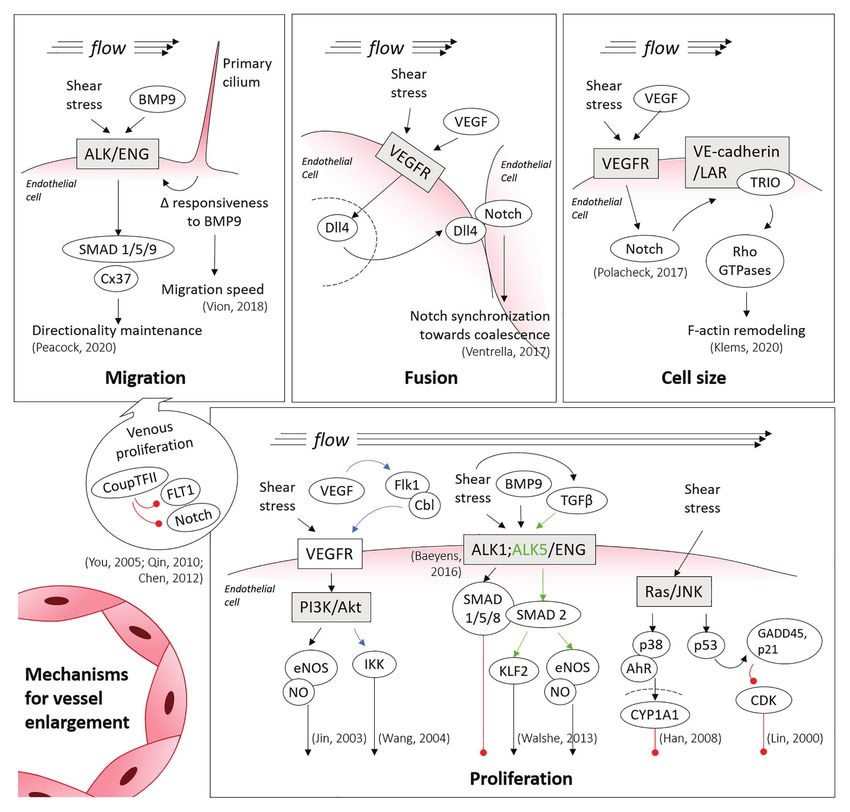
Gifre-Renom and Jones                                                                                                             Vessel Enlargement

proper perfusion (Schaper and Ito, 1996). Vessel can dilate on             and of collateral vessels. Differences in the type of vessel which
the short term, but can also undergo outward remodeling such               is enlarging can also account for differences in mechanism
that vessel diameter changes are permanent (Silver and Vita,               highlighted below.
2006). As such, it is primarily vessel enlargement that prevents
excessive cell death.
   Though occlusive vascular diseases are important pathologies            Migration
where inducing vessel enlargement could provide therapeutic                Migration is currently the most well accepted mechanism for
benefits, when vessels enlarge ectopically, there can be devastating       vessel enlargement in developmental models. In this mechanism,
negative consequences. Arterial-venous malformations (AVMs)                vessel enlargement occurs by a reshuffling of existing vessels
are a form of anomalous vessel enlargement where enlarged                  guided by changes in shear stress. Shear stress is the force
shunts bypass the capillary bed and directly connect arteries              per unit area exerted by flow, expressed either in N/m2 (i.e.,
and veins. Because the venous network is then exposed to                   Pascals) or in dyne/cm2. Shear stress can be thought of as
arterial blood pressure, AVMs are prone to hemorrhage, which               friction against the endothelium. It relates both to the speed
can be fatal depending on the organ where they occur. For                  of the blood flow and vessel geometry. In the migration
instance, cerebral AVMs account for 50% of childhood strokes               paradigm of vessel enlargement, endothelial cells migrate against
(Meyer-Heim and Boltshauser, 2003). Though AVMs are the                    flow when shear stress levels are decreased compared to
most serious example of pathological vessel enlargement, these             physiological normal levels (which are 15 dyn/cm2 in humans
are not the only example of such a process. Varicose veins                 or 30 dyn/cm2 in mouse) and stop migrating in the presence
also represent a form of pathological vessel enlargement (Jacobs           of physiological levels (Franco et al., 2015; Tabibian et al.,
et al., 2017) that can cause itching and discomfort, and are               2020). Therefore, vessels with the lowest flow rates regress
one of the most common reported medical conditions in                      increasing the pool of available endothelial cells. Furthermore,
Western countries (Beebe-Dimmer et al., 2005).                             because endothelial cells stop migrating in high shear stress
   Despite the importance of vessel enlargement, we are just               vessels, they accumulate in regions of high shear stress (Figure 1).
beginning to understand how diameter changes occur. It was                 This idea was first proposed approximately 20 years ago (Hughes
initially assumed that vessels enlarged by proliferation and               and Chang-Ling, 2000) but gained significantly more interest
while this may be true in some vascular beds, proliferation                as imaging technology improved, allowing individual endothelial
is a slow process and therefore could not restore blood flow               cell tracking (Udan et al., 2013; Franco et al., 2015).
on the timescales needed after stroke or heart attack. Vessel                  In support of this mechanism, multiple groups have shown
dilation, followed by outward remodeling can, at least partially,          that there is very little proliferation or apoptosis during vessel
mitigate the slow process of cell proliferation. More recently,            remodeling and vessel enlargement, either in the retina, in
however, migration and capillary fusion have been proposed                 zebrafish embryo or in mouse embryo (Hughes and Chang-
as methods by which a vascular network can increase the                    Ling, 2000; Udan et al., 2013; Franco et al., 2015). Myosin
diameter of vessels. It is important to note, however, that it             light chain 2a (MLC2a)−/− embryos, which have severely reduced
is unlikely that any of these processes happen in isolation. As            flow and do not undergo vessel enlargement, have the same
such, vascular networks likely use a combination of means to               levels of endothelial cell proliferation and apoptosis as control
increase vessel diameters. We therefore review here the processes          wild-type embryos (Udan et al., 2013). Furthermore, time-lapse
and pathways by which vessels enlarge, and highlight differences           microscopy of developing embryos has shown that there is a
and similarities between vascular development, vasculopathy                stunning amount of endothelial cell migration occurring during
and enlargement in adult vascular beds.                                    vascular remodeling (Sato et al., 2010; Cui et al., 2013). Using
                                                                           a quail embryo that has Yellow Fluorescent Protein (YFP)-
                                                                           labeled endothelial cells, Cui et al. (2013) imaged vitelline
MECHANISMS OF VESSEL                                                       artery formation (see especially movie S4). Endothelial cells
ENLARGEMENT                                                                can be observed migrating against flow, towards the embryo
                                                                           proper, as a mass collective movement. Similarly, using time-
Four mechanisms of vessel enlargement have been identified,                lapse microscopy of whole mouse embryos between embryonic
though the prevalence or relative importance of each of these              day (E) E8.5 and E9.5, Udan et al. (2013) showed that endothelial
mechanisms is still not clear. First, endothelial cell migration           cells leave low flow capillaries towards enlarging vessels, whereas
can lead to the accumulation of cells in one region, leading               in the MLC2a−/− mutant embryos, endothelial cells lose their
to regional enlargement of a vessel. Second, smaller vessels               ability to undergo directional migration.
can fuse with each other, thereby rapidly increasing the diameter.             Our group has recently used the migration-induced
Vessels can also increase in diameter because of either local              mechanism of vessel enlargement to develop computational
proliferation of endothelial cells (third mechanism), or by local          models of remodeling and vessel enlargement (Tabibian et al.,
hypertrophy of endothelial cells (fourth). Vessel enlargement              2020). In vitro, we found that there is indeed a bell-shaped
occurring in developmental vascular networks refers to the                 pattern of migration with respect to shear stress. Endothelial
enlargement of capillaries to form arterioles/venules. In the              cells do not migrate in static or at extremely low shear stress
adult, vessel enlargement has mostly only been studied with                levels. Endothelial cells exposed to shear stress levels between
respect to enlargement of larger vessels (arterioles and venules)          0.5 and 5 dyn/cm2 show the highest level of migration, with

Frontiers in Physiology | www.frontiersin.org                          2                                    February 2021 | Volume 12 | Article 639645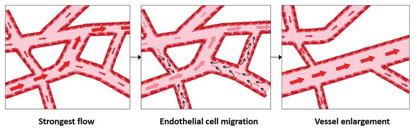
Gifre-Renom and Jones                                                                                                                                   Vessel Enlargement

  FIGURE 1 | Migration-induced vessel enlargement. Under low shear stress, endothelial cells migrate against flow and stop or reduce migration when exposed to
  shear stresses at or above their shear stress physiological levels. In this way, endothelial cells accumulate in vessels under higher shear stress, leading to vessel
  enlargement.

a peak at 1 dyn/cm2. Higher shear stress levels return migration                         in a zebrafish AVM model lacking ALK1, respectively. Recently,
rates to static levels (Tabibian et al., 2020). This information                         our group has identified that, under shear stress, SMAD1/5/9
was then used to build the computational model where shear-                              (transcription factors in this pathway) control the expression
stress levels defined the speed of migration, but the direction                          of the gene (GJA4) for the gap junction protein Cx37
of migration was influenced both by the direction of the flow                            (Connexin37; Peacock et al., 2020). We also found that Cx37
and a requirement for collective cell movement. This alone                               has a critical role in the maintenance of the directionality
gave modest predictive ability, which was improved by the                                of endothelial cell migration under flow. Interestingly,
addition of growth of avascular regions and, more surprisingly,                          mechanotransduction by the primary cilium also functions
by the addition of endothelial cell elongation in the direction                          through the ALK1 pathway, with the primary cilium increasing
of flow. Although the role of cell elongation in remodeling is                           BMP9 responsiveness of endothelial cells under low shear
currently unexplored, studies on mice where endothelial cells                            stress, thereby decreasing their migration speed. This may
cannot elongate did not report vessel enlargement defects                                prevent premature vascular regression under low shear stress
(Baeyens et al., 2014; Corti et al., 2019). Those results, however,                      processes, such as during the initial remodeling of the retinal
also found that shear stress magnitude is sensed apart from                              vascular network (Vion et al., 2018).
shear stress direction (Baeyens et al., 2014), consistent with                               Though regressing cells are a source of endothelial cells for
our computational results.                                                               enlarging vessels, the venous vascular bed also contributes cells.
   In order to migrate against flow, endothelial cells are first                         Notably, proliferation is higher in venous endothelial cells than
polarized against the direction of flow (Franco et al., 2015).                           in arterial endothelial cells (Red-Horse et al., 2010; Ehling et al.,
Labeling the Golgi and nucleus allows endothelial cell polarization                      2013) and, therefore, venous cells provide a source of cells for
to be visualized. By comparing polarization to predicted flow                            the migration model of vessel enlargement (Figure 2). Coup-
patterns, endothelial cells were shown to be polarized against                           TFII, which is one of the most important venous transcription
flow in the developing retina (Franco et al., 2015). A key player                        factors, can repress the expression of Fms Related Receptor
in the maintenance of the endothelial polarization, first identified                     Tyrosine Kinase 1 (FLT1; also known as VEGFR1, Vascular
in the retina, is the primary cilium of endothelial cells (Vion                          Endothelial Growth Factor Receptor 1) and Notch, thereby
et al., 2018). Primary cilia are present on endothelial cells when                       promoting endothelial cell proliferation (You et al., 2005; Qin
exposed to low shear stress, whereas higher levels of shear stress                       et al., 2010; Chen et al., 2012). In the mouse embryonic heart,
often cause disassembly of the cilia (Iomini et al., 2004; Egorova                       for instance, coronary arteries have been shown to form from
et al., 2011; Ten Dijke et al., 2012). By a genetic deletion of                          venous endothelial cells (Su et al., 2018). In the mouse retina,
the essential cilia component intraflagellar transport protein 88                        labeling tip cells permitted to observe the integration of the
(IFT88), Vion et al. (2018) described a random and premature                             labeled cells in growing arteries, but rarely into the venous vascular
regression of blood vessels related to an increase in migration                          bed (Xu et al., 2014; Pitulescu et al., 2017). This has led to the
and a reduced polarization of the endothelial cells.                                     proposal that endothelial cells proliferate in veins, migrate from
   The Bone Morphogenetic Protein/Activin receptor-like                                  low shear stress veins through the capillary bed and then ultimately
kinase 1/Endoglin (BMP/ALK1/ENG) pathway is also required                                stop migrating in the arterial vascular bed because of high shear
for polarized migration in response to flow (Figure 2).                                  stress levels. They therefore accumulate in this region, inducing
Mutations in the ENG and ACVRL1 genes (encoding for                                      vessel enlargement (Red-Horse and Siekmann, 2019).
ENG and ALK1) cause Hereditary Hemorrhagic Telangiectasia
(HHT), which is a genetic form of AVM development
(McAllister et al., 1994; Johnson et al., 1996). Loss of directional                     Fusion
migration has been reported for ENG (Jin et al., 2017) and                               Vessel fusion was first described over three decades ago but
ALK1 (Rochon et al., 2016) in mouse ENG-knockouts and                                    has received less attention than other mechanisms of vessel

Frontiers in Physiology | www.frontiersin.org                                        3                                           February 2021 | Volume 12 | Article 639645
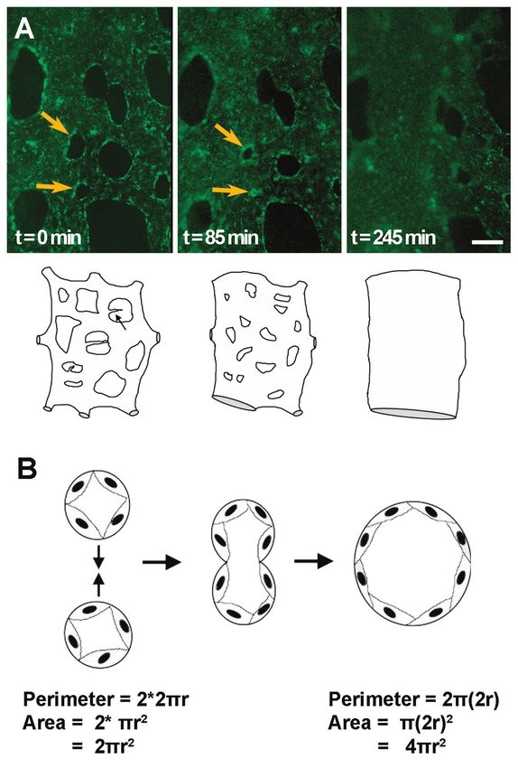
Gifre-Renom and Jones                                                                                                                                    Vessel Enlargement

  FIGURE 2 | Main mechanisms involved in vessel enlargement. Summary of the shear-inducible mechanotransduction pathways involved in migration, fusion,
  hypertrophy and proliferation of endothelial cells, the processes directing vessel enlargement. Blue and green arrows are colored to clarify the trigger of specific
  downstream pathways, being shear stress involved in both cases. Red lines denote inhibition (thus, cell cycle arrest, in the proliferation panel). Discontinued lines
  denote the nuclei membranes.

enlargement. Drake and Little (1995) were first to describe                              reported to form by fusion of smaller capillaries in these
this process. By time-lapsing dorsal aorta development in avian                          transgenic quails (Sato et al., 2010).
embryos, they showed that a capillary bed initially formed                                  Though the initial reports on fusion mainly focused on
along the length of the embryo proper and that with the onset                            the dorsal aorta in avian embryos, this process was later shown
of blood flow, these capillaries merge together forming larger                           to also occur in other models and other vascular beds.
and larger vessels (Figure 3A; Rupp et al., 2004). These                                 In mouse, time-lapse microscopy showed that fusion is the
observations were further confirmed with the development of                              main mechanism by which yolk sac vessel enlarge, leading to
transgenic quail embryos that allowed clear visualizations of                            periodic jumps in vessel diameter rather than smooth linear
the forming dorsal aorta [see movies S3 and S6, (Sato et al., 2010)].                    increases in vessel diameter (Udan et al., 2013). Here, fusion
Moreover, both the vitelline artery and vitelline vein were also                         processes could be identified in the formation of both the

Frontiers in Physiology | www.frontiersin.org                                        4                                            February 2021 | Volume 12 | Article 639645
Gifre-Renom and Jones Vessel Enlargement
capillary vessels joining and fusing into one large diameter
vessel (Azizoglu et al., 2016).
Fusion is a much faster process to enlarge a vessel than
either migration or proliferation. Within hours, two small
vessels can merge leading to a doubling in the radius of the
vessel, but a 4-fold increase in the cross-sectional area for
flow (Figure 3B). This may explain why reports of fusion
have been limited to embryonic vascular beds, where remodeling
must occur much more rapidly than in more mature systems.
It is also possible, thus, that difficulties in detecting fusion
prevent its identification in other vascular beds. Morphologically,
as vessels fuse, the avascular region between the two vessels
becomes smaller and smaller, eventually creating “pillars” of
avascular tissue that are identical to the pillars present in
intussusceptive angiogenesis. As such, static images cannot
differentiate fusion from intussusceptive angiogenesis. Indeed,
fusion may only have been identified in embryonic vasculatures
because these are the vascular beds where time-lapse microscopy
at the resolution of capillaries is possible.
Just as for migration, flow is essential for fusion to occur.
In the aforementioned SEMA3E−/− mutants, the timing of
resolution of the unfused phenotype correlates to the onset
of blood flow (Meadows et al., 2013). In normal vascular
development, fusion occurs in regions with the highest flow,
such as the region where the vitelline artery and vein are
forming (Sato et al., 2010; Chouinard-Pelletier et al., 2013;
Udan et al., 2013). Unexpectedly, if flow patterns are altered
to reduce shear stress, an increased number of fusion events
is observed (Chouinard-Pelletier et al., 2013). If shear stress
levels are increased instead, the opposite occurs, and less fusion
is present. Though these results may appear paradoxical, the
increased flow in the region of the forming vitelline artery
FIGURE 3 | Vessel enlargement by fusion. (A) Capillary plexus vessels and vein do not necessarily mean that increased shear stress
merge to form a larger vessel, as shown by time-lapse microscopy of AcLDL
labeled vitelline vessels in quail embryos. Yellow arrows indicate avascular
drives fusion. Shear stress relates not only to the flow velocity
regions that become smaller and smaller until two adjacent vessels fuse. but also to geometry of the vessels. If an avascular region
Cartoon exemplifies changes that can be observed by time-lapse microscopy. between two fusing vessels acts as an obstruction in the middle
(B) Schematic of the fusion process in transverse view. The same number of of a fast-flowing stream, then, as the velocity of that stream
cells, or perimeter length, leads to a doubling in the area for flow after fusion.
increases, shear stress will be increased on the upstream side
Cartoons adapted from (Drake and Little, 1999).
of the avascular region but decreased on the downstream side
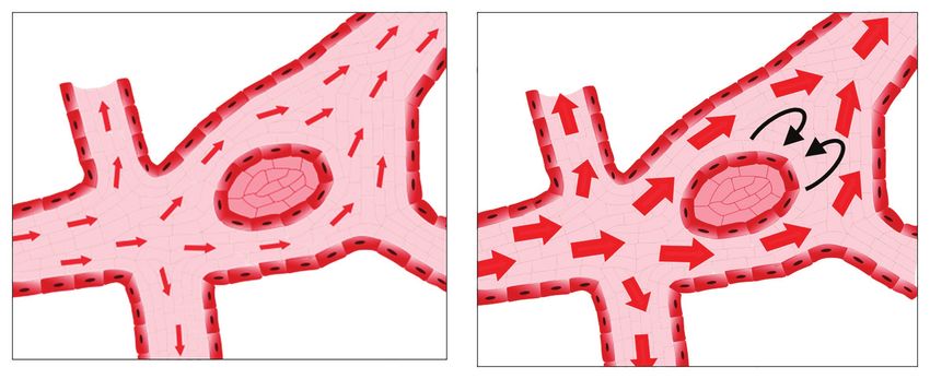
(Figure 4). As such, gradients of shear stress may drive fusion
vitelline artery and the vitelline vein in mouse. Quantification events rather than just increases in the total amount or velocity
of sprouting/regression/fusion/splitting events during remodeling of flow.
of the yolk sac vasculature shows that fusion is more common Given the difficulties in identifying fusion events, very little
than vessel regression in the embryo, but slightly less common is known about the mechanism of fusion. There are various
than angiogenesis (Chouinard-Pelletier et al., 2013). The overall phenotypes, however, that present with a hyperfused vascular
number of fusion events is on the same order of magnitude plexus. Inhibition of Notch in the embryonic yolk sac results
as sprouting and regression events. The coalesce of a capillary in increased number of fusion events (Chouinard-Pelletier
bed along the embryonic midline has never been reported et al., 2013; Caolo et al., 2018). The increased amount of
for the mouse dorsal aorta formation. However, the Semaphorin fusion observed during Notch inhibition can be rescued by
3E (SEMA3E)−/− embryo, involved in repulsive endothelial cell increasing shear stress levels (Caolo et al., 2018). In the chick
guidance, shows a transient phenotype whereby a plexus is embryo yolk sac, exogenous vascular endothelial growth factor
present that eventually coalesces to paired dorsal aortae. This (VEGF) induced increased vascular fusion (Drake and Little,
suggests that vessel coalescence may be retarded in these 1995). Sema3E signaling induces the expression of sFLT1
mutants and, therefore, observable (Meadows et al., 2013). (Zygmunt et al., 2011) and, as such, the SEMA3E−/− should
Vessel enlargement in other organs has also been reported to have reduced VEGF signaling. Though the interplay of VEGF
occur by fusion, or coalescence, of smaller vessels, such as and Notch signaling is well studied for sprouting angiogenesis
the central pancreatic duct, which develops from smaller (Eichmann and Simons, 2012), why they would induce fusion
Frontiers in Physiology | www.frontiersin.org 5 February 2021 | Volume 12 | Article 639645Gifre-Renom and Jones Vessel Enlargement
FIGURE 4 | Possible low shear stress regions in the presence of increased flow rates. As flow increases, avascular regions can act as obstacles to the flow,
resulting in regions of low shear stress and/or recirculation downstream from the avascular region.
in other situations is a mystery. The key may lie in the role hypoxia (Deindl et al., 2001). In fact, although experimentally
of VEGF in synchronizing Notch signaling (Figure 2). In inhibited VEGF in femoral artery ligation (an occlusion
somitogenesis, Notch is involved in synchronizing cells such model) results in inhibited arteriogenesis (Jacobi et al., 2004;
that all cells cycle together and express the same genes together Lloyd et al., 2005; Toyota et al., 2005), VEGF is not expressed
(Jiang et al., 2000). In endothelial cells, levels of Notch targeting in the tissue near the growing collaterals (Lee et al., 2004).
genes also oscillate, but in an unsynchronized manner. Under Thus, although endothelial cells clearly proliferate within
higher levels of VEGF, however, Notch-induced gene expression the growing arterioles (Scholz et al., 2000), the role of VEGF
synchronizes endothelial cells, favoring vessel enlargement rather is unlikely to be needed for this local proliferation. Instead,
than extension (Ubezio et al., 2016). The act of favoring “self ” VEGF may be needed to induce proliferation distal from
(staying together) rather than “other” (extending a sprout) has the site of arteriogenesis, though it is also possible that
previously been proposed for the role of Notch target genes VEGF could be responsible for the release of blood-marrow
in boundary formation (Ventrella et al., 2017). In this model, derived cells.
Notch synchronization would create a situation where vessel In the case of flow-induced remodeling, VEGF itself is also
coalesce with each other (i.e., fuse), as they prefer adhering not a necessary ligand to activate PI3K/MAPK signaling pathways.
to one another rather than remaining separate. Laminar flow induces a transphosphorylation of VEGF receptor
2 (VEGFR2) that activates the downstream PI3K pathway in
Proliferation a ligand-independent manner (Jin et al., 2003). This leads to
Though no significant proliferation is observed during vessel the phosphorylation of Akt that has pleiotropic effects on
enlargement in developmental models, whether the retina or proliferation. Akt phosphorylation by flow is highest at
the embryo, this is not the case for vessel enlargement in physiological shear stress levels between 10 and 20 dyn/cm2
adult vascular beds. Chronic changes in flow in the adult (Dimmeler et al., 1998; Li et al., 2009), however, these levels
lead to expansion of collateral blood vessels that restore of shear stress are known to induce quiescence and not
normal blood flow levels. This process was termed arteriogenesis proliferation in endothelial cells. VEGFR2 and Akt
(Scholz et al., 2001). After partial coronary ligation model, phosphorylation are, however, short-lived events that occur
endothelial cell proliferation is observed in enlarging vessels, within the first 1–2 h of a change in shear stress (Shay-Salit
peaking 3 weeks after implantation of the constrictor (Schaper et al., 2002; Guo et al., 2007). Oscillatory flow, which in contrast
et al., 1971). In hind limb ischemia models, significant to unidirectional flow does induce increased proliferation, leads
endothelial cell and smooth muscle cell proliferation is observed to prolonged VEGFR2 and Akt phosphorylation (Guo et al.,
in the enlarging vessels within 2–3 days of ligation (Scholz 2007). Furthermore, unidirectional flow also activates other
et al., 2000). Vessel enlargement by proliferation occurs over factors such as AMP-activated protein kinase (AMPK), which
a period of days and weeks, not hours. Though endothelial counteract the pro-proliferative signals (Guo et al., 2007).
cells are clearly proliferating within the growing arterioles Shear stress can also modulate endothelial cell proliferation
(Scholz et al., 2000), this does not exclude a role for migration through other signaling pathways. The phosphorylation of
in collateral enlargement. endothelial Nitric Oxide Synthase (eNOS) and its increased
Vascular endothelial growth factor is most well known activity induced by shear stress was one of the first studied
as a mitogen for endothelial cells proliferation. VEGF acts effects of mechanotransduction (Buga et al., 1991; Kuchan and
by binding to VEGF receptors, and this phosphorylates Frangos, 1994). Nitric oxide (NO) not only induces vasodilation
protein kinases that activate downstream Phosphoinositide but also induces proliferation. In vasodilation, NO is produced
3-kinase (PI3K) and Mitogen-activated protein kinase (MAPK) by endothelial cells, diffuses to smooth muscle cells where it
pathways promoting proliferation (Figure 2). VEGF expression activates soluble guanylyl cyclase by binding to the heme group
is triggered by hypoxia. The proliferation process involved (Zhao et al., 1999). Estimates vary concerning the concentration
in arteriole enlargement, however, does not respond to at which this occurs, however, most publications have placed
Frontiers in Physiology | www.frontiersin.org 6 February 2021 | Volume 12 | Article 639645Gifre-Renom and Jones Vessel Enlargement this between 5 and 100 nM (Chen et al., 2008). As a pro-proliferative Hypertrophy compound, NO acts by controlling protein activation by reacting Endothelial cells have an amazing ability to change their cell with cysteine residues to induce S-nitrosylation. NO leads to size. Vessel enlargement by hypertrophy results in an increase S-nitrosylation of MAPK phosphatase 7 (MKP7), rendering it in the size of individual endothelial cells and can extremely inactive which then prevents the inactivation of c-Jun N-terminal rapidly increase vessel diameter. Endothelial cell density in Kinase 3 (JNK3; Pi et al., 2009). The concentrations at which vessels is very high, meaning that there is a large potential NO induce proliferation are extremely low, in the pico to for growth purely by altering their size. nanomolar ranges that occur due to release of NO by macrophages In normal embryonic development, no differences in cell and endothelial cells (Ridnour et al., 2005; Thomas et al., 2008; density are observed along vessel growth based on somite stage. Figure 2). At concentrations in the micromolar range, NO inhibits Nonetheless, between large and small vessels a 30% reduction proliferation and induces cell cycle arrest in several cell types in endothelial cell density was reported (Udan et al., 2013). (Gooch et al., 1997; Heller et al., 1999). The difference, however, was not large enough to explain the Though the ALK1/ENG complex affects endothelial cell difference in vessel diameter, suggesting that size of endothelial migration, this signaling pathway also has a key role in regulating cells might be involved. Similarly, in the retina there is no the proliferation of endothelial cells (Goumans et al., 2002; overall change in endothelial cells density as large vessels form, David et al., 2007). The ALK1/ENG complex is, thus, a key but in this case, the endothelial cell density was not compared component through which shear stress can block endothelial between large and small vessels (Franco et al., 2015). In cells from entering the cell cycle (Baeyens et al., 2016; Figure 2). transgenic zebrafish embryos with constitutive or inducible By recognizing both BMP9 and flow, the ALK1/ENG complex expression of Placental Growth Factor (PLGF) under control allows the modulation of vascular morphogenesis in response of a somite muscle-specific promoter (PLGFmusc), cell size was to flow (Baeyens et al., 2016). Mutations in SMAD4 and in found to contribute to vessel enlargement but, importantly, the Growth differentiation factor 2 (GDF2) genes (encoding alone could not account for the diameter increase (Klems et al., for SMAD4 and BMP9) had been also reported later on to 2020). As such, in normal vascular development as well, cause variants of HHT. Both the endothelial-specific ablation endothelial cell hypertrophy appears to contribute to vessel of SMAD4 (a transcription factor in the ALK1/ENG pathway) enlargement but never acts alone. and of ENG show increased proliferation of endothelial cells Though endothelial cell size increases appear to play a within the developing shunt (Ola et al., 2018; Tual-Chalot lesser role in developmental vessel enlargement, this process et al., 2020). The later, however, has been described to involve could still contribute to pathological vessel enlargement. In the VEGF signaling pathway (Tual-Chalot et al., 2020). an AVM model of constitutive expression of active Notch4, Transforming Growth Factor-β (TGF-β) is involved in the pathological vessel enlargement occurs not due to an increase maintenance of the endothelium in a nonactivated state (Walshe in endothelial cell density nor proliferation but, instead, due et al., 2009) and protecting it from aberrant permeability to an increased size of individual endothelial cells (Murphy and perfusion, and from apoptosis (Walshe et al., 2011). et al., 2014). In zebrafish embryos, TRIO (Trio Rho Guanine Interestingly, experiments on HUVECs demonstrated that Nucleotide Exchange Factor) activation, which in turn activates shear stress activates TGF-β, leading to downstream activation Ras homologous (Rho) GTPases, also leads to increased cell of Krüppel-Like Factor 2 (KLF2) and eNOS in an ALK5 size causing an enlargement in arterial caliber (Klems et al., dependent manner (Walshe et al., 2013; Figure 2). Moreover, 2020). Interestingly, these results may be linked because shear- TGF-β malfunction through the SMAD signaling pathway induced Notch activation has been shown to regulate and has been linked to diverse cerebrovascular diseases related activate the assembly of a VE-Cadherin/LAR (leukocyte to aberrant endothelial cell proliferation (including HHT), antigen-related protein tyrosine phosphatase)/TRIO complex as reviewed by Zhang and Yang (2020). (Polacheck et al., 2017; Figure 2). Other AVM models, such Flow can also modulate the endothelial cell cycle through as the endothelial cell specific knockout of ENG (Choi et al., other pathways. For example, in bovine aortic endothelial cells, 2014), also present with an increase in endothelial cell size, 24 h of laminar shear stress (3–12 dyn/cm2) activated the both in zebrafish and mouse embryos (Sugden et al., 2017). phosphorylation of p53 protein through the JNK pathway. The Overall, however, the contribution of cell size changes to increased levels of p53 in turn activated Growth Arrest and vessel growth is rarely assessed. DNA Damage 45 (GADD45) and p21 proteins, inhibiting the Cyclin-Dependent Kinase (CDK) and, thus, arresting endothelial cell proliferation (Lin et al., 2000; Figure 2). Transcription ENLARGEMENT OF VEINS factor Aryl hydrocarbon receptor (AhR) is also sensitive to shear stress. In this case, laminar shear stress between 6 and The process of vessel enlargement is referred to as arteriogenesis 15 dyn/cm2 induced –likely also through the JNK/p38 pathway– when it occurs in the mature arterial vascular network, but the expression and the translocation of AhR into the nucleus. veins can also increase in diameter. The process of increasing In the nucleus, AhR promotes an increase in Cytochrome P450 venous diameter is so understudied that it lacks a name, though Family 1 Subfamily A Member 1 (CYP1A1) expression and it is occasionally referred to as venogenesis. the subsequent shear stress-induced arrest of the cell cycle In vascular development, fusion has been reported as the (Han et al., 2008; Figure 2). predominant mechanism of venous vessel enlargement in both Frontiers in Physiology | www.frontiersin.org 7 February 2021 | Volume 12 | Article 639645
Gifre-Renom and Jones                                                                                                               Vessel Enlargement

mouse and chicken embryos (le Noble et al., 2004; Udan et al., 2013).        immune cell recruitment, increased vessel leakiness and loss
In the chick embryo, the vitelline vein arises from a region that            of smooth muscle cells (Segiet et al., 2015). Varicose veins
is genetically arterial before vessel enlargement begins (le Noble           have upregulated Notch pathway genes like Delta Like Canonical
et al., 2004). In the developing retina, endothelial specific ablation       Notch Ligand 4 (DLL4), Hairy/enhancer-of-split related with
of Cell Division Control protein 42 (CDC42 homolog) impairs                  YRPW motif protein 2 (HEY2), and EPHB2 (Surendran et al.,
migration, and enlarged veins and capillaries form without arterial          2016). Smooth muscle cells become enlarged and surrounded
enlargement (Lavina et al., 2018). This increased diameter was               by an increased amount of extracellular matrix, suggesting
attributed to the presence of an increase in the number of                   that they de-differentiate into a synthetic phenotype (Wali and
endothelial cells per vessel length, without an increase in                  Eid, 2001). Matrix Metalloproteinases (MMPs) and Tissue
venous proliferation.                                                        Inhibitors of Metalloproteinases (TIMPs) play an important
    Understanding the process of vein enlargement requires                   role in the development of varicose veins. Increases in TIMP-1
mutants that specifically show changes in the diameter of                    levels and in the TIMP/MMP-2 ratio lead to an increase in
veins. As such, somatic mutations that lead to venous                        extracellular matrix deposition and a decrease in degradation
malformations can be especially informative. Venous                          processes (Badier-Commander et al., 2000). The mechanism
malformations are enlarged veins that present with few mural                 of enlargement for varicose vein is, therefore, much more akin
cells. The most well studied somatic mutations in venous                     to collateral vessel enlargement than to vessel enlargement in
malformation are the ones related to the Tyrosine-protein                    developmental models.
kinase (Tie2) receptor (Limaye et al., 2009; Soblet et al., 2013).
Constitutive ablation of TIE2 leads to embryonic lethality at
E10.5 (Sato et al., 1995). If TIE2 is ablated at later stages,               DIFFERENCES BETWEEN VESSEL
however, arteries continue to form but veins do not (Chu                     ENLARGEMENT IN THE ADULT AND
et al., 2016). This is associated with a loss of venous markers
[EphB4 (Ephrin type-B receptor 4), APJ (apelin receptor)]
                                                                             EMBRYO
without any increase in arterial markers, indicating that this               Vessel enlargement occurs during embryonic development but
may be a defect in venous specification rather than vessel                   continues to occur in the adult vascular networks. Any time
enlargement. Constitutively active forms of Tie2 that replicate              the vasculature is exposed to a chronic change in flow, the
somatic TIE2 mutations found in venous malformations, cause                  vasculature adapts through either enlargement or inward
increased migration but loss of proper polarization in endothelial           remodeling to accommodate the altered flow. The most common
cells in vitro (Cai et al., 2019). However, it is not clear whether          experimental model for vessel enlargement in the adult is the
loss of proper cell identity or improper migration lead to                   remodeling of collateral vessels after occlusion, whether by
vessel enlargement in venous malformations.                                  partial occlusion of a coronary artery or by ligation of the
    Though venous malformations arise from veins, some argue                 femoral or middle cerebral artery. Other models, such as
that all AVMs originate from venous endothelial cells. In                    arteriovenous fistulas, have also been used to study the
biopsies from patients with telangiectasias, the enlargement of              mechanisms of vessel enlargement. Vessel enlargement post-
post-capillary venules precedes AVM formation (Braverman                     natally is inherently different than in developmental models,
et al., 1990). In mouse models, exogenous expression of activated            such as the retina, since post-natal enlargement requires the
Notch4 induces AVM formation (Murphy et al., 2014). However,                 degradation of an existing basement membrane as well as the
when this expression is limited to arteries, no AVMs form                    detachment and proliferation of mural cells. Furthermore, the
(Murphy et al., 2014). Deletion of the ENG only in venous                    remodeling of adult vessels, such as the collaterals, is initiated
and capillary endothelial cells results in the same rate of AVM              by endothelial cell activation, leukocyte invasion, and proliferation
formation as for deletion in all endothelial cells (Singh et al.,            of vascular cells (Ma and Bai, 2020). This leads to the question,
2020). Conversely, in retinas of an endothelial-specific ENG                 what parallels exist between vessel enlargement in developmental
knockout model, imaging of developing AVMs showed that                       models and in adult models? And though there may be more
these initiated in arterioles and grew toward the venous                     than one way in which a vessel enlarges, it is likely that there
vasculature (Jin et al., 2017). Although this would suggest an               will be common components from which we can gain
arterialization of the capillaries, in mouse models of AVM                   significant insight.
formation, the AVMs themselves express venous markers and
downregulate arterial marker (Ola et al., 2018).
    Another venous malformation that leads to increased                      Endothelial Cell Activation
venogenesis is varicose vein development. Though most research               In femoral artery ligation, arteriogenesis occurs far away from
on vessel enlargement focuses on the role of shear stress as                 where ischemia is occurring (Resnick et al., 2003; Pipp et al.,
the stimuli for enlargement, varicose vein development occurs                2004). As such, just as with developmental angiogenesis, it is
due to defective valves leading to an increase in hydrodynamic               a process driven by shear stress. Just after coronary occlusion,
pressure (Welkie et al., 1992; Nicolaides et al., 1993). It should           the endothelial cells in the collateral vessel lose alignment
be noted, however, that the increased pressure in veins also                 with flow and take on a bulged appearance (Cai and Schaper,
leads to altered shear stress. On a cellular level, varicose vein            2008). These cells increase DNA synthesis (Schaper et al., 1971;
development involves activation of endothelial cells leading to              Pasyk et al., 1982) and proliferation, as indicated by

Frontiers in Physiology | www.frontiersin.org                            8                                    February 2021 | Volume 12 | Article 639645
Gifre-Renom and Jones Vessel Enlargement
Bromodeoxyuridine (BrdU) incorporation or Ki67 staining recruitment in the adult impairs arteriogenesis during collateral
(Arras et al., 1998b; Wolf et al., 1998). Interestingly, these are remodeling (Heil et al., 2002, 2004). Recruited monocytes produce
all behaviors associated with low or recirculating shear stress Tumor necrosis factor-α (TNF-α) and VEGF, which induces
patterns and not with increased shear stress levels. Adult endothelial and smooth muscle cell mitoses (Schaper and Ito, 1996).
endothelial cells, however, are adapted to the flow to which Though essential when vessel enlarge post-natally, functional
they are exposed and become activated in response to altered immune cells may not be as present in vessel enlargement
flow (Ward et al., 2020). Furthermore, though physiological occurring just after the onset of flow in the embryo. The first
shear stress reduces activation and proliferation, when shear immune cells form at E8.5 in the form of erythromyeloid
levels are extremely elevated (above 30 dyn/cm2 in humans), progenitor and primitive macrophages (Gomez Perdiguero et al.,
the response is outward remodeling (Dolan et al., 2013). Thus, 2015), and no functional role for these progenitors has been
difference between developmental and adult remodeling may established until much later in development. At Hamburger
arise from either one of the following facts. On the one hand, Hamilton stage 18 in avian embryos (equivalent to E12.5–13.5 in
developmental vasculature is naïve and therefore, it is not mouse), circulating phagocytic cells are recruited to sites of
adapting to its “expected” flow; on the other hand, the stimulus vascular remodeling (Al-Roubaie et al., 2012). In the zebrafish
for remodeling is a physiological level of shear stress (i.e., embryo, depletion of myeloid cells using a pu.1 morpholino
15 dyn/cm2) for vessel enlargement during development, but inhibits collateral growth in the gridlock mutant embryo (Gray
an acute non-physiological level of shear stress (above 30 dyn/ et al., 2007). However, both these reports were for embryos
cm2) in models of vessel enlargement in the adult. at stages much older than the ones that gave the results showing
The activated endothelium produces NO that is essential vessel enlargement by migration and/or fusion (Chouinard-
for collateral growth. Both eNOS and iNOS (respectively, Pelletier et al., 2013). The retinal vasculature does form post-
endothelial and inducible NOS) are upregulated in remodeling natally, when resident myeloid cells are present in the retina
collateral vessel (Cai et al., 2004; Yang et al., 2015). When (Haupt et al., 2019). Ablation of macrophages using chlodronate
NO production is inhibited with L-NAME [N(G)-nitro liposomes results in a dramatic loss of vascular density, which
L-arginine methyl ester], there is an almost complete inhibition makes it difficult to assess whether vessel enlargement itself
of collateral enlargement (Eitenmuller et al., 2006; Park et al., is affected (Checchin et al., 2006).
2010). It is not clear, however, whether this is due to a true One of the roles of immune cells in arteriogenesis is to
inhibition of growth or related to increased vasoconstriction degrade the basement membrane. During arteriogenesis, the
(Cai and Schaper, 2008). Endothelial NOS itself is involved elastic lamina is broken down by MMPs to give the vessels
in maintaining collateral vessel under physiological conditions. room to expand (Haas et al., 2007; Dodd et al., 2011).
Mice that lack eNOS are born with a normal number of Inflammatory cells are also an important source of MMPs, as
collateral vessels in the brain, but the number of these vessels well as of other proteases. Macrophages secrete cytokines that
decreases over the first 6 months of life as compared to induce MMP expression by endothelial cells (Galis et al., 1994a).
age-matched controls (Dai and Faber, 2010). The primary Vessel enlargement in response to arteriovenous fistula induces
role of NO in vessel enlargement is in the recruitment of a more than 1700-fold increase in MMP-2, along with a 12–60-
immune cells (Park et al., 2010). Delivery of NO donors fold increase in MMP-9, Membrane-type 1 MMP (MT1-MMP),
induces VE-Cadherin disassembly that is necessary for immune and TIMP-2 (Sho et al., 2002). These increases correlate to
cell recruitment. Conversely, NO inhibitor L-NAME prevents the timing of elastic lamina degradation (Sho et al., 2002).
increased vessel permeability and immune cell recruitment Notably, MMPs are produced as inactive zymogens and, in
after vessel occlusion (Yang et al., 2015). Though NO is critical addition, TIMPs can inhibit their activity. Therefore, increased
in vessel enlargement in post-natal stages, it has not been expression of MMPs does not necessarily indicate increased
extensively investigated during development. The triple knockout activity. Indeed, many MMPs are constitutively expressed by
of NOS enzymes is born at normal mendelian frequency endothelial cells and smooth muscle cells (Hanemaaijer et al.,
with no reported vascular defects at birth (Morishita et al., 1993; Galis et al., 1994a) but show no enzymatic activity until
2005). Conversely, however, culture of E8.5 mouse embryos activated by disease (Galis et al., 1994b, 1995). Though essential
with the NO inhibitor L-NMMA [N(G)-monomethyl L-arginine] in adult remodeling, no single mutant of MMPs has shown
prevents the formation of large vessels in the yolk sac vasculature defects during embryogenesis. The double mutant of MMP2
(Nath et al., 2004). NO influences endothelial cell proliferation and MT1-MMP does die perinatally, with a defect in the
(Morbidelli et al., 1996) and migration (Noiri et al., 1998). formation of vessels with wider diameters (Oh et al., 2004).
As such, NO could have a role in several of the mechanisms This is, however, a very late stage of vascular development,
by which vessel enlarge. after initial vascular remodeling has occurred.
One of the effects of immune cell recruitment and matrix
Immune Cells and Matrix Degradation degradation is an increase in permeability. Middle cerebral artery
Another difference between adult and developmental vessel occlusion leads to an increase in permeability in the blood-
enlargement is the involvement of immune cells. Immune brain-barrier to large molecules such as fibrinogen,
cells, especially monocytes and macrophages, are essential Immunoglobulin G (IgG) or nanoparticles within 2–4 h of the
for adult collateral growth and vessel enlargement in general occlusion (Okada et al., 1994; Fischer et al., 2002). Degradation
(Arras et al., 1998a; Voskuil et al., 2003). Inhibiting monocyte is necessary for this increase in permeability, as inhibiting MMPs
Frontiers in Physiology | www.frontiersin.org 9 February 2021 | Volume 12 | Article 639645Gifre-Renom and Jones Vessel Enlargement
with BB-1101 prevents permeability increases immediately after along all the vasculature lifetime, dysregulations may entail
middle cerebral artery occlusion (Rosenberg et al., 1998). critical pathologies. However, more and more pathways and
The presence of this extensive basement membrane is one molecular interconnections are being uncovered, shedding light
of the main reasons that post-natal vessel enlargement is unlike to a better understanding and control over these pathologies.
to occur by vascular fusion. The presence of extensive matrix
and mural cells from the arterioles onward would be a physical
barrier for fusion. Hence, vascular fusion could only occur AUTHOR CONTRIBUTIONS
on the capillary level allowing arterioles to grow, which would
then have to further increase in diameter by combination with LG-R and EAVJ contributed to writing, editing, and making
another mechanism. figures. All authors contributed to the article and approved
the submitted version.
CONCLUSION
FUNDING
Vessel enlargement plays a critical role both during development
as well as in the adult vasculature, with a high capacity for This work was supported by the Fonds Wetenschappelijk
adapting to flow changes. Through an extremely fast Onderzoek (G091018N and G0B5920N) and by internal funding
responsiveness and interconnected processes such as fusion, from the KU Leuven (IDN/19/031 and C14/19/095). This project
endothelial cell migration and proliferation, vessels reshape in has also received funding from the European Union’s Horizon
order to accommodate changes in flow rates such as to restore 2020 research and innovation program under grant agreement
physiological levels. Because these processes are so much present No 848109.
REFERENCES Cai, W., and Schaper, W. (2008). Mechanisms of arteriogenesis. Acta Biochim.
Biophys. Sin. 40, 681–692. doi: 10.1093/abbs/40.8.681
Al-Roubaie, S., Hughes, J. H., Filla, M. B., Lansford, R., Lehoux, S., and Cai, Y., Schrenk, S., Goines, J., Davis, G. E., and Boscolo, E. (2019). Constitutive
Jones, E. A. (2012). Time-lapse microscopy of macrophages during embryonic active mutant TIE2 induces enlarged vascular lumen formation with loss
vascular development. Dev. Dyn. 241, 1423–1431. doi: 10.1002/dvdy.23835 of Apico-basal polarity and Pericyte recruitment. Sci. Rep. 9:12352. doi:
Arras, M., Ito, W. D., Scholz, D., Winkler, B., Schaper, J., and Schaper, W. 10.1038/s41598-019-48854-2
(1998a). Monocyte activation in angiogenesis and collateral growth in the Caolo, V., Peacock, H. M., Kasaai, B., Swennen, G., Gordon, E., Claesson-Welsh, L.,
rabbit hindlimb. J. Clin. Invest. 101, 40–50. doi: 10.1172/JCI119877 et al. (2018). Shear stress and VE-cadherin. Arterioscler. Thromb. Vasc. Biol.
Arras, M., Strasser, R., Mohri, M., Doll, R., Eckert, P., Schaper, W., et al. 38, 2174–2183. doi: 10.1161/ATVBAHA.118.310823
(1998b). Tumor necrosis factor-alpha is expressed by monocytes/macrophages Checchin, D., Sennlaub, F., Levavasseur, E., Leduc, M., and Chemtob, S. (2006).
following cardiac microembolization and is antagonized by cyclosporine. Potential role of microglia in retinal blood vessel formation. Invest. Ophthalmol.
Basic Res. Cardiol. 93, 97–107. doi: 10.1007/s003950050069 Vis. Sci. 47, 3595–3602. doi: 10.1167/iovs.05-1522
Azizoglu, D. B., Chong, D. C., Villasenor, A., Magenheim, J., Barry, D. M., Chen, K., Pittman, R. N., and Popel, A. S. (2008). Nitric oxide in the vasculature:
Lee, S., et al. (2016). Vascular development in the vertebrate pancreas. Dev. where does it come from and where does it go? A quantitative perspective.
Biol. 420, 67–78. doi: 10.1016/j.ydbio.2016.10.009 Antioxid. Redox Signal. 10, 1185–1198. doi: 10.1089/ars.2007.1959
Badier-Commander, C., Verbeuren, T., Lebard, C., Michel, J. B., and Jacob, M. P. Chen, X., Qin, J., Cheng, C. M., Tsai, M. J., and Tsai, S. Y. (2012). COUP-
(2000). Increased TIMP/MMP ratio in varicose veins: a possible explanation TFII is a major regulator of cell cycle and notch signaling pathways. Mol.
for extracellular matrix accumulation. J. Pathol. 192, 105–112. doi: Endocrinol. 26, 1268–1277. doi: 10.1210/me.2011-1305
10.1002/1096-9896(2000)9999:99993.0.CO;2-1 Choi, E. J., Chen, W., Jun, K., Arthur, H. M., Young, W. L., and Su, H. (2014).
Baeyens, N., Larrivee, B., Ola, R., Hayward-Piatkowskyi, B., Dubrac, A., Huang, B., Novel brain arteriovenous malformation mouse models for type 1 hereditary
et al. (2016). Defective fluid shear stress mechanotransduction mediates hemorrhagic telangiectasia. PLoS One 9:e88511. doi: 10.1371/journal.pone.0088511
hereditary hemorrhagic telangiectasia. J. Cell Biol. 214, 807–816. doi: 10.1083/ Chouinard-Pelletier, G., Jahnsen, E. D., and Jones, E. A. (2013). Increased
jcb.201603106 shear stress inhibits angiogenesis in veins and not arteries during vascular
Baeyens, N., Mulligan-Kehoe, M. J., Corti, F., Simon, D. D., Ross, T. D., development. Angiogenesis 16, 71–83. doi: 10.1007/s10456-012-9300-2
Rhodes, J. M., et al. (2014). Syndecan 4 is required for endothelial alignment Chu, M., Li, T., Shen, B., Cao, X., Zhong, H., Zhang, L., et al. (2016). Angiopoietin
in flow and atheroprotective signaling. Proc. Natl. Acad. Sci. U. S. A. 111, receptor Tie2 is required for vein specification and maintenance via regulating
17308–17313. doi: 10.1073/pnas.1413725111 COUP-TFII. elife 5:e21032. doi: 10.7554/eLife.21032
Beebe-Dimmer, J. L., Pfeifer, J. R., Engle, J. S., and Schottenfeld, D. (2005). Corti, F., Wang, Y., Rhodes, J. M., Atri, D., Archer-Hartmann, S., Zhang, J.,
The epidemiology of chronic venous insufficiency and varicose veins. Ann. et al. (2019). N-terminal syndecan-2 domain selectively enhances 6-O heparan
Epidemiol. 15, 175–184. doi: 10.1016/j.annepidem.2004.05.015 sulfate chains sulfation and promotes VEGFA165-dependent neovascularization.
Braverman, I. M., Keh, A., and Jacobson, B. S. (1990). Ultrastructure and Nat. Commun. 10:1562. doi: 10.1038/s41467-019-09605-z
three-dimensional organization of the telangiectases of hereditary hemorrhagic Cui, C., Filla, M. B., Jones, E. A., Lansford, R., Cheuvront, T., Al-Roubaie, S.,
telangiectasia. J. Invest. Dermatol. 95, 422–427. doi: 10.1111/1523-1747. et al. (2013). Embryogenesis of the first circulating endothelial cells. PLoS One
ep12555569 8:e60841. doi: 10.1371/journal.pone.0060841
Buga, G. M., Gold, M. E., Fukuto, J. M., and Ignarro, L. J. (1991). Shear Dai, X., and Faber, J. E. (2010). Endothelial nitric oxide synthase deficiency
stress-induced release of nitric oxide from endothelial cells grown on beads. causes collateral vessel rarefaction and impairs activation of a cell cycle
Hypertension 17, 187–193. doi: 10.1161/01.hyp.17.2.187 gene network during arteriogenesis. Circ. Res. 106, 1870–1881. doi: 10.1161/
Cai, W. J., Kocsis, E., Luo, X., Schaper, W., and Schaper, J. (2004). Expression CIRCRESAHA.109.212746
of endothelial nitric oxide synthase in the vascular wall during arteriogenesis. David, L., Mallet, C., Mazerbourg, S., Feige, J. J., and Bailly, S. (2007). Identification
Mol. Cell. Biochem. 264, 193–200. doi: 10.1023/b:mcbi.0000044388.27953.a0 of BMP9 and BMP10 as functional activators of the orphan activin receptor-
Frontiers in Physiology | www.frontiersin.org 10 February 2021 | Volume 12 | Article 639645Gifre-Renom and Jones Vessel Enlargement like kinase 1 (ALK1) in endothelial cells. Blood 109, 1953–1961. doi: 10.1182/ activated protein kinase and Akt pathways. Circ. Res. 100, 564–571. doi: blood-2006-07-034124 10.1161/01.RES.0000259561.23876.c5 Deindl, E., Buschmann, I., Hoefer, I. E., Podzuweit, T., Boengler, K., Vogel, S., Haas, T. L., Doyle, J. L., Distasi, M. R., Norton, L. E., Sheridan, K. M., and et al. (2001). Role of ischemia and of hypoxia-inducible genes in arteriogenesis Unthank, J. L. (2007). Involvement of MMPs in the outward remodeling after femoral artery occlusion in the rabbit. Circ. Res. 89, 779–786. doi: of collateral mesenteric arteries. Am. J. Physiol. Heart Circ. Physiol. 293, 10.1161/hh2101.098613 H2429–H2437. doi: 10.1152/ajpheart.00100.2007 Dimmeler, S., Assmus, B., Hermann, C., Haendeler, J., and Zeiher, A. M. (1998). Han, Z., Miwa, Y., Obikane, H., Mitsumata, M., Takahashi-Yanaga, F., Morimoto, S., Fluid shear stress stimulates phosphorylation of Akt in human endothelial et al. (2008). Aryl hydrocarbon receptor mediates laminar fluid shear stress- cells: involvement in suppression of apoptosis. Circ. Res. 83, 334–341. induced CYP1A1 activation and cell cycle arrest in vascular endothelial Dodd, T., Jadhav, R., Wiggins, L., Stewart, J., Smith, E., Russell, J. C., et al. cells. Cardiovasc. Res. 77, 809–818. doi: 10.1093/cvr/cvm095 (2011). MMPs 2 and 9 are essential for coronary collateral growth and are Hanemaaijer, R., Koolwijk, P., Le Clercq, L., de Vree, W. J., and van Hinsbergh, V. W. prominently regulated by p38 MAPK. J. Mol. Cell. Cardiol. 51, 1015–1025. (1993). Regulation of matrix metalloproteinase expression in human vein doi: 10.1016/j.yjmcc.2011.08.012 and microvascular endothelial cells. Effects of tumour necrosis factor alpha, Dolan, J. M., Kolega, J., and Meng, H. (2013). High wall shear stress and interleukin 1 and phorbol ester. Biochem. J. 296, 803–809. doi: 10.1042/ spatial gradients in vascular pathology: a review. Ann. Biomed. Eng. 41, bj2960803 1411–1427. doi: 10.1007/s10439-012-0695-0 Haupt, F., Krishnasamy, K., Napp, L. C., Augustynik, M., Limbourg, A., Drake, C. J., and Little, C. D. (1995). Exogenous vascular endothelial growth Gamrekelashvili, J., et al. (2019). Retinal myeloid cells regulate tip cell factor induces malformed and hyperfused vessels during embryonic selection and vascular branching morphogenesis via notch ligand Delta-like 1. neovascularization. Proc. Natl. Acad. Sci. U. S. A. 92, 7657–7661. doi: 10.1073/ Sci. Rep. 9:9798. doi: 10.1038/s41598-019-46308-3 pnas.92.17.7657 Heil, M., Ziegelhoeffer, T., Pipp, F., Kostin, S., Martin, S., Clauss, M., et al. Drake, C. J., and Little, C. D. (1999). VEGF and vascular fusion: implications (2002). Blood monocyte concentration is critical for enhancement of collateral for normal and pathological vessels. J. Histochem. Cytochem. 47, 1351–1356. artery growth. Am. J. Physiol. Heart Circ. Physiol. 283, H2411–H2419. doi: Egorova, A. D., Khedoe, P. P., Goumans, M. J., Yoder, B. K., Nauli, S. M., 10.1152/ajpheart.01098.2001 Ten Dijke, P., et al. (2011). Lack of primary cilia primes shear-induced Heil, M., Ziegelhoeffer, T., Wagner, S., Fernandez, B., Helisch, A., Martin, S., endothelial-to-mesenchymal transition. Circ. Res. 108, 1093–1101. doi: 10.1161/ et al. (2004). Collateral artery growth (arteriogenesis) after experimental CIRCRESAHA.110.231860 arterial occlusion is impaired in mice lacking CC-chemokine receptor-2. Ehling, M., Adams, S., Benedito, R., and Adams, R. H. (2013). Notch controls Circ. Res. 94, 671–677. doi: 10.1161/01.RES.0000122041.73808.B5 retinal blood vessel maturation and quiescence. Development 140, 3051–3061. Heller, R., Polack, T., Grabner, R., and Till, U. (1999). Nitric oxide inhibits doi: 10.1242/dev.093351 proliferation of human endothelial cells via a mechanism independent of Eichmann, A., and Simons, M. (2012). VEGF signaling inside vascular endothelial cGMP. Atherosclerosis 144, 49–57. cells and beyond. Curr. Opin. Cell Biol. 24, 188–193. doi: 10.1016/j. Hughes, S., and Chang-Ling, T. (2000). Roles of endothelial cell migration and ceb.2012.02.002 apoptosis in vascular remodeling during development of the central nervous Eitenmuller, I., Volger, O., Kluge, A., Troidl, K., Barancik, M., Cai, W. J., et al. system. Microcirculation 7, 317–333. doi: 10.1080/mic.7.5.317.333 (2006). The range of adaptation by collateral vessels after femoral artery Iomini, C., Tejada, K., Mo, W., Vaananen, H., and Piperno, G. (2004). Primary occlusion. Circ. Res. 99, 656–662. doi: 10.1161/01.RES.0000242560.77512.dd cilia of human endothelial cells disassemble under laminar shear stress. J. Fischer, S., Wobben, M., Marti, H. H., Renz, D., and Schaper, W. (2002). Cell Biol. 164, 811–817. doi: 10.1083/jcb.200312133 Hypoxia-induced hyperpermeability in brain microvessel endothelial cells Jacobi, J., Tam, B. Y., Wu, G., Hoffman, J., Cooke, J. P., and Kuo, C. J. involves VEGF-mediated changes in the expression of zonula occludens-1. (2004). Adenoviral gene transfer with soluble vascular endothelial growth Microvasc. Res. 63, 70–80. doi: 10.1006/mvre.2001.2367 factor receptors impairs angiogenesis and perfusion in a murine model Franco, C. A., Jones, M. L., Bernabeu, M. O., Geudens, I., Mathivet, T., Rosa, A., of hindlimb ischemia. Circulation 110, 2424–2429. doi: 10.1161/01. et al. (2015). Dynamic endothelial cell rearrangements drive developmental CIR.0000145142.85645.EA vessel regression. PLoS Biol. 13:e1002125. doi: 10.1371/journal.pbio.1002125 Jacobs, B. N., Andraska, E. A., Obi, A. T., and Wakefield, T. W. (2017). Galis, Z. S., Muszynski, M., Sukhova, G. K., Simon-Morrissey, E., Unemori, E. N., Pathophysiology of varicose veins. J. Vasc. Surg. Venous Lymphat. Disord. Lark, M. W., et al. (1994a). Cytokine-stimulated human vascular smooth 5, 460–467. doi: 10.1016/j.jvsv.2016.12.014 muscle cells synthesize a complement of enzymes required for extracellular Jiang, Y. J., Aerne, B. L., Smithers, L., Haddon, C., Ish-Horowicz, D., and matrix digestion. Circ. Res. 75, 181–189. Lewis, J. (2000). Notch signalling and the synchronization of the somite Galis, Z. S., Sukhova, G. K., Lark, M. W., and Libby, P. (1994b). Increased segmentation clock. Nature 408, 475–479. doi: 10.1038/35044091 expression of matrix metalloproteinases and matrix degrading activity in Jin, Y., Muhl, L., Burmakin, M., Wang, Y., Duchez, A. C., Betsholtz, C., et al. vulnerable regions of human atherosclerotic plaques. J. Clin. Invest. 94, (2017). Endoglin prevents vascular malformation by regulating flow-induced 2493–2503. cell migration and specification through VEGFR2 signalling. Nat. Cell Biol. Galis, Z. S., Sukhova, G. K., and Libby, P. (1995). Microscopic localization of 19, 639–652. doi: 10.1038/ncb3534 active proteases by in situ zymography: detection of matrix metalloproteinase Jin, Z. G., Ueba, H., Tanimoto, T., Lungu, A. O., Frame, M. D., and Berk, B. C. activity in vascular tissue. FASEB J. 9, 974–980. (2003). Ligand-independent activation of vascular endothelial growth factor Gomez Perdiguero, E., Klapproth, K., Schulz, C., Busch, K., Azzoni, E., Crozet, L., receptor 2 by fluid shear stress regulates activation of endothelial nitric et al. (2015). Tissue-resident macrophages originate from yolk-sac-derived oxide synthase. Circ. Res. 93, 354–363. doi: 10.1161/01.RES.0000089257. erythro-myeloid progenitors. Nature 518, 547–551. doi: 10.1038/nature13989 94002.96 Gooch, K. J., Dangler, C. A., and Frangos, J. A. (1997). Exogenous, basal, and Johnson, D. W., Berg, J. N., Baldwin, M. A., Gallione, C. J., Marondel, I., flow-induced nitric oxide production and endothelial cell proliferation. J. Yoon, S. J., et al. (1996). Mutations in the activin receptor-like kinase 1 Cell. Physiol. 171, 252–258. gene in hereditary haemorrhagic telangiectasia type 2. Nat. Genet. 13, Goumans, M. J., Valdimarsdottir, G., Itoh, S., Rosendahl, A., Sideras, P., and 189–195. Ten Dijke, P. (2002). Balancing the activation state of the endothelium via Klems, A., van Rijssel, J., Ramms, A. S., Wild, R., Hammer, J., Merkel, M., two distinct TGF-beta type I receptors. EMBO J. 21, 1743–1753. doi: 10.1093/ et al. (2020). The GEF Trio controls endothelial cell size and arterial emboj/21.7.1743 remodeling downstream of Vegf signaling in both zebrafish and cell models. Gray, C., Packham, I. M., Wurmser, F., Eastley, N. C., Hellewell, P. G., Nat. Commun. 11:5319. doi: 10.1038/s41467-020-19008-0 Ingham, P. W., et al. (2007). Ischemia is not required for arteriogenesis in Kuchan, M. J., and Frangos, J. A. (1994). Role of calcium and calmodulin in zebrafish embryos. Arterioscler. Thromb. Vasc. Biol. 27, 2135–2141. doi: flow-induced nitric oxide production in endothelial cells. Am. J. Phys. 266, 10.1161/ATVBAHA.107.143990 C628–C636. Guo, D., Chien, S., and Shyy, J. Y. (2007). Regulation of endothelial cell cycle Lavina, B., Castro, M., Niaudet, C., Cruys, B., Alvarez-Aznar, A., Carmeliet, P., by laminar versus oscillatory flow: distinct modes of interactions of AMP- et al. (2018). Defective endothelial cell migration in the absence of Cdc42 Frontiers in Physiology | www.frontiersin.org 11 February 2021 | Volume 12 | Article 639645
You can also read