Immune Profiling and Precision Medicine in Systemic Lupus Erythematosus - MDPI
←
→
Page content transcription
If your browser does not render page correctly, please read the page content below
cells
Review
Immune Profiling and Precision Medicine in Systemic
Lupus Erythematosus
Yasuo Nagafuchi * , Hirofumi Shoda and Keishi Fujio
 Department of Allergy and Rheumatology, Graduate School of Medicine, The University of Tokyo,
 113-8655 Tokyo, Japan; shoda-tky@umin.ac.jp (H.S.); kfujio-tky@umin.ac.jp (K.F.)
 * Correspondence: nagafuchi-tky@umin.ac.jp; Tel.: +81-3-3815-5411; Fax: +81-3-3815-5954

 Received: 28 December 2018; Accepted: 9 February 2019; Published: 10 February 2019

 Abstract: Systemic lupus erythematosus (SLE) is an autoimmune disorder with a wide range
 of clinical symptoms. Enormous progress has been made in the immunological and genetic
 understanding of SLE. However, the biology of disease heterogeneity in SLE has remained largely
 unexplored. Human immune profiling studies, helped by recent technological advances especially
 in single-cell and “omics” analyses, are now shedding light on the cellular and molecular basis of
 clinical symptoms and disease flares in individual patients. Peripheral blood immunophenotyping
 analysis with flow cytometry or mass cytometry are identifying responsible cell subsets and markers
 characteristic of disease heterogeneity. Transcriptome analysis is discovering molecular networks
 responsible for disease activity, disease subtype and future relapse. In this review, we summarize
 recent advances in the immune profiling analysis of SLE patients and discuss how they will be used
 for future precision medicine.

 Keywords: systemic lupus erythematosus; immune profiling; single-cell analysis; transcriptome;
 precision medicine

1. Introduction
      Systemic lupus erythematosus (SLE) is a chronic autoimmune disease characterized by a broad
spectrum of clinical symptoms and arrays of autoantibodies, such as antinuclear antibodies (ANA)
and anti-double strand DNA (dsDNA) antibodies. Both genetic and environmental factors contribute
to the onset of SLE. Concordance rates of 20 to 30% in monozygotic twins imply the importance of
environmental factors in genetically predisposed individuals [1]. SLE primarily affects women of
childbearing age. The female-to-male ratio of about 9:1 suggest a role of female sex hormones [2].
Exposure to ultraviolet light exacerbates skin disease in SLE and can trigger disease onset by induction
of apoptosis in the skin [3]. Other environmental factors including cigarette smoking, chemicals, drugs,
viral infections and gut microbiota [4] can also act as environmental triggers for SLE.
      In the pathophysiology of SLE, there is an imbalance between cellular apoptosis and disposal
of apoptotic material [5]. During the course of apoptosis, nuclear antigens and nucleic acids become
accessible to the immune system. Persistent apoptotic debris can stimulate an inflammatory response
through the activation of nucleic acid recognition receptors, such as members of the Toll-like receptor
(TLR) family. Nucleic acid recognition receptors are strongly associated with type I interferon (IFN)
production. Type I IFNs and other cytokines promote B-cell differentiation and loss of tolerance. B
cells respond to nucleic acids or nuclear antigens and produce autoantibodies against them. Tolerance
checkpoints can be bypassed when B cells are activated by TLR9 [6] or BLyS/ BAFF, a cytokine
promoting B-cell survival and development [7,8].
      In the 1950s, corticosteroids were introduced for treatment of SLE, and the 5-year survival of SLE
in developed countries gradually increased to >95%, plateauing in the 1990s [9]. Major causes of death

Cells 2019, 8, 140; doi:10.3390/cells8020140                                    www.mdpi.com/journal/cells
Cells 2019, 5, x 2 of 15
In the 1950s, corticosteroids were introduced for treatment of SLE, and the 5-year survival of
Cells 2019, 8, 140 2 of 15
SLE in developed countries gradually increased to >95%, plateauing in the 1990s [9]. Major causes of
death in SLE patients shifted from lupus activity to infection or cardiovascular events that can be
in SLE patients
related to treatment shifted from
[10]. In lupus activity toactive
most patients, infection or cardiovascular
disease is controlled by events that can betreatment
corticosteroid related to
treatment [10].
combined with In most patients, activedrugs
immunosuppressant disease
such is controlled by corticosteroid
as cyclophosphamide treatment
introduced combined
in the with
1970s, with
immunosuppressant
mycophenolate mofetil drugs such asavailable
becoming cyclophosphamide introduced
in 2000s. Treatment in the 1970s,
guidelines with mycophenolate
or recommendations are
mofetil becoming
available from academic available in 2000s.
societies around Treatment
the world guidelines
[11–14]. or recommendations
Treat-to-target has been areproposed,
available with
from
aacademic societies
target of clinical around or
remission thelow
world [11–14].
disease activityTreat-to-target
that predicts has been proposed,
favourable long-termwith a target
outcomes [15].of
clinical remission
However, or low
controlling diseaseactivity
disease activity life-long
that predicts favourable
without or withlong-term
use of outcomes
a minimal [15]. However,
amount of
controlling disease activity
immunosuppressants life-long
is a clinical without or
challenge. The with
goaluse
ofof a minimalremission
prolonged amount of orimmunosuppressants
low disease activity
is achieved
is a clinical challenge. The goal
only in a small of prolonged
proportion remission
of patients or low
[16,17]. disease activity
Unpredictable is achieved
and frequent only inofa
relapses
small proportion
active SLE, flares, of andpatients [16,17].
difficulties Unpredictable
in maintaining and frequent
a state relapses
of remission haveofpersuades
active SLE, flares, and
patients and
difficulties to
physicians in continue
maintaining a state of remission
immunosuppressive have persuades
treatment patients and side
(with accompanying physicians
effects)totocontinue
control
immunosuppressive
disease activity. treatment (with accompanying side effects) to control disease activity.
In addition
In addition to to disease
disease activity,
activity, SLESLE patients
patients are are stratified
stratified and
and treated
treated according
according to to organ
organ
involvement, such as the kidney. The pattern of autoantibodies
involvement, such as the kidney. The pattern of autoantibodies is also correlated to some SLE is also correlated to some SLE
phenotypes [18].
phenotypes [18]. For
Forexample,
example,anti-dsDNA
anti-dsDNAantibodiesantibodiesare areassociated
associatedwith withlupus
lupusnephritis
nephritis[19,20].
[19,20].
However, these
However, these clinical
clinical stratifications do not necessarily reflect the biological heterogeneity heterogeneity of of SLE.
SLE.
Belimumab, aa monoclonal
Belimumab, monoclonal antibody
antibody against
against BLyS/
BLyS/BAFF BAFFwas wasapproved
approved as as the
the first
first targeted
targeted
biological therapy
biological therapyfor for SLE
SLE in in 2011
2011 [21–23].
[21–23]. At At least
least 74
74 targeted
targeted treatments
treatments utilizing
utilizing IFN,
IFN, cytokines,
cytokines,
chemokines, and
chemokines, and B-cells
B-cells are
are under
under clinical
clinical trial
trial for
for SLE
SLE [24]. With
With increasing
increasing treatment
treatment options,
options,
identification of
identification of biomarkers
biomarkers that that stratify
stratify SLE
SLE patients
patients to to adequate
adequate treatment is an urgent need.
AA biomarker
biomarker isis aa characteristic
characteristic that thatcan
can bebe objectively
objectivelymeasured
measuredas asananindicator
indicatorof of aa normal
normal or or
pathological biological
pathological biological processes,
processes, or or asas an
an indicator
indicator of of response
response to to therapy
therapy [25].
[25]. Immune
Immuneprofiling,
profiling, oror
immune cell
immune cell profiling,
profiling, realized
realized by by technological
technological advances
advances in in multi-dimensional
multi-dimensional analysis analysis of of immune
immune
cells, will
cells, will enable deeper understanding
understanding of ofdisease
diseasepathogenesis
pathogenesisand andheterogeneity
heterogeneityand andthethediscovery
discovery of
of novel
novel biomarkers
biomarkers [26].[26]. Prognostic
Prognostic biomarkers
biomarkers to forecast
to forecast the natural
the natural course ofcourse of theorpatient
the patient predictive or
biomarkersbiomarkers
predictive to forecast the to response
forecast to thetherapy
response are necessary
to therapy forare
precisely
necessarytailoredfor treatment,
precisely precision
tailored
treatment,
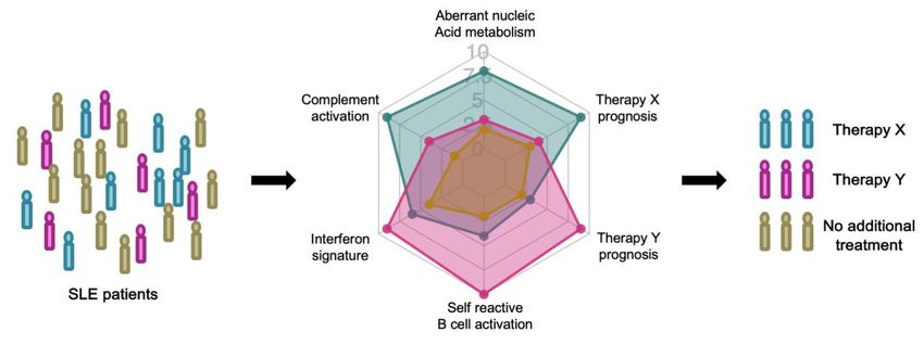
medicine, of precision medicine,
SLE (Figure of SLE (Figure 1) [27].
1) [27].
Figure 1. Stratified treatment of systemic lupus erythematosus (SLE) patients based on prognostic and
Figure 1. Stratified treatment of systemic lupus erythematosus (SLE) patients based on
predictive biomarkers.
prognostic and predictive biomarkers.
In the coming era of precision medicine, SLE patients will be stratified by immune profiling. Each
In the
patient will coming era of precision
be longitudinally medicine,
evaluated SLE patients
with prognostic will be stratified
biomarkers by immune
that predict profiling.
the natural course
Each patient will be longitudinally evaluated with prognostic biomarkers that predict
of the disease. Prognostic biomarkers reflect immunological characteristics, such as a high interferon the
natural course of the disease. Prognostic biomarkers reflect immunological characteristics,
signature. When disease relapse is predicted with high confidence, the patient’s treatment can be such
as abyhigh
guided interferon
predictive signature.
biomarkers thatWhen
projectdisease relapsetoiseach
the response predicted
therapy.with high confidence, the
patient’s treatment can be guided by predictive biomarkers that project the response to each
therapy.Cells 2019, 8, 140 3 of 15
2. Genetics of SLE and Immunological Insights
2.1. Monogenic SLE
Identification of causal mutations in rare monogenic lupus families or lupus-like autoimmune
syndrome gave crucial insights into the pathogenesis of SLE. Lupus-prone families with primary
defects of classical complement pathways (C1q, C1r/s, C2, C4A and C4B) revealed the importance
of this pathway for lupus pathogenesis [28]. Complement is essential for opsonization and clearance
of immune complexes and apoptotic cells. In addition, gene mutations involved in DNA processing
during apoptosis cause lupus-like systemic autoimmunity [29]. Pediatric brain disease with a lupus-like
symptom, Aicardi-Goutieres syndrome (AGS) is a prototypic example. AGS involves mutations in
RNases (RNASEH2A, RNASEH2B, RNASEH2C), a DNase (TREX1), double-stranded RNA (dsRNA)
editing (ADAR), dsRNA recognition and binding (IFIH1), and activation of the innate immune
system (SAMHD1, IFIH1) [30]. Twelve of 20 AGS patients presented SLE-like characteristics, such as
antinuclear antibodies (ANA), skin disease, thrombocytopenia, leukocytopenia, and arthritis [31]. AGS
is now considered as a ‘type I interferonopathy’ because type I IFN production is constitutively
upregulated in AGS patients and is directly relevant to pathogenesis [32]. Decreased DNase 1
activity by heterozygous nonsense DNASE1 mutation has been reported in 2 of 20 SLE patients [33].
Additionally, DNASE1L3 mutation causes familial SLE with positive ANA, anti-double strand DNA
(dsDNA) antibodies, and anti-neutrophil cytoplasmic antibodies (ANCA) and low C3/C4 [34].
A homozygous missense mutation in PRKCD, encoding protein kinase δ (PKCδ) was reported in
a family of juvenile-onset SLE [35]. PKCδ is a serine/threonine kinase that has been implicated in the
negative selection of self-reactive B cells and control of cell proliferation in mice [36,37]. The missense
mutation of PRKCD in affected families resulted in reduced activity of PKCδ, leading to resistance to
apoptosis and increased B-cell proliferation.
These insights from Mendelian SLE or related syndrome families highlight the central role of
nucleic acid metabolism, the complement pathway, and self-reactive B cells in human SLE pathogenesis.
2.2. Polygenic SLE
A genome-wide association study (GWAS) consists of hypothesis-free screening for linkage
between loci and common multifactorial diseases, such as SLE. The association between
GWAS-identified common single nucleotide polymorphisms (SNPs) and targeted traits is statistically
robust. However, the effect size of most individual loci is small, as shown by typical odds ratios
of identified loci ranging from 1.1 to 1.5 in large scale GWASs. Most of the identified SNPs lie in
noncoding regions and affect gene expression through transcriptional or epigenetic modifications.
More than 100 loci have been shown to be robustly associated to SLE, especially in European
and/or Asians GWASs [38,39]. Some of the reported genes are related to aberrant recognition of
self-nucleic acid (NCF1, NCF2, FCGR2A, ITGAM and others), type I IFN overproduction/TLR signaling
(IFIH1, IRF5, TNFAIP3 and others) and defective immune cell signaling (BLK, TNFSF13B and others).
In other cases, the immunological function is unknown.
The human leukocyte antigen (HLA) region encodes more than 120 functional genes, such as
HLA molecules involved in antigen presentation, complement components C2 and C4 and cytokine
TNF-α [38,39]. Most of the genes in this region are immune-related and have a strong linkage
equilibrium. HLA-DR and -DQ loci are consistently associated with SLE in different ethnic populations.
The involvement of non-HLA class III region genes has also been strongly supported by GWAS
results [40–42].
Expression quantitative trait loci (eQTL) analysis links each locus to variations of gene expression
in each cell or tissue type. eQTL analysis of GWAS-identified loci with cell type-specific regulation
of disease loci, such as BLK in B cells and JAZF1 in T cells [40]. SLE GWAS SNPs are enriched for
B cell- and T-cell-specific gene expression and epigenetic enhancer marks [41,43]. Genetic risk score
calculated by adding cumulative SLE-associated risk alleles weighted by SLE risk odds ratios revealed aCells 2019, 8, 140 4 of 15
higher genetic risk in non-European than European individuals, which may help explain the increased
prevalence of SLE in non-Europeans [44].
SLE is a clinically heterogeneous disease and some phenotype-related loci have been reported,
such as PDGRFA in lupus nephritis and ITGAM in arthritis [45,46]. However, the genetic architecture of
subphenotypes of SLE is not fully elucidated. Reanalysis of existing GWAS with clinical subphenotypes
may identify novel loci in association.
3. Immune Profiling of SLE
SLE is an autoimmune disease mediated by both innate and adaptive immune systems. Therefore,
profiling of immune cells is a promising approach for biomarker discovery. Immunological memory
enables immune systems to specifically and efficiently recognize antigens that they encountered,
sometimes for a lifetime. Memory T cells and B cells that are long-lived and specific for particular
antigens are the classical cells responsible for immune memory [47,48]. Defects in immune tolerance
cause this efficient immune system to provoke autoimmunity that typically lasts a lifetime. Profiling of
immune cells can reflect its history (for example, by analyzing autoantibody repertoire). Additionally,
immune profiling can reflect the current status of an immunological system related to disease activity
and future responses to treatment (for example, by analyzing IFN signature or cell subset frequencies).
Several methods have been developed to identify human immune cells. Flow cytometry or mass
cytometry, targeting pre-specified marker proteins, allows quantification of immune cell composition
at single-cell resolution. Transcriptome analysis, by microarray or RNA-sequencing (RNA-seq), allows
genome-wide messenger RNA expression level quantification, methods that are fruitful for identifying
pathways or modules of genes that are related to disease activity or prognosis. Proteome analysis
can profile every protein or targeted proteins, such as autoantigens, thus reflecting immune system
activation in SLE.
3.1. Flow Cytometry
Flow cytometry is a popular and powerful tool to characterize immune cell populations and
functions. It utilizes fluorescent markers to label cells, and each cell is subjected to laser illumination to
characterize expressed antigens/fluorochromes. Today, multicolor flow cytometry can simultaneously
analyze more than 10 protein markers and a maximum of 50 markers on a single cell. Cell membrane
permeabilization enables detection of not only cell surface markers, but also of cytokines and
transcription factors in the cytoplasm and nucleus.
Kubo et al. performed flow cytometric phenotyping of circulating T cells, B cells and dendritic
cell from 143 SLE patients and classified the patients into three clusters [49]. Patients with a high
percentage of follicular helper T cells (Tfh) were more resistant to treatment with cyclophosphamide,
mycophenolate mofetil, or calcineurin inhibitors, in addition to high-dose glucocorticoids. Tfh cells are
essential for B-cell maturation and autoantibody production [50]. The proportions of CXCR5+ CCR7low
PD-1high Tfh cells or CXCR5high ICOShigh PD-1high Tfh cells were also reported to be associated with
disease activity in SLE [51,52]. OX40 ligand on myeloid antigen-presenting cells, induced by immune
complexes containing RNA, is reportedly involved in the mechanism promoting Tfh responses in
SLE [53].
Rituximab, an anti-CD20 monoclonal antibody targeting B cells, is a treatment option for severe
lupus. However, randomized control trials did not confirm the efficacy [54,55]. Yusof et al. reported
that B-cell depletion at 6 weeks predicts clinical response to rituximab treatment in SLE with an odds
ratio of 3.22 in multivariate analysis [56]. Monitoring of B-cell depletion and individually tailored
rituximab treatment was also tried in ANCA-associated vasculitis and resulted in fewer rituximab
infusions with similar clinical efficacy [57]. Measuring disease-relevant cell population frequency is a
candidate approach to the monitoring of immune system status or to predict treatment response.
Additional immune cell subpopulations are gaining attention in the pathophysiology of SLE.
Low-density granulocytes constitute a distinct pro-inflammatory neutrophil subset found in SLE,Cells 2019, 8, 140 5 of 15
and they exhibit enhanced spontaneous NETosis [58]. SLE low-density granulocytes release
pro-inflammatory oxidized mitochondrial DNA in a mitochondrial reactive oxygen species- (ROS)
dependent manner [59]. NETs opsonized by autoantibodies can stimulate plasmacytoid dendritic
cells (pDCs) to synthesize IFN-α. In healthy individuals, pDCs drive the differentiation of CD19+
CD24hi CD38hi immature B cells into IL-10-producing CD24+ CD38hi regulatory B (Breg) cells and
plasmablasts by the release of IFN-α and CD40 engagement [60]. In SLE, pDCs fail to induce Breg cells
because of the high IFN-α concentration. CXCR5− CXCR3+ PD1hi CD4+ helper T cells (Th10 cells),
distinct from Tfh cells, help B cells through interleukin-10 and succinate and are expanded in SLE
blood and kidneys [61]. A subset of CD19+ IgD+ CD27− naïve B cells persist in the circulation for
months and differentiate into autoantibody secreting cells [62]. Additionally, autoreactive CD27− IgD−
CXCR5− CD11c+ (DN2) B cells were recently shown to be disease-relevant in SLE patients [63]. DN2
cells, characterized by high transcription factor T-bet expression, are derived from naïve B cells and
generate autoreactive plasma cells likely through extrafollicular differentiation. They show similarities
to previously reported age- or autoimmune-associated B cells (ABCs) [64,65].
Diamond et al. developed a flow cytometric assay to identify ANA-reacting B-cells using
biotinylated nuclear extracts [66,67]. Malkiel et al. showed an impairment of anergy induction
in ANA+ naïve B cells in SLE patients, assessed by the percentage of IgMlow ANA+ naïve B cells, and
restoration with belimumab treatment [66]. Suurmond et al. observed increased numbers of ANA+
IgG+ plasma cells in SLE patients, as well as in lupus-prone MRL/lpr and NZB/W mice. This increase
was suggested to be the result of aberrant IgG+ plasma cell expansion, not by impaired antigen-specific
tolerance checkpoints [67]. While the clinical utility of ANA+ B-cell analysis has not been assessed,
profiling of autoantibody-specific B-cells is an attractive approach to monitoring B-cell immunity
in SLE.
3.2. Mass Cytometry
As an alternative to flow cytometry, mass cytometry, also known as Cytometry by time-of-flight
(CyTOF), uses heavy metal isotopes to label antibodies, and the labeled cells are analyzed by
high-throughput spectrometry on a single-cell level. Typically, it analyzes >40 markers simultaneously
and significantly augments the ability to evaluate complex cellular systems in high dimension. In
flow cytometry, fluorophore emission spectra overlap, which makes it difficult to resolve the colors in
multicolor analysis. This feature limits the number of measurable parameters and “compensation” is
needed to account for spillover of light among them. In contrast, mass cytometry is able to discriminate
isotopes of different atomic weights with high accuracy and the need for compensation is reduced [68].
However, it cannot recover living cells after analysis because cells are atomized and ionized for analysis.
Application of this new technology to leukemia and carcinoma has successfully identified
potential biomarkers to forecast prognosis [69–71]. Mass cytometry is also being used in studies
of rheumatology. Mass cytometric analysis of peripheral blood or synovial cells of rheumatoid arthritis
patients identified candidate disease-associated T-cell populations, PD-1hi CXCR5− peripheral helper
T cells and CD27− HLA-DR+ effector memory T cells [72,73].
Using mass cytometry, O’Gorman et al. performed systematic ex vivo Toll-like receptor activation
analysis of SLE [74]. Among various immune cell subsets analyzed, CD14hi monocytes exhibited the
most polyfunctional cytokine expression patterns, with more than 80 distinct cytokine combinations.
Eight newly diagnosed untreated SLE patients shared a distinct monocytic chemokine signature
compared to healthy volunteers. They also analyzed 10 newly diagnosed and untreated pediatric SLE
patients with mass cytometry and identified a distinct monocyte signature characterized by MCP1,
Mip1β and IL-1RA [75]. The cytokine signature was induced in monocytes from healthy volunteers
by adding plasma from clinically active SLE patients and was abrogated by selective Janus kinase
(JAK) inhibition. This signature was partially abrogated by interferon-α/β receptor (IFNAR) blockade,
suggesting a role of IFN pathway for this monocyte signature. These promising results show the utilityCells 2019, 8, 140 6 of 15
of mass cytometry in unveiling the pathophysiology of SLE. Larger sample size analysis is needed to
dissect disease heterogeneity and discover prognostic and predictive biomarkers in SLE.
3.3. Microarray and RNA-seq
Initially, microarrays were used to study the transcriptome, the full range of messenger RNAs
(mRNAs), or all RNAs in various cell populations [76]. It utilizes hybridization between fluorescently
labelled complementary DNA (cDNA) with custom-made microarrays or commercial high-density
oligo microarrays. They are relatively inexpensive, and they rely on known genomic sequences.
RNA-seq directly determines cDNA sequences with next generation sequencers. RNA-seq offers
several advantages over microarrays. RNA-seq is not dependent on prior knowledge of genomic
sequences, and the dynamic range to quantify gene expression level is superior to microarrays. It can
also be applied to analyze different isoforms and allele-specific expression [77]. RNA-seq is expected
to substitute for microarrays in the near future. Most of the existing SLE transcriptome data are based
upon microarray analysis.
Early microarray analysis led to the development of gene expression signatures, such as the
IFN signature, i.e., the group of transcripts modified by IFN exposure. In 2003, two groups
almost simultaneously identified IFN-induced genes by microarray comparisons of peripheral blood
mononuclear cells (PBMC) in active SLE [78,79]. The expression of IFN signature genes was correlated
to disease activity, as confirmed by later cross-sectional analyses [80–82]. An IFN signature is inducible
by type I IFNs (IFNα or IFNβ). Anifrolumab, a type I IFN receptor antagonist, showed significant,
although not strong, decreases of disease activity in moderate to severe SLE in a phase II clinical
trial [83]. Anifrolumab showed a greater effect in patients with a high IFN signature. This clinical
efficacy confirmed the role of IFN signaling as a disease-relevant pathway of SLE. The importance
of this pathway is also noted in other autoimmune diseases, such as rheumatoid arthritis, Sjogren
syndrome and systemic sclerosis [84–86].
Chaussabel et al. applied modular analysis that identified sets of coordinately expressed
transcripts to PBMC microarray data from 239 individuals and found two SLE disease activity-related
transcriptional modules, IFN-inducible and neutrophil genes [87]. Their analysis also revealed complex
IFN signatures in SLE that are composed of three modules and involve both IFNα signature and also
IFNβ and IFNγ [88]. Recently, a two-score system for IFN status has also been proposed, based on
factor analysis of 31 IFN-stimulated genes [89,90]. Large scale IFN signaling network analysis (by the
Immunological Genome Project Consortium) showed five regulatory modules of IFN signaling [91].
Most SLE-associated genes were from one cluster and appeared to require TYK2. These genes were
sensitive to TYK2 deletion in mice. TYK2 is a JAK kinase activated by IFN binding to the IFNAR
receptor and one of the GWAS-identified locus for SLE [38].
Banchereau et al. transcriptionally profiled 156 pediatric SLE patients longitudinally [92]. With 924
whole blood samples analyzed with microarrays, the plasmablast signature was the most robust disease
activity biomarker in their analysis. While IFN response and plasmablast signature were involved in all
patients, the neutrophil module was only activated in patients with active nephritis. They proposed a
model of gradual disease progression, with early increases in IFN response and differentiation of B cells
into plasmablasts, and late kidney disease and full-blown systemic inflammation fueled by myeloid
cells, including neutrophils. They stratified SLE patients into seven clusters based on personalized
immune-monitoring correlates of disease activity. Interestingly, the disease activity-related correlates
differed from cluster to cluster, showing high molecular heterogeneity in SLE patients.
One limitation of whole blood transcriptome analysis is the fact that the differences in
transcriptomes are largely influenced by the composition of immune cells in each sample. For example,
a plasmablast signature can reflect increased frequency of plasmablasts and it is difficult to assess
the qualitative difference in B-cell differentiation. To address qualitative changes in each cell subset,
analysis of purified populations is needed. Lyons et al. isolated CD4 and CD8 T cells, B cells, monocytes
and neutrophils from SLE and vasculitis patients and performed microarrays together with wholeCells 2019, 8, 140 7 of 15
PBMC analysis. A substantial number of differentially expressed genes was only identified with
purified cells and discrimination between patient groups was improved with purified monocytes [93].
McKinney et al. analyzed microarrays of purified CD8 and CD4 T cells with network and module
analysis. They found that CD8 T-cell exhaustion negatively correlated with CD4 T-cell co-stimulation
and that it indicated a better prognosis in SLE and in ANCA-associated vasculitis patients [94,95]. A
candidate prognosis marker in the transcriptome of unseparated PBMCs, KAT2B, was also identified
in their analysis. Their study emphasizes the utility of transcriptomic analysis of purified cell subsets.
To guide the initial therapy of active lupus nephritis, kidney biopsy and histological evaluation
is the gold-standard [96]. Parikh et al. analyzed the expression of 511 immune-response genes with
NanoString in kidney biopsies from 19 patients [97]. Genes responsible for clinical responses included
IFN pathway genes, while complement genes were mainly found in non-responders. Transcriptome
profiling of biopsy specimens may provide additional biomarkers for clinical histology, although access
to affected organs, such as kidney, is more limited than blood sampling.
Recently, several groups in this field have shown the utility of RNA-seq. Shi et al. performed
RNA-seq of both coding and noncoding RNAs of monocytes from 9 SLE patients [98]. They found
significantly altered splicing patterns and novel transcripts. Rai et al. compared peripheral blood
RNA-seq data from 28 SLE patients [99]. Multiple cytokine signaling pathways were specifically
dysregulated in anti-dsDNA+ patients, whereas IFN signaling was predominantly dysregulated in
anti-extractable nuclear antigen (ENA)+ patients. Zhang et al. performed RNA-seq in combination
with H3K4me3 Chip-seq in T cells, B cells and monocytes from 6 SLE patients [100]. Although their
analysis was limited by the small number of participants, it showed both shared and cell-specific
changes in gene expression and epigenetics.
3.4. Single-Cell Expression Profiles and RNA-seq
Application of RNA-seq to single cells (single-cell RNA-seq) allows comprehensive analysis of
immune cells at single-cell resolution [101,102]. In combination with plate-based or droplet-based
single-cell isolation, hundreds, thousands, and more cells can be simultaneously analyzed for their
transcriptome. It can be used to identify and characterize disease related immune cell subsets.
The transcriptional signatures of these immune cells enable the identification of novel biomarkers.
Additionally, immune repertoire analysis of single cells can identify T-cell or B-cell receptor pairs.
Jin et al. analyzed single-cell gene expression profiles using a monocyte-related transcription
panel to assess SLE patient monocytes [103]. Unsupervised hierarchical clustering of SLE monocytes
demonstrated that independent clusters of cells were related to disease activity, type I IFN and
medication use. Accelerating Medicines Partnership (AMP) in Rheumatoid Arthritis and Lupus
Network is a public-private partnership created to develop new ways of identifying and validating
promising biological targets for diagnostics and drug development. In an early report from the AMP
project, Der et al. applied single-cell RNA-seq to kidney and skin biopsy samples from 16 lupus
nephritis patients [81]. Keratinocytes from the skin of those patients also revealed upregulation of
IFN-inducible genes. With greater sample sizes, their analysis may help to identify disease-relevant or
disease subtype-related populations that only exist in affected organs.
3.5. Autoantibody Repertoire and Proteomics
More than 180 autoantibodies have been found in the blood of SLE patients and different
patients may exhibit different autoantibody profiles [104]. Autoantigen arrays are a high-throughput
autoantibody screening platform based on antigen-antibody reactions. The arrays are produced by
immobilizing hundreds or more diverse autoantigens on the coated surface of glass slides. The arrays
are reacted with diluted samples, such as serum, and the autoantibodies bound to their corresponding
antigens are detected with the fluorophore-conjugated second antibodies. Li et al. constructed protein
microarray bearing about 30 antigens known to be expressed in the kidney and identified five distinct
clusters of IgG autoreactivity in the sera of lupus patients, and two of the clusters showed associationCells 2019, 8, 140 8 of 15
with disease activity [105]. Huang et al. used protein arrays containing over 5000 recombinant human
proteins to profile the autoantibodies in the sera of SLE patients. Four novel antigens including CLIC2,
were identified as potential targets of autoantibodies in SLE [106]. ELISA experiments confirmed the
presence of autoantibody to CLIC2 in 28% of SLE patients. Kinloch et al. screened cloned activated B
cells isolated from renal biopsy specimens of lupus nephritis patients. They found that vimentin was
a dominant autoantigen targeted in lupus tubulointerstitial nephritis [107]. These results show that
protein arrays can contribute to the discovery of novel autoantibodies. Detection of certain patterns of
autoantibodies may help to forecast disease activity or response to therapy.
Autoantibodies, such as anti-dsDNA antibodies and anti-Ro antibodies, are present many years
before the diagnosis of SLE and progressively accumulate until clinical onset [108]. Recent analysis
with multiplex bead-based antigen detection for 398 antigens, showed that the number of epitopes
recognized by autoantibodies generally do not change over six years in established SLE patients [109].
dsDNA and histone H3 autoantibodies were notable exceptions and constituted disease activity
markers. An increase in antibodies to the U1-RNP epitopes at the time of new organ involvement was
noted, suggesting intramolecular epitope spreading may be more sensitive to disease activity.
Of note, autoantigen microarrays and bead-based multiplex analysis are both “biased” proteomic
analyses to the extent that they target only known peptides [110]. Typical “unbiased” proteomic
analysis with mass spectrometry first separates proteins using gel-based techniques and identifies
proteins with mass spectrometry [111]. More than 241 SLE candidate proteomic biomarkers have
been discovered with mass spectrometry analysis and 28 candidate biomarkers were validated in
independent cohorts or studies. Ferreira et al. identified candidate lupus diagnosis and activity
markers with two-dimensional differential gel electrophoresis and mass spectrometry [112]. Limited
numbers of lupus participants, typically 5 to 20, and variable experimental conditions in each study
warrant validation of these candidate protein biomarkers.
4. Toward Precision Medicine in SLE
At present, no validated biological biomarker exists to predict disease course and treatment
response with high reliability and reproducibility [113]. Conventional disease assessment methods,
including the use of acute phase markers, such as erythrocyte sedimentation rate (ESR) and C-reactive
protein (CRP), and anti-dsDNA antibodies are of limited sensitivity and specificity.
Future enhancements of prognostic and predictive biomarkers will improve the assessment and
clinical management of SLE (Figure 1). It will help the diagnosis, evaluation of disease activity and
therapeutic decisions. Precise biological evaluation of disease activity by use of prognostic biomarkers
will quantitatively evaluate the possibility of future relapse or organ damage. In that way, clinical
decisions regarding intensification or tapering of treatment can be improved. Predictive biomarkers
will forecast the clinical response and possible adverse reactions and will aid clinical decision making
based upon the available treatment options.
Transcriptome analysis of whole blood or targeted immune cells is a promising way to identify
gene expression biomarkers. There have been recent successes in discovering gene modules closely
related to disease activity, disease subtype, or future relapse [92,94,95]. The bulk transcriptome
reflects the average expression of individual cells. A small cell fraction may be responsible for the
transcriptome difference. For example, a CD8 T-cell exhaustion signature can be the result of an
expanded small fraction of exhausted CD8 T cells. To fully characterize a disease-relevant small
cell subset that is responsible for transcriptome change, deeper immune phenotyping, probably by
mass cytometry or single-cell RNA-seq in combination with bulk transcriptome analysis is needed.
Additionally, quantifying the expression of a small number of mRNAs, as is done in IFN signature
assays [89,90], may serve as an alternative approach to monitor transcriptome signatures.
Reproducibility is one of the most important issues for clinical application of biomarkers to
precision medicine. Thus, candidate biomarkers must be assessed with a standardized protocol.
For example, standardization of immune cell phenotyping has been proposed [114]. However,Cells 2019, 8, 140 9 of 15
researchers continue to identify new candidate immune cell populations that drive SLE including
Th10 cells and DN2/ABCs described above. These examples of the diversity of candidate biomarker
populations provoke a dilemma between standardized and exploratory research protocols. Exploratory
immune profiling “omics” analysis needs to be followed by standardized multi-center validation
studies, targeting a small number of candidate biomarkers. Identification of reliable and reproducible
biomarkers is the key to realize precision or personalized medicine in SLE.
5. Conclusions
The heterogeneous nature of SLE is being elucidated by human immune profiling studies.
Disease-relevant immune subsets identified by immunophenotyping analysis, or gene network
signatures identified by transcriptome analysis, if validated by independent study, may serve as
novel biomarkers to realize future precision medicine in SLE. Prognostic biomarkers will reflect
immunological abnormalities in each patient and will forecast their natural course. Importantly,
predictive biomarkers will forecast the response to treatment options. These biomarkers will improve
and guide future clinical management of SLE.
Author Contributions: Y.N., H.S., and K.F. contributed to writing the manuscript.
Funding: This research received no external funding.
Conflicts of Interest: K.F. received financial support or fees from Astellas, BMS, Daiichi-Sankyo, Mitsubishi
Tanabe, Pfizer, Ayumi, Takeda, Chugai, Eisai, Taisho Toyama, UCB, Janssen, Eli Lilly and NIPPON KAYAKU. All
other authors declare no conflicts of interest.
References
1. Wahren-Herlenius, M.; Dorner, T. Immunopathogenic mechanisms of systemic autoimmune disease. Lancet
2013, 382, 819–831. [CrossRef]
2. Lisnevskaia, L.; Murphy, G.; Isenberg, D. Systemic lupus erythematosus. Lancet 2014, 384, 1878–1888.
[CrossRef]
3. Zandman-Goddard, G.; Solomon, M.; Rosman, Z.; Peeva, E.; Shoenfeld, Y. Environment and lupus-related
diseases. Lupus 2012, 21, 241–250. [CrossRef] [PubMed]
4. Manfredo Vieira, S.; Hiltensperger, M.; Kumar, V.; Zegarra-Ruiz, D.; Dehner, C.; Khan, N.; Costa, F.R.C.;
Tiniakou, E.; Greiling, T.; Ruff, W.; et al. Translocation of a gut pathobiont drives autoimmunity in mice and
humans. Science 2018, 359, 1156–1161. [CrossRef] [PubMed]
5. Tsokos, G.C.; Lo, M.S.; Costa Reis, P.; Sullivan, K.E. New insights into the immunopathogenesis of systemic
lupus erythematosus. Nat. Rev. Rheumatol. 2016, 12, 716–730. [CrossRef] [PubMed]
6. Guerrier, T.; Youinou, P.; Pers, J.O.; Jamin, C. TLR9 drives the development of transitional B cells towards the
marginal zone pathway and promotes autoimmunity. J. Autoimmun. 2012, 39, 173–179. [CrossRef]
7. Lesley, R.; Xu, Y.; Kalled, S.L.; Hess, D.M.; Schwab, S.R.; Shu, H.B.; Cyster, J.G. Reduced competitiveness of
autoantigen-engaged B cells due to increased dependence on BAFF. Immunity 2004, 20, 441–453. [CrossRef]
8. Thien, M.; Phan, T.G.; Gardam, S.; Amesbury, M.; Basten, A.; Mackay, F.; Brink, R. Excess BAFF rescues
self-reactive B cells from peripheral deletion and allows them to enter forbidden follicular and marginal
zone niches. Immunity 2004, 20, 785–798. [CrossRef]
9. Tektonidou, M.G.; Lewandowski, L.B.; Hu, J.; Dasgupta, A.; Ward, M.M. Survival in adults and children
with systemic lupus erythematosus: A systematic review and Bayesian meta-analysis of studies from 1950 to
2016. Ann. Rheum. Dis. 2017, 76, 2009–2016. [CrossRef]
10. Ocampo-Piraquive, V.; Nieto-Aristizabal, I.; Canas, C.A.; Tobon, G.J. Mortality in systemic lupus
erythematosus: Causes, predictors and interventions. Expert Rev. Clin. Immunol. 2018, 14, 1043–1053.
[CrossRef]
11. Bertsias, G.; Ioannidis, J.P.; Boletis, J.; Bombardieri, S.; Cervera, R.; Dostal, C.; Font, J.; Gilboe, I.M.;
Houssiau, F.; Huizinga, T.; et al. EULAR recommendations for the management of systemic lupus
erythematosus. Report of a Task Force of the EULAR Standing Committee for International Clinical Studies
Including Therapeutics. Ann. Rheum. Dis. 2008, 67, 195–205. [CrossRef] [PubMed]Cells 2019, 8, 140 10 of 15
12. Hahn, B.H.; McMahon, M.A.; Wilkinson, A.; Wallace, W.D.; Daikh, D.I.; Fitzgerald, J.D.; Karpouzas, G.A.;
Merrill, J.T.; Wallace, D.J.; Yazdany, J.; et al. American College of Rheumatology guidelines for screening,
treatment, and management of lupus nephritis. Arthritis Care Res. 2012, 64, 797–808. [CrossRef] [PubMed]
13. Bertsias, G.K.; Tektonidou, M.; Amoura, Z.; Aringer, M.; Bajema, I.; Berden, J.H.; Boletis, J.; Cervera, R.;
Dorner, T.; Doria, A.; et al. Joint European League Against Rheumatism and European Renal
Association-European Dialysis and Transplant Association (EULAR/ERA-EDTA) recommendations for the
management of adult and paediatric lupus nephritis. Ann. Rheum. Dis. 2012, 71, 1771–1782. [CrossRef]
[PubMed]
14. Mok, C.C.; Yap, D.Y.; Navarra, S.V.; Liu, Z.H.; Zhao, M.H.; Lu, L.; Takeuchi, T.; Avihingsanon, Y.; Yu, X.Q.;
Lapid, E.A.; et al. Overview of lupus nephritis management guidelines and perspective from Asia. Int. J.
Rheum. Dis. 2013, 16, 625–636. [CrossRef] [PubMed]
15. Van Vollenhoven, R.F.; Mosca, M.; Bertsias, G.; Isenberg, D.; Kuhn, A.; Lerstrom, K.; Aringer, M.; Bootsma, H.;
Boumpas, D.; Bruce, I.N.; et al. Treat-to-target in systemic lupus erythematosus: Recommendations from an
international task force. Ann. Rheum. Dis. 2014, 73, 958–967. [CrossRef] [PubMed]
16. Zen, M.; Iaccarino, L.; Gatto, M.; Bettio, S.; Nalotto, L.; Ghirardello, A.; Punzi, L.; Doria, A. Prolonged
remission in Caucasian patients with SLE: Prevalence and outcomes. Ann. Rheum. Dis. 2015, 74, 2117–2122.
[CrossRef] [PubMed]
17. Urowitz, M.B.; Feletar, M.; Bruce, I.N.; Ibanez, D.; Gladman, D.D. Prolonged remission in systemic lupus
erythematosus. J. Rheumatol. 2005, 32, 1467–1472. [PubMed]
18. To, C.H.; Petri, M. Is antibody clustering predictive of clinical subsets and damage in systemic lupus
erythematosus? Arthritis Rheum. 2005, 52, 4003–4010. [CrossRef] [PubMed]
19. Okamura, M.; Kanayama, Y.; Amastu, K.; Negoro, N.; Kohda, S.; Takeda, T.; Inoue, T. Significance of
enzyme linked immunosorbent assay (ELISA) for antibodies to double stranded and single stranded DNA
in patients with lupus nephritis: Correlation with severity of renal histology. Ann. Rheum. Dis. 1993, 52,
14–20. [CrossRef] [PubMed]
20. Isenberg, D.A.; Garton, M.; Reichlin, M.W.; Reichlin, M. Long-term follow-up of autoantibody profiles in
black female lupus patients and clinical comparison with Caucasian and Asian patients. Br. J. Rheumatol.
1997, 36, 229–233. [CrossRef] [PubMed]
21. Navarra, S.V.; Guzmán, R.M.; Gallacher, A.E.; Hall, S.; Levy, R.A.; Jimenez, R.E.; Li, E.K.; Thomas, M.;
Kim, H.Y.; León, M.G.; et al. Efficacy and safety of belimumab in patients with active systemic lupus
erythematosus: A randomised, placebo-controlled, phase 3 trial. Lancet 2011, 377, 721–731. [CrossRef]
22. Furie, R.; Petri, M.; Zamani, O.; Cervera, R.; Wallace, D.J.; Tegzová, D.; Sanchez-Guerrero, J.; Schwarting, A.;
Merrill, J.T.; Chatham, W.W.; et al. A phase III, randomized, placebo-controlled study of belimumab, a
monoclonal antibody that inhibits B lymphocyte stimulator, in patients with systemic lupus erythematosus.
Arthritis Rheum. 2011, 63, 3918–3930. [CrossRef] [PubMed]
23. Tesar, V.; Hruskova, Z. Belimumab in the management of systemic lupus erythematosus—An update.
Expert Opin. Biol. Ther. 2017, 17, 901–908. [CrossRef] [PubMed]
24. Felten, R.; Dervovic, E.; Chasset, F.; Gottenberg, J.E.; Sibilia, J.; Scher, F.; Arnaud, L. The 2018 pipeline of
targeted therapies under clinical development for Systemic Lupus Erythematosus: A systematic review of
trials. Autoimmun. Rev. 2018, 17, 781–790. [CrossRef]
25. Biomarkers Definitions Working Group. Biomarkers and surrogate endpoints: Preferred definitions and
conceptual framework. Clin. Pharmacol. Ther. 2001, 69, 89–95. [CrossRef] [PubMed]
26. Ermann, J.; Rao, D.A.; Teslovich, N.C.; Brenner, M.B.; Raychaudhuri, S. Immune cell profiling to guide
therapeutic decisions in rheumatic diseases. Nat. Rev. Rheumatol. 2015, 11, 541–551. [CrossRef]
27. Mirnezami, R.; Nicholson, J.; Darzi, A. Preparing for precision medicine. N. Engl. J. Med. 2012, 366, 489–491.
[CrossRef]
28. Ghodke-Puranik, Y.; Niewold, T.B. Immunogenetics of systemic lupus erythematosus: A comprehensive
review. J. Autoimmun. 2015, 64, 125–136. [CrossRef]
29. Saeed, M. Lupus pathobiology based on genomics. Immunogenetics 2017, 69, 1–12. [CrossRef]
30. Crow, Y.J.; Chase, D.S.; Lowenstein Schmidt, J.; Szynkiewicz, M.; Forte, G.M.; Gornall, H.L.; Oojageer, A.;
Anderson, B.; Pizzino, A.; Helman, G.; et al. Characterization of human disease phenotypes associated with
mutations in TREX1, RNASEH2A, RNASEH2B, RNASEH2C, SAMHD1, ADAR, and IFIH1. Am. J. Med.
Genet. A 2015, 167a, 296–312. [CrossRef]Cells 2019, 8, 140 11 of 15
31. Ramantani, G.; Kohlhase, J.; Hertzberg, C.; Innes, A.M.; Engel, K.; Hunger, S.; Borozdin, W.; Mah, J.K.;
Ungerath, K.; Walkenhorst, H.; et al. Expanding the phenotypic spectrum of lupus erythematosus in
Aicardi-Goutieres syndrome. Arthritis Rheum. 2010, 62, 1469–1477. [CrossRef] [PubMed]
32. Crow, Y.J.; Manel, N. Aicardi-Goutieres syndrome and the type I interferonopathies. Nat. Rev. Immunol. 2015,
15, 429–440. [CrossRef] [PubMed]
33. Yasutomo, K.; Horiuchi, T.; Kagami, S.; Tsukamoto, H.; Hashimura, C.; Urushihara, M.; Kuroda, Y. Mutation
of DNASE1 in people with systemic lupus erythematosus. Nat. Genet. 2001, 28, 313–314. [CrossRef]
[PubMed]
34. Al-Mayouf, S.M.; Sunker, A.; Abdwani, R.; Abrawi, S.A.; Almurshedi, F.; Alhashmi, N.; Al Sonbul, A.;
Sewairi, W.; Qari, A.; Abdallah, E.; et al. Loss-of-function variant in DNASE1L3 causes a familial form of
systemic lupus erythematosus. Nat. Genet. 2011, 43, 1186–1188. [CrossRef]
35. Belot, A.; Kasher, P.R.; Trotter, E.W.; Foray, A.P.; Debaud, A.L.; Rice, G.I.; Szynkiewicz, M.; Zabot, M.T.;
Rouvet, I.; Bhaskar, S.S.; et al. Protein kinase cdelta deficiency causes mendelian systemic lupus
erythematosus with B cell-defective apoptosis and hyperproliferation. Arthritis Rheum. 2013, 65, 2161–2171.
[CrossRef] [PubMed]
36. Mecklenbrauker, I.; Saijo, K.; Zheng, N.Y.; Leitges, M.; Tarakhovsky, A. Protein kinase Cdelta controls
self-antigen-induced B-cell tolerance. Nature 2002, 416, 860–865. [CrossRef]
37. Miyamoto, A.; Nakayama, K.; Imaki, H.; Hirose, S.; Jiang, Y.; Abe, M.; Tsukiyama, T.; Nagahama, H.; Ohno, S.;
Hatakeyama, S.; et al. Increased proliferation of B cells and auto-immunity in mice lacking protein kinase
Cdelta. Nature 2002, 416, 865–869. [CrossRef]
38. Deng, Y.; Tsao, B.P. Updates in Lupus Genetics. Curr. Rheumatol. Rep. 2017, 19, 68. [CrossRef]
39. Cui, Y.; Sheng, Y.; Zhang, X. Genetic susceptibility to SLE: Recent progress from GWAS. J. Autoimmun. 2013,
41, 25–33. [CrossRef]
40. Bentham, J.; Morris, D.L.; Graham, D.S.C.; Pinder, C.L.; Tombleson, P.; Behrens, T.W.; Martin, J.; Fairfax, B.P.;
Knight, J.C.; Chen, L.; et al. Genetic association analyses implicate aberrant regulation of innate and adaptive
immunity genes in the pathogenesis of systemic lupus erythematosus. Nat. Genet. 2015, 47, 1457–1464.
[CrossRef]
41. Sun, C.; Molineros, J.E.; Looger, L.L.; Zhou, X.J.; Kim, K.; Okada, Y.; Ma, J.; Qi, Y.Y.; Kim-Howard, X.;
Motghare, P.; et al. High-density genotyping of immune-related loci identifies new SLE risk variants in
individuals with Asian ancestry. Nat. Genet. 2016, 48, 323–330. [CrossRef] [PubMed]
42. Armstrong, D.L.; Zidovetzki, R.; Alarcón-Riquelme, M.E.; Tsao, B.P.; Criswell, L.A.; Kimberly, R.P.;
Harley, J.B.; Sivils, K.L.; Vyse, T.J.; Gaffney, P.M.; et al. GWAS identifies novel SLE susceptibility genes and
explains the association of the HLA region. Genes Immun. 2014, 15, 347–354. [CrossRef] [PubMed]
43. Farh, K.K.; Marson, A.; Zhu, J.; Kleinewietfeld, M.; Housley, W.J.; Beik, S.; Shoresh, N.; Whitton, H.; Ryan, R.J.;
Shishkin, A.A.; et al. Genetic and epigenetic fine mapping of causal autoimmune disease variants. Nature
2015, 518, 337–343. [CrossRef]
44. Morris, D.L.; Sheng, Y.; Zhang, Y.; Wang, Y.F.; Zhu, Z.; Tombleson, P.; Chen, L.; Cunninghame Graham, D.S.;
Bentham, J.; Roberts, A.L.; et al. Genome-wide association meta-analysis in Chinese and European
individuals identifies ten new loci associated with systemic lupus erythematosus. Nat. Genet. 2016, 48,
940–946. [CrossRef]
45. Chung, S.A.; Brown, E.E.; Williams, A.H.; Ramos, P.S.; Berthier, C.C.; Bhangale, T.; Alarcon-Riquelme, M.E.;
Behrens, T.W.; Criswell, L.A.; Graham, D.C.; et al. Lupus nephritis susceptibility loci in women with systemic
lupus erythematosus. J. Am. Soc. Nephrol. 2014, 25, 2859–2870. [CrossRef]
46. Taylor, K.E.; Chung, S.A.; Graham, R.R.; Ortmann, W.A.; Lee, A.T.; Langefeld, C.D.; Jacob, C.O.; Kamboh, M.I.;
Alarcon-Riquelme, M.E.; Tsao, B.P.; et al. Risk alleles for systemic lupus erythematosus in a large case-control
collection and associations with clinical subphenotypes. PLoS Genet. 2011, 7, e1001311. [CrossRef]
47. Ahmed, R.; Gray, D. Immunological memory and protective immunity: Understanding their relation. Science
1996, 272, 54–60. [CrossRef]
48. Farber, D.L.; Netea, M.G.; Radbruch, A.; Rajewsky, K.; Zinkernagel, R.M. Immunological memory: Lessons
from the past and a look to the future. Nat Rev Immunol. 2016, 16, 124–128. [CrossRef]
49. Kubo, S.; Nakayamada, S.; Yoshikawa, M.; Miyazaki, Y.; Sakata, K.; Nakano, K.; Hanami, K.; Iwata, S.;
Miyagawa, I.; Saito, K.; et al. Peripheral Immunophenotyping Identifies Three Subgroups Based on T Cell
Heterogeneity in Lupus Patients. Arthritis Rheumatol. 2017, 69, 2029–2037. [CrossRef]Cells 2019, 8, 140 12 of 15
50. Craft, J.E. Follicular helper T cells in immunity and systemic autoimmunity. Nat. Rev. Rheumatol. 2012, 8,
337–347. [CrossRef]
51. Choi, J.Y.; Ho, J.H.; Pasoto, S.G.; Bunin, V.; Kim, S.T.; Carrasco, S.; Borba, E.F.; Goncalves, C.R.; Costa, P.R.;
Kallas, E.G.; et al. Circulating follicular helper-like T cells in systemic lupus erythematosus: Association
with disease activity. Arthritis Rheumatol. 2015, 67, 988–999. [CrossRef] [PubMed]
52. He, J.; Tsai, L.M.; Leong, Y.A.; Hu, X.; Ma, C.S.; Chevalier, N.; Sun, X.; Vandenberg, K.; Rockman, S.; Ding, Y.;
et al. Circulating precursor CCR7(lo)PD-1(hi) CXCR5(+) CD4(+) T cells indicate Tfh cell activity and promote
antibody responses upon antigen reexposure. Immunity 2013, 39, 770–781. [CrossRef] [PubMed]
53. Jacquemin, C.; Schmitt, N.; Contin-Bordes, C.; Liu, Y.; Narayanan, P.; Seneschal, J.; Maurouard, T.; Dougall, D.;
Davizon, E.S.; Dumortier, H.; et al. OX40 Ligand Contributes to Human Lupus Pathogenesis by Promoting T
Follicular Helper Response. Immunity 2015, 42, 1159–1170. [CrossRef] [PubMed]
54. Merrill, J.T.; Neuwelt, C.M.; Wallace, D.J.; Shanahan, J.C.; Latinis, K.M.; Oates, J.C.; Utset, T.O.; Gordon, C.;
Isenberg, D.A.; Hsieh, H.J.; et al. Efficacy and safety of rituximab in moderately-to-severely active systemic
lupus erythematosus: The randomized, double-blind, phase II/III systemic lupus erythematosus evaluation
of rituximab trial. Arthritis Rheum. 2010, 62, 222–233. [CrossRef] [PubMed]
55. Rovin, B.H.; Furie, R.; Latinis, K.; Looney, R.J.; Fervenza, F.C.; Sanchez-Guerrero, J.; Maciuca, R.; Zhang, D.;
Garg, J.P.; Brunetta, P.; et al. Efficacy and safety of rituximab in patients with active proliferative lupus
nephritis: The Lupus Nephritis Assessment with Rituximab study. Arthritis Rheum. 2012, 64, 1215–1226.
[CrossRef] [PubMed]
56. Md Yusof, M.Y.; Shaw, D.; El-Sherbiny, Y.M.; Dunn, E.; Rawstron, A.C.; Emery, P.; Vital, E.M. Predicting and
managing primary and secondary non-response to rituximab using B-cell biomarkers in systemic lupus
erythematosus. Ann. Rheum. Dis. 2017, 76, 1829–1836. [CrossRef] [PubMed]
57. Charles, P.; Terrier, B.; Perrodeau, E.; Cohen, P.; Faguer, S.; Huart, A.; Hamidou, M.; Agard, C.; Bonnotte, B.;
Samson, M.; et al. Comparison of individually tailored versus fixed-schedule rituximab regimen to maintain
ANCA-associated vasculitis remission: Results of a multicentre, randomised controlled, phase III trial
(MAINRITSAN2). Ann. Rheum. Dis. 2018, 77, 1143–1149. [CrossRef]
58. Villanueva, E.; Yalavarthi, S.; Berthier, C.C.; Hodgin, J.B.; Khandpur, R.; Lin, A.M.; Rubin, C.J.; Zhao, W.;
Olsen, S.H.; Klinker, M.; et al. Netting neutrophils induce endothelial damage, infiltrate tissues, and expose
immunostimulatory molecules in systemic lupus erythematosus. J. Immunol. 2011, 187, 538–552. [CrossRef]
59. Lood, C.; Blanco, L.P.; Purmalek, M.M.; Carmona-Rivera, C.; De Ravin, S.S.; Smith, C.K.; Malech, H.L.;
Ledbetter, J.A.; Elkon, K.B.; Kaplan, M.J. Neutrophil extracellular traps enriched in oxidized mitochondrial
DNA are interferogenic and contribute to lupus-like disease. Nat. Med. 2016, 22, 146–153. [CrossRef]
60. Menon, M.; Blair, P.A.; Isenberg, D.A.; Mauri, C. A Regulatory Feedback between Plasmacytoid Dendritic
Cells and Regulatory B Cells Is Aberrant in Systemic Lupus Erythematosus. Immunity 2016, 44, 683–697.
[CrossRef]
61. Caielli, S.; Veiga, D.T.; Balasubramanian, P.; Athale, S.; Domic, B.; Murat, E.; Banchereau, R.; Xu, Z.;
Chandra, M.; Chung, C.H.; et al. A CD4(+) T cell population expanded in lupus blood provides B cell help
through interleukin-10 and succinate. Nat. Med. 2018. [CrossRef]
62. Tipton, C.M.; Fucile, C.F.; Darce, J.; Chida, A.; Ichikawa, T.; Gregoretti, I.; Schieferl, S.; Hom, J.; Jenks, S.;
Feldman, R.J.; et al. Diversity, cellular origin and autoreactivity of antibody-secreting cell population
expansions in acute systemic lupus erythematosus. Nat. Immunol. 2015, 16, 755–765. [CrossRef] [PubMed]
63. Jenks, S.A.; Cashman, K.S.; Zumaquero, E.; Marigorta, U.M.; Patel, A.V.; Wang, X.; Tomar, D.; Woodruff, M.C.;
Simon, Z.; Bugrovsky, R.; et al. Distinct Effector B Cells Induced by Unregulated Toll-like Receptor
7 Contribute to Pathogenic Responses in Systemic Lupus Erythematosus. Immunity 2018, 49, 725–739.
[CrossRef] [PubMed]
64. Tipton, C.M.; Hom, J.R.; Fucile, C.F.; Rosenberg, A.F.; Sanz, I. Understanding B-cell activation and
autoantibody repertoire selection in systemic lupus erythematosus: A B-cell immunomics approach.
Immunol. Rev. 2018, 284, 120–131. [CrossRef] [PubMed]
65. Phalke, S.; Marrack, P. Age (autoimmunity) associated B cells (ABCs) and their relatives. Curr. Opin. Immunol.
2018, 55, 75–80. [CrossRef] [PubMed]
66. Malkiel, S.; Jeganathan, V.; Wolfson, S.; Manjarrez Orduno, N.; Marasco, E.; Aranow, C.; Mackay, M.;
Gregersen, P.K.; Diamond, B. Checkpoints for Autoreactive B Cells in the Peripheral Blood of Lupus Patients
Assessed by Flow Cytometry. Arthritis Rheumatol. 2016, 68, 2210–2220. [CrossRef] [PubMed]Cells 2019, 8, 140 13 of 15
67. Suurmond, J.; Atisha-Fregoso, Y.; Marasco, E.; Barlev, A.N.; Ahmed, N.; Calderon, S.A.; Wong, M.Y.;
Mackay, M.C.; Aranow, C.; Diamond, B. Loss of an IgG plasma cell checkpoint in lupus. J. Allergy
Clin. Immunol. 2018. [CrossRef] [PubMed]
68. Spitzer, M.H.; Nolan, G.P. Mass Cytometry: Single Cells, Many Features. Cell 2016, 165, 780–791. [CrossRef]
[PubMed]
69. Chevrier, S.; Levine, J.H.; Zanotelli, V.R.T.; Silina, K.; Schulz, D.; Bacac, M.; Ries, C.H.; Ailles, L.; Jewett, M.A.S.;
Moch, H.; et al. An Immune Atlas of Clear Cell Renal Cell Carcinoma. Cell 2017, 169, 736–749. [CrossRef]
[PubMed]
70. Levine, J.H.; Simonds, E.F.; Bendall, S.C.; Davis, K.L.; Amir el, A.D.; Tadmor, M.D.; Litvin, O.; Fienberg, H.G.;
Jager, A.; Zunder, E.R.; et al. Data-Driven Phenotypic Dissection of AML Reveals Progenitor-like Cells that
Correlate with Prognosis. Cell 2015, 162, 184–197. [CrossRef] [PubMed]
71. Simoni, Y.; Becht, E.; Fehlings, M.; Loh, C.Y.; Koo, S.L.; Teng, K.W.W.; Yeong, J.P.S.; Nahar, R.; Zhang, T.;
Kared, H.; et al. Bystander CD8(+) T cells are abundant and phenotypically distinct in human tumour
infiltrates. Nature 2018, 557, 575–579. [CrossRef] [PubMed]
72. Rao, D.A.; Gurish, M.F.; Marshall, J.L.; Slowikowski, K.; Fonseka, C.Y.; Liu, Y.; Donlin, L.T.; Henderson, L.A.;
Wei, K.; Mizoguchi, F.; et al. Pathologically expanded peripheral T helper cell subset drives B cells in
rheumatoid arthritis. Nature 2017, 542, 110–114. [CrossRef] [PubMed]
73. Fonseka, C.Y.; Rao, D.A.; Teslovich, N.C.; Korsunsky, I.; Hannes, S.K.; Slowikowski, K.; Gurish, M.F.;
Donlin, L.T.; Lederer, J.A.; Weinblatt, M.E.; et al. Mixed-effects association of single cells identifies an
expanded effector CD4(+) T cell subset in rheumatoid arthritis. Sci. Transl. Med. 2018, 10. [CrossRef]
[PubMed]
74. O’Gorman, W.E.; Hsieh, E.W.; Savig, E.S.; Gherardini, P.F.; Hernandez, J.D.; Hansmann, L.; Balboni, I.M.;
Utz, P.J.; Bendall, S.C.; Fantl, W.J.; et al. Single-cell systems-level analysis of human Toll-like receptor
activation defines a chemokine signature in patients with systemic lupus erythematosus. J. Allergy
Clin. Immunol. 2015, 136, 1326–1336. [CrossRef] [PubMed]
75. O’Gorman, W.E.; Kong, D.S.; Balboni, I.M.; Rudra, P.; Bolen, C.R.; Ghosh, D.; Davis, M.M.; Nolan, G.P.;
Hsieh, E.W. Mass cytometry identifies a distinct monocyte cytokine signature shared by clinically
heterogeneous pediatric SLE patients. J. Autoimmun. 2017. [CrossRef]
76. Hyatt, G.; Melamed, R.; Park, R.; Seguritan, R.; Laplace, C.; Poirot, L.; Zucchelli, S.; Obst, R.; Matos, M.;
Venanzi, E.; et al. Gene expression microarrays: Glimpses of the immunological genome. Nat. Immunol. 2006,
7, 686–691. [CrossRef]
77. Wang, Z.; Gerstein, M.; Snyder, M. RNA-Seq: A revolutionary tool for transcriptomics. Nature Rev. Genet.
2009, 10, 57–63. [CrossRef]
78. Bennett, L.; Palucka, A.K.; Arce, E.; Cantrell, V.; Borvak, J.; Banchereau, J.; Pascual, V. Interferon and
granulopoiesis signatures in systemic lupus erythematosus blood. J. Exp. Med. 2003, 197, 711–723. [CrossRef]
79. Baechler, E.C.; Batliwalla, F.M.; Karypis, G.; Gaffney, P.M.; Ortmann, W.A.; Espe, K.J.; Shark, K.B.;
Grande, W.J.; Hughes, K.M.; Kapur, V.; et al. Interferon-inducible gene expression signature in peripheral
blood cells of patients with severe lupus. Proc. Natl. Acad. Sci. USA 2003, 100, 2610–2615. [CrossRef]
80. Nikpour, M.; Dempsey, A.A.; Urowitz, M.B.; Gladman, D.D.; Barnes, D.A. Association of a gene expression
profile from whole blood with disease activity in systemic lupus erythaematosus. Ann. Rheum. Dis. 2008, 67,
1069–1075. [CrossRef]
81. Feng, X.; Wu, H.; Grossman, J.M.; Hanvivadhanakul, P.; FitzGerald, J.D.; Park, G.S.; Dong, X.; Chen, W.;
Kim, M.H.; Weng, H.H.; et al. Association of increased interferon-inducible gene expression with disease
activity and lupus nephritis in patients with systemic lupus erythematosus. Arthritis Rheum. 2006, 54,
2951–2962. [CrossRef] [PubMed]
82. Crow, M.K. Type I interferon in the pathogenesis of lupus. J. Immunol. 2014, 192, 5459–5468. [CrossRef]
[PubMed]You can also read