PHF2 regulates homology-directed DNA repair by controlling the resection of DNA double strand breaks - idUS
←
→
Page content transcription
If your browser does not render page correctly, please read the page content below
Nucleic Acids Research, 2020 1
                                                                                                                           doi: 10.1093/nar/gkaa196

PHF2 regulates homology-directed DNA repair by
controlling the resection of DNA double strand breaks
Ignacio Alonso-de Vega1,2 , Maria Cristina Paz-Cabrera1 , Magdalena B. Rother3 ,
Wouter W. Wiegant3 , Cintia Checa-Rodrı́guez4 , Juan Ramón Hernández-Fernaud1 ,
Pablo Huertas 4 , Raimundo Freire 1,2,5 , Haico van Attikum 3 and
Veronique A.J. Smits 1,2,5,*

                                                                                                                                                                  Downloaded from https://academic.oup.com/nar/article-abstract/doi/10.1093/nar/gkaa196/5813805 by guest on 21 April 2020
1
 Unidad de Investigación, Hospital Universitario de Canarias, Tenerife, Spain, 2 Instituto de Tecnologı́as Biomédicas,
Universidad de La Laguna, Tenerife, Spain, 3 Department of Human Genetics, Leiden University Medical Center,
Leiden, The Netherlands, 4 Centro Andaluz de Biologı́a Molecular y Medicina Regenerativa-CABIMER, Sevilla, Spain
and 5 Universidad Fernando Pessoa Canarias, Las Palmas de Gran Canaria, Spain

Received August 16, 2019; Revised March 12, 2020; Editorial Decision March 13, 2020; Accepted March 27, 2020

ABSTRACT                                                                          INTRODUCTION
Post-translational histone modifications and chro-                                The DNA damage response (DDR), which detects, signals
matin remodelling play a critical role controlling the                            and repairs DNA lesions, is essential in the maintenance
integrity of the genome. Here, we identify histone ly-                            of genome integrity and functions as a first defence in the
sine demethylase PHF2 as a novel regulator of the                                 early stages of cancer development (1). Among the different
                                                                                  types of DNA lesions, DNA double strand breaks (DSBs)
DNA damage response by regulating DNA damage-
                                                                                  are particularly hazardous to the cell as, if not repaired ade-
induced focus formation of 53BP1 and BRCA1, crit-                                 quately, they can lead to chromosomal rearrangements (2).
ical factors in the pathway choice for DNA double                                    Mammalian cells have developed different pathways to
strand break repair. PHF2 knockdown leads to im-                                  repair DSBs. Non-homologous end joining (NHEJ) is a
paired BRCA1 focus formation and delays the res-                                  fast, and efficient, but also error-prone pathway in which
olution of 53BP1 foci. Moreover, irradiation-induced                              the broken DNA-ends are directly ligated. On the other
RPA phosphorylation and focus formation, as well                                  hand, during homologous recombination (HR), the sister
as localization of CtIP, required for DNA end resec-                              chromatid is used as a repair template and thereby results
tion, to sites of DNA lesions are affected by deple-                              in more accurate repair. Finally, the less efficient alterna-
tion of PHF2. These results are indicative of a defec-                            tive non-homologous end joining (Alt-NHEJ), also called
tive resection of double strand breaks and thereby                                microhomology-mediated end joining (MMEJ), uses short
an impaired homologous recombination upon PHF2                                    microhomologous sequences during the alignment of bro-
                                                                                  ken ends (3). Several factors are important in DSB repair
depletion. In accordance with these data, Rad51 fo-                               pathway choice, in which the availability of homologous se-
cus formation and homology-directed double strand                                 quence, and therefore the cell cycle stage, plays a critical
break repair is inhibited in cells depleted for PHF2.                             role. Two DDR proteins that have an important influence
Importantly, we demonstrate that PHF2 knockdown                                   on this decision are 53BP1 and BRCA1, that together con-
decreases CtIP and BRCA1 protein and mRNA lev-                                    trol DNA end resection, the degradation of the DNA end in
els, an effect that is dependent on the demethylase                               the 5 to 3 direction, resulting in singe-stranded (ss) DNA
activity of PHF2. Furthermore, PHF2-depleted cells                                that is critical for DSB repair by HR (4). Whereas 53BP1,
display genome instability and are mildly sensitive to                            together with its partner RIF1 and the Shieldin complex
the inhibition of PARP. Together these results demon-                             (5,6), blocks DNA end-resection and thus stimulates repair
strate that PHF2 promotes DNA repair by homolo-                                   through NHEJ, BRCA1 promotes DNA end-resection and
gous recombination by controlling CtIP-dependent                                  the removal of 53BP1 from sites of DNA damage, thereby
                                                                                  switching repair from NHEJ to HR (7,8). DNA end resec-
resection of double strand breaks.                                                tion is initiated by endonuclease CtIP, in cooperation with
                                                                                  the Mre11/Rad50/Nbs1 (MRN) complex, and thereafter
                                                                                  extended by the EXO1 and DNA2 nucleases (9). The re-
                                                                                  sulting ssDNA is protected by immediate coating with the

* To   whom correspondence should be addressed. Tel: +34 922 678107; Email: vsmits@ull.edu.es

C The Author(s) 2020. Published by Oxford University Press on behalf of Nucleic Acids Research.
This is an Open Access article distributed under the terms of the Creative Commons Attribution Non-Commercial License
(http://creativecommons.org/licenses/by-nc/4.0/), which permits non-commercial re-use, distribution, and reproduction in any medium, provided the original work
is properly cited. For commercial re-use, please contact journals.permissions@oup.com
2 Nucleic Acids Research, 2020
RPA1-3 complex, which is replaced by the Rad51 protein Cells were treated with camptothecin (CPT, 2 M), hy-
that then mediates strand invasion (10). droxyurea (HU, 10 mM), ultra violet light (UV, 40 J/m2 ),
Efficient DNA repair requires the correct and timely co- etoposide (ETP, 20 M) or ionizing radiation using a Cell-
ordination of a multitude of signalling events, in which post- Rad (Faxitron) or a WOMed superficial X-Ray Therapy
translational modifications play a critical role. As DNA le- Unit and harvested 1 h later, unless stated otherwise.
sions occur and are repaired in the context of chromatin,
it is not surprising that chromatin modifications impact on
this process (11). One of the earliest modifications upon siRNA oligos, plasmids and transfection
the induction of a DSB is the phosphorylation of his-
tone H2AX at serine 139 (named ␥ H2AX) by the cen- siRNA oligonucleotides (Sigma, Microsynth AG) were
Downloaded from https://academic.oup.com/nar/article-abstract/doi/10.1093/nar/gkaa196/5813805 by guest on 21 April 2020
tral DDR kinase ATM, on either side of the lesion (12). transfected into cells using Lipofectamine RNAiMax (In-
H2AX phosphorylation triggers the initiation of protein vitrogen). Sequences of oligonucleotides were as follows:
ubiquitination by the RNF8 E3 ubiquitin ligase, which is
Luc CGUACGCGGAAUACUUCGA
recruited to DSBs via binding to MDC1, a direct reader of Non-Target UGGUUUACAUGUCGACUAA
␥ H2AX (13). Ubiquitination of histones and other proteins PHF2#1 GCUGGAAAUUCGAGAGCAA
by RNF8 and RNF168, another E3 ligase that is subse- PHF2#2 GCUAGAGAAGUCGCCUCUA
quently bound, serves to recruit additional proteins, among PHF2#3 CCACUUUAAGGACAGCCUU
which are 53BP1 and BRCA1 (14,15). Also, the modifica- CtIP GCUAAAACAGGAACGAAUC
BRCA1 AAGGAACCUGUCUCCACAAAG
tion of histone and non-histone proteins by methylation
and acetylation are involved in the regulation of DNA re- Plasmid DNA was transfected into cells using
pair. For example, the recruitment of 53BP1 depends on the Polyethylenimine (Sigma Aldrich) or Lipofectamine
methylation of H4K20, the RNF8-dependent degradation 2000 (Invitrogen).
of competing H4K20me readers, deacetylation of H4K16 p3FLAG murine PHF2 was kindly provided by Dr Xi-
and RNF168-mediated H2AK15 ubiquitination (16). In aobing Shi (Department of Epigenetics and Molecular Car-
addition to promoting the direct recruitment of repair pro- cinogenesis, The University of Texas M.D. Anderson Can-
teins, chromatin modifications can physically facilitate the cer Center, Houston, USA). Three silent mutations (in cap-
accessibility of regulatory proteins to the lesion. An exam- ital, gctggaGatCAgGgagcaa) were introduced in the Flag-
ple is lysine acetylation, which opens up the chromatin (17). PHF2 plasmid) to make it resistant to siRNA oligonu-
Finally, histone methylation regulates gene expression, by cleotide PHF2#1 (Flag-PHF2* ). A demethylase inactive
recruiting proteins involved in this process or by inhibiting version of PHF2 containing the mutations H249A/D251A
the binding of transcription factors to DNA. Lysine methy- (catalytic inactive, CI) and PHF2 PHD domain mutant
lation of histone H3 and H4 is associated to both transcrip- with W29E/H31A/C34A (PHDm) were generated in Flag-
tional activation and repression, depending on the methy- PHF2* . For mutagenesis, the QuikChange Site-Directed
lation site (18). Mutagenesis Kit (Agilent Technologies) was used and mu-
Interestingly, defects in the regulation of chromatin mod- tants were verified by Sanger-Sequencing. GFP-CtIP and
ifying enzymes were described to be linked to genome in- mCherry-NBS1 expressing plasmids have been previously
stability and tumour development (19). Here, we identified described (25,26).
PHD Finger Protein 2 (PHF2), also known as KDM7C
or JHDM1E, member of the KDM7 family of histone
demethylases, in a search for additional chromatin modi-
Antibodies and western blot
fications and regulators involved in DNA repair. PHF2 was
described as a transcriptional activator by demethylating Antibodies obtained from commercial sources were as fol-
H3K9me2, although PHF2 was also shown to demethylate lowing: -actin and Histone H3 from Genscript, Ku86
H4K20me3, another transcriptional repression mark, and (C-20) and p53 (DO-1) from Santa Cruz Biotechnology,
suggested to demethylate non-histone proteins (20–23). Our 53BP1 (Ab172580) and NBS1 (Ab175800) from Abcam,
results show that PHF2 regulates the resection of DSBs by pSer139-H2AX (clone: JWB301), BRCA1 (clone MS110)
controlling the levels of CtIP and BRCA1. By demonstrat- from Merck-Millipore, pSer345-CHK1 and PHF2 from
ing that PHF2 regulates DNA DSB repair in this way, we Cell Signalling, PHF2 and pSer4/8-RPA2 from Bethyl,
identify a novel function for this lysine-specific demethylase. RPA2 from Novus Biologicals, Rad51 by Invitrogen and
CtIP from Active Motif.
MATERIALS AND METHODS Also, antibodies against CtIP and Mre11 were generated
by injecting rabbits with a His-tagged antigen (amino acids
Cell culture and treatments
150–500 and 182–480, respectively) that were obtained by
293T, U2OS and HeLa cells were grown in DMEM supple- expression in bacteria and purification with a Ni-NTA resin
mented with 10% FBS and penicillin–streptomycin. U2OS (Qiagen) following manufacturers recommendations.
DR-GFP, U2OS EJ5-GFP and U2OS SA-GFP reporter Cell were lysed in Laemmli sample buffer. Lysates con-
stable cell lines were maintained in medium supplemented taining equal amounts of protein, measured by the BCA
with 1 g/ml puromycin. Rap80 knock out U2OS cells method (Thermo Scientific), were subjected to SDS-PAGE.
were kindly provided by Dr. Daniel Durocher (Lunenfeld- The chemiluminescent images were obtained using the Im-
Tanenbaum Research Institute, Mount Sinai Hospital, ageQuant LAS 4000 mini and quantified using the Image-
Toronto, Canada) and have been validated previously (24). Quant TL software (GE Healthcare).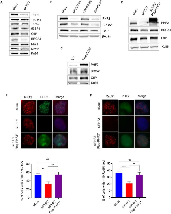
Nucleic Acids Research, 2020 3
Comet assay PBS + 0.5% FCS, immunostained with antibodies as indi-
cated and mounted with DAPI. For RPA2, BRCA1 and
Single Cell Gel Electrophoresis (SCGE) was carried out us-
Rad51 focus formation, cells were pre-extracted (20 mM
ing the CometAssay® ES II kit (Trevigen) according to
HEPES pH 8, 20 mM NaCl, 5 mM MgCl2 , 0.5% NP40)
the manufacturer’s instructions. Images were taken using a
for 5 min at 4◦ C before fixation.
Zeiss Cell Observer fluorescent microscope and the tail mo-
Images of cells were taken using a Zeiss Cell Observer flu-
ment of at least 50 cells per experiment was analysed with
orescent microscope equipped with a 63× NA 1.3 objective
the TriTek CometScore software.
and ZEN imaging software. The number of foci was evalu-
ated using the ImageJ software. In all instances, >100 cells
RT-PCR were analysed for each point and error bars on graphs rep-
Downloaded from https://academic.oup.com/nar/article-abstract/doi/10.1093/nar/gkaa196/5813805 by guest on 21 April 2020
RNA was isolated using the RiboZol Extraction Reagent resent the standard deviation of three independent experi-
(VWR) according to the manufacturer’s instructions. ments. Cells with >10 foci were scored as positive.
cDNA synthesis and PCR amplification were carried out
in the same tube using the qScript One-Step SYBR Green Laser micro-irradiation
RT-qPCR Kit (Quantabio) in a LightCycler480 II (Roche).
All reactions were performed in triplicate. Transcript levels Multiphoton laser micro-irradiation was essentially per-
were normalized in parallel with test genes GAPDH. The formed as described previously (31). Cells, grown on cov-
primers used were the following: erslips, were placed in a Chamlide CMB magnetic cham-
BRCA1-F: ACCTTGGAACTGTGAGAACTCT ber and the regular culture medium was replaced by
BRCA1-R: TCTTGATCTCCCACACTGCAATA CO2 -independent Leibovitz’s L15 supplemented with 10%
CtIP-F: CAGGAACGAATCTTAGATGCACA FBS and penicillin-streptomycin. Laser micro-irradiation
CtIP-R: GCCTGCTCTTAACCGATCTTCT was carried out on a Leica SP5 confocal microscope
GAPDH-F: GGAGCGAGATCCCTCCAAAAT equipped with an environmental chamber set to 37◦ C. DSB-
GAPDH-R: GGCTGTTGTCATACTTCTCATGG containing tracks (1.5 m width) were generated with a
Mre11-F: ATGCAGTCAGAGGAAATGATACG Mira mode-locked titanium-sapphire (Ti:Sapphire) laser
Mre11-R: CAGGCCGATCACCCATACAAT ( = 800 nm, pulse length = 200 fs, repetition rate = 76
RPA2-F: GCACCTTCTCAAGCCGAAAAG MHz, output power = 80 mW) using a UV-transmitting
RPA2-R: CCCCACAATAGTGACCTGTGAAA 63× 1.4 NA oil immersion objective (HCX PL APO; Le-
ica). Confocal images were recorded before and after laser
irradiation at 5 or 10 s time intervals over a period of 5–10
DR-GFP, SA-GFP and EJ5-GFP assays min.
U2OS cells bearing a single copy integration of the reporters To examine accumulation of Flag-PHF2, a different field
DR-GFP (HR (27)), SA-GFP (SSA (28)) or EJ5-GFP was irradiated every minute for 20 min, after which the cells
(NHEJ (28)) were used to analyse the different DSB repair were fixed for immunofluorescence.
pathways. Cells were seeded in 6-well plates in duplicate and
transfected with the indicated siRNA oligonucleotide. The FokI assays
next day, cells were infected with lentiviral particles con-
taining I-SceI–BFP expression construct at MOI 10 using U2OS 2-6-3 cells expressing inducible FokI-mCherry-LacR
8 g/ml polybrene. Forty eight hours later, cells were col- (32) were treated with 300 nM 4-OHT and 1 M Shield-I
lected by trypsinization and fixed with 4% paraformalde- for 5 h for inducing stabilization and nuclear localization of
hyde for 20 min. Samples were analysed with a BD FAC- the expressed product. Subsequently, cells were fixed with
SAria with the BD FACSDiva Software. PFA and immunostained with the indicated antibodies as
The number of GFP-positive cells from at least 10 000 described above.
events positive for blue fluorescence (infected with the I-
SceI–BFP construct) was scored. The average of both du- Cell cycle analysis
plicates was calculated for each sample and normalized
to siRNA control. At least three independent experiments For cell cycle analysis, cells were fixed in 70% ethanol at 4◦ C
were carried out for each condition and the mean and stan- o/n. After fixation, cells were washed with PBS, and the
dard deviation of the three experiments represented. DNA was stained with propidium iodide (PI). Cells were
analysed using a Macsquant Analyzer with Macsquantify
software (Miltenyi).
Chromatin fractionation
Biochemical fractionation of cells was performed as pre- Clonogenic survival
viously described (29,30). Soluble cytoplasmic and soluble
nuclear fractions were pooled to one soluble fraction. To determine cellular sensitivity to DNA damaging agents,
HeLa cells were transfected with the indicated siRNA
oligonucleotides. Forty eight hours later, 500 cells were
Immunofluorescence
seeded in six-well dishes and treated with the indicated con-
For immunostaining, cells were fixed in 4% paraformalde- centrations of Olaparib (Cayman Chemical) or doses of IR.
hyde for 15 min and then permeabilized with PBS + 0.2% Following 7–10 days in culture, cells were fixed, stained and
Triton X-100 for 10 min at RT. Samples were blocked in colonies were counted. Triplicate cultures were scored for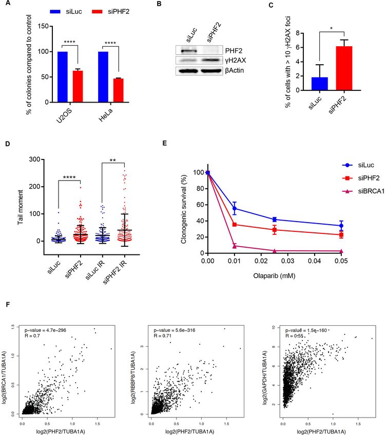
4 Nucleic Acids Research, 2020
each treatment. Shown is the relative survival as compared preventing the initiation of DNA resection (Figure 1A).
to the undamaged control and the error bars present the In contrast, overexpression of Flag-PHF2 prevented IR-
standard error of three independent experiments. induced focus formation of 53BP1 (Supplementary Figure
S1B). To examine the effect of modulating PHF2 levels on
Gene expression correlation analysis HR, we monitored the recruitment of BRCA1 in Rap80
knock out U2OS cells, in which Rap80-mediated recruit-
Gene expression correlation analysis was performed us- ment of BRCA1, subsequent BRCA1-A complex forma-
ing the Gene Expression Profiling Interactive Analysis tion and suppression of HR are prevented (31,34). Whereas
(GEPIA), a web-based tool containing high-throughput treating control siRNA transfected cells with IR led to effi-
RNA sequencing data (TCGA and GTEx databases) (33). cient focus formation of BRCA1 that increased in time, de-
Downloaded from https://academic.oup.com/nar/article-abstract/doi/10.1093/nar/gkaa196/5813805 by guest on 21 April 2020
A pairwise gene correlation analysis for given tumour data pletion of PHF2 by siRNA impaired IR-induced BRCA1
sets of TGCA was performed, with PHF2 as ‘gene A’ and foci (Figure 1B). The observed altered dynamics of BRCA1
a correlation against ‘gene B’: RBBP8 (CtIP), BRCA1 or and 53BP1 focus formation upon modulating the levels of
GAPDH. All genes were normalized to TUBA1A (Tubu- PHF2 suggests that this demethylase regulates DSB repair.
lin alpha-1A). The correlation coefficient was calculated As a consequence, depletion of PHF2 led to increased fo-
using Spearman rank correlation test against the tumour cus formation of phosphorylated H2AX (␥ H2AX), used as
dataset, which included a range different tumours of the fe- marker for DNA damage, as compared to control-depleted
male reproductive tract (cervical squamous cell carcinoma cells (Supplementary Figure S1C).
and endocervical adenocarcinoma (CESC, n = 306), ovar-
ian serous cystadenocarcinoma (OV, n = 426), uterine cor-
pus endometrial carcinoma (UCEC, n = 174) and uterine Depletion of PHF2 affects DSB resection and DNA repair
carcinosarcoma (UCS, n = 57)), and breast invasive carci- We continued to study what process in the DNA damage
noma (BRCA, n = 1085). response is affected by PHF2. To this end, we first exam-
ined DNA damage-induced checkpoint activation by ex-
Statistical analysis and reproducibility posing U2OS cells to different types of DNA damage and
monitoring the phosphorylation of CHK1, a critical ef-
Gaussian (normal) distribution of the data was assessed us- fector kinase of this response (35). Although DNA dam-
ing a D’ Agostino–Pearson omnibus normality test. In case aging agents triggered efficient CHK1 phosphorylation in
of normal distribution, an unpaired Student’s t-test was control depleted cells, PHF2 depletion reduced the DNA
used to determine whether the difference between the means damage-induced phosphorylation of CHK1, especially af-
of two sets of values was significant. In case the distribution ter IR, camptothecin and etoposide (Figure 1C). These
was not normal, a Mann–Whitney test was used. * P < 0.1 treatments result in a more efficient DSB formation com-
**
P < 0.01, *** P < 0.001, **** P < 0.0001. P < 0.05 was con- pared to UV and HU, which only resulted in a moderate
sidered statistically significant. NS = not significant. Unless effect, substantiating a possible role for PHF2 in the DSB
stated otherwise, representative experiments are shown out response. In addition, these treatments need DNA end re-
of at least two independent ones and depicted is the mean section to generate ssDNA, subsequently covered by the
± standard deviation. Additional details are listed in the in- RPA1–3 complex, that triggers the activation of ATR and
dividual figure legends. phosphorylation of its substrates such as CHK1 (36,37).
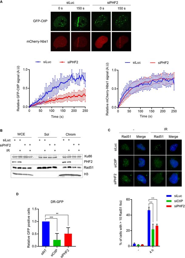
We therefore examined if PHF2 depletion affected DNA
RESULTS end resection, by studying RPA2 phosphorylation using
western blot analysis (38). Downregulation of PHF2 led
The demethylase PHF2 controls the DNA damage response
to lower levels of RPA2 phosphorylation on Ser4/8 and
To identify novel enzymes that regulate DSB repair by reduced levels of total RPA2 phosphorylation, as demon-
NHEJ and HR, we analysed ionizing radiation (IR)- strated by a lower mobility shift using an antibody against
induced focus formation of 53BP1 and BRCA1, that total RPA2, in response to camptothecin and etoposide, as
together control the choice between these two DSB compared to control transfected cells (Figure 1D). To cor-
repair pathways, in cells depleted for individual en- roborate these findings, we also measured IR-induced fo-
zymes involved in post-translation modifications of hi- cus formation of RPA2 by immunofluorescence. As previ-
stones by siRNA. Modulation of the expression level ously published, depletion of CtIP completely inhibited fo-
of PHF2/KDM7C/JHDM1E, a lysine-specific histone cus formation of RPA2 (Figure 1E). Notably, although less
demethylase hereafter called PHF2, changed the dynamics pronounced when compared to that after CtIP knockdown,
of 53BP1 focus formation in response to IR. Whereas irra- the downregulation of PHF2 also significantly reduced IR-
diating U2OS cells triggered efficient 53BP1 focus forma- induced focus formation of RPA (Figure 1E).
tion, the number of cells with 53BP1 foci and the number DSB resection is initiated by CtIP, together with the
of 53BP1 foci per cell stayed high at later time points (4– MRN complex. Given the effect of PHF2 on DNA end re-
7 h) in cells depleted for PHF2 by siRNA whereas at these section, we wondered if PHF2 functions at the level of CtIP.
time points 53BP1 foci decreased again in control trans- We consequently monitored the recruitment of GFP-CtIP
fected populations (Figure 1A and Supplementary Figure to DSB-containing tracks in U2OS cells by laser micro-
S1A). The effect of PHF2 depletion on 53BP1 focus res- irradiation (25). An inhibition in the accumulation of GFP-
olution was the same as that of downregulation of CtIP, CtIP to the laser tracks was observed upon depletion of
which served as a control for negatively regulating HR by PHF2, whereas the recruitment of mCherry-Nbs1, a com-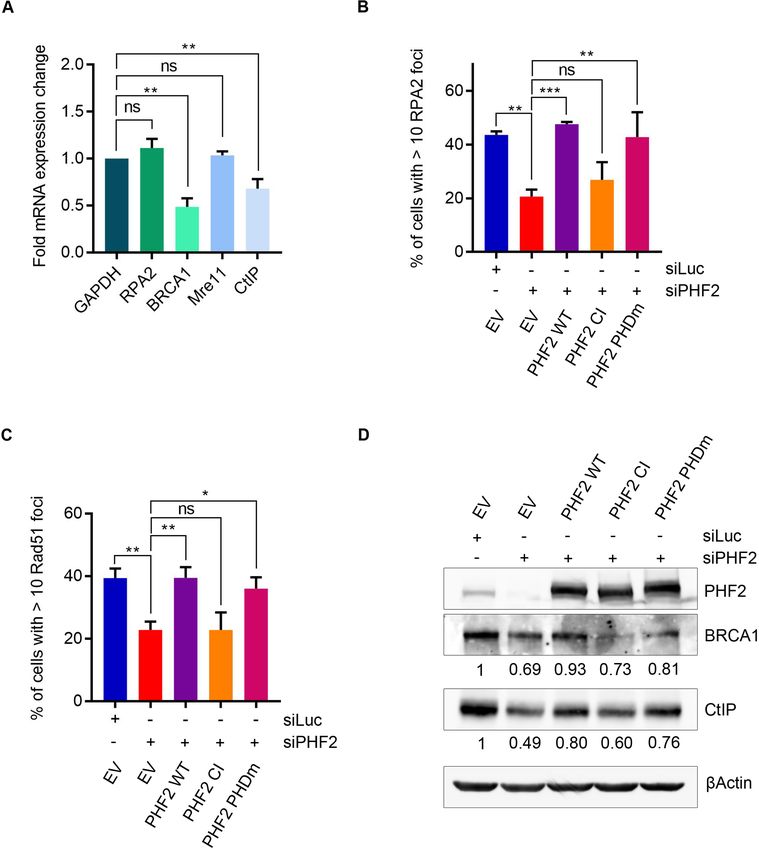
Nucleic Acids Research, 2020 5

                                                                                                                                                          Downloaded from https://academic.oup.com/nar/article-abstract/doi/10.1093/nar/gkaa196/5813805 by guest on 21 April 2020

Figure 1. PHF2 knockdown alters 53BP1 and BRCA1 focus dynamics in response to IR. (A) U2OS cells were depleted for Luciferase (Luc), CtIP or
PHF2 by siRNA. After 48 h, cells were treated with IR (10 Gy) and fixed after 1, 4 or 7 h. 53BP1 focus formation was analysed by immunofluorescence.
Left panel: representative images. Right panel: quantification of three independent experiments with each at least 100 cells. (B) U2OS Rap80 knockout
cells were depleted for Luc or PHF2. Cells were treated with IR (10 Gy), fixed at the indicated time points and ␥ H2AX (positive control for DNA damage
induction) and BRCA1 focus formation was analysed as in (A). Left panel: representative images (IR, 4 h). (C) U2OS cells depleted for Luc or PHF2 were
subjected to different DNA damaging agents and lysed after 1 h. Extracts were analysed by western blot using the indicated antibodies. (D) U2OS cells
were transfected as in (C), treated with CPT or ETP and analysed by western blot with the indicated antibodies. (E) U2OS cells were depleted for CtIP
or PHF2 by siRNA. Cells were treated with IR (3 Gy), fixed at the indicated time points and RPA2 focus formation was analysed as in (A). Left panel:
representative images (IR, 1 h).
6 Nucleic Acids Research, 2020
ponent of the MRN complex, was unaffected in these con- as well as BRCA1, while leaving Rad51, RPA2, 53BP1,
ditions (Figure 2A). To investigate the consequences of the Nbs1 and Mre11 unaffected (Figure 3A). Downregulating
effect of PHF2 on DNA end resection, we examined the IR- PHF2 by two additional siRNA oligonucleotides resulted
induced accumulation of HR protein Rad51 on the chro- in the same phenotype as seen before, namely a diminished
matin. Downregulation of PHF2 prevented this accumula- abundance of CtIP and BRCA1 protein (Figure 3B). In con-
tion (Figure 2B). In accordance, when analysing Rad51 fo- trast, overexpression of Flag-PHF2 had the opposite effect:
cus formation in response to IR by immunofluorescence, we both BRCA1 and CtIP protein levels increased under these
observed that knockdown of PHF2, as well as depletion of conditions (Figure 3C). Furthermore, the lower protein lev-
CtIP, inhibited focus formation by Rad51 (Figure 2C). To- els of BRCA1 and CtIP upon depletion of PHF2 were par-
gether these results indicate that PHF2 promotes the DSB tially rescued by expressing an siRNA-resistant version of
Downloaded from https://academic.oup.com/nar/article-abstract/doi/10.1093/nar/gkaa196/5813805 by guest on 21 April 2020
response by regulating DNA resection and strongly suggest Flag-PHF2 (Figure 3D). In addition, we could comple-
that the subsequent DSB repair is affected by PHF2 deple- ment the effects of PHF2 depletion on focus formation of
tion. RPA2, Rad51 and 53BP1 by expressing siRNA-resistant
DNA end resection is a critical step in homology-directed Flag-PHF2 (Figure 3E, F and Supplementary Figure S3A,
DSB repair (4). To address whether PHF2 impacts on respectively). Together these data demonstrate that the ef-
DSB repair by homologous recombination, we used a DR- fects of modulating PHF2 levels are genuinely due to the de-
GFP reporter assay in U2OS. In these cells, depletion of pletion of PHF2 instead of an off-target effect of the siRNA
PHF2 caused a significant decrease in HR efficiency (Fig- oligonucleotides used.
ure 2D). The efficiency of Single Strand Annealing (SSA), We next examined if PHF2 regulates CtIP and BRCA1 at
another form of homology-directed repair that also de- the transcriptional level by investigating the effect of PHF2
pends on CtIP-mediated DNA end resection but is Rad51- depletion on CtIP and BRCA1 mRNA in U2OS by quanti-
independent (39), measured using an SA-GFP reporter as- tative PCR. The mRNA levels of CtIP and BRCA1, but not
say, was also dramatically affected by PHF2 knockdown those of Mre11 and RPA2, were decreased under conditions
(Supplementary Figure S1D). Surprisingly, depletion of of PHF2 depletion when compared to control transfected
PHF2 also slightly inhibited NHEJ, not dependent on DNA cells (Figure 4A), indicating that PHF2 controls homology-
end resection, as measured by the EJ5-GFP reporter (Sup- directed DSB repair by regulating the mRNA levels of CtIP
plementary Figure S1E). and BRCA1, two proteins critical for DNA end-resection
Cell cycle is a major determinant of DSB repair path- and thereby initiation of HR.
way choice. As HR depends on the availability of a sister As mentioned before, PHF2 harbours an N-terminal
chromatid as repair template, this type of repair is restricted PHD domain, a chromatin reader module, in addition to
to the S and G2 phases of the cell cycle (40). Importantly, the enzymatically active Jumonji-C (JmjC) domain (41).
PHF2 depletion only very slightly decreased the percentage To gain more insight into the mechanism of how PHF2
of cells in S phase, making it unlikely that the decrease in controls DSB repair, we investigated if the effect of PHF2
DSB repair efficiency upon PHF2 knockdown is due to an on DNA end resection depends on the lysine demethylase
indirect effect on the cell cycle (Supplementary Figure S1F). activity and/or on the presence of a functional PHD do-
main. To do so, mutants of these domains were generated
by introducing changes in critical residues within these re-
PHF2 controls CtIP and BRCA1 mRNA levels by means of
gions in siRNA resistant Flag-PHF2 (42,43). These mu-
its demethylase activity
tants were subsequently expressed in PHF2-depleted cells
We next set out to address if PHF2 affects DSB repair in and IR-induced focus formation of RPA2 and Rad51 were
a direct manner by acting at sites of DNA damage. To this studied. The defect in RPA2 and Rad51 focus formation
end, the accumulation of Flag-PHF2 at sites of DNA dam- caused by knockdown of PHF2 could be rescued by the wild
ages generated by laser micro-irradiation was examined. Al- type (WT) and PHD mutant (PHDm) version of PHF2 but
though mCherry-Nbs1 and ␥ H2AX were detected at dam- not with catalytic inactive (CI) PHF2 (Figure 4B and C). In
aged regions upon laser micro-irradiation, we did not ob- accordance with these data, whereas WT and PHDm PHF2
serve detectable Flag-PHF2 accumulation to such laser- could complement the decrease in BRCA1 and CtIP protein
tracks (Supplementary Figure S2A). In addition, also no levels caused by PHF2 depletion, expressing PHF2 CI did
accumulation of Flag-PHF2 was observed to a DSB cre- not rescue the reduced BRCA1 and CtIP levels (Figure 4D).
ated in a single genomic locus containing an array of LacO Together these results demonstrate that the lysine demethy-
repeats following expression and tethering of an mCherry- lase activity is required for the effect of PHF2 in resection.
LacI-FokI nuclease fusion (Supplementary Figure S2B). In contrast, the PHD chromatin reader domain seems to be
Together these results could be an indication that PHF2 reg- dispensable.
ulates DSB repair in an indirect and more global manner.
Interestingly, PHF2 contains a zinc finger-like PHD
PHF2 downregulation causes genome instability
(plant homeodomain) finger, a motif found in proteins that
are involved in transcriptional regulation, possibly by recog- As DNA repair is also important in unperturbed cells, de-
nizing chromatin modifications (41), which led to the sug- fective DSB repair caused by downregulation of PHF2 is
gestion that PHF2 might regulate DNA repair in a tran- likely to affect genome stability and cell survival in these
scriptional manner. Western blot analysis of the levels of conditions. Consistent with such a scenario, the colony
proteins involved in DSB repair demonstrated that down- forming capacity of both U2OS and HeLa cells decreased
regulation of PHF2 led to diminished protein levels of CtIP dramatically after depletion of PHF2 as compared to Lu-Nucleic Acids Research, 2020 7
Downloaded from https://academic.oup.com/nar/article-abstract/doi/10.1093/nar/gkaa196/5813805 by guest on 21 April 2020
Figure 2. Depletion of PHF2 impairs DSB repair by homologous recombination. (A) U2OS cells, transfected with Luc or PHF2 siRNA oligos, and 24 h
later transfected with GFP-CtIP and mCherry-Nbs1, were laser-irradiated and analysed by time-lapse imaging. Upper panel: representative images of the
indicated time points. Lower panel: relative fluorescence (left: GFP-CtIP, right: mCherry-Nbs1) at laser stripes of at least 50 cells per experiment. (B) U2OS
cells were depleted for Luc or PHF2 by siRNA, treated with IR (10 Gy) and subjected to chromatin fractionation (WCE: whole cell extracts, Sol: soluble
and Chrom: chromatin). Samples were analysed by western blot using the indicated antibodies. (C) U2OS cells were depleted for Luc, CtIP or PHF2 by
siRNA. Cells were treated with IR (3 Gy), fixed after 4 h and Rad51 focus formation was analysed by immunofluorescence. Top panel: representative
images. Bottom panel: quantification from three independent experiments with each at least 100 cells. (D) U2OS cells stably expressing a single copy of
the DR-GFP reporter construct were depleted of CtIP, PHF2 or control (non-target, NT). After 48 h, GFP fluorescence was analysed by flow cytometry.
Presented is the relative fluorescence as compared to the control cells, of three independent experiments.8 Nucleic Acids Research, 2020
Downloaded from https://academic.oup.com/nar/article-abstract/doi/10.1093/nar/gkaa196/5813805 by guest on 21 April 2020
Figure 3. PHF2 regulates HR by modulating CtIP and BRCA1 levels. (A) U2OS cells were depleted for Luc or PHF2 by siRNA. Forty eight hours later,
the cells were lysed and extracts were analysed by western blot with the indicated antibodies. (B) U2OS cells were depleted for PHF2 using three different
siRNA oligonucleotides and subsequently analysed by western blot using the indicated antibodies. (C) U2OS cells were transfected with empty vector (EV)
or a Flag-PHF2 expression vector, followed by western blot analysis with the indicated antibodies. (D) U2OS cells were depleted for Luc or PHF2 by siRNA
and 24 h later transfected with EV or siRNA-resistant Flag-PHF2 (Flag-PHF2* ). The day after, extracts were prepared and analysed by western blot with
the indicated antibodies. (E) U2OS cells were depleted for PHF2 and transfected with Flag-PHF2* the day after. One day later, cells were treated with
IR (3 Gy) and fixed for IF after 1 h. RPA2 focus formation of Flag-positive cells was analysed by immunofluorescence. Top panel: representative images.
Bottom panel: quantification of three independent experiments, counting at least 50 cells each. (F) U2OS cells were depleted for PHF2 and transfected
with Flag-PHF2* the day after. One day later, cells were treated with IR (3 Gy) and fixed for IF after 4 h. Rad51 focus formation of Flag-positive cells was
analysed as in (E).Nucleic Acids Research, 2020 9
Downloaded from https://academic.oup.com/nar/article-abstract/doi/10.1093/nar/gkaa196/5813805 by guest on 21 April 2020
Figure 4. PHF2 controls the expression of CtIP and BRCA1 through demethylase activity. (A) U2OS were downregulated with Luc or PHF2 siRNA
oligos. 72 h later, RNA was isolated and mRNA levels of GAPDH, RPA2, BRCA1, Mre11 and CtIP were determined by RT-qPCR. Shown is the fold
mRNA change of PHF2-depleted samples as compared to the Luc control. (B) U2OS cells were depleted for PHF2 and transfected with EV, siRNA
resistant Flag-PHF2 wild type (WT), demethylase inactive Flag-PHF2 (CI) or PHD domain mutant Flag-PHF2 (PHDm) the day after. One day later, cells
were treated with IR (5 Gy) and fixed for IF after 1 h. RPA2 focus formation of Flag-positive cells was analysed by immunofluorescence. Quantification
of three independent experiments with at least 50 cells each is shown. (C) U2OS cells were transfected as in (B). One day later, cells were treated with IR
(10 Gy) and fixed for IF after 4 h. Rad51 focus formation of Flag-positive cells was analysed by immunofluorescence. Quantification of three independent
experiments with at least 50 cells each. (D) U2OS cells were transfected as in (B) and the day after, extracts were prepared and analysed by western blot
with the indicated antibodies. Quantifications of BRCA1 and CtIP levels, compared to the loading control, are shown below each panel.
ciferase knockdown control cells (Figure 5A). Interestingly, sence of exogenous damage (Figure 5B), and this was con-
although downregulation of CtIP also affected clonogenic firmed by an elevated percentage of cells with ␥ H2AX foci
survival, the effect was less severe than after PHF2 deple- upon PHF2 knockdown as compared to Luc control cells
tion, suggesting that the effect of PHF2 on cell survival is by immunofluorescence (Figure 5C). To directly asses the
not solely due to its function in controlling HR (Supplemen- appearance of DNA breaks resulting from downregulation
tary Figure S3B). Western blot analysis demonstrated an in- of PHF2 in individual cells, we employed the alkaline comet
crease in ␥ H2AX in PHF2-depleted U2OS cells in the ab- assay. Compared to control depleted cells, decreasing PHF210 Nucleic Acids Research, 2020
Downloaded from https://academic.oup.com/nar/article-abstract/doi/10.1093/nar/gkaa196/5813805 by guest on 21 April 2020
Figure 5. PHF2 controls genome stability. (A) U2OS and HeLa cells were depleted for PHF2 by siRNA. Equal numbers of cells were seeded and incubated
for 10 days for colonies to form. Bar graph shows the number of colonies compared to control depleted cells from three independent experiments. (B) U2OS
cells were depleted for Luc or PHF2 by siRNA. 48 h later the cells were lysed and analysed by western blot with the indicated antibodies. (C) As in (B), but
now ␥ H2AX focus formation was analysed by IF. Quantification of three independent experiments with each at least 100 cells. (D) U2OS cells, depleted
for PHF2 by siRNA, were left untreated or irradiated with 5 Gy and processed after 1 h for comet assay analysis. Depicted is the tail moment of three
independent experiments (at least 50 cells each). (E) Clonogenic survival assays of HeLa cells that were depleted for Luc, PHF2 or BRCA1 by siRNA and
incubated with the indicated concentrations of Olaparib. Shown is the relative survival as compared to the undamaged control. Error bars represent the
SEM of three individual experiments. (F) Correlation analysis between expression of PHF2 and expression of BRCA1, RBBP8 and GAPDH by GEPIA.
Scatter plots represent the correlation between gene expression of PHF2 and BRCA1 (left), RBBP8 (middle) or GAPDH (right) in each tumour (n = 2048),
with R as the correlation coefficient.Nucleic Acids Research, 2020 11
by siRNA led to a significant tail moment increase in U2OS of CtIP is additionally required for CtIP chromatin bind-
and Hela cells, in the absence of exogenous DNA damage ing and damage-induced focus formation (49), which likely
as well as after IR (Figure 5D and Supplementary Figure explains the reported defect in the accumulation of GFP-
S3C). Together these data demonstrate that regulated lev- CtIP at interstrand crosslinks induced by micro-irradiation
els of PHF2 are important for the maintenance of genome (50). Our observation that depletion of PHF2 inhibits the
stability. accumulation of GFP-CtIP to the laser damage (Figure 2A)
Notably, HR deficiency is exploited in the treatment is therefore likely to be an indirect effect of the diminished
of tumours with BRCA1/BRCA2 mutations as cells in BRCA1 levels in these conditions. However, we cannot ex-
such conditions are sensitive to inhibition of poly(ADP- clude the possibility that PHF2 regulates CtIP on several
ribose) polymerase (PARP) (44). As PHF2 depletion affects levels, like the recently reported splicing complex SF3B, that
Downloaded from https://academic.oup.com/nar/article-abstract/doi/10.1093/nar/gkaa196/5813805 by guest on 21 April 2020
HR, downregulation of this protein is likewise expected controls CtIP both at the level of mRNA abundance and the
to result in sensitivity to PARP inhibition. Indeed, deple- recruitment of CtIP to the chromatin in response to DNA
tion of PHF2 mildly sensitized HeLa cells to inhibition of damage (51).
PARP1/2 by Olaparib (Figure 5E), which is in accordance Interestingly, we also observed a modest decrease in the
to its effect on BRCA1 levels. Co-depletion of PHF2 and efficiency of NHEJ after knockdown of PHF2. A possible
BRCA1 did not increase the sensitivity to PARP1/2 inhibi- explanation for this result could be the decreased BRCA1
tion as compared to BRCA1 knockdown alone, suggesting levels in these conditions, as depletion of BRCA1 has been
that the Olaparib sensitivity after PHF2 depletion is due to reported to affect NHEJ as well as HR (52,53). Depletion of
its effect on BRCA1 (Supplementary Figure S3D). In ad- PHF2 also affected cell growth and led to genome instability
dition, downregulation of PHF2 also caused sensitivity to in unperturbed cells, as demonstrated by H2AX phospho-
IR (Supplementary Figure S3E). Together these data un- rylation and comet assay analysis, an effect also recently re-
derscore the importance of PHF2 in HR. ported by others in mouse neural stem cells (54). Although
Finally, we analysed the gene expression of PHF2, these results could be due to DNA repair defects, these ob-
BRCA1 and RBBP8, the gene encoding CtIP, in a broad servations might also reflect a separate phenotype of PHF2.
range of tumours of breast and the female reproductive Indeed, PHF2 has been reported to be involved in the con-
tract by GEPIA (33). As shown in Figure 5F, the expression trol of cell cycle regulation, cell differentiation and the in-
of PHF2 correlated with that of BRCA1 and RBBP8 in a flammatory response (21,54,55).
high proportion of the tumours (R ≥ 0.7), whereas the cor- The KDM7 family of histone demethylases, to which
relation with GAPDH was lower (R = 0.55). These data are PHF2 belongs, can remove the methylation from H3K9,
in accordance with our previous experiments and strongly H3K27 or H4K20, which are responsible for transcriptional
suggest that the mechanism of regulation of BRCA1 and repression, presumably by the concerted action of their
CtIP expression by PHF2 also occurs in tumours. PHD methyl reader domain and the enzymatically active
JmjC-domain (41,56). Indeed, PHF2 was described to regu-
DISCUSSION late transcription by removing the dimethylated H3K9 and,
to a lesser extent, trimethylated H4K20 (20–23). We hypoth-
In this study, we show that the demethylase PHF2 con- esize that PHF2 controls CtIP and BRCA1 gene transcrip-
tributes to the maintenance of genome integrity by con- tion by erasing the transcription repression mark from the
trolling DNA repair, predominantly through homology- respective promoters (23). PHF2 was shown to stimulate
directed DSB repair. Specifically, we showed that downreg- the expression of genes driven by the transcription factors
ulation of PHF2 affects the DNA damage-induced focus HNF4, CEBP␣, p53 and NF-B (20–23). Interestingly, NF-
formation by BRCA1 and 53BP1, which together determine B was shown to regulate HR by controlling BRCA1-CtIP
the choice between DSB repair by homologous recombina- complexes, although this effect is mediated by protein stabi-
tion or non-homologous end joining. Concomitant with a lization of BRCA1 rather than by transcriptional regulation
decrease in BRCA1 focus formation, PHF2 depletion also of BRCA1 and CtIP (57).
affected the accumulation of CtIP to sites of DNA lesions. The fact that a catalytic inactive version of PHF2 can-
CtIP is known to initiate DNA end resection to generate ss- not rescue the defect in DNA end resection and the lower
DNA ends that are a prerequisite for HR (10,25). BRCA1, BRCA1/CtIP levels caused by PHF2 depletion suggests
that forms a complex with CtIP, collaborates in ssDNA-end that PHF2 controls the transcription of CtIP and BRCA1
formation (39,45,46). Indeed, impaired DSB resection was mRNA by direct demethylation of H3K9me2 at the respec-
observed after PHF2 downregulation, as demonstrated by tive promotors. Interestingly however, although biochemi-
decreased RPA phosphorylation and focus formation. Con- cal data indicate that PHF2 demethylates H3K9me2 upon
sequently, PHF2 depletion compromised IR-induced focus interaction with methylated H3K4 through its PHD do-
formation of Rad51 and resulted in a diminished efficiency main (58), our experiments show that the PHD domain of
of HR. Impaired DSB repair by HR in response to PHF2 PHF2 is dispensable for its function on DSB resection and
depletion is explained by our data showing that PHF2 con- control of BRCA1/CtIP levels. It is therefore possible that
trols the DSB response by regulating CtIP and BRCA1 PHF2 controls BRCA1/CtIP mRNA levels in another, pos-
mRNA and protein levels. That PHF2 regulates expression sibly indirect way. Moreover, as dimethylation of H4K20 is
of CtIP and BRCA1 simultaneously is in accordance to in- critical in the recruitment of 53BP1 to sites of DNA lesions
dications in the literature that BRCA1 supports the con- (59,60), interfering with this methyl mark and/or compe-
trol of CtIP. CtIP was reported to control its own tran- tition for binding to methylated H4K20 by PHF2 could
scription, possibly via interaction with BRCA1 through its also contribute to the phenotype in disturbing homology-
BRCT domains (47,48). BRCA1-dependent ubiquitination directed DSB repair observed in this study. We consider this12 Nucleic Acids Research, 2020
possibility less likely though, since at this moment, we have 5. Noordermeer,S.M., Adam,S., Setiaputra,D., Barazas,M., Pettitt,S.J.,
no indications that PHF2 might function directly at the sites Ling,A.K., Olivieri,M., Álvarez-Quilón,A., Moatti,N.,
of DNA lesions. Zimmermann,M. et al. (2018) The shieldin complex mediates
53BP1-dependent DNA repair. Nature, 560, 117–121.
By controlling DNA DSB repair, PHF2 emerges as a pu- 6. Mirman,Z., Lottersberger,F., Takai,H., Kibe,T., Gong,Y., Takai,K.,
tative important regulator in maintaining genomic integrity. Bianchi,A., Zimmermann,M., Durocher,D. and de Lange,T. (2018)
Therefore, our data might have an importance in patholo- 53BP1-RIF1-shieldin counteracts DSB resection through CST- and
gies in which PHF2 levels are changed or PHF2 is mutated. Pol␣-dependent fill-in. Nature, 560, 112–116.
7. Bunting,S.F., Callén,E., Wong,N., Chen,H.-T., Polato,F., Gunn,A.,
For example, high PHF2 levels were found in oesophageal Bothmer,A., Feldhahn,N., Fernandez-Capetillo,O., Cao,L. et al.
carcinoma and renal cell carcinoma (61,62), whereas the (2010) 53BP1 inhibits homologous recombination in Brca1-deficient
PHF2 gene is deleted or hypermethylated in its promoter re- cells by blocking resection of DNA breaks. Cell, 141, 243–254.
Downloaded from https://academic.oup.com/nar/article-abstract/doi/10.1093/nar/gkaa196/5813805 by guest on 21 April 2020
gion in breast cancer (63). In addition, PHF2 mutants were 8. Chapman,J.R., Taylor,M.R.G. and Boulton,S.J. (2012) Playing the
reported in gastric and colon cancer (64). Together these end game: DNA double-strand break repair pathway choice. Mol.
Cell, 47, 497–510.
observations suggest that PHF2 plays a role in the develop- 9. Symington,L.S. (2016) Mechanism and regulation of DNA end
ment and/or progression of cancer. Interestingly, the use of resection in eukaryotes. Crit. Rev. Biochem. Mol. Biol., 51, 195–212.
histone demethylases as therapeutic targets by pharmaco- 10. Jasin,M. and Rothstein,R. (2013) Repair of strand breaks by
logical inhibitors is currently being investigated and might homologous recombination. Cold Spring Harb. Perspect Biol., 5,
a012740.
open new strategies for tumour therapy (65,66). Our results 11. Sulli,G., Di Micco,R. and d’Adda di Fagagna,F. (2012) Crosstalk
demonstrating that depletion of PHF2 affects the sensitiv- between chromatin state and DNA damage response in cellular
ity to inhibition of PARP, together with the observed corre- senescence and cancer. Nat. Rev. Cancer, 12, 709–720.
lation between PHF2 and CtIP/BRCA1 levels in tumours, 12. Burma,S., Chen,B.P., Murphy,M., Kurimasa,A. and Chen,D.J. (2001)
might suggest that targeting PHF2 could be particularly ef- ATM phosphorylates histone H2AX in response to DNA
double-strand breaks. J. Biol. Chem., 276, 42462–42467.
fective in breast and ovarian cancers without mutations in 13. Kolas,N.K., Chapman,J.R., Nakada,S., Ylanko,J., Chahwan,R.,
BRCA1/2 or other known HR proteins. Sweeney,F.D., Panier,S., Mendez,M., Wildenhain,J., Thomson,T.M.
et al. (2007) Orchestration of the DNA-damage response by the
RNF8 ubiquitin ligase. Science, 318, 1637–1640.
SUPPLEMENTARY DATA 14. Doil,C., Mailand,N., Bekker-Jensen,S., Menard,P., Larsen,D.H.,
Pepperkok,R., Ellenberg,J., Panier,S., Durocher,D., Bartek,J. et al.
Supplementary Data are available at NAR Online. (2009) RNF168 binds and amplifies ubiquitin conjugates on damaged
chromosomes to allow accumulation of repair proteins. Cell, 136,
435–446.
ACKNOWLEDGEMENTS 15. Stewart,G.S., Panier,S., Townsend,K., Al-Hakim,A.K., Kolas,N.K.,
We thank Drs Daniel Durocher and Xiaobing Shi for shar- Miller,E.S., Nakada,S., Ylanko,J., Olivarius,S., Mendez,M. et al.
(2009) The RIDDLE syndrome protein mediates a
ing reagents, Esperanza Hernández for valuable assistance ubiquitin-dependent signaling cascade at sites of DNA damage. Cell,
and Antonio Catalán and Luciano Benı́tez (Servicio de 136, 420–434.
Fı́sica Médica) from the Hospital Universitario de Canarias 16. Zhao,Y., Brickner,J.R., Majid,M.C. and Mosammaparast,N. (2014)
for use of the WOmed X-Ray Therapy Unit. Crosstalk between ubiquitin and other post-translational
modifications on chromatin during double-strand break repair.
Trends Cell Biol., 24, 426–434.
FUNDING 17. Kouzarides,T. (2000) Acetylation: a regulatory modification to rival
phosphorylation? EMBO J., 19, 1176–1179.
Ministerio de Ciencia e Innovación [SAF2016-80626-R to 18. Greer,E.L. and Shi,Y. (2012) Histone methylation: a dynamic mark in
V.A.J.S./R.F., SAF2016-74855-P to P.H. and BFU2017- health, disease and inheritance. Nat. Rev. Genet., 13, 343–357.
19. Luijsterburg,M.S. and van Attikum,H. (2011) Chromatin and the
90889-REDT to V.A.J.S./P.H.], co-funded by EU-ERDF; DNA damage response: the cancer connection. Mol. Oncol., 5,
Fundación Canaria Instituto de Investigación Sanitaria de 349–367.
Canarias (FIISC) [PIFUN16/18 to V.A.J.S.]; I.A.V. is sup- 20. Baba,A., Ohtake,F., Okuno,Y., Yokota,K., Okada,M., Imai,Y.,
ported by a predoctoral fellowship from the Gobierno de Ni,M., Meyer,C.A., Igarashi,K., Kanno,J. et al. (2011)
Canarias; J.R.H.F. by the Asociación Española Contra el PKA-dependent regulation of the histone lysine demethylase complex
PHF2-ARID5B. Nat. Cell Biol., 13, 668–675.
Cáncer; H.v.A. by an ERC Consolidator grant from the 21. Stender,J.D., Pascual,G., Liu,W., Kaikkonen,M.U., Do,K.,
European Research Council and a VICI grant from the Spann,N.J., Boutros,M., Perrimon,N., Rosenfeld,M.G. and
Netherlands Organisation for Scientific Research. Funding Glass,C.K. (2012) Control of proinflammatory gene programs by
for open access charge: FIISC [PIFUN16/18]; Ministerio regulated trimethylation and demethylation of histone H4K20. Mol.
Cell, 48, 28–38.
de Ciencia e Innovación [SAF2016-80626-R]. 22. Okuno,Y., Ohtake,F., Igarashi,K., Kanno,J., Matsumoto,T.,
Conflict of interest statement. None declared. Takada,I., Kato,S. and Imai,Y. (2013) Epigenetic regulation of
adipogenesis by PHF2 histone demethylase. Diabetes, 62, 1426–1434.
23. Lee,K.-H., Park,J.-W., Sung,H.-S., Choi,Y.-J., Kim,W.H., Lee,H.S.,
REFERENCES Chung,H.-J., Shin,H.-W., Cho,C.-H., Kim,T.-Y. et al. (2015) PHF2
1. Bartek,J., Bartkova,J. and Lukas,J. (2007) DNA damage signalling histone demethylase acts as a tumor suppressor in association with
guards against activated oncogenes and tumour progression. p53 in cancer. Oncogene, 34, 2897–2909.
Oncogene, 26, 7773–7779. 24. Rabl,J., Bunker,R.D., Schenk,A.D., Cavadini,S., Gill,M.E.,
2. Aguilera,A. and Gómez-González,B. (2008) Genome instability: a Abdulrahman,W., Andrés-Pons,A., Luijsterburg,M.S.,
mechanistic view of its causes and consequences. Nat. Rev. Genet., 9, Ibrahim,A.F.M., Branigan,E. et al. (2019) Structural basis of
204–217. BRCC36 function in DNA repair and immune regulation. Mol. Cell,
3. Hiom,K. (2010) Coping with DNA double strand breaks. DNA 75, 483–497.
Repair (Amst.), 9, 1256–1263. 25. Sartori,A.A., Lukas,C., Coates,J., Mistrik,M., Fu,S., Bartek,J.,
4. Huertas,P. (2010) DNA resection in eukaryotes: deciding how to fix Baer,R., Lukas,J. and Jackson,S.P. (2007) Human CtIP promotes
the break. Nat. Struct. Mol. Biol., 17, 11–16. DNA end resection. Nature, 450, 509–514.Nucleic Acids Research, 2020 13
26. Luijsterburg,M.S., de Krijger,I., Wiegant,W.W., Shah,R.G., 48. Liu,F. and Lee,W.-H. (2006) CtIP activates its own and cyclin D1
Smeenk,G., de Groot,A.J.L., Pines,A., Vertegaal,A.C.O., promoters via the E2F/RB pathway during G1/S progression. Mol.
Jacobs,J.J.L., Shah,G.M. et al. (2016) PARP1 links CHD2-Mediated Cell. Biol., 26, 3124–3134.
chromatin expansion and H3.3 deposition to DNA repair by 49. Yu,X., Fu,S., Lai,M., Baer,R. and Chen,J. (2006) BRCA1
non-homologous end-joining. Mol. Cell, 61, 547–562. ubiquitinates its phosphorylation-dependent binding partner CtIP.
27. Pierce,A.J., Johnson,R.D., Thompson,L.H. and Jasin,M. (1999) Genes Dev., 20, 1721–1726.
XRCC3 promotes homology-directed repair of DNA damage in 50. Duquette,M.L., Zhu,Q., Taylor,E.R., Tsay,A.J., Shi,L.Z.,
mammalian cells. Genes Dev., 13, 2633–2638. Berns,M.W. and McGowan,C.H. (2012) CtIP is required to initiate
28. Bennardo,N., Cheng,A., Huang,N. and Stark,J.M. (2008) replication-dependent interstrand crosslink repair. PLoS Genet., 8,
Alternative-NHEJ is a mechanistically distinct pathway of e1003050.
mammalian chromosome break repair. PLoS Genet., 4, e1000110. 51. Prados-Carvajal,R., López-Saavedra,A., Cepeda-Garcı́a,C.,
29. Méndez,J. and Stillman,B. (2000) Chromatin association of human Jimeno,S. and Huertas,P. (2018) Multiple roles of the splicing
Downloaded from https://academic.oup.com/nar/article-abstract/doi/10.1093/nar/gkaa196/5813805 by guest on 21 April 2020
origin recognition complex, cdc6, and minichromosome maintenance complex SF3B in DNA end resection and homologous
proteins during the cell cycle: assembly of prereplication complexes in recombination. DNA Repair (Amst.), 66-67, 11–23.
late mitosis. Mol. Cell. Biol., 20, 8602–8612. 52. Fouquin,A., Guirouilh-Barbat,J., Lopez,B., Hall,J., Amor-Guéret,M.
30. Smits,V.A.J., Reaper,P.M. and Jackson,S.P. (2006) Rapid and Pennaneach,V. (2017) PARP2 controls double-strand break
PIKK-dependent release of Chk1 from chromatin promotes the repair pathway choice by limiting 53BP1 accumulation at DNA
DNA-damage checkpoint response. Curr. Biol., 16, 150–159. damage sites and promoting end-resection. Nucleic Acids Res., 45,
31. Typas,D., Luijsterburg,M.S., Wiegant,W.W., Diakatou,M., 12325–12339.
Helfricht,A., Thijssen,P.E., van den Broek,B., van de Broek,B., 53. Hu,Y., Wang,C., Huang,K., Xia,F., Parvin,J.D. and Mondal,N.
Mullenders,L.H. and van Attikum,H. (2015) The de-ubiquitylating (2014) Regulation of 53BP1 protein stability by RNF8 and RNF168
enzymes USP26 and USP37 regulate homologous recombination by is important for efficient DNA double-strand break repair. PLoS
counteracting RAP80. Nucleic Acids Res., 43, 6919–6933. ONE, 9, e110522.
32. Tang,J., Cho,N.W., Cui,G., Manion,E.M., Shanbhag,N.M., 54. Pappa,S., Padilla,N., Iacobucci,S., Vicioso,M., Álvarez de la
Botuyan,M.V., Mer,G. and Greenberg,R.A. (2013) Acetylation limits Campa,E., Navarro,C., Marcos,E., la Cruz,X. and
53BP1 association with damaged chromatin to promote homologous Martı́nez-Balbás,M.A. (2019) PHF2 histone demethylase prevents
recombination. Nat. Struct. Mol. Biol., 20, 317–325. DNA damage and genome instability by controlling cell cycle
33. Tang,Z., Li,C., Kang,B., Gao,G., Li,C. and Zhang,Z. (2017) GEPIA: progression of neural progenitors. Proc. Natl. Acad. Sci. U.S.A., 116,
a web server for cancer and normal gene expression profiling and 19464–19473.
interactive analyses. Nucleic Acids Res., 45, W98–W102. 55. Yang,J., Ma,J., Xiong,Y., Wang,Y., Jin,K., Xia,W., Chen,Q.,
34. Hu,Y., Scully,R., Sobhian,B., Xie,A., Shestakova,E. and Huang,J., Zhang,J., Jiang,N. et al. (2018) Epigenetic regulation of
Livingston,D.M. (2011) RAP80-directed tuning of BRCA1 megakaryocytic and erythroid differentiation by PHF2 histone
homologous recombination function at ionizing radiation-induced demethylase. J. Cell. Physiol., 233, 6841–6852.
nuclear foci. Genes Dev., 25, 685–700. 56. Jaskelioff,M. and Peterson,C.L. (2003) Chromatin and transcription:
35. Smits,V.A.J. and Gillespie,D.A. (2015) DNA damage control: histones continue to make their marks. Nat. Cell Biol., 5, 395–399.
regulation and functions of checkpoint kinase 1. FEBS J., 282, 57. Volcic,M., Karl,S., Baumann,B., Salles,D., Daniel,P., Fulda,S. and
3681–3692. Wiesmüller,L. (2012) NF-B regulates DNA double-strand break
36. Zou,L. and Elledge,S.J. (2003) Sensing DNA damage through ATRIP repair in conjunction with BRCA1-CtIP complexes. Nucleic Acids
recognition of RPA-ssDNA complexes. Science, 300, 1542–1548. Res., 40, 181–195.
37. Shiotani,B. and Zou,L. (2009) Single-stranded DNA orchestrates an 58. Wen,H., Li,J., Song,T., Lu,M., Kan,P.-Y., Lee,M.G., Sha,B. and
ATM-to-ATR switch at DNA breaks. Mol. Cell, 33, 547–558. Shi,X. (2010) Recognition of histone H3K4 trimethylation by the
38. Maréchal,A. and Zou,L. (2015) RPA-coated single-stranded DNA as plant homeodomain of PHF2 modulates histone demethylation. J.
a platform for post-translational modifications in the DNA damage Biol. Chem., 285, 9322–9326.
response. Cell Res., 25, 9–23. 59. Botuyan,M.V., Lee,J., Ward,I.M., Kim,J.-E., Thompson,J.R., Chen,J.
39. Yun,M.H. and Hiom,K. (2009) CtIP-BRCA1 modulates the choice of and Mer,G. (2006) Structural basis for the methylation state-specific
DNA double-strand-break repair pathway throughout the cell cycle. recognition of histone H4-K20 by 53BP1 and Crb2 in DNA repair.
Nature, 459, 460–463. Cell, 127, 1361–1373.
40. Huertas,P. and Jackson,S.P. (2009) Human CtIP mediates cell cycle 60. Pei,H., Zhang,L., Luo,K., Qin,Y., Chesi,M., Fei,F., Bergsagel,P.L.,
control of DNA end resection and double strand break repair. J. Biol. Wang,L., You,Z. and Lou,Z. (2011) MMSET regulates histone
Chem., 284, 9558–9565. H4K20 methylation and 53BP1 accumulation at DNA damage sites.
41. Fortschegger,K. and Shiekhattar,R. (2011) Plant homeodomain Nature, 469, 124–128.
fingers form a helping hand for transcription. Epigenetics, 6, 4–8. 61. Sun,L.-L., Sun,X.-X., Xu,X.-E., Zhu,M.-X., Wu,Z.-Y., Shen,J.-H.,
42. Horton,J.R., Upadhyay,A.K., Hashimoto,H., Zhang,X. and Wu,J.-Y., Huang,Q., Li,E.-M. and Xu,L.-Y. (2013) Overexpression of
Cheng,X. (2011) Structural basis for human PHF2 Jumonji domain Jumonji AT-rich interactive domain 1B and PHD finger protein 2 is
interaction with metal ions. J. Mol. Biol., 406, 1–8. involved in the progression of esophageal squamous cell carcinoma.
43. Fortschegger,K., de Graaf,P., Outchkourov,N.S., van Schaik,F.M.A., Acta Histochem., 115, 56–62.
Timmers,H.T.M. and Shiekhattar,R. (2010) PHF8 targets histone 62. Lee,C., Kim,B., Song,B. and Moon,K.C. (2017) Implication of PHF2
methylation and RNA polymerase II to activate transcription. Mol. expression in clear cell renal cell carcinoma. J. Pathol. Transl. Med.,
Cell. Biol., 30, 3286–3298. 51, 359–364.
44. Jackson,S.P. and Helleday,T. (2016) DNA REPAIR. Drugging DNA 63. Sinha,S., Singh,R.K., Alam,N., Roy,A., Roychoudhury,S. and
repair. Science, 352, 1178–1179. Panda,C.K. (2008) Alterations in candidate genes PHF2, FANCC,
45. Chen,L., Nievera,C.J., Lee,A.Y.-L. and Wu,X. (2008) Cell PTCH1 and XPA at chromosomal 9q22.3 region: pathological
cycle-dependent complex formation of BRCA1.CtIP.MRN is significance in early- and late-onset breast carcinoma. Mol. Cancer, 7,
important for DNA double-strand break repair. J. Biol. Chem., 283, 84.
7713–7720. 64. Lee,J.H., Yoo,N.J., Kim,M.S. and Lee,S.H. (2017) Histone
46. Nakamura,K., Kogame,T., Oshiumi,H., Shinohara,A., Sumitomo,Y., demethylase gene PHF2 is mutated in gastric and colorectal cancers.
Agama,K., Pommier,Y., Tsutsui,K.M., Tsutsui,K., Hartsuiker,E. Pathol. Oncol. Res., 23, 471–476.
et al. (2010) Collaborative action of Brca1 and CtIP in elimination of 65. Højfeldt,J.W., Agger,K. and Helin,K. (2013) Histone lysine
covalent modifications from double-strand breaks to facilitate demethylases as targets for anticancer therapy. Nat. Rev. Drug
subsequent break repair. PLoS Genet., 6, e1000828. Discov., 12, 917–930.
47. Yu,X., Wu,L.C., Bowcock,A.M., Aronheim,A. and Baer,R. (1998) 66. Morera,L., Lübbert,M. and Jung,M. (2016) Targeting histone
The C-terminal (BRCT) domains of BRCA1 interact in vivo with methyltransferases and demethylases in clinical trials for cancer
CtIP, a protein implicated in the CtBP pathway of transcriptional therapy. Clin. Epigenetics, 8, 57.
repression. J. Biol. Chem., 273, 25388–25392.You can also read