Two Undervalued Functions of the Golgi Apparatus: Removal of Excess Ca2+ and Biosynthesis of Farnesol-Like Sesquiterpenoids, Possibly as Ca2+-Pump ...
←
→
Page content transcription
If your browser does not render page correctly, please read the page content below
REVIEW
                                                                                                                                         published: 15 October 2020
                                                                                                                                    doi: 10.3389/fphys.2020.542879

                                                Two Undervalued Functions of the
                                                Golgi Apparatus: Removal of Excess
                                                Ca2+ and Biosynthesis of
                                                Farnesol-Like Sesquiterpenoids,
                                                Possibly as Ca2+-Pump Agonists and
                                                Membrane “Fluidizers–Plasticizers”
                                                Arnold De Loof* and Liliane Schoofs
                                                Research Group of Functional Genomics and Proteomics, Department of Biology, KU Leuven, Leuven, Belgium

                                                The extensive literature dealing with the Golgi system emphasizes its role in protein
                           Edited by:           secretion and modification, usually without specifying from which evolutionary ancient
            Ovidiu Constantin Baltatu,
                                                cell physiological necessity such secretion originated. Neither does it specify which
                    Khalifa University,
                United Arab Emirates            functional requirements the secreted proteins must meet. From a reinterpretation
                      Reviewed by:              of some classical and recent data gained mainly, but not exclusively, from (insect)
                        Pablo Munoz,            endocrinology, the view emerged that the likely primordial function of the rough
      Universidad de Valparaiso, Chile
                       Karel - Slama,           endoplasmic reticulum (RER)–Golgi complex in all eukaryotes was not the secretion
      Confederation of Industry of the          of any type of protein but the removal of toxic excess Ca2+ from the cytoplasm. Such
             Czech Republic, Czechia
                                                activity requires the concurrent secretion of large amounts of Ca2+ -carrying/transporting
                 *Correspondence:
                                                proteins acting as a micro-conveyor belt system inside the RER–Golgi. Thus, (fitness
                     Arnold De Loof
       arnold.deloof@bio.kuleuven.be            increasing) protein secretion is subordinate to Ca2+ removal. Milk with its high content
                                                of protein and Ca2+ (60–90 mM vs. 100 nM in unstimulated mammary gland cells)
                   Specialty section:
         This article was submitted to
                                                is an extreme example. The sarco(endo)plasmatic reticulum Ca2+ -ATPases (SERCAs)
                Integrative Physiology,         and SPCA1a Ca2+ /Mn2+ transport ATPases are major players in Ca2+ removal through
               a section of the journal         the Golgi. Both are blocked by the sesquiterpenoid thapsigargin. This strengthens the
               Frontiers in Physiology
                                                hypothesis (2014) that endogenous farnesol-like sesquiterpenoids (FLSs) may act as
          Received: 14 March 2020
      Accepted: 16 September 2020               the long sought for but still unidentified agonist(s) for Ca2+ -pumps in both the ER and
        Published: 15 October 2020              Golgi. A second putative function also emerges. The fusion of both the incoming and
                            Citation:           outgoing transport vesicles, respectively, at the cis- and trans- side of Golgi stacks,
     De Loof A and Schoofs L (2020)
  Two Undervalued Functions of the
                                                with the membrane system requiring high flexibility and fast self-closing of the involved
 Golgi Apparatus: Removal of Excess             membranes. These properties may—possibly partially—be controlled by endogenous
              Ca2+ and Biosynthesis             hydrophobic membrane “fluidizers” for which FLSs are prime candidates. A recent
  of Farnesol-Like Sesquiterpenoids,
   Possibly as Ca2+ -Pump Agonists              reexamination of unexplained classical data suggests that they are likely synthesized by
                      and Membrane              the Golgi itself. This game-changing hypothesis is endorsed by several arguments and
             “Fluidizers–Plasticizers”.
          Front. Physiol. 11:542879.
                                                data, some of which date from 1964, that the insect corpus allatum (CA), which is the
   doi: 10.3389/fphys.2020.542879               major production site of farnesol-esters, has active Golgi systems. Thus, in addition to

Frontiers in Physiology | www.frontiersin.org                                       1                                      October 2020 | Volume 11 | Article 542879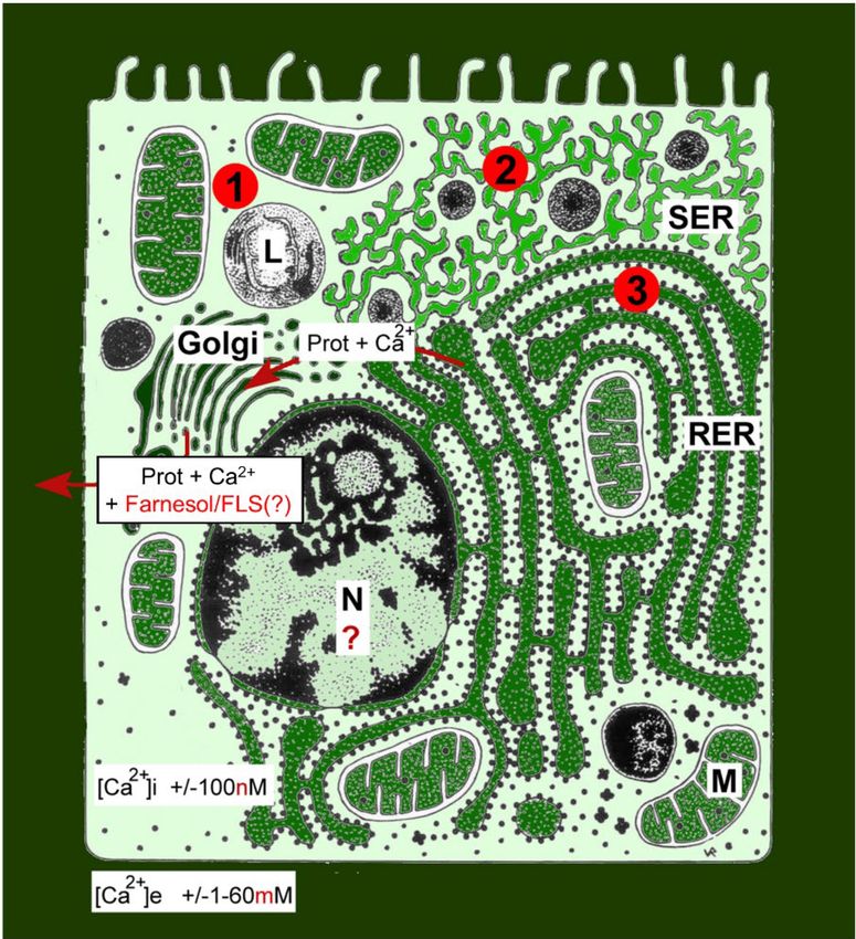
De Loof and Schoofs                                                                                                    Undervalued Functions of Golgi Apparatus

                                                secreting FLS, in particular juvenile hormone(s), it also secretes a protein(s) or peptide(s)
                                                with thus far unknown function. This paper suggests answers to various open questions
                                                in cell physiology and general endocrinology.
                                                Keywords: Golgicrine activity, juvenile hormone, SERCA, SPCA, thapsigargin, corpus allatum, hormone receptor,
                                                calcitox

INTRODUCTION                                                                          likely to meet with initial skepticism. At first sight, the reason
                                                                                      for skepticism seems very logical and understandable, but it is
In cell- and organismal animal physiology, Ca2+ is best known                         nonetheless erroneous. The “milk example” illustrates why. If
for its beneficial effects, e.g., in the construction of calcareous                   people are asked why female mammals after having given birth
skeletons in various animals, as a secondary messenger in                             start producing milk, the almost unanimous answer is: “To feed
signaling pathways, in neuronal activity, in muscle contraction,                      and provide immune protection to their young!” But if one next
etc. It is less known that Ca2+ is the most abundant toxin on                         asks if the female deliberately plans to engage in milk production,
earth, and that exactly this toxicity—at increasing cytoplasmic                       hesitation emerges. This hesitation turns into negation upon
concentrations to above 100 nM—forms the basis for the cited                          asking: “Do the mammary gland cells ‘know’ that they have to
beneficial effects. The huge Ca2+ concentration gradients over                        produce milk because a young/baby is waiting to be fed?” Of
the plasma membrane, ranging from an average of about 1–2 mM                          course not. But why is galactopoiesis initiated at all, if there is
extracellularly vs. 100 nM in the cytoplasm of unstimulated cells,                    no “goal” at all to do so? The right answer is that females start
in combination with the fact that the plasma membrane is not                          secreting milk because of hormonally controlled physiological
fully impermeable to Ca2+ (mainly due to Ca2+ channels and                            necessity. The “milk case” illustrates that there is no “goal” in
pumps), result in a constant drive for Ca2+ to enter the cell.                        evolution (De Loof, 2015, 2017b), perhaps some exceptions not
Figure 1 gives a visual impression of how Ca2+ is gradient-                           taken into account (Pookottil, 2013), and that the ways followed
wise distributed not only over the plasma membrane, but                               to increase fitness can be very ingenious.
intracellularly as well. It illustrates the challenges a eukaryotic cell
faces due to the fact that it has to maintain a low concentration
of free intracellular Ca2+ in an environment in which the                             “FROM PHYSIOLOGICAL NECESSITY”:
extracellular Ca2+ concentration is at least 20,000 times higher.                     WHICH ROLE FOR Ca2+
   When for whatever reason(s) excess Ca2+ enters the cell, this
excess has to be removed as quickly as possible. Small amounts
                                                                                      HOMEOSTASIS?
of Ca2+ can be removed by plasma membrane ATPases and                                 The drop in progesterone and estrogen titers prior to birth giving,
functionally related enzymes (Mechanism 1 in Figure 1) and                            combined with increased release of some brain hormones such as
by temporary storing excess Ca2+ in the lumina of intracellular                       prolactin, which facilitates the entry of Ca2+ into the mammary
membrane systems, in particular the ER and the mitochondria                           gland cells, and oxytocin, which is instrumental to milk ejection,
(Mechanism 2 in Figure 1; Orrenius et al., 2003; Groenendyk                           is causal to this “necessity.” Secondarily, the beneficial effect of
et al., 2004). When these mechanisms do not suffice, a third                          milk, with its high concentrations of proteins, Ca2+ , lipids, etc.,
system is mobilized, namely, removal of Ca2+ through the rough                        increases the fitness of the lactating female and the population to
endoplasmic reticulum (RER)–Golgi system (Mechanism 3 in                              which she belongs. Lactation got conserved in evolution because
Figure 1). The secretion of Ca2+ along with milk proteins from                        it increases fitness.
mammary gland cells is an extreme example (see section “‘From                             The “milk case” raises several cell physiological issues. One
Physiological Necessity’: Which Role For Ca2+ Homeostasis?”).                         concerns the high concentration of Ca2+ in milk, up to
If even this mechanism fails, the Ca2+ overload will induce                           60–100 mM (Wuytack et al., 2003). This concentration is
cell death/apoptosis (Nicotera and Orrenius, 1998; Orrenius                           about 600,000 to 1 million times higher than the free Ca2+
et al., 2003). Selective massive (programmed) cell death that                         concentration in the cytoplasm of unstimulated cells. Again, the
is physiologically and developmentally controlled occurs in,                          usual answer to the question why milk is so extremely rich
e.g., the gut epithelium, during complete metamorphosis of                            in Ca2+ is: “Because the developing baby/young needs lots of
insects, and in humans in the later stages of Alzheimer’s disease                     Ca2+ , among other things, for the construction of its calcareous
(De Loof and Schoofs, 2019a).                                                         endoskeleton.” And next: “Do the mammary gland cells add so
                                                                                      much Ca2+ to the milk fluid with this “intention?” Of course
                                                                                      not. They do so to get rid of the massive amounts that enter
ALWAYS KEEP IN MIND THAT THERE IS                                                     the gland cells resulting from the increase in permeability to
NO “GOAL” IN EVOLUTION, ONLY                                                          Ca2+ , which is caused by the “lactation-promoting hormones”
“PHYSIOLOGICAL NECESSITY”                                                             produced by the brain of lactating females. The ultimate reason
                                                                                      for secreting Ca2+ is that excess Ca2+ is very toxic (section “Basic
Our counterintuitive proposal that protein secretion by the RER–                      Mechanisms in Ca2+ Homeostasis”) and has to be eliminated
Golgi is subordinate to its role in removal of excess Ca2+ is                         as quickly as possible. The suckling young survives being fed

Frontiers in Physiology | www.frontiersin.org                                     2                                    October 2020 | Volume 11 | Article 542879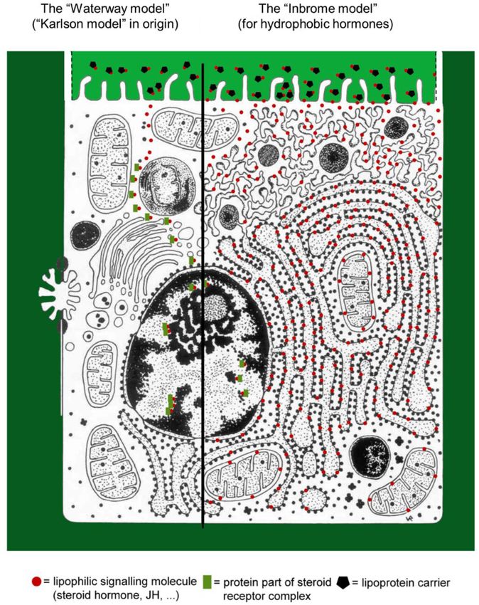
De Loof and Schoofs                                                                                                    Undervalued Functions of Golgi Apparatus

                                                                                        Carafoli and Krebs, 2016). On one hand, it is essential to the
                                                                                        correct functioning of cell processes, but, if not carefully
                                                                                        controlled spatially and temporally within cells, it generates
                                                                                        various severe cell dysfunctions, and even cell death (Nicotera
                                                                                        and Orrenius, 1998; Orrenius et al., 2003; Carafoli and Krebs,
                                                                                        2016). The very origin of the Ca2+ homeostasis system dates back
                                                                                        to ancestral prokaryotes as a survival system preventing Ca2+ -
                                                                                        mediated cell damage (Case et al., 2007). It further developed
                                                                                        at the unicellular stage of eukaryote evolution (Plattner and
                                                                                        Verkhratsky, 2015, 2016). Later on, mechanisms of signaling
                                                                                        became diversified, reflecting multiplication and specialization
                                                                                        of Ca2+ -regulated cellular activities (Figure 2). They have been
                                                                                        very well conserved in evolution (Plattner and Verkhratsky,
                                                                                        2015). The toxicity of Ca2+ and the counterbalancing by
                                                                                        endogenous farnesol-like sesquiterpenoids (FLSs) (De Loof et al.,
                                                                                        2015; De Loof and Schoofs, 2019e) resemble the story of the
                                                                                        evolution of O2 toxicity, the underlying free radical [reactive
                                                                                        oxygen species (ROS)] mechanisms and the role of antioxidants
                                                                                        (Zhu et al., 2016).
                                                                                            The Ca2+ homeostasis system with its numerous molecular
                                                                                        players (Ca2+ pumps, Ca2+ channels, calmodulin, etc., Figure 2)
                                                                                        produced a steep (see section “‘From Physiological Necessity’:
                                                                                        Which Role For Ca2+ Homeostasis?”) concentration gradient
                                                                                        between extracellular and intracellular compartments, which had
  FIGURE 1 | Schematic representation of the Ca2+ gradients (adapted from               both signaling function and survival importance. The latter is
  De Loof, 2017a). The different shades of green are not meant to give an exact         illustrated by the fact that even relatively (prolonged) moderate
  representation of differences in Ca2+ concentration. L, lysosome; N, nucleus;
  M, mitochondrion; RER, rough endoplasmic reticulum; SER, smooth
                                                                                        increases in cytosolic Ca2+ concentrations above 100 nM are
  endoplasmic reticulum. By just looking at the ultrastructure of cells and by          incompatible with life. Thus, the most basic activity of the
  evaluating how abundant the SER and RER are, one can make plausible                   Ca2+ homeostasis system is to keep cytosolic Ca2+ very low, not
  guesses about the gross outline of their Ca2+ homeostasis system, as well as          higher than 100 nM. Remarkably, which mechanisms control
  of some of its non-genomic effects on those enzyme systems that are                   this “keeping Ca2+ low” are only partially understood, in
  involved in lipid, steroid, and protein syntheses. This is due to the fact that
  numerous enzymes, the activity of which is (partially) controlled by the Ca2+
                                                                                        particular, the role of farnesol-like endogenous sesquiterpenoids
  gradient over their membrane, are anchored in these SER and RER                       remains undervalued until to date (De Loof and Schoofs,
  membrane systems. The red dots with 1, 2, and 3 correspond to the                     2019e; this paper).
  mechanisms 1–3 for keeping [Ca2+ ]i low (see text). Copyright permission:                 Rising concentrations of Ca2+ can alter the 3D conformation
  Own work (De Loof, 2017a), licensed under Creative Commons license.
                                                                                        of some types of macromolecules, in particular, of proteins as
                                                                                        well of chromatin/DNA. This forms the basis of the potential
                                                                                        toxicity of Ca2+ , as well as for the fact that Ca2+ can act as
with a potentially toxic nutrient because he or she neutralizes                         a secondary messenger in various signaling systems (De Loof,
the excess incoming Ca2+ by uploading it in deposits of Ca2+                            2017a; De Loof and Schoofs, 2019b). Muscle contraction is an
that are no longer toxic, in particular in the developing skeleton                      example of the effect of a sudden Ca2+ increase resulting from
and in intracellular Ca2+ stores, such as the lumina/cisternae of                       its release from the lumen of the smooth endoplasmic reticulum.
the endoplasmic reticulum, the mitochondria, and Golgi. The                             Maintaining “livable gradients” of Ca2+ is a most important task
mammary gland cells do not succumb under the high Ca2+                                  for all eukaryotic cells. We next focus on some novel insights
concentrations because they make use of the conveyor belt of                            in the role of the Golgi system and of endogenous FLSs in
transport proteins produced in the RER and next secreted by the                         achieving this goal.
Golgi, packed into vesicles.
                                                                                        The Golgi Apparatus: A Complex System
                                                                                        According to Barlow et al. (2018), the Golgi’s characteristic
BASIC MECHANISMS IN Ca2+                                                                morphology of multiple differentiated compartments organized
HOMEOSTASIS                                                                             into stacked flattened cisternae (Figure 3) fused together in a
                                                                                        continuous ribbon structure (Gosavi and Gleeson, 2017) is one of
If one is not familiar with the principles of Ca2+ homeostasis,                         the most recognizable features of modern eukaryotic cells. How
the statement that Ca2+ is the most abundant toxic pollutant in                         it originated and how it is maintained are not well understood.
the aqueous environment of cells but that it nonetheless became                         It may reasonably be assumed that the composition and lateral
the best communicator may sound strange at first encounter.                             organization of the Golgi membranes are not less complex
Calcium is an ambivalent messenger (Krebs et al., 2015;                                 than those of the plasma membrane (Jacobson et al., 2019).

Frontiers in Physiology | www.frontiersin.org                                       3                                  October 2020 | Volume 11 | Article 542879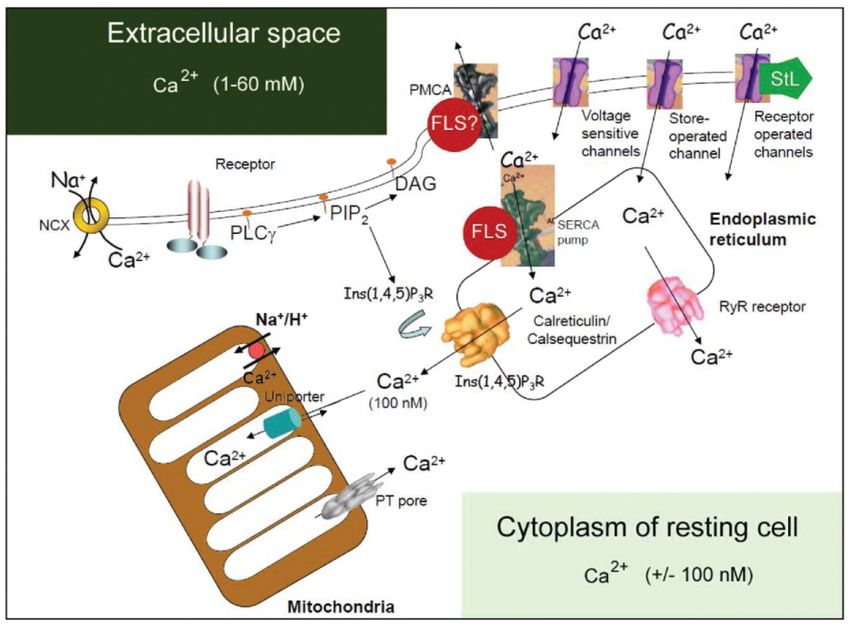
De Loof and Schoofs Undervalued Functions of Golgi Apparatus
FIGURE 2 | Classical view of the major players in the system that regulates intracellular Ca2+ compartmentalization. Cellular Ca2+ import through the plasma
membrane occurs largely by receptor-operated (e.g., glutamate receptors), voltage-sensitive and store-operated channels. Once inside the cell, Ca2+ can either
interact with Ca2+ -binding proteins or become sequestered into the endoplasmic reticulum (ER) or mitochondria. The largest Ca2+ store in cells is found in the ER
or sarcoplasmic reticulum, with local Ca2+ concentrations reaching millimolar levels. Ca2+ levels in the ER are affected by the relative distribution of
sarco(endo)plasmatic reticulum Ca2+ -ATPase (SERCA) pumps and of inositol-1, 4, 5-triphosphate [Ins(1, 4, 5)P3 ] receptors [Ins(1, 4, 5)P3 Rs] and ryanodine
receptors (RYRs), as well as by the relative abundance of Ca2+ -binding proteins (calreticulin, calsequestrin) in the ER or sarcoplasmic reticulum. The cytosolic Ca2+
concentration in unstimulated cells is kept at approximately 100 nM by both uptake into the ER and Ca2+ extrusion into the extracellular space by the plasma
membrane Ca2+ -ATPase (PMCA). ER Ca2+ release is triggered by agonist stimulation through the generation of Ins(1, 4, 5)P3 through hydrolysis of
phosphatidylinositol-4,5-biphosphate [PtdIns(4,5)P2 ] operated by a phospholipase C (PLCγ). The mitochondria take up Ca2+ electrophoretically through a uniport
transporter and can release it again through three different pathways: reversal of the uniporter, Na+ /H+ -dependent Ca2+ exchange, or as a consequence of
permeability transition pore (PTP) opening. The PTP can also flicker to release small amounts of Ca2+ . Ca2+ efflux from cells is regulated primarily by the PMCA,
which binds calmodulin and has a high affinity for Ca2+ . Ca2+ efflux might also be mediated by the Na+ /Ca2+ exchanger (NCX). [Ca2+ ], calcium concentration;
DAG, diacylglyceride. This figure was kindly provided by Prof. S. Orrenius. It served as the template for a figure in Orrenius et al. (2003). Regulation of cell death: the
calcium-apoptosis link. Nature Rev. Cell Biol. 2003; 4:552–565; https://doi.org/10.1038/nrm1150. The figure was slightly modified by adding the red dots with
farnesol-like sesquiterpenoid (FLS), suggesting a role for endogenous sesquiterpenoids (FLSs) as agonists for Ca2+ -ATPases. Such role is more probable for
sarco(endo)plasmatic reticulum Ca2+ -ATPases (SERCAs) than for PMCAs (hence the question mark). In 2003, the role of the Golgi system in Ca2+ homeostasis was
not yet well documented and therefore not represented in this figure. With copyright permission for both the figure and the legend from the publisher and from Prof.
S. Orrenius. This modified figure was published before in De Loof (2017a), with copyright permission (Open Access).
According to Barlow et al. (2018), golgins, which are omnipresent meeting point for the endocytotic and exocytotic systems in
in eukaryotes, are prominent proteins implicated in Golgi eukaryotic cells (Barlow et al., 2018). Last but not least, through
structure, and this since the common eukaryotic ancestor. The logical deduction, De Loof and Schoofs (2019b,d) came to the
Golgi apparatus has many functions (Makhoul et al., 2018). conclusion that the Golgi system is the most probable site of
Its role in protein secretion, a process that usually includes synthesis of endogenousFLSs) (section “The Golgi Apparatus as
protein modification, is best known. Golgi membranes harbor a Probable Subcellular Site of Synthesis of Farnesol/Farnesol-Like
a set of glycosylating enzymes (Figure 3C) that attach various Sesquiterpenoid: Origin of This Idea”).
sugar monomers to proteins moving through the apparatus.
Other functions include mitosis, DNA repair, stress responses,
Ca2+ homeostasis (Zhang et al., 2020), lysosome production
Pumping Excess Ca2+ Out of the Cell:
(Nakano and Luini, 2010), autophagy, apoptosis, inflammation, The Golgi System as Part of the
and prostaglandin synthesis (Smith and Malkowski, 2019). Ca2+ Homeostasis System
The Golgi apparatus also interacts with the microtubule and The best known Ca2+ pumping systems (active transporters)
actin networks (Kulkarni-Gosavi et al., 2019). It is a central are the plasma membrane Ca2+ -ATPases (PMCAs) and the
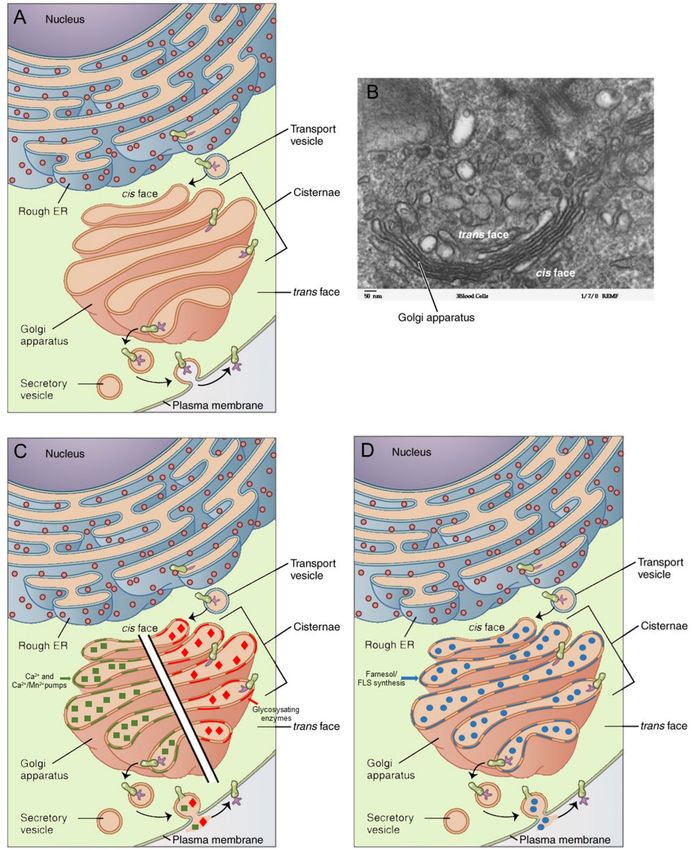
Frontiers in Physiology | www.frontiersin.org 4 October 2020 | Volume 11 | Article 542879De Loof and Schoofs Undervalued Functions of Golgi Apparatus FIGURE 3 | Golgi apparatus in context of the secretory pathway: (A) Schematic representation (Golgi in salmon pink). (B) Electron micrograph. (C) Schematic representation of the Ca2+ - and the Ca2+ /Mn2+ pumps as well as of glycosylating enzymes in the membranes. (D) The Golgi membranes also contain enzymes for the synthesis of farnesol/farnesol-like sesquiterpenoid (FLS). The major function of the rough endoplasmic reticulum (RER)–Golgi apparatus is the removal of excess Ca2+ from the cytoplasm, an activity that requires the synthesis of cell type-specific transport proteins in the RER. From here, proteins are sent to the Golgi apparatus, which organizes, modifies (e.g., by glycosylation), packages, and tags them. Some of these products are transported to other areas of the cell, and some are exported from the cell through exocytosis. Enzymatic proteins are packaged as new lysosomes (or packaged and sent for fusion with existing lysosomes) (Nakano and Luini, 2010). A reanalysis of classical data from insect endocrinology (see text) revealed that the Golgi apparatus is the likely site of synthesis of farnesol and farnesol-like sesquiterpenoids (FLSs), the juvenile hormones of insects being the best known ones. Copyright permission: (A–D) All under Creative Commons license CC BY 4.0. (A,B) Original files unchanged from Wikipedia File 0314 Golgi Apparatus.jpg (Created May 18, 2016). (C,D) Slightly modified from this file. Frontiers in Physiology | www.frontiersin.org 5 October 2020 | Volume 11 | Article 542879
De Loof and Schoofs                                                                                      Undervalued Functions of Golgi Apparatus

sarco/endoplasmic-reticulum Ca2+ -transport ATPases (SERCAs              Orrenius et al. (2003) advanced it as a causal link about
with various splice variants). Figure 2 illustrates the complexity       30 years later. Ablation of the CA not only affects the Golgi
of the system. In addition, animal cells also contain a                  systems but nearly all aspects of cell physiology, including
less characterized P-type Ca2+ -transport ATPase, PMR1/SPCA              lipid, protein, and ecdysteroid biosynthesis, the multiplication
Ca2+ /Mn2+ -transport ATPase (Van Baelen et al., 2002), which            of mitochondria, etc. It visualized in a few images that farnesol
is encoded by two genes that have various splice variants. This          esters are indeed the master hormone(s) in insect physiology
P-type Ca2+ -transport ATPase is mainly targeted to the Golgi            and development (De Loof and Schoofs, 2019b,d). Both
apparatus (Wuytack et al., 2003). It provides the Golgi with the         reimplantation of active CA and administration of synthetic JH
Ca2+ and Mn2+ necessary for the production and processing                (dissolved in an oil to act as a slow-release formula) temporarily
of secretory proteins. An N-terminal Ca2+ -binding motif in the          (= within a few days) rescued the observed phenotype and
Ca2+ /Mn2+ -transport ATPase SPCA1 regulates the secretory               thus demonstrated that absence of farnesol/JH is the real
pathway in particular in cells with a high Ca2+ load (e.g.,              cause of the cell physiological effects induced by allatectomy
mammary gland cells during lactation) or in cells with a low ATP         (De Loof and Lagasse, 1970).
content such as keratinocytes (Chen et al., 2019). Phylogenetic
analysis showed that SPCA1 may be older than the SERCA                   Another “Spark” of Insight: The
pump and related to a putative ancestral Ca2+ pump (Wuytack              Sarco(endo)plasmatic Reticulum
et al., 2003). The Golgi system can function as a Ca2+ store,            Ca2+ -ATPase Pump Blocker
which can be involved in setting up cytosolic Ca2+ oscillations
                                                                         Thapsigargin Is, Like Farnesol/Juvenile
(Wuytack et al., 2003).
                                                                         Hormones, a Sesquiterpenoid
                                                                         Sarco(endo)plasmatic reticulum Ca2+ -ATPases can interact with
                                                                         a variety of molecules. The number of interacting proteins
THE LINK BETWEEN Ca2+ PUMPING BY                                         keeps growing (Vandecaetsbeek et al., 2011). One of the
SARCO(ENDO)PLASMATIC RETICULUM                                           well-known agents is thapsigargin, a potent blocker of both
Ca2+ -ATPases AND ENDOGENOUS                                             the SERCA pump (Rogers et al., 1995) and the SPCA1a
                                                                         Ca2+ /Mn2+ transport ATPase present in the Golgi apparatus
SESQUITERPENOIDS
                                                                         (Chen et al., 2017, 2019; Smaardijk et al., 2017). Thapsigargin
                                                                         is extracted from the plant Thapsia garganica, hence its name.
Fifty Years Ago: Arrest of                                               Structurally, it is classified as a sesquiterpene lactone. By
Farnesol/Juvenile Hormone Production                                     blocking the ability of the cell to pump calcium into the
Impairs Golgicrine Secretory Activity in                                 sarcoplasmic and endoplasmic reticula, thapsigargin raises the
an Insect. Temporary Rescue Possible                                     cytosolic (intracellular) Ca2+ concentration. Store depletion
By electron microscopy, De Loof and Lagasse (1970) discovered            can secondarily activate plasma membrane calcium channels,
that the ultrastructure of fat body cells of the Colorado                allowing an influx of calcium into the cytosol. Depletion of ER
potato beetle (Leptinotarsa decemlineata) changes when the               calcium stores leads to ER stress and ultimately leads to cell death
farnesol/juvenile hormone (JH) synthesizing glands, the corpora          (Zhang et al., 2020).
allata (CA), are inactivated by microsurgical removal. In                    The similarity of cell death induction in a wide variety
addition, they also observed this phenotype when the beetles             of eukaryotes by administration of thapsigargin as well as by
were raised in conditions with a short photoperiod (= less than          silencing the production of JH by the CA of insects with a
12-hour light per day). Other tissues were not investigated, and         complete metamorphosis (= Holometabola) triggered the search
sites of JH synthesis other than the CA were not known at that           for the cause of this similarity. Is, perhaps, thapsigargin toxic
time (see later).                                                        because it displaces a natural sesquiterpenoid ligand from its
    The most drastic effect in the fat body cells was on the             binding site on Ca2+ -pump(s) thereby blocking them (De Loof
functioning of the Golgi systems: the secretion of vesicles              et al., 2014)? It is known since long that farnesol and its
was totally disturbed. The outgoing transport vesicles no                JH-esters (Röller et al., 1967; Röller and Dahm, 1968) are
longer fused with the plasma membrane but instead fused                  sesquiterpenoids, but for thapsigargin, this property is only
with one another, thereby forming large protein bodies (for              rarely mentioned. Despite intensive research, the natural ligand
figure, see De Loof and Schoofs, 2019b, or De Loof and                   for this binding still remains unknown. We think, without
Schoofs, 2019a). At that time, this effect was (erroneously: see         having experimental proof, that for engaging in Ca2+ pumping,
sections “‘From Physiological Necessity’: Which Role For Ca2+            SERCAs and other Ca2+ pumps need the presence of an
Homeostasis?” and “From Physiological Necessity”: Which Role             endogenous FLS as an agonist of the pumps (De Loof et al., 2014;
For Ca2+ Homeostasis?) interpreted as being physiologically              De Loof, 2017a).
relevant and beneficial for depositing reserves in preparation of
hibernation/diapause.                                                    Some Ca2+ Channel Types Are
    We now believe that the effect is in fact a step toward              Receptors for Farnesol
programmed cell death. However, at that time, the role of                In addition to a suggested but not yet experimentally proven
Ca2+ in the induction of apoptosis was not yet known.                    role of FLS as agonists of SERCAs and possible other

Frontiers in Physiology | www.frontiersin.org                        6                                   October 2020 | Volume 11 | Article 542879
De Loof and Schoofs Undervalued Functions of Golgi Apparatus
Ca2+ pumps (De Loof, 2017a), direct experimental proof A STILL CONTINUING SEARCH: HOW
for a receptor role of farnesol for a voltage-gated Ca2+ MANY SITES OF
channel has been described by the electrophysiologists Roullet
et al. (1999) and Luft et al. (1999). For more details, see
FARNESOL/FARNESOL-LIKE
De Loof and Schoofs (2019a,b). SESQUITERPENOID SYNTHESIS ARE IN
In combination, the picture emerges that farnesol/FLSs act THE BODY?
on keeping intracellular Ca2+ low by concurrently inhibiting the
influx of Ca2+ through (some types of) Ca2+ channels and acting The Situation in Insects Is Best
as agonists of Ca2+ pumps that remove excess Ca2+ . This made Documented
De Loof and Schoofs (2019b) state that the membrane receptor(s) There are several reasons why more experimental data on the site
of farnesol/JH is the integrated Ca2+ homeostasis system in its of synthesis and the functions of farnesol have been advanced
totality. Even ecdysteroids and hydrophobic vertebrate steroid in insects and not in any chordate/vertebrate species. First,
hormones may act in a similar way. until recently (De Loof et al., 2015; De Loof and Schoofs,
2019e), farnesol was known neither as a hormone nor as an
Nuclear Receptor(s) for Juvenile “inbrome” (De Loof et al., 2015) in vertebrates. Its only role
Hormones: Methoprene-Tolerant (Met), in chordates was thought to be one of the intermediates in the
the Putative Insect Nuclear Juvenile mevalonate biosynthetic pathway that leads to the synthesis of
Hormone Receptor, Remains squalene, and next to cholesterol and steroids, without having
any function on its own. Logically, that should have changed
Controversial when the electrophysiologists Roullet et al. (1999) and Luft et al.
Yamamoto (1988) suggested, based on solid experimental results, (1999) demonstrated that farnesol is a potent blocker of N-type
that a membrane protein mediates an effect of JH that involves voltage-gated Ca2+ channels in the brain of some rodents and
calcium and kinase C. Much later, Jindra et al. (2015a,b) of humans. It did not. Hence, the question where endogenous
advanced the view that the transcription factor Methoprene- FLSs are synthesized never became a topic worth investigating
tolerant is, in their opinion, the key/master receptor for JH: in chordates. In contrast, in insects, it started with the pioneering
no need for a membrane receptor, Ca2+ , and kinase C. work of Kopec (1922) about a century ago, with his discovery that
This view gained a rather wide acceptance among molecular the insect brain is an endocrine gland. Later, key pioneers were
biologists, but not among cell biologists. The major objection sir Vincent Wigglesworth with his discovery of JH synthesized by
is that, up to the present day, it has not been experimentally the CA, and Carroll M. Williams with the serendipitous discovery
demonstrated that farnesol/JHs, which are highly hydrophobic that the abdomen of male Hyalophora cecropia moths was a
molecules, ever enter the nucleus with its internal hydrophilic repository of high amounts of JH (references in Riddiford, 1994;
watery environment. As long as the in situ binding between Watt and Davey, 1996; Nijhout, 1998). It took many years before
JH and its Met-transcription factor inside the nucleus has not it became clear that the CA were not the only site of synthesis
been experimentally proven, Met does not meet the required of farnesol/JHs. An indirect indication in favor of multiple sites
conditions to be classified as a “genuine receptor.” According of synthesis farnesol/JHs was advanced by Castillo-Gracia and
to De Loof and Schoofs (2019a,d,e), there is no problem to Couillaud (1999). They found that the catalytic site of the enzyme
classify MET as a JH target. Even more, it is probable that farnesyl-diphosphate synthase, which plays a role in isoprenoid
Met is a Ca2+ -sensitive target (De Loof and Schoofs, 2019e, biosynthesis, was present in the brain, ovary, fat body, and
and this paper). CA samples (= all tissues with active Golgi systems), but not
Farnesol/JH influences many other targets, most of them in muscle, a tissue that does not engage in substantial protein
residing in membranes as cited before (section “The Link secretion. In the CA, the encoding gene for farnesyl-diphosphate
Between Ca2+ Pumping by Sarco(Endo)Plasmatic Reticulum synthase is overexpressed.
Ca2+ -Atpases And Endogenous Sesquiterpenoids”). To our
knowledge, no data are available on nuclear receptors for
farnesol in chordates. Wigglesworth (1969) suggested that the Endocrine-, Exocrine-, and Golgicrine
difference in biological activity of some 40 compounds he Farnesol/Juvenile Hormone Synthesis
tested in a typical JH bioassay was quantitative rather than The successive steps in this development have recently been
qualitative, and that such compounds first act at the plasma reviewed by De Loof and Schoofs (2019d,e). In short, up to
membrane, with secondary effect in the nucleus as a result. 1995, the CA were assumed to be the sole site of synthesis
Wigglesworth (1969) also reported that JH analogs are not of JHs. That changed when Borovsky et al. (1994a,b) reported
active in a watery environment or in the presence of wetting that, in addition to the CA, both male and female gonads of
agents. This brings the hydrophobicity issue, which is very mosquitoes also synthesized JHs de novo. Another two decades
relevant for sesquiterpenoids and some steroid hormones, in later, Paroulek and Sláma (2014) refined this addition by showing
focus (section “The Hydrophobicity Issue in Farnesol/Farnesol- that in the moth H. cecropia, the model insect from which JH
Like Sesquiterpenoid Transport and Mode of Action. The I had been originally extracted and chemically identified, JH is
“Waterway Mode” Versus the Lipid Membrane Way or synthesized de novo in the male accessory glands (= to some
“Inbrome Way””). extent the counterpart of the mammalian prostate gland) and
Frontiers in Physiology | www.frontiersin.org 7 October 2020 | Volume 11 | Article 542879De Loof and Schoofs Undervalued Functions of Golgi Apparatus
this in a developmental stage in which the CA are inactive. only indirect arguments can be advanced. Short Neuropeptide
They named it “exocrine JH synthesis.” They also reported that F might be a good candidate (arguments and references in De
this male accessory gland (MAG) JH was not secreted into the Loof and Schoofs, 2019b; Fadda et al., 2019). When sNPF peptide
hemolymph, but instead left the male body along with secretory was injected into adult female locusts, the stimulation of ovarian
proteins that during copulation are transferred into the female’s development was so pronounced that Cerstiaens et al. (1999)
genital system. This selectivity, or better unidirectionality, in thought that they discovered a potent growth hormone of insects
JH secretion triggered De Loof and Schoofs (2019d) to search or, alternatively, a releasing hormone for such growth hormone.
for an explanation for this at first sight unusual transport Later research showed that it is a much more complex issue
of a hydrophobic/lipophilic hormone. When upon analyzing and that sNPF plays, among other functions, a prominent role
Schmialek’s (1961) experimental data, the idea emerged that the in the control of feeding (Carlson et al., 2013; Fadda et al.,
most plausible and logical answer to the question how farnesol 2019). In addition, changes in microRNAs may also play a role
can end up in the lumen of the alimentary canal of larval (Nouzova et al., 2018a).
mealworms, was that this requires a key contribution of Golgi
systems of the gut’s epithelial cells. Indeed, the Golgi system with
its membranes that harbor various enzymes including some P450 THE GOLGI APPARATUS AS A
enzymes, a family of enzymes that plays important roles in both PROBABLE SUBCELLULAR SITE OF
the biosynthesis and inactivation of 20-hydroxyecdysone and JHs SYNTHESIS OF
(Iga and Kataoka, 2012), is the only cell organelle that can explain
the unidirectional transport of JH along with secretory proteins
FARNESOL/FARNESOL-LIKE
in Hyalophora. This way, the idea of “Golgicrine farnesol/FLS SESQUITERPENOID: ORIGIN OF
synthesis and secretion” was born. Immediately, the question THIS IDEA
emerged whether such “Golgicrine activity” is compatible with
JH synthesis by the CA. Not only the question which protein/peptide is secreted by the
Golgi of the CA, but also the questions where exactly farnesol/FLS
is synthesized in the gland cells and how it leaves the CA cells
Gland Cells of the Corpora Allata Have remain unanswered and deserve innovative thinking.
Golgi Systems: A Problem Emerges
The first mention of the presence of active Golgi systems in Peter Schmialek’s Identification of
the CA was published by Scharrer (1964) in the cockroach Farnesol and Farnesal in the Mealworm’s
Leucophaea maderae. Similar results were later published by Excrements
Bradley and Edwards (1979) in CA of the house cricket Acheta Schmialek (1961) was the first to chemically characterize two
domesticus. Although it was known since long that Golgi systems compounds that were active in the Tenebrio assay for detecting JH
serve a role in the secretion of proteins, the possibility that activity, namely, the sesquiterpenoids farnesol and farnesal. They
the “juvenilizing factor” secreted by the CA might be a protein were not extracted from tissue or whole-body extracts but from
was not considered at that time. The farnesol line of thinking excrements of the mealworm Tenebrio molitor. The question how
(Schmialek, 1961) dominated the scene at that time. In fact, these compounds could end up in the lumen of the alimentary
it continues to do so until today. Hence, the question: “What canal was asked, but could not be answered. The hypothesis was
type of protein/peptide do the Golgi of the CA secrete?” that their transport was achieved through the Malpighian tubule
still remains unanswered. It cannot be excluded that the key system as degradation products of the true—but at that time
function of the CA is not the secretion of JH, but of an still unidentified—JH. But the Malpighian tubule system does
as yet unknown sort of proteinaceous/peptidic growth factor. not transport such hydrophobic compounds. Upon reexamining
In particular, in the recent decade, several analyses of the Schmialek’s data, De Loof and Schoofs (2019d) logically deduced
peptidome of the CA have been undertaken. Liu et al. (2012) that the more probable explanation is that the Golgi system of the
could assign a total of 41 ion masses present in the CC–CA epithelial cells of the gut play a role in the unidirectional transport
complex of fifth instar Bombyx mori but no clear-cut candidate of endogenous sesquiterpenoids into the gut’s lumen.
for a peptide that is only secreted by the Golgi of the CA
was found in this or other (e.g., Nouzova et al., 2018b) mass Paroulek and Sláma: In the Male
spectrometric studies.
One should remember that Sláma (2013, 2015) since long
Accessory Glands of H. cecropia,
postulated the possibility that the “real JH” might be the Exocrine Juvenile Hormone I Is
protein/peptide secreted by the Golgi of the CA. Almost nobody Unidirectionally Transported
listened. In our opinion, answering this question is of prime As already cited before, Paroulek and Sláma (2014) solved the
importance for avoiding that some researchers continue working problem of the site of synthesis of the high amounts of JH-active
along wrong premises and hypotheses. material in the MAGs of H. cecropia. It had been thought for
Maybe a putative “Golgi-growth stimulating peptide” has not many years that the MAGs only function as a repository for the
yet been found because it could be totally absent from early JHs that were already synthesized in the CA (Shirk et al., 1976).
metamorphosis on up to adult emergence. In such situation, However, the authors proved that not the CA but the MAGs
Frontiers in Physiology | www.frontiersin.org 8 October 2020 | Volume 11 | Article 542879De Loof and Schoofs Undervalued Functions of Golgi Apparatus
themselves synthesize the JH they accumulate, and that they
do not release it into the hemolymph. The consequence is that
MAG JH I should not have been denominated as a “hormone.”
Later, when it was shown that the CA also produce JH I,
which they do release into the hemolymph, it rightly acquired
the status of “hormone,” at least for JHs that end up in the
hemolymph. An intriguing issue remained unanswered: How to
explain that MAG-JH does not diffuse into the hemolymph, but
that instead it is unidirectionally transported along with secretory
proteins produced by the MAGs into the female genital system
during copulation.
The Hydrophobicity Issue in
Farnesol/Farnesol-Like Sesquiterpenoid
Transport and Mode of Action. The
“Waterway Mode” Versus the Lipid
Membrane Way or “Inbrome Way”
To date, the almost general consensus is that, after its synthesis
somewhere in the gland cells of the CA, JH freely diffuses
through the cytoplasm toward the plasma membrane of the
gland cells, next inserts itself into its lipid bilayer, and next
“jumps” to lipoproteins circulating in the hemolymph. This
view neglects the consequences of a most important chemical
property of farnesol/JHs, namely, that it is only very moderately
soluble in water. Thus, without a hydrophobic carrier, it
cannot freely diffuse through a watery/hydrophilic environment.
Recent textbooks (e.g., Raven et al., 2019) pay attention to FIGURE 4 | Two models for the entry and intracellular transport of small
the hydrophobicity issue. The “exocrine JH” denomination of non-proteinaceous/peptidic hormones in cells. (Left panel) According to
Karlson and Sekeris (1966) the ecdysteroid molting hormone of insects
MAG-JH by Paroulek and Sláma (2014) suggests another way
(20-hydroxy ecdysone or 20E), which is moderately water-soluble, could pass
of transport of JH. After its synthesis in the Golgi system itself, the plasma membrane by simple diffusion without eliciting any signaling (e.g.,
JH gets attached to hydrophobic parts of (transport) proteins through Ca2+ ) at the level of this membrane. Next, it would diffuse through
that are on their way from the RER to the cell’s exterior—to end the cytoplasm and end up in the nucleus. Here, it would (in)activate specific
up in the hemolymph. Apparently, this hydrophobic interaction genes. In the 1960s–1970s, this concept was expanded toward being also
valid for lipohilic/hydrophobic molecules such as some vertebrate-type sex
is sufficiently strong to prevent the diffusion of JH through the steroids and to insect juvenile hormone(s). Later, this model got
membranes of the Golgi system. In such scenario, exocrine JH complemented with cytoplasmic protein transporters for the transport of
would also have an endocrine function, which is not the case lipophilic/hydrophobic molecules through the cytoplasm into the nucleus. This
of the MAGs of Hyalophora, because MAG-JH does not end up model, despite justified criticisms, is still widely used to date. (Right panel)
in and is not transported by the hemolymph. Thus, by logical The “intramembrane transport model” or “inbrome model.” In particular for
hydrophobic small hormones, e.g., the juvenile hormone esters of farnesol,
deduction, the conclusion can be reached that farnesol/FLS, after the inbrome mode of action model was originally advanced by De Loof (2015).
their synthesis in membranes of the Golgi (not yet experimentally It says that such hydrophobic hormones are transported in the
proven!), distributes itself between the membranes of the Golgi blood/hemolymph by a lipoprotein that delivers the hormone at the plasma
and hydrophobic stretches of amino acids present in the Golgi’s membrane, in which the hormone will enter through hydrophobic interaction.
Next, the hormone will start diffusing through the entire system of all
lumen where they can be further modified. Because both the
connected membrane systems: plasma membrane, smooth endoplasmic
membrane and the content of the lumen of the Golgi move reticulum (SER)–rough endoplasmic reticulum (RER), nuclear envelope, Golgi
toward the trans side where pinching off happens, farnesol/FLS apparatus, mitochondria, etc. These lipid bilayer membranes are liquid and
also move along. We propose the term “lipid membrane way act as a solvent and transport system for hydrophobic molecules. During the
mode of transport” or “inbrome way of transport” (De Loof diffusion, the hydrophobic molecules will encounter many proteins that are
anchored in the membranes. They all have hydrophobic transmembrane
et al., 2015) of small hydrophobic molecules for such transport
stretches of amino acid chains. Some membrane proteins may act as genuine
(Figure 4). In fact, the membrane system of cells acts as a receptors. The interaction with actors of the Ca2+ homeostasis system is
liquid hydrophobic solvent system through which hydrophobic important. The lighter green color at the top of this figure does not mean that
hormones and other solutes can freely diffuse. This oil canal here the Ca2+ concentration is lower, but to make the details readable.
system complements the “the waterway mode” for the transport Intracellular Ca2+ gradients (Figure 1) are not represented. Own work,
modified after De Loof (2017a, Open Access): Copyright permission under
of, e.g., water-soluble hormones: through the blood or through Creative Commons License.
the hydrophilic cytoplasm.
Frontiers in Physiology | www.frontiersin.org 9 October 2020 | Volume 11 | Article 542879De Loof and Schoofs Undervalued Functions of Golgi Apparatus
FUNCTION(S) OF In addition, they can be anchored in the lipid bilayer part
FARNESOL/FARNESOL-LIKE of membranes as shown for the membrane attachment of
Ras by Schafer et al. (1989). Already in Barber et al. (1981)
SESQUITERPENOIDS IN THE GOLGI reported that JH and two JH analogs can modulate the physical
MEMBRANES properties of phospholipid bilayers. This raised the prospect
that some of their physiological effects may be edited through
Sarco(endo)plasmatic Reticulum their influence on the molecular organization of the membrane
Ca2+ -ATPase and SPCA1a Ca2+ /Mn2+ lipids in particular. Such effect, e.g., on viscosity, for naturally
Transport ATPase Have a Binding Site for occurring polyisoprenoid alcohols (including farnesol/FLS) was
further documented by Murgolo et al. (1989), who determined
Some Sesquiterpenoids the horseshoe shape of such compounds as well as their high
We only elaborate on the plant toxin thapsigargin because
rotatable bond count, two properties of farnesol/JHs that have
it is a sesquiterpenoid that, in our opinion, likely acts by
only recently been mentioned as being functionally important
displacing a natural endogenous sesquiterpenoid ligand(s) from
in the literature on insect endocrinology (De Loof and Schoofs,
its binding site on the SERCA pump. That ligand could
2019c,e). Murgolo et al. (1989) (also) suggested a role in protein
be farnesol itself or a farnesol-like sesquiterpenoid (FLS)
glycosylation, another important activity in Golgi systems.
(De Loof, 2017a). As already mentioned in section “Another
Thus, in our opinion, farnesol/FLS in the Golgi may serve
“Spark” of Insight: The Sarco(endo)plasmatic Reticulum Ca2+ -
the role of membrane “liquidizer–plasticizer.” Maybe they can
ATPase Pump Blocker Thapsigargin Is, Like Farnesol/Juvenile
be viewed as the biological counterparts of the phthalates
Hormones, a Sesquiterpenoid,” both the SERCAs and the
that are used as plasticizers in the polyvinylchloride (PVC)
Ca2+ /Mn2+ Ca2+ -transport ATPases have a binding site for the
plastic industry. Farnesol/FLS may also have a role in protein
plant toxin thapsigargin, which blocks these pumps. This toxin
glycosylation in the Golgi.
is a sesquiterpene lactone. It binds to SERCA in a hydrophobic
In a former paper, De Loof and Schoofs (2019c) proposed
funnel-like cavity open to the cytosol formed by TM3, TM5, and
a model for another, transcription-independent, role of
TM7 transmembrane loops of the SERCA pump and prevents
farnesol, through the mechanism of prenylation,” for getting
the movement of the helices relative to each other. Nine out of
inserted in between some of the helices of G protein-coupled
the 10 transmembrane loops of SERCA undergo rearrangements
receptors. It involves the high flexibility of farnesol/FLS.
during a pumping cycle, enabling the release of Ca2+ into the
The self-closing issue of membranes is of course also
lumen of the ER and Golgi and, on the cytoplasmic side, to create
essential during mitosis, a process that requires Golgi activity
a pathway for entry of new Ca2+ ions (for references, see De Loof,
(Smith and Malkowski, 2019).
2017a). To date (2020), experimental validation of this hypothesis
or the identification of other putative endogenous ligands is still
missing. The fact that the Golgi apparatus seems to be a major site Why Are Multiple Sites of
of synthesis of farnesol/FLS strengthens the hypothesis that such Farnesol/Farnesol-Like
compounds may be, one way or another, agonists of the Ca2+ Sesquiterpenoids Synthesis in the Body
pumps that play prominent roles in the RER–Golgi secretory Required?
pathway. Because thapsigargin acts as a SERCA blocker in various
The bigger an animal, the higher its membrane turnover. In
eukaryotes and because SERCAs are very well conserved in
insects, the CA in larvae may produce enough JH to lubricate
evolution, it can be assumed that the binding is well conserved
the plasma membranes of all cells. This may be facilitated
as well. However, this awaits experimental validation.
by the fact that the farnesol esters with JH activity (which
is orders of magnitude than the biological activity of farnesol
Facilitating Membrane Self-Closing? itself) ensure that only very low amounts of JHs are needed to
Farnesol as Membrane accommodate the whole body.
“Liquidizer–Plasticizer”? A problem arises when the CA stop secreting JH while
A prominent feature of Golgi membranes is their high flexibility internally some tissues continue a function that requires a
and capacity of self-closing. In active Golgi systems, vesicles high membrane turnover. This is the case with the MAGs of
that are pinched off from the RER will fuse with membranes H. cecropia. But even in the adult stage, when the CA are active, an
of the Golgi at its cis side. At the trans side, the opposite additional supply of farnesol may be required, e.g., in the gonads
happens. The trans vesicles will, in their turn, fuse with the with their numerous microvilli and secretory vesicles in some cell
plasma membrane and discharge their content into the cell’s types in both males and females (Roth and Porter, 1964; Borovsky
exterior. All these fusion and pinching off require instant closing et al., 1994a,b).
of the membranes involved; otherwise, the electrical properties Our hypothesis is that in insects, CA farnesol secretion
of the secreting cells would get severely impaired by the loss suffices of accommodating the needs for plasma membrane
of inorganic ions, which are gradient-wise distributed over flexibility for endocytosis and exocytosis, e.g., in immune
the membranes. By their nature, sesquiterpenoids are highly reactions. For intensive protein synthesis and release through
flexible molecules as indicated by their high rotatable bond the RER–Golgi system, additional sites of farnesol/FLS synthesis
count (7 according to PubChem) and horseshoe-shape form. seem to be required.
Frontiers in Physiology | www.frontiersin.org 10 October 2020 | Volume 11 | Article 542879De Loof and Schoofs Undervalued Functions of Golgi Apparatus
Farnesol/Farnesol-Like DISCUSSION
Sesquiterpenoids: The Most Ubiquitous
Farnesol was first detected in floral extracts, which are ingredients
Animal Hormone(s)? for the perfume industry. The name “farnesol” is derived
Yes, that is the unavoidable conclusion from this review paper. from Vachellia farnesiana that had been planted in the garden
Why was this never proposed before? First, for historical reasons. of Odoardo Farnese in Rome. To our knowledge, the exact
The very first identification of farnesol was not in animals but in physiological role of farnesol in plants is still unknown to date.
extracts of flowers to be used in perfumes. Its physiological role in But such ignorance about the functionality of farnesol/FLS also
plants has not yet been thoroughly investigated. Similar functions covers the great majority of all eukaryotes. They all synthesize
as in animal cells (this paper) are likely. Flowers are structures farnesol. Even in vertebrates, early 2019 farnesol was named “a
in which massive cell division takes place, an activity for which noble unknown” (De Loof and Schoofs, 2019e). It was thought
plasma membrane flexibility and turnover is, by definition, a that its only function was to act as one of the intermediates of the
major issue. Second, a function as “membrane liquidizer” or well-documented mevalonate–cholesterol biosynthetic pathway,
“membrane lubricant” is the last thing most researchers would without having a function by itself in cell physiology. One of
think about. Yet, as we know from machine functioning, moving the reasons for this way of thinking was that farnesol is not
parts need to be lubricated. But this flexibility/lubricant problem known as a hormone or inbrome or any other type of signaling
is not restricted to animal cells only, but to all eukaryotic ones. molecule in vertebrates. Neither are vertebrates known to have a
That may be an explanation for the omnipresence of farnesol/FLS stage in their development in which the body is totally devoid
synthesis by the ubiquitous mevalonate biosynthetic pathway. of farnesol/FLS. Finally, neither has it as yet been investigated
Third, it is only recently that the key role of the mevalonate whether the sesquiterpenoid lactone thapsigargin, upon binding
biosynthetic pathway in Ca2+ homeostasis is put into the picture to SERCA or Ca2+ /Mn2+ calcium transport proteins, displaces a
(De Loof and Schoofs, 2019e). putative endogenous possibly FLS ligand.
This contrasts sharply with the situation in insects. They do
Farnesol: Also Part of the Animal’s have such a stage in their development. At the onset the last larval
Pheromone System Like in Plants? instar of holometabolous insects, the only gland that synthesizes
For a long time, farnesol was best known as a component farnesol/FLS (the CA) gets fully inactivated with a drop of
of flower extracts to be used in the perfume industry. Its farnesol/FLS titers to zero as a result. The hormone secreted
basic physiological role in plants is still poorly documented. by the CA that, when absent, induced metamorphosis had been
In animal physiology, a possible role as a component of the named “juvenile hormone.” Röller et al. (1967) and Röller and
“smell” of (terrestrial) animals has not yet been considered. In Dahm (1968) identified it as an ester of farnesol. Farnesol itself
our opinion, there is no reason to assume that with respect to has biological activity in typical bioassays for detecting JH activity
“the (physiologically secondary) perfume aspect,” the situation in (for a picture of a positive reaction, see De Loof et al., 2014,
animals should differ very much from that in plants. open access). Its activity is weak compared to the much higher
activity of its esters.
In rodents and man, farnesol, which is biosynthesized in the
PERSPECTIVES FOR FUTURE brain and other tissues, acts as a potent blocker of N-type voltage-
RESEARCH gated Ca2+ channels (Roullet et al., 1999) and Luft et al. (1999).
This discovery causally linked farnesol to the control of Ca2+
Only a few possible topics are listed: homeostasis. However, for one reason or another, these data did
not draw the attention of the vertebrate endocrine community,
In Vertebrates but they did, with some delay, of insect endocrinologists who
The key question: Is farnesol/FLS a hormone in vertebrates? searched for the nature of the membrane receptor(s) of JH in
Which stereoisomers do occur? Do SERCAs and SPCA pumps insects (De Loof and Schoofs, 2019b). That is the reason why in
need a farnesol/FLS-like agonist? How would such agonists be this review paper, the majority of data and models come from
attached: by hydrophobic interaction or by prenylation or still insect endocrinology: data in vertebrates are almost non-existent.
in another way? Do all tissues with active Golgi synthesize The fact that all eukaryotes, both uni- and multicellular
farnesol/FLS? Does the “inbrome model” of transport also apply ones, produce farnesol is compatible with a key role in Ca2+
to hydrophobic sex steroids? homeostasis. Indeed, Ca2+ is the most ubiquitous toxin on earth
for all eukaryotes; they all have to counter the influx of excess
In Insects and Other Protostomes Ca2+ from the extracellular environment of cells.
Identify the protein or peptide secreted by the Golgi present The idea of a functional link between the Golgi and
in the CA. Are enzymes involved in JH synthesis anchored farnesol/FLS emerged from a serendipitous reexamination (De
in the membranes of the Golgi? Why does “exocrine Loof and Schoofs, 2019d) of a paper by Schmialek (1961). It
JH” not diffuse into the hemolymph compartment? Does triggered the idea that the most logical explanation for the
farnesol/FLS, due to its hydrophobic nature, ever end fact that farnesol is found in substantial concentrations in
up in the nucleus? How is exocrine JH complementary to excrements of the mealworm Tenebrio was that farnesol must be
endocrine JH? unidirectionally transported from the gut’s epithelial cells into the
Frontiers in Physiology | www.frontiersin.org 11 October 2020 | Volume 11 | Article 542879De Loof and Schoofs Undervalued Functions of Golgi Apparatus
gut’s lumen. The only cell organelle that can enable this is the two decades against disagreeing researchers: no way to publish
RER–Golgi system. Paroulek and Sláma (2014) showed for the “aberrant” experimental data. Today’s situation is very different.
first time that JH I that turned out to be synthesized by the MAGs If the two major functions of the Golgi, namely, roles in Ca2+
themselves of the silk moth H. cecropia, and not in their CA as homeostasis and in farnesol/FLS production, are accepted, the
had been assumed before, is unidirectionally transported, not into novel paradigm says that it is normal and almost self-evident
the hemolymph, but into the genital system of the female during that all protein-secreting cells and tissues may produce their own
copulation. This is a strange way of transport of a hydrophobic FLSs. Whether there are quantitative and qualitative (types of
hormone that had been assumed to freely, not unidirectionally, isomers) differences remains to be investigated. The data that
diffuse in cells. It raised the question: Is, perhaps, the Golgi system farnesyldiphosphate synthase is only abundant in the CA and not
involved in the production and transport of all cells with an in any other tissue known to synthesize farnesol/FLS (Castillo-
active Golgi? Gracia and Couillaud, 1999) suggest that the CA are indeed the
Upon reexamining the older literature, it became apparent master gland that influences all cells of the body. Apparently,
that the presence of active Golgi in the CA was already well some (intra)cellular functions, in particular in the case of active
documented since 1964 (Scharrer, 1964; Bradley and Edwards, protein secretion by the RER–Golgi, the massive Ca2+ removal
1979), but these data got buried under the massive number of and membrane turnover in the Golgi require an additional local
novel developments in the endocrine field. Yet, the presence site of synthesis.
of active, protein-secreting Golgi in the CA deserves to be With this review, we intend to stimulate research in both
reanalyzed. It could, perhaps, yield a very potent growth factor, basic and medical sciences into the full role of the Golgi.
the importance of which might exceed that of JH, which may Malfunctioning of the Golgi system is implicated in several
rather be an adjuvant than a growth factor by itself, an idea diseases, e.g., breast cancer and Haily–Haily disease (Chen
already formulated by Sláma (2013). Our knowledge of Ca2+ et al., 2019). De Loof and Schoofs (2019a) also suggested that
homeostasis in the CA is limited (Allen et al., 1992a,b; Granger a dysfunctional mevalonate pathway may disturb Golgicrine
et al., 1995) and needs further exploration. activity (as it does in insects) and be a major inducer of
Farnesol and its esters are not at all the only hydrophobic Alzheimer’s disease.
hormones present in animals. Some sex steroids, e.g., testosterone We also intend to contribute to eliminate the goal-oriented
and estradiol are also poorly soluble in water (see PubChem). thinking on evolution, which is still unconsciously propagated in
It looks plausible to assume that the major mechanisms of their textbooks: see the “milk production case” in this paper in which
transport and mode of action at the level of the plasma membrane many people still mistake result for physiological necessity.
may be similar to those of farnesol/JHs.
The conclusions of this paper are based on a combination
of experimental data and logical deduction. We fully agree with AUTHOR CONTRIBUTIONS
the objection that, for the moment being, too few experimental
data are available to fully generalize “insect data” to ubiquitous Both authors jointly conceived and wrote the manuscript.
mechanisms that we suppose operate in all eukaryotes. We
nevertheless maintain our generalizations because we assume
that the Golgi system has been well conserved in evolution for
FUNDING
billions of years, along with the mevalonate biosynthetic pathway We thank the Research Foundation of the University of Leuven
that is also omnipresent in eukaryotes because both serve an (KU Leuven, Belgium) for the financial support.
essential function, namely, in Ca2+ homeostasis. Otherwise, they
would have disappeared in some subdivisions of the eukaryotes,
but they did not. ACKNOWLEDGMENTS
In insect endocrinology, the idea that the CA is the unique
site of synthesis of JH(s) was vigorously “defended” for over We thank Marijke Christiaens for their help with the figures.
REFERENCES Barlow, L. D., Nýyvltová, E., Aguillar, M., Dacks J. B. (2018). A sophisticated,
differentiated Golgi in the ancestor of eukaryote. BMC Biol. 16:27. doi: 10.1186/
Allen, C. U., Herman, B., and Granger, N. A. (1992a). Fura-2 measurement of s12915-018-0492499
cytosolic free Ca2+ concentration in corpus allatum cells of larval Manduca Borovsky, D., Carlson, D. A., Hancock, R. G., Rembold, H., and van Handel, E.
sexta. J. Exp. Biol. 166, 253–266. (1994a). De novo biosynthesis of juvenile hormone III and I by the accessory
Allen, C. U., Janzen, W. P., and Granger, N. A. (1992b). Manipulation of glands of the male mosquito. Insect. Biochem. Mol. Biol. 24, 437–444. doi:
intracellular calcium affects in vitro juvenile hormone synthesis by larval 10.1016/0965-1748(94)9003890038
corpora allata of Manduca sexta. Mol. Cell. Endocrinol. 84, 227–241. doi: 10. Borovsky, D., Carlson, D. A., Ujváry, I., and Prestwich, G. D. (1994b). Biosynthesis
1016/0303-7207(92)9003490034 of (10R) juvenile hormone III from farnesoic acid by Aedes aegypti ovary. Arch.
Barber, R. F., McKersie, B. D., Downer, R. G., and Thompson, J. E. Insect. Biochem. Physiol. 27, 11–25. doi: 10.1002/arch.940270104
(1981). Perturbation of phospholipid membranes by juvenile hormone. Bradley, J. T., and Edwards, J. S. (1979). Ultrastructure of the corpus cardiacum
Biochem. Biophys. Acta 643, 593–600. doi: 10.1016/0005-2736(81)90355 and corpus allatum of the house cricket Acheta domesticus. Cell Tissue Res. 198,
90352 201–208. doi: 10.1007/bf00232004
Frontiers in Physiology | www.frontiersin.org 12 October 2020 | Volume 11 | Article 542879You can also read