Topical Application of the PI3Kβ-Selective Small Molecule Inhibitor TGX-221 Is an Effective Treatment Option for Experimental Epidermolysis ...
←
→
Page content transcription
If your browser does not render page correctly, please read the page content below
ORIGINAL RESEARCH
                                                                                                                                         published: 07 September 2021
                                                                                                                                        doi: 10.3389/fmed.2021.713312

                                              Topical Application of the
                                              PI3Kβ-Selective Small Molecule
                                              Inhibitor TGX-221 Is an Effective
                                              Treatment Option for Experimental
                                              Epidermolysis Bullosa Acquisita
                                              Hannah Zillikens 1 , Anika Kasprick 1 , Colin Osterloh 1 , Natalie Gross 1 , Michael Radziewitz 1 ,
                                              Cindy Hass 2 , Veronika Hartmann 1 , Martina Behnen-Härer 3 , Nancy Ernst 1 ,
                                              Katharina Boch 1 , Gestur Vidarsson 4 , Remco Visser 4 , Tamás Laskay 3 , Xinhua Yu 2 ,
                                              Frank Petersen 2 , Ralf J. Ludwig 1 and Katja Bieber 1*
                                              1
                                                Lübeck Institute of Experimental Dermatology and Center for Research on Inflammation of the Skin, University of Lübeck,
                                              Lübeck, Germany, 2 Priority Area Asthma and Allergy, Research Center Borstel, Airway Research Center North, German
                                              Center for Lung Research, Borstel, Germany, 3 Department for Infectious Diseases and Microbiology, University of Lübeck,
                            Edited by:        Lübeck, Germany, 4 Sanquin Research and Landsteiner Laboratory, Amsterdam, Netherlands
                         Oleg E. Akilov,
University of Pittsburgh, United States
                                              Class I phosphoinositide 3-kinases (PI3K) have been implemented in pathogenesis of
                      Reviewed by:
                       Hiroaki Iwata,         experimental epidermolysis bullosa acquisita (EBA), an autoimmune skin disease caused
           Hokkaido University, Japan         by type VII collagen (COL7) autoantibodies. Mechanistically, inhibition of specific PI3K
                       Hiroshi Koga,
          Kurume University School of         isoforms, namely PI3Kβ or PI3Kδ, impaired immune complex (IC)-induced neutrophil
                    Medicine, Japan           activation, a key prerequisite for EBA pathogenesis. Data unrelated to EBA showed
                   *Correspondence:           that neutrophil activation is also modulated by PI3Kα and γ, but their impact on the
                            Katja Bieber
                                              EBA has, so far, remained elusive. To address this and to identify potential therapeutic
                  katja.bieber@uksh.de
                                              targets, we evaluated the impact of a panel of PI3K isoform-selective inhibitors (PI3Ki)
                    Specialty section:        on neutrophil function in vitro, and in pre-clinical EBA mouse models. We document that
          This article was submitted to
                                              distinctive, and EBA pathogenesis-related activation-induced neutrophil in vitro functions
                           Dermatology,
                a section of the journal      depend on distinctive PI3K isoforms. When mice were treated with the different PI3Ki,
                   Frontiers in Medicine      selective blockade of PI3Kα (alpelisib), PI3Kγ (AS-604850), or PI3Kβ (TGX-221) impaired
             Received: 22 May 2021            clinical disease manifestation. When applied topically, only TGX-221 impaired induction
         Accepted: 17 August 2021
      Published: 07 September 2021
                                              of experimental EBA. Ultimately, multiplex kinase activity profiling in the presence of
                               Citation:
                                              disease-modifying PI3Ki identified unique signatures of different PI3K isoform-selective
   Zillikens H, Kasprick A, Osterloh C,       inhibitors on the kinome of IC-activated human neutrophils. Collectively, we here identify
       Gross N, Radziewitz M, Hass C,
                                              topical PI3Kβ inhibition as a potential therapeutic target for the treatment of EBA.
         Hartmann V, Behnen-Härer M,
         Ernst N, Boch K, Vidarsson G,        Keywords: neutrophils, bullous skin diseases, signaling, PI3K, immune-complex induced autoimmunity
  Visser R, Laskay T, Yu X, Petersen F,
        Ludwig RJ and Bieber K (2021)
               Topical Application of the
        PI3Kβ-Selective Small Molecule
                                              INTRODUCTION
       Inhibitor TGX-221 Is an Effective
    Treatment Option for Experimental
                                              Class I phosphoinositide 3-kinases (PI3Ks) are heterodimers consisting of one regulatory and one
       Epidermolysis Bullosa Acquisita.       homologous p110 catalytic subunit. The p110α and p110β isoforms are ubiquitously expressed
                  Front. Med. 8:713312.       and mice lacking p110α or p110β die during embryonic development (1, 2). The p110δ subunit
     doi: 10.3389/fmed.2021.713312            is primarily found in leukocytes (3, 4), and has essential roles in development and function of

Frontiers in Medicine | www.frontiersin.org                                          1                                       September 2021 | Volume 8 | Article 713312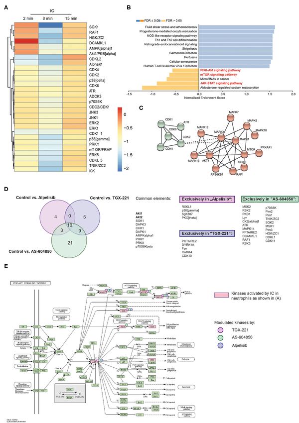
Zillikens et al.                                                                                                         PI3K in Experimental EBA

lymphocytes, mast cells and possibly neutrophils (5–7). The                an immunological synapse by binding to the immune complexes
p110γ subunit is highly expressed in the immune system but                 located at the dermal-epidermal junction, and ultimately release
also in other tissues, including heart and central nervous system          ROS, we evaluated the impact of the PI3Ki on (i) IL-8-induced
(8, 9). Regarding its role in immune homeostasis, aberrant                 neutrophil migration, (ii) IC-induced neutrophil spreading, and
PI3K signaling may lead to hematologic malignancies or chronic             (iii) IC-induced ROS release form neutrophils. For pre-clinical
inflammatory diseases (10, 11).                                            translation, the impact of systemic and topical PI3Ki treatment
    In pemphigoid diseases, a group of autoimmune skin diseases,           was evaluated in antibody transfer-induced EBA.
neutrophils are recruited to the sites of autoantibody deposits and
are activated by binding to the tissue-bound immune complexes
(ICs) (12). Binding to ICs is facilitated by FcγR expressed by             MATERIALS AND METHODS
neutrophils. Signaling downstream of activating FcγR is initiated          Studies With Human Biomaterial
by receptor clustering, leading to activation of Src family kinases        For isolation of PMNs and peripheral blood mononuclear cells
and subsequently SYK. Next, downstream kinases, including                  (PBMCs), normal human blood was obtained. Healthy controls
PI3K, are activated (13–15). Neutrophils express all 4 class I             gave their written informed consent prior to study participation.
PI3K isoforms (16, 17). Of these, PI3Kβ/γ/δ, but not p110α,                All of the experiments using human samples were approved
have been implicated to regulate neutrophil activation. More               by the local ethics committee (University of Lübeck, Lübeck,
specifically, PI3Kβ (16) and PI3Kδ (18) were shown to be crucial           Germany, AZ 09-140 and AZ 20-341) and were performed in
for IC-induced neutrophil activation, specifically the release of          accordance with the Declaration of Helsinki.
reactive oxygen species (ROS). PI3Kγ controls ROS release from
fMLP-stimulated human and murine neutrophils (19), as well as              Mice
neutrophil migration toward GM-CSF (17). PI3Kδ inhibition also
                                                                           C57BL/6J mice (Charles River, Sulzfeld, Germany) were bred
leads to selective impairment of neutrophil functions, including
                                                                           in a specific pathogen free environment and provided standard
IC-, C5a-, and fMLP-induced ROS release, while no impact on
                                                                           mouse chow and acidified drinking water ad libitum. Gender-
PMA-induced ROS release is observed. Furthermore, IL-8- or
                                                                           matched mice were used for experimental EBA models at the
fMLP-induced migration are impaired by PI3Kδ inhibition, while
                                                                           age of 8–10 weeks. Animal experiments were approved by
C5a-induced migration remains unaffected (18). Furthermore,
                                                                           local authorities of the Animal Care and Use Committee
TNF- and LPS-induced cytokine release from neutrophils (19, 20)
                                                                           (Kiel, Germany) and performed by certified personnel
and extravasation (7) are controlled by PI3Kδ.
                                                                           [AZ122.5(108/08-15)] following the ARIVE guidelines.
    Epidermolysis bullosa acquisita (EBA) is a chronic
autoimmune pemphigoid-like disease characterized and caused
by autoantibodies targeting type VII collagen (COL7) (21, 22).             Chemicals
Two distinct clinical EBA manifestations have been described: (i)          If not otherwise stated, all other chemicals were applied from
mechano-bullous EBA and (ii) inflammatory EBA. Overall, EBA                Merck or Sigma. All PI3K blocker were supplied by Selleckchem
is notoriously difficult to treat—combined immunosuppressive               (Houston, Tx, USA). A list of used drugs including IC50
treatment induces remissions after 9 months (23–25). Therefore,            values and concentration of oral administration is prepared
the implementation of novel therapeutic strategies would be of             as Supplementary Table 1. For in vitro assays, 10 mM were
great benefit. Insights from model systems of the disease that             dissolved in DMSO and further diluted in RPMI media. Final
reflect the inflammatory variant of EBA (26) showed that IC                concentration for all in vitro assays were 1,000, 100, 10, 1, and
activation of neutrophils is a key prerequisite to induce tissue           0.1 nM. For all in vitro assays, the final DMSO concentration was
pathology in EBA (21). In line with this notion, inhibition of class       adjusted to 0.01%. All in vitro data were recorded in technical
I PI3K has been shown to dampen clinical disease manifestation             duplicates, and the mean of the results was normalized to the
in pre-clinical EBA models. More specifically, PI3Kβ deficient             value obtained from stimulated cells without PI3K inhibition.
mice are almost completely protected from induction of                        For in vivo experiments (oral administration), PI3K inhibitors
experimental EBA by transfer of COL7 antibodies (16), and                  were dissolved in 10% DMSO/0.8% methylcellulose/0.1% Tween-
pharmacological PI3Kδ inhibition has therapeutic effects in                80. For topical administration to the ear 100 µl 0.9 mg/ml PI3K
immunization-induced EBA (18). The impact of the PI3Kα and                 inhibitors in DMSO: Acetone, 1: 25 were administered daily
γ-isoforms on EBA has, however remained elusive. Systemic                  by dropping.
blockade of the PI3K signaling pathway is associated with severe
adverse events, i.e., neutropenia and pulmonary infections (27).           Human Polymorphonuclear (PMN)
Thus, for the treatment of EBA, topical application of PI3K                Purification
inhibitors (PI3Ki) would be ideal in terms of efficacy and safety.         For the PamGene analysis, FACS, NET formation, cytokine
To address these questions, we here used a panel of 7 PI3Ki with           release, and ROS release assays, human PMNs were isolated
selectivity profiles covering all PI3K isoforms in experimental            from whole blood samples using a PolymorphPrepTM (Progen,
models of EBA.                                                             Heidelberg, Germany) gradient according to the manufacturer’s
    Based on the current understanding of autoantibody-induced             instructions. The purity of the PMNs was evaluated by
tissue pathology in the inflammatory variant of EBA (12, 21),              flow cytometry (MACSQuant R Analyzer 10, Miltenyi) using
where neutrophils migrate form the blood into the skin, form               fluorescent staining with anti-human CD14 (clone HCD14,

Frontiers in Medicine | www.frontiersin.org                            2                                September 2021 | Volume 8 | Article 713312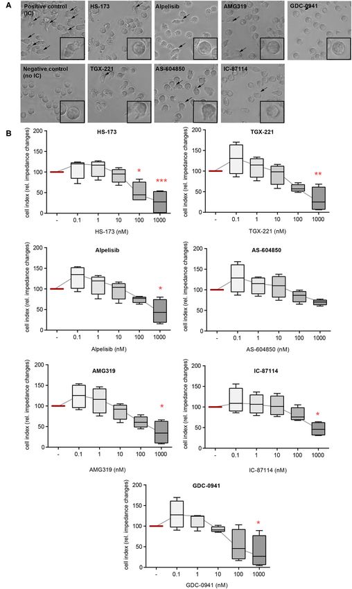
Zillikens et al.                                                                                                           PI3K in Experimental EBA

Biolegend) and anti-human CD15 antibodies (clone VIMC6,                    mediated neutrophil adhesion. It is expressed as cell index in
Miltenyi). For all experiments, the purity of PMNs was >85%.               arbitrary units. As microscopic control, cells were stimulated in
   For chemotaxis assay, PMNs were isolated from normal                    addition with ICs consisting of human COL7E-F antigen at a final
human blood using FicollTM (28). In detail, blood was diluted              concentration of 2.5 µg/ml and anti-human COL7 IgG1 antibody
1:3 in HES/PBS (1:1) and allowed to sediment for 45 min.                   at a final concentration of 1.8 µg/ml for 2 h at 37◦ C and treated
Supernatant (20 ml) was slowly added to 8 ml of FicollTM and               w/o 10 µM of the respective PI3K inhibitors.
centrifuged at 850 × g for 25 min (RT) without braking. The
sediment (containing granulocytes) was erythrocyte lysed with              IC-Induced Stimulation of PMNs and
Aq. dest. for 45 s, and the reaction was stopped with the same
volume of 2xPBS After washing, the cells were diluted to 4 ×
                                                                           Determination of Cytokine Concentrations
106 /ml in RPMI 1640 (without phenol red) containing 0.5%                  and Toxicity
bovine serum albumin (BSA).                                                A high binding 96-well-plate (Thermo Fisher Scientific) was
   For spreading assay, human blood is mixed 1: 2 with 1%                  coated with ICs consisting of human COL7E-F antigen at a final
Polyvinylalcohol at RT, and left at RT for 30–45 min and slowly            concentration of 2.5 µg/ml and anti-human COL7 IgG1 antibody
layered onto Pancoll (PAN biotech, Aidenbach, Germany). The                at a final concentration of 1.8 µg/ml, as described previously (29).
tube is centrifuged for 20 min without brake at 850 × g at                 A total of 2 × 10⁵ cells were added per well w/o PI3K inhibitors at
RT. The supernatant is discarded and the PMNs are transferred              the indicated concentrations. As controls, antigens or antibodies
into a fresh 50 ml tube by prior resuspension and erythrocyte              alone were added to the wells. Cells were incubated for 2 h at
lysis with 1 ml of distilled water for a total of 45 s. The lysis is       37◦ C in an atmosphere containing 5% CO2 . The supernatant was
stopped with 2xPBS and the tube is filled up with 1xPBS (without           analyzed by Legendplex (Biolegend, San Diego, USA) for TNF
Ca/Mg). After centrifugation, the cell pellet is washed and then           and IL-6 among others (for a list see Supplementary Table 2)
resuspended with RPMI 1640 (without phenol red) containing                 following the manufacturer’s protocol. For FACS analysis, single
mit Glutamin, 2 g/L NaHCO3,1% FCS, 2 g/L Glucose 25 mM                     cells were stained for the following surface markers using
HEPES at a concentration of 2 × 10⁶ cells/ml.                              standard FACS procedures: anti-CD15 (clone VIMC6) and
                                                                           anti-CD45 (clone H130) both from Biolegend or Miltenyi.
IC-Induced ROS Release From PMNs                                           For live/dead staining, Annexin V and PI were used in
A LumiTrackTM high binding 96-well-plate (Thermo                           accordance with the manufacturer’s protocol. Cells were first
Fisher Scientific, Waltham, MA, USA) was coated with                       gated for scatter (SSC-A/FSC-A) and singlets (FSC-H/FSC-A).
immunocomplexes (ICs) consisting of human COL7E-F antigen                  The CD45+/CD15+ gates were further analyzed for double-
at a final concentration of 2.5 µg/ml and anti-human COL7                  positive staining of PI and Annexin V for the measurement
IgG1 antibody at a final concentration of 1.8 µg/ml, as described          of toxicity/survival (CD15pos CD45pos PIneg Annexin Vneg cells
previously (29). A total of 2 × 10⁵ cells were added per well              are identified as live cells). Measurements were performed on
in the presence/absence of PI3K inhibitors at the indicated                a Miltenyi MacsQuant10, and data were analyzed with the
concentrations. As controls, antigens or antibodies alone were             MACSQuantifyTM Software (Version 2.11).
added to the wells. Just before the measurement was taken,
luminol was added to the wells, and the chemiluminescence                  IL-8-Induced Neutrophil Chemotaxis
resulting from the ROS production was measured immediately                 Chemotaxis was measured using a 48-well Boyden chamber
in a luminescence reader (Perkin Elmer GloMax). The ROS                    (NeuroProbe Inc., Cabin John, MD) as described previously (31).
release was measured for 1 s per well, sixty-six times for a period        The chamber was pretreated as described in the manufacturer’s
of ∼2 h at a constant temperature of 37◦ C (29).                           protocol. The bottom wells were blocked for 1 h with blocking
                                                                           buffer (1% BSA in 0.1 M NaHCO3 /0.1 M Na2 CO3 , pH = 9)
Neutrophil Adhesion and Spreading on IC                                    at 37◦ C. The buffer was carefully removed, and the chamber
A 96-well E-plate (ACEA bioscience, Bremen, Germany) was                   was dried at 37◦ C. The bottom wells were filled with 30 µl of
coated with 0.5 µg/well mCOL7 and incubated overnight at                   6–12 nmol/L human IL-8 (Peprotech, Hamburg, Germany) in
4◦ C. The E-plate is washed with PBS/1%BSA/0.05% Tween20                   0.5% BSA/PBS (with Ca/Mg). The chambers were covered with
and dried for 2 h at 37◦ C with 5% CO2 . Thereafter, the plate             a polycarbonate membrane (pore size, 3 µm; Costar Nucleopore
was incubated with 1.8 µg/ml anti mCOL7-IgG for 1 h at 37◦ C               GmbH, Tübingen, Germany) and incubated for 30 min at 37◦ C.
to perform immobilized ICs. The plate is then washed again                 Finally, the top wells received 50–80 µl of the isolated PMNs
and incubated with the cell suspension (2 × 10⁶ /ml) w/o PI3K              containing the appropriate concentrations of PI3K inhibitors and
inhibitors. IC-stimulated with vehicle cells serve as positive             were incubated for 1 h at 37◦ C in an atmosphere containing
control and the negative control consists of unstimulated cells,           5% CO2 .
antigen or antibodies and cells. Adhesion was measured for                    The assay was stopped by replacing the cell suspension in the
2 h. Neutrophil adhesion was monitored by phase contrast                   upper well for another 5 min to completely detach migrated cells
microscopy quantified by real-time impedance measurement                   from the bottom side of the filters. Then, filters were removed,
by using the xCELLigence system (Roche, Penzberg, Germany)                 and the migrated cells were transferred from the bottom wells
(30). The electrical impedance reflects the surface area covered           to a microtiter plate. Residual cells in the bottom wells were
by cells, and is therefore associated with the immune complex              lysed by adding 25 µl of 0.2% hexadecyltrimethylammonium

Frontiers in Medicine | www.frontiersin.org                            3                                  September 2021 | Volume 8 | Article 713312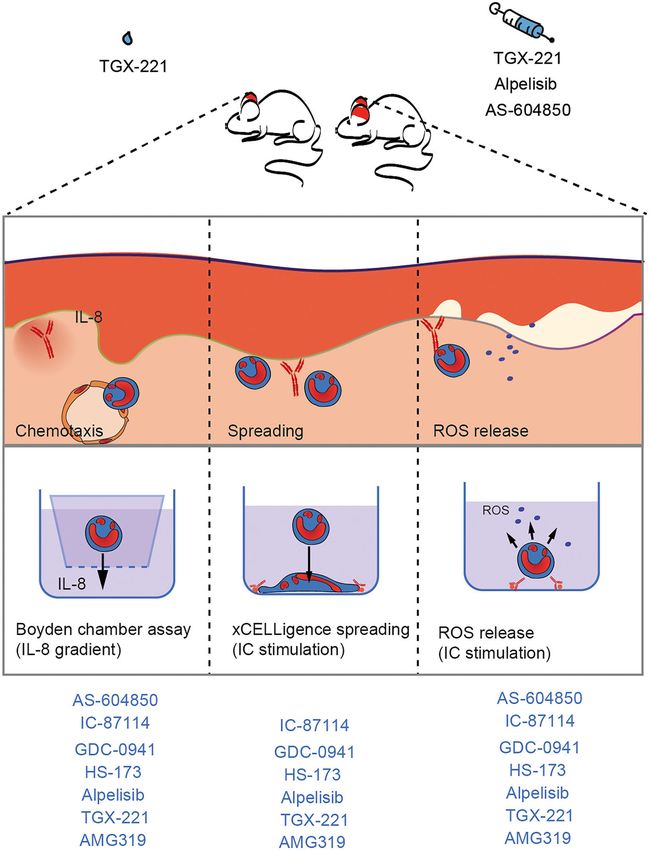
Zillikens et al.                                                                                                        PI3K in Experimental EBA

bromide/0.1% BSA in PBS, and were combined with the cells               they led to a 50% reduction of ROS release in IC-stimulated
transferred to the microtiter plate; cell lysis was continued for       PMNs. Flat bottom 6-well-plates were coated with immune
15 min. Then, 50 µl of TMB solution (Thermo Fisher Scientific)          complexes consisting of human COL7E-F antigen at a final
was added until a color reaction was visible, and the enzymatic         concentration of 10 µg/ml and anti-human COL7 IgG1 antibody
reaction was stopped by adding 50 µl 1 M H2 SO4 . The color             at a final concentration of 2 µg/ml (29), and 107 PMNs/1.5 ml
reaction was determined at 450 nm in a microplate reader. The           per well (containing the respective inhibitors or vehicle) were
number of migrated cells was calculated from a standard of lysed        added. The plates were incubated at 37%, 5% CO2 for 0, 2, 8,
cells run in parallel.                                                  or 15 min, respectively. After incubation, the plates were placed
                                                                        on ice immediately, and the cell solution from each well was
Measurement of C5a and IL-8 Release                                     transferred to a separate 15 ml tube. Each well was also flushed
From Keratinocytes                                                      with 1 ml of ice cold DPBS to detach cells adhered to the plate
Affinity purified antibodies against hCOL7 E-F (EBA) were               surface. The cell solutions were centrifuged for 5 min at 700
isolated from patient immunapheresis material as described              × g (4◦ C). The supernatant was discarded, and the cell pellet
(32, 33). Briefly, antigen stock solutions were prediluted to           used to prepare lysates as described in the protocol provided
a concentration of 1.5 mg/ml in stock solvent (PBS), then               by PamGene (Protocol #1160 for preparation of Lysates of Cell
further diluted 1:3 in 0.1 M Borat buffer (pH 9.5) (final volume        Lines or Purified Cells) (100 µl of lysis buffer per pellet). Protein
12 ml). Antigens were subsequently coupled to NHS-Sepharose             concentrations in the lysates (above the recommended level
Fast Flow4 columns (GE Healthcare, Chicago, USA) following              >0.5 µg/µL) were determined using the Pierce BCA Protein
manufacturer’s instructions. Patient immunapheresis material            Assay Kit (Waltham, MA, USA) according to the manufacturer‘s
was added to the corresponding column and incubated for                 instructions (microplate procedure).
30 min at 4◦ C. Afterwards, the column was washed with PBS until           The Serine-Threonine Kinase (STK) and Protein Tyrosine
OD280 of the flow through was lower than 0.01, and the bound            Kinase (PTK) microarray assays were performed according to
antibody was eluted in glycine buffer (pH 2.8). The eluate was          the manufacturer‘s instructions. Samples originating from the
adjusted to pH 7 using 1M tris base solution (pH 9.0).                  same donor and experimental setup (n = 3) were placed on
   HaCaT cells (34) were grown to confluency in 48-well plates          the same microarray chip to enable more accurate comparisons.
in keratinocyte growth medium-2 (KGM-2) (PromoCell GmbH,                A FITC-conjugated anti-phosphotyrosine antibody was used for
Heidelberg, Germany) supplemented with medium supplement                visualization during and after the pumping of lysates through
and calcium chloride solution provided by the supplier at 37◦ ,         the three-dimensional surface of the array. The capture of
5% CO2 . After confluence was reached, cells were incubated             substrate phosphorylation signals was enabled by a computer-
with different PI3Ki for 5 min. After incubation, 4 0µg/ml              controlled CCD camera and measured repeatedly during a
of EBA antibody solutions was added to the designated wells             1-h kinetic protocol using the Evolve software (PamGene
for 24 h. The following controls were included (antibody only,          International B.V.). The 1-h kinetic protocol showed a linear
antibody/vehicle, normal human IgG only (Intratect 50 g/L,              increase in the signal intensity for the majority of the peptide
Biotest AG, Dreieich, Germany, the IgG was re-buffered before           substrates, indicating that kinome profiling was run without
use to PBS using Amicon R Ultra-15 Centrifugal Filters), and            methodological complications. The analysis of the images was
normal human IgG/vehicle. Cells from two different passages             performed using the BioNavigator Software (Ver. 6.3), with
were used and EBA-IgG from 2 donors each, leading to 4                  the predesigned protocols “STK Image Analysis,” “PTK Image
data points.                                                            Analysis,” “STK Basic Processing,” and “PTK Basic Processing.”
   The C5a content of the supernatants was measured using               Additional applications used were “Fit and apply a combat
the Human Complement Component C5a DuoSet ELISA                         model”, “STK upstream kinase analysis,” and “PTK upstream
(R&D Systems, Minneapolis, USA) according to manufacturer               kinase analysis.” After visual check and quality control, endpoint
instructions, with changes to detection using TMB One solution          signal intensities minus background signals were calculated by
(Promega Corporation, Wisconsin, USA) and 0.9 M H2 SO4 to               BioNavigator for each spot representing each kinase peptide
stop the color reaction. IL-8 release was measured using the            substrate per array. Subsequently, the data were log2 -transformed
ELISA MAXTM human IL-8 standard set (Biolegend, San Diego,              before mean replicate signal intensity within each experiment was
CA, USA) according to manufacturer instructions.                        calculated for each peptide substrate. A kinase was considered
                                                                        to be modulated (either activated or inhibited) if it had a
PamGene Measurement                                                     mean specificity score (= negative decadic logarithm of the
Kinase activity profiles were determined using the PamChip R            likelihood of obtaining a higher difference between the groups
4 protein tyrosine (PTK) peptide microarray system                      when assigning peptides to kinases randomly) of 1 (p = 0.1)
from PamGene International B.V. (BJ‘s-Hertogenbosch,                    and a significance score (= likelihood of obtaining a higher
The Netherlands). Human PMNs were diluted to a final                    difference for random assignment of values to treatment- and
concentration of 6.7 × 10⁶ cells/ml in RPMI 1640 (without               control groups) of 0.5 (p = 0.32). To detect changes caused by the
phenol red) containing 5% FCS and pre-incubated with a                  tested compounds, samples for each timepoint and compound
final concentration of 5 µM AS-604850, 1 µM alpelisib, 10 µM            were compared with the control samples for the same timepoint.
TGX or vehicle (RPMI 1640 without phenol red/ 5%FCS, 0.1%               To ascertain the impact of the stimulation itself, control samples
DMSO) for 5 min (RT). These concentrations were selected as             from before stimulation were compared to control samples at

Frontiers in Medicine | www.frontiersin.org                         4                                  September 2021 | Volume 8 | Article 713312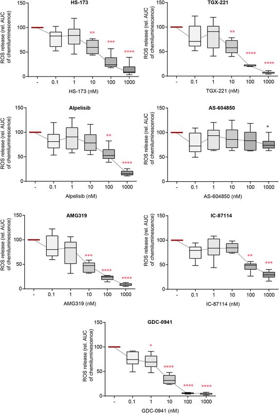
Zillikens et al.                                                                                                       PI3K in Experimental EBA

each timepoint (2, 8, and 15 min). The mean kinase statistic            (Kruskal-Wallis) was applied followed by a Bonferroni t-test
(= calculated by averaging the difference between the signal            for multiple comparisons. In all tests, p < 0.05 was considered
intensity of a sample and its control value, normalized against         indicative of significance. All statistical analyses were performed
a pooled estimate of the standard deviation in each sample,             using SigmaPlot 13.0 (Systat Software, Erkrath, Germany).
for each peptide assigned to a specific kinase) was used for               For the in vivo studies a sample size calculation was
further analysis:                                                       performed. Assuming a minimal detectable difference in means
   Kinome Trees were created using Coral (35). Venn diagram             of 0.5, a standard deviation of 0.25, and aiming at a power of 0.8
analyses were performed in Venny2.0 (https://bioinfogp.cnb.             and an α of 0.05, 8 mice per group have to be included when using
csic.es/tools/venny/). Heatmaps were created using in R studio          ANOVA with an appropriate post-test. Hence, 8 mice per group
(Ver. 1.3.1093) the heatmap package (“pheatmap” Ver. 1.0.12).           were included in the in vivo studies.
Enrichment analysis was performed using Webgestalt Gene Set
Enrichment Analysis (36). Settings were as follows: minimum
                                                                        RESULTS
number of genes for a category: 3, maximum: 1000, Significance
Level: Top 10. Number of Permutations: 1000. Pathway graphs             IL-8-Induced Chemotaxis of PMNs Is
were created using KEGG Mapper (Ver. 4.2) and sourced from              Mainly Impaired by PI3Ki Selective for the
KEGG (Release 95.2) or STRING database (37).
                                                                        β-, γ-, and δ-Isoforms
Induction of Experimental EBA in Mice and                               The directed chemotaxis of polymorphonuclear leukocytes
                                                                        (PMNs) to sites of local inflammation is an early-step in
Treatment Protocol                                                      and a key prerequisite for their (patho)-physiological function
Rabbits were immunized with a fragment of murine type VII
                                                                        in EBA (41). Within this multi-step process, cytokines, such
collagen NC1 (mCOL7C ) in CFA/IFA by Eurogentec, Köln,
                                                                        as IL-8, are required to facilitate neutrophil extravasation,
Germany. Specific anti-mCOL7C IgG from immune serum
                                                                        as well as their activation (42–44). We thus evaluated the
was isolated as previously described (26, 31, 38). Mice were
                                                                        impact of inhibition of the selected PI3Ki on IL8-induced
injected in the ear base once with 30–100 µg of specific rabbit
                                                                        PMN chemotaxis. All investigated inhibitors reduced IL-8-
anti-mCOL7 IgG per mouse. The respective dose to induce
                                                                        induced chemotaxis. PI3K inhibitors mainly selective for PI3Kδ-
a disease score of 25–50% of the affected ear surface area
                                                                        (AMG319 and IC87114) as well as PI3Kγ-selective (AS-
was determined in advance. PI3K inhibitors were administered
                                                                        604850 and AMG319) and PI3Kβ-selective inhibitors (TGX-221)
orally (Supplementary Table 1), or topically applied to the ear
                                                                        inhibited chemotaxis at concentrations within the described IC50
1 day prior to the initial anti-mCOL7C IgG injection and
                                                                        values (Figure 1). Of note, the pan-PI3K inhibitor GDC-0941
was performed every day (in total 4 times). The ear thickness
                                                                        had the most pronounced inhibitory effects on IL-8-induced
measurement, using a Mitutoyo 7301 dial thickness gauge
                                                                        neutrophil migration, with an efficacy at 0.1 nM. Also, PI3Kα-
(Neuss, Germany), and scoring was performed by a blinded
                                                                        selective inhibitors (HS-173 and alpelisib) reduced chemotaxis at
person. For anesthesia, a mixture of 15 µg/g body weight Xylazin
                                                                        concentrations above the known IC50 values.
and 100 µg/g body weight Ketamin was injected i.p. into mice
(26, 39).
                                                                        PMN Adhesion and Spreading on ICs Are
Histopathology and Direct                                               Predominantly Impaired by PI3Kδ- and
Immunofluorescence Staining                                             PI3Kβ-Selective Inhibitors
Biopsies of lesional and perilesional skin were obtained on             In addition to ROS release, FcγR-dependent binding of
day 3 of the antibody transfer-induced EBA and prepared for             neutrophils also induces their β2 integrin-dependent spreading
examination by histopathology and immunofluorescence (IF)               and adhesion to IC-coated surfaces. This creates an enclosed
microscopy, as described previously (26, 31, 38). In brief, the         space between the neutrophils and the target tissue, allowing a
biopsies collected from mice were fixed in 4% PBS-buffered              directional release of proteases and ROS. If neutrophil adhesion
formalin, and subsequently, sections of paraffin-embedded               to IC-coated surfaces is blocked, the tissue is protected from
tissues were stained with hematoxylin and eosin. IgG deposits           the detrimental effects of proteases and ROS (30). We hence
were detected by direct IF microscopy in frozen sections                evaluated the impact of the PI3Ki on PMN spreading on IC-
prepared from tissue biopsies using fluorescein isothiocyanate          coated surfaces. Interestingly, PI3K inhibition had only marginal
(FITC)-labeled antibodies specific to rabbit IgG (Dako, Glostrup,       effects on IC-induced spreading and adhesion. Mostly, inhibitors
Denmark) as previously described (40).                                  with a PI3Kδ- and PI3Kβ-selectivity reduced PMN spreading
                                                                        (IC-87114, TGX-221, and AMG319, Figure 2). As AMG319
Statistical Analysis                                                    inhibits also p110γ, the effects on spreading could be also
Unless otherwise noted, data are presented as the mean ±                partially mediated by PI3Kγ. Yet, the second PI3Kγ-selective
SD or Tukey’s box-and-whisker plots; the dots represent actual          inhibitor (AS-604850) did not have an effect on IC-induced
results for each sample. For comparisons of more than 2 groups,         spreading of PMNs within the investigated concentration range.
ANOVA was used. For equally distributed data, one-way ANOVA             Furthermore, a statistically significant inhibition was observed
followed by a Bonferroni t-test for multiple comparisons was            for the p110α inhibitor HS-173 at concentrations of 0.1 and
used; if the data were non-parametric, ANOVA on ranks                   1.0 µM. HS-173, GCD-0941 and alpelisib inhibited neutrophil

Frontiers in Medicine | www.frontiersin.org                         5                                 September 2021 | Volume 8 | Article 713312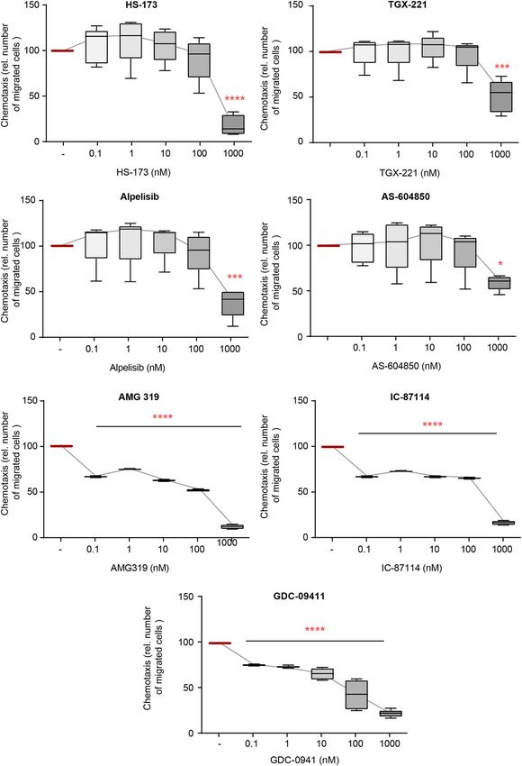
Zillikens et al.                                                                                                                               PI3K in Experimental EBA

  FIGURE 1 | IL-8-induced chemotaxis from PMNs depends on PI3K. Chemotaxis of PMNs was induced by IL-8 in the presence of either one of the PI3K
  isoform-selective inhibitors. The attracted cell number during a time period of 60 min is shown. Data were normalized to positive control (chemotaxis induced by IL-8
  in the presence of solvent). Data are shown as Tukey’s box-and-whisker plots. n = 5. ANOVA on ranks (Kruskal-Wallis) was applied followed by a Bonferroni t-test for
  multiple comparisons. *p < 0.05, ***p < 0.001, ****p < 0.0001.

Frontiers in Medicine | www.frontiersin.org                                         6                                       September 2021 | Volume 8 | Article 713312
Zillikens et al.                                                                                                                              PI3K in Experimental EBA

  FIGURE 2 | IC-induced spreading of PMNs mainly depends on PI3Kδ. Freshly isolated human blood PMNs were activated with immobilized ICs in the presence of
  either one of the PI3K isoform-selective inhibitors. (A) Microscopic appearance of PMNs 2 h after IC-stimulation. IC-stimulated PMNs show clear spreading on surface
  (arrows). The highest concentration of all inhibitors (1 µM) reduced the adhesion of PMNs. (B) Spreading was monitored live for 2 h and the area under curve (AUC)
  was analyzed. Data are shown as Tukey’s box-and-whisker plots. n = 4. ANOVA on ranks (Kruskal-Wallis) was applied followed by a Bonferroni t-test for multiple
  comparisons. Data were normalized to IC-stimulated cells. *p < 0.05, **p < 0.01, ***p < 0.001.

Frontiers in Medicine | www.frontiersin.org                                        7                                       September 2021 | Volume 8 | Article 713312
Zillikens et al.                                                                                                                               PI3K in Experimental EBA

  FIGURE 3 | Immune complex (IC)-induced reactive oxygen species (ROS) release from PMNs depends on different PI3K isoforms. Human PMNs were activated with
  immobilized ICs in the presence of either one of the PI3K isoform-selective inhibitors. Release of ROS was tracked for 2 h and the area under curve (AUC) was
  calculated. Data were normalized to positive control (IC-stimulated PMNs with solvent). Data are shown as Tukey’s box-and-whisker plots. n = 6. ANOVA on ranks
  (Kruskal-Wallis) was applied followed by a Bonferroni t-test for multiple comparisons. Data were normalized to stimulated cells. *p < 0.05, **p < 0.01, ***p < 0.001,
  ****p < 0.0001.

Frontiers in Medicine | www.frontiersin.org                                         8                                       September 2021 | Volume 8 | Article 713312
Zillikens et al. PI3K in Experimental EBA
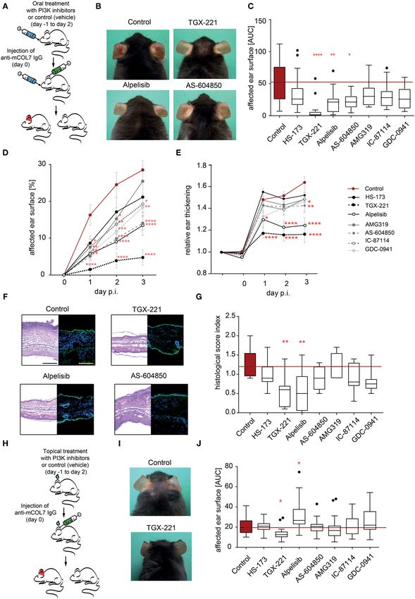
FIGURE 4 | Treatment with TGX-221, alpelisib or AS-604850 impairs induction of experimental EBA. (A) Schematic illustration of experimental work flow. Anti-mCOL7
IgG was injected into the ears of C57BL/6J mice and the mice were orally treated with the indicated PI3K isoform selective inhibitors. (B) Representative clinical
images of the mice on day 3. (C) The cumulative affected ear surface area (AUC) in mice treated with solvent or the indicated PI3K inhibitors. Data are shown
(Continued)
Frontiers in Medicine | www.frontiersin.org 9 September 2021 | Volume 8 | Article 713312Zillikens et al. PI3K in Experimental EBA
FIGURE 4 | as Tukey’s box-and-whisker plots. ANOVA on ranks (Kruskal-Wallis) was applied followed by a Bonferroni t-test for multiple comparisons. (D) Percentage
of ear surface area affected by EBA skin lesions at days 0–3 of the same experiment. Data are shown as mean ± SD. Two way-ANOVA with Bonferroni post test. (E)
Relative ear swelling at days 0–3 of the same experiment. Data are normalized to day 0 of the experiment and shown as mean ± SD. Two way-ANOVA with Bonferroni
post test. (F) Left panel: Representative H&E stained ear skin at day 3 after IgG-injection. Right panel: Representative direct IF microscopy of a skin biopsy, stained for
anti-rabbit IgG (green) and nuclei (DAPI, blue). (G) The cumulative histological score index (AUC of skin infiltration, epidermal thickening and split formation at the DEJ)
was analyzed in ear skin at day 3 of the experiment. Data are shown as Tukey’s box-and-whisker plots. ANOVA on ranks (Kruskal–Wallis) was applied followed by a
Bonferroni t-test for multiple comparisons. Data in panels (C–E,G) is based on 8 mice/group. (H) Schematic illustration of experimental work flow for topical treatment
with PI3K isoform-selective inhibitors. Anti-mCOL7 IgG was injected into the ears of C57BL/6J mice and the mice were topically treated daily with the indicated PI3K
isoform-selective inhibitors. (I) Representative clinical images on day 3 after topical treatment with solvent or TGX-221. (J) Cumulative affected ear surface area (AUC
of percentage of day 0–3) in mice with topical PI3K inhibitor treatment. Data are shown as Tukey’s box-and-whisker plots. ANOVA on ranks (Kruskal-Wallis) was
applied followed by a Bonferroni t-test for multiple comparisons. Data in panels (I,J) is based on 8 mice/group. *p < 0.05, **p < 0.01, ***p < 0.001, ****p < 0.0001.
spreading and adhesion only at the 1 µM dose, which are relative Selective Pharmacological Inhibition of
high doses (Figure 2). PI3K Reduced Inflammation in
Experimental EBA
So far, we had demonstrated a selective contribution of
IC-Induced ROS Release From PMNs Is PI3K to distinct neutrophil functions in vitro. Pathogenesis
Sensitive to Blockade of All PI3K Isoforms of inflammation in autoantibody-induced and neutrophil-
Activation of neutrophils and subsequent ROS release by FcγR- dependent diseases, is, however, a multistep process that involves
dependent binding to IC initiates local inflammation across all (if not more) of the above tested steps (12). Furthermore, in
many autoantibody-mediated diseases, including EBA (12, 45, vivo pharmacokinetics of the PI3Ki are also key determinants
46). Herein, we thus used IC to activate PMN and evaluated of their potential disease-modifying activity. Thus, prediction
the impact of selective PI3Ki on IC-induced ROS release. which PI3K inhibitor has anti-inflammatory effects in pre-
Here, all inhibitors, with the exception of PI3Kα subclass- clinical models of EBA is challenging. To address this, we used the
selective inhibitors, impaired ROS release from IC-activated mouse model of antibody transfer-induced EBA, in which disease
PMNs at doses close to the reported IC50 (Figure 3). The pan- manifestation depends on the presence of COL7 (auto)antibodies
PI3K inhibitor GDC-0941 had the most profound inhibitory and a subsequent FcγR-dependent neutrophil activation (21).
effects, with efficacy at 1.0 nM. All effects were independent of In a first set of experiments, mice, in which EBA was induced
any cytotoxic effects on neutrophils as none of the inhibitors by anti-COL7-IgG injection, were orally treated with PI3K-
effected Annexin V/propidium iodide staining in IC-stimulated selective inhibitors in a concentration that was recently shown
neutrophils (Supplementary Figure 1). to be effective known effective for other applications (50–56)
(Figure 4). The p110α-selective inhibitor alpelisib and the p110γ-
selective inhibitor AS-604850 showed impaired disease induction
(ear thickness increase and affected ear surface area) by 50–60%
Selective Pharmacological Inhibition of (Figures 4D,E). Treatment with the p110β-selective inhibitor
PI3K Did Not Influence C5a and IL-8 TGX-221 almost completely abolished the pathogenic activity
of the anti-mCOL7 antibodies. A minor effect was observed
Release From Keratinocytes
for GDC-0941 and IC-87114 as visible by a reduced affected
So far, we had demonstrated a selective contribution of PI3K
surface area (Figure 4D). All other tested inhibitors (HS-173 and
to distinct neutrophil functions in vitro. To evaluate possible
AMG319) had no effect on clinical disease manifestation at the
effects on other disease-relevant cell types, we stimulated human
selected doses (Figures 4A–G).
keratinocytes with EBA-IgG. We hence evaluated the impact
of the PI3Ki on C5a and IL-8 release after 24 h of stimulation
(Supplementary Figure 2) as complement activation and IL-
Topical Application of the PI3Kβ-Selective
8 release are key events in the effector phase of EBA and Inhibitor TGX-221 Impairs Induction of
other pemphigoid diseases (21, 47–49). Compared to normal Experimental EBA
human IgG, EBA-IgG significantly increased IL-8 and C5a As systemic PI3K inhibition in humans may be associated
concentrations in the supernatants of HaCaT cells. Yet, none with a relative high number of (serious) adverse events (27,
of the analyzed inhibitors had an inhibitory effect on EBA-IgG 57), we next evaluated if topical application of the PI3Ki
activated C5a release. Of note, after stimulation with EBA-IgG, has an impact on disease manifestation in experimental
several inhibitors (TGX-221, alpelisib, and IC-87115) showed EBA. Of the seven inhibitors, the p110β-selective inhibitor
a slight tendency even to increase the C5a release if used in TGX-221 again profoundly impaired the induction of skin
low concentrations. Nevertheless, higher concentrations of the inflammation (Figures 4H–J). By contrast, all other PI3Ki did
inhibitors did not affect C5a release. Interestingly, release of IL- not reduce clinical disease manifestation in experimental EBA
8 was slightly inhibited by alpelisib at concentrations above the at selected concentration. During all experiments in the pre-
known IC50 values (1 µM) whereas none of the other inhibitors clinical EBA mouse model, neither oral nor topical treatment
had any effect. with one of the PI3K inhibitors led to an increased suffering
Frontiers in Medicine | www.frontiersin.org 10 September 2021 | Volume 8 | Article 713312Zillikens et al. PI3K in Experimental EBA
FIGURE 5 | Unique impact of different PI3K inhibitors on downstream kinase activation. Freshly isolated human blood PMNs were activated with IC for 0, 2, 8, or
15 min. Cells were lysed and the PTK and STK activity was measured by PamGene. (A) Heatmap of mean kinase statistic in comparison to unstimulated cells (0 min).
Blue: Kinase activity decreased/Red: Kinase activity increased. (B) Enriched KEGG pathways (as shown by Webgestalt) after IC-stimulation. (C) String network for
(Continued)
Frontiers in Medicine | www.frontiersin.org 11 September 2021 | Volume 8 | Article 713312Zillikens et al. PI3K in Experimental EBA
FIGURE 5 | IC-stimulated kinases (MCL clustering, inflation parameter 3) (D) Venn diagram and summary of the common and uniquely regulated kinase activities after
use of three PI3K isoform-selective inhibitors that were effective in experimental EBA (alpelisib, AS-604850 and TGX-221). The comparison was performed for all time
points with direct comparison to the kinase activity of ICs activated PMNs treated with solvent. (E) Influence of PI3K inhibition on PI3K/Akt and mTOR pathways,
kinases regulated by ICs (cumulative stimulation for 2, 8, and 15 min using PamGene) are shown in bright red. Kinases, that are regulated by the respective kinase
inhibitors are marked with a reddish dot (TGX-221), green dot (AS-604850), and purple dot (alpelisib). Data is based on 3 replicates per group.
(weight reduction, general condition and behavior) in the mice We demonstrate that the PI3Kβ-selective TGX-221 has the most
(Supplementary Tables 2, 3). pronounced disease-modifying activity, especially when applied
topically. Of note, this superior in vivo effect of TGX-221 could
Kinome Analysis of Identified Unique not be derived from the in vitro characterization of the PI3Ki
Downstream Signaling Pathways of the evaluated herein. More specifically, TGX-221 impaired IL-8-
induced chemotaxis only at relatively high concentrations, while
Different, Disease-Modifying
AMG 319 and IC-87114 dose-dependently and at quite low
PI3K-Selective Inhibitors concentrations, reduced PMN chemotaxis. PMN spreading to IC,
At this point, using the EBA mouse model, we had demonstrated as well as IC-induced ROS release from PMN was equally well
that TGX-221, alpelisib or AS-604850 impaired induction of inhibited by HS-173, TGX-221, as well as several more PI3Ki
experimental EBA. To obtain more detailed insights into in the IC-induced ROS release. Thus, in vivo disease models are
the orchestration of IC-induced signal transduction pathways required to demonstrate efficacy.
selectively inhibited by these 3 therapeutically promising Hence, while this and previous (16, 18) reports demonstrate
drugs, we performed a multiplex kinase activity profiling the fundamental role of PI3Kβ and PI3Kδ in the pathogenesis
using PamGene (58). We incubated human PMNs with PI3Ki of skin inflammation in EBA, our data presented here does
(alpelisib, AS-604850 or TGX-221) for 5 min and subsequently not rule out that other class I PI3K isoforms contribute to
stimulated PMNs with IC for 0, 2, 8, or 15 min. As EBA pathogenesis—especially PI3Kα, one PI3Ki selective for
expected, IC-stimulation of PMNs induced, among others, the this isoform (alpelisib) impaired induction of experimental
PI3K/Akt, JAK/STAT and mTOR pathways (Figures 5A,B and EBA. Interestingly, alpelisib was blocking chemotaxis and ROS
Supplementary Table 4). In addition to these known pathways, release only if used at rather high concentrations but in
we found a strong activity of several cyclin-dependent kinases contrast reduced IL-8 release from human keratinocytes. Still,
(CDKs, Figure 5C). These are enriched in the KEGG pathway for the kinase activation profiles indicate a specific inhibition of
“cellular senescence” (Supplementary Figure 3). Other relevant the PI3K pathway by alpelisib. In addition to these in vitro
pathways that were regulated by IC-stimulation are summarized findings, the in vivo pharmacokinetics of a given drug are
in Supplementary Figures 3, 4 (mTOR signaling, FoxO signaling crucial for its activity. For alpelisib, over 50% are absorbed,
and FcRγ-mediated phagocytosis). with a T(max) of 2 h and an elimination half-life from plasma
To obtain more detailed insights into the mode of action of 13.7 h (59) which indicates a good bioavailability that
of the effective inhibitors, we compared the kinase activity in could contribute to the effectivity of alpelisib in murine
PMNs after PI3K inhibition with the corresponding control experimental EBA.
time points (Figure 5D). Inhibition of PI3K by TGX-221 Dissection of the contribution of different PI3K isoforms
(Supplementary Figure 5), alpelisib (Supplementary Figure 6) to EBA pathogenesis, would require the use of PI3K isoform-
or AS-604850 (Supplementary Figure 7) led to a decreased deficient mice (1, 2, 60–62). We here, however, specifically
activity of Akt1/2 and the downstream kinases SGK, p70S6Kbeta focused on pharmacological PI3K inhibition allowing a better
(S6K1) and AMPK (Figures 5D,E). Interestingly, all 3 clinical translation.
inhibitors also had unique effects on several kinases and In the pathogenesis of the inflammatory type of EBA, PMNs
KEGG pathways that were independent of the known PI3K exert their pathogenic effects in a stepwise manner that is
network (Figure 5D). More specifically, inhibition using initiated by attracting them into the tissue (chemotaxis) and
AS-604850 (Supplementary Figure 7) identified a total of 21 ultimately results in the release of pro-inflammatory substances,
kinases that were exclusively de- or increased in their activity, such as ROS (Figure 6). Extravasation of PMNs to sites of
indicating additional effects on different targets. By contrast, inflammation is an early key step driving EBA (63, 64). Within
only 5 unique kinases were detected in TGX-221-treated PMN this multi-step process, cytokines, such as IL-8, are required to
and 4 unique kinases in alpelisib treated PMNs (Figure 5 and facilitate neutrophil chemotaxis, as well as activation (42–44).
Supplementary Tables 5–7). We here demonstrate that PI3Kγ- and PI3Kδ- selective inhibitors
block PMN chemotaxis. By contrast, selective inhibitors of PI3Kα
DISCUSSION have no major impact on IL-8-induced PMN migration. These
findings support earlier notions demonstrating that in fMLP-
We here systematically investigated the impact of seven PI3Ki stimulated neutrophils, the PI3Kδ inhibitor IC-87114 affected
with different isoform-selectivity on neutrophil functions in polarized morphology of neutrophils, PIP3 production and
vitro, as well as their potential to reduce autoantibody-induced, chemotaxis, but did not block F-actin synthesis or neutrophil
neutrophil-driven inflammation in preclinical models of EBA. adhesion (65). Regarding PI3Kγ, G-protein-coupled receptor
Frontiers in Medicine | www.frontiersin.org 12 September 2021 | Volume 8 | Article 713312Zillikens et al. PI3K in Experimental EBA FIGURE 6 | Unique impact of different PI3K inhibitors on neutrophil-dependent processes in EBA. In the pathogenesis of IC-induced EBA, PMN exert their pathogenic effects in a stepwise manner that is initiated by attracting them into the tissue (chemotaxis), spreading, and adhesion to surfaces of IC deposits and ultimately results in the release of pro-inflammatory substances, such as ROS. As these processes strongly depend to PI3K, inhibition leads to improvement of EBA. (GPCR) stimulation of PI3Kγ induced PMN migration (and the rolling to firm adhesion, a process depending on β2 integrins oxidative burst) (66). Therefore, both PI3Kγ and δ seem to be (70). We here show that β2 integrin-dependent neutrophil important to mediate PMN chemotaxis across several stimuli. adhesion to immobilized IC mostly depends on PI3Kγ /δ. By creating a protected space between the neutrophil and Regarding ROS release from IC-activated neutrophils, we here the target tissue, neutrophil spreading and adhesion to surfaces show that this requires PI3Kβ, γ, and δ. These findings are of IC deposits is a prerequisite for autoantibody-induced tissue consisted with published data for PI3Kβ and δ (16, 18), which damage (30). This process is regulated by PI3K activation (67, may even act synergistically in this context (16). Interestingly, 68). Previous data demonstrated that the pan-PI3K inhibitor while important for TNF/fMLP-induced ROS release from wortmannin or the PI3Kα/β/δ inhibitor LY294002 impaired neutrophils (19), as shown here, PI3Kγ-selective inhibition only neutrophil binding to ICAM-1 after fMLP stimulation (69). has a marginal impact on IC-induced ROS release. In line with Other data on the PI3K isoforms involved in these processes is this, pan-PI3K inhibition completely abolished the IC-induced sparse. PI3Kγ has been shown to mediate the transition from ROS release. Frontiers in Medicine | www.frontiersin.org 13 September 2021 | Volume 8 | Article 713312
Zillikens et al.                                                                                                                      PI3K in Experimental EBA

    All of the above neutrophil functions contribute to local                        further    inquiries    can           be      directed       to       the
inflammation in EBA (21). Yet, the in vitro inhibitory effects of                    corresponding author/s.
PI3K isoform-selective drugs do not allow to predict their impact
on the complex disease pathogenesis in vivo. To investigate                          ETHICS STATEMENT
if inhibition of specific PI3K isoforms has an impact on in
vivo inflammation provoked by tissue-bound IC, we used the                           The studies involving human participants were reviewed and
antibody transfer model of EBA. We specifically selected a skin                      approved by University of Lübeck, Lübeck, Germany, AZ 09-
inflammation mouse model because topical application, which                          140. The patients/participants provided their written informed
presumably leads to less adverse events, is possible in chronic                      consent to participate in this study. The animal study was
inflammatory skin diseases. Out of 7 tested PI3K inhibitors, AS-                     reviewed and approved by Animal Care and Use Committee
604850 (PI3Kγ-selective), alpelisib (PI3Kα-selective) and TGX-                       (Kiel, Germany)—AZ122.5(108/08-15).
221 (PI3Kß-selective) showed a significant inhibition of ear
thickening and led to a more than 50% reduction of the affected
                                                                                     AUTHOR CONTRIBUTIONS
ear surface area. One of these inhibitors, TGX-221 was even
effective if applied topically. Given that EBA is notoriously                        RL and KBi: conceptualization and methodology and writing—
difficult to treat, achieving remissions after 9 month of systemic                   original draft. HZ, AK, NG, CO, CH, MR, VH, NE, and
immunosuppressive treatment (23), addition of a (preferably                          RV: investigation. KBi and CO: data curation. RL: funding
topically applied) PI3Ki could potentially lead to a more rapid                      acquisition and project administration. RL, GV, FP, TL, and
induction of remission.                                                              XY: resources. MB-H, XY, FP, RL, and KBi: supervision.
    Previously, we had documented that induction of skin                             HZ, AK, CO, NG, MR, CH, VH, MB-H, NE, KBo, GV,
inflammation is hampered in PI3Kβ-deficient mice or in mice                          RV, TL, XY, FP, RL, and KBi: writing—review and editing.
treated with a PI3Kδ-selective inhibitor (16, 18). Overall, these                    All authors contributed to the article and approved the
data and the results presented herein point toward a non-                            submitted version.
redundant role of these 2 class I PI3K isoforms.
    Next, we here evaluated the impact of the three PI3K
isoform-specific inhibitors on downstream signaling cascades,                        FUNDING
allowing detailed insights into PI3K isoform-specific signaling
                                                                                     This study was funded by Research Training Group Modulation
pathways in human PMNs, which may provide additional and
                                                                                     of Autoimmunity (GRK 1727), Clinical Research Unit
more downstream targets to block IC-induced PMN activation.
                                                                                     Pemphigoid Diseases – Molecular Pathways and their
Unbiased analysis of kinase activity in IC-activated PMN treated
                                                                                     Therapeutic Potential (KFO303), and Cluster of Excellence
with either TGX-221, alpelisib or AS-604850 identified a number
                                                                                     Precision Medicine in Chronic Inflammation (EXC2167), all
of shared downstream kinases like Akt1/2 and the downstream
                                                                                     from the Deutsche Forschungsgemeinschaft (DFG). RL was
kinases SGK, p70S6Kbeta (S6K1) and AMPK. However, the
                                                                                     supported by the Schleswig-Holstein Excellence-Chair Program
majority of changes in kinase activity after treatment of PMN
                                                                                     (State of Schleswig-Holstein).
with either of the compounds was specific for each inhibitor, and
most pronounced for AS-604850.
    Collectively, we here defined the differential impact of PI3K                    ACKNOWLEDGMENTS
isoforms on immune complex-induced neutrophil signaling and
function, and identify the PI3Kβ-selective TGX-221 as a potential                    We thank Claudia Kauderer, Alexandra Wobig, Daniela Rieck,
topical treatment for the inflammatory type of EBA and other                         and Astrid Fischer for their excellent technical support.
autoimmune skin blistering diseases with a similar pathogenesis.
                                                                                     SUPPLEMENTARY MATERIAL
DATA AVAILABILITY STATEMENT
                                                                                     The Supplementary Material for this article can be found
The       original     contributions presented in the study                          online at: https://www.frontiersin.org/articles/10.3389/fmed.
are       included      in the article/Supplementary Material,                       2021.713312/full#supplementary-material

REFERENCES                                                                           3. Chantry D, Vojtek A, Kashishian A, Holtzman DA, Wood C, Gray
                                                                                        PW, et al. p110delta, a novel phosphatidylinositol 3-kinase catalytic
 1. Bi L, Okabe I, Bernard DJ, Wynshaw-Boris A, Nussbaum RL. Proliferative              subunit that associates with p85 and is expressed predominantly in
    defect and embryonic lethality in mice homozygous for a deletion in                 leukocytes. J Biol Chem. (1997) 272:19236–41. doi: 10.1074/jbc.272.31.
    the p110alpha subunit of phosphoinositide 3-kinase. J Biol Chem. (1999)             19236
    274:10963–8. doi: 10.1074/jbc.274.16.10963                                       4. Vanhaesebroeck B, Welham MJ, Kotani K, Stein R, Warne PH, Zvelebil
 2. Bi L, Okabe I, Bernard DJ, Nussbaum RL. Early embryonic lethality in mice           MJ, et al. P110delta, a novel phosphoinositide 3-kinase in leukocytes.
    deficient in the p110beta catalytic subunit of PI 3-kinase. Mamm Genome.            Proc Natl Acad Sci USA. (1997) 94:4330–5. doi: 10.1073/pnas.94.9.
    (2002) 13:169–72. doi: 10.1007/s00335-001-2123-x                                    4330

Frontiers in Medicine | www.frontiersin.org                                     14                                   September 2021 | Volume 8 | Article 713312
Zillikens et al. PI3K in Experimental EBA
5. Clayton E, Bardi G, Bell SE, Chantry D, Downes CP, Gray A, et 27. Dreyling M, Santoro A, Mollica L, Leppa S, Follows GA, Lenz
al. A crucial role for the p110delta subunit of phosphatidylinositol 3- G, et al. Phosphatidylinositol 3-Kinase inhibition by copanlisib in
kinase in B cell development and activation. J Exp Med. (2002) 196:753– relapsed or refractory indolent lymphoma. J Clin Oncol. (2017)
63. doi: 10.1084/jem.20020805 35:3898–905. doi: 10.1200/JCO.2017.75.4648
6. Ali K, Bilancio A, Thomas M, Pearce W, Gilfillan AM, Tkaczyk C, et al. 28. Skoog WA, Beck WS. Studies on the fibrinogen, dextran and
Essential role for the p110delta phosphoinositide 3-kinase in the allergic phytohemagglutinin methods of isolating leukocytes. Blood. (1956)
response. Nature. (2004) 431:1007–11. doi: 10.1038/nature02991 11:436–54. doi: 10.1182/blood.V11.5.436.436
7. Puri KD, Doggett TA, Douangpanya J, Hou Y, Tino WT, Wilson T, 29. Recke A, Trog LM, Pas HH, Vorobyev A, Abadpour A, Jonkman MF,
et al. Mechanisms and implications of phosphoinositide 3-kinase delta et al. Recombinant human IgA1 and IgA2 autoantibodies to type VII
in promoting neutrophil trafficking into inflamed tissue. Blood. (2004) collagen induce subepidermal blistering ex vivo. J Immunol. (2014) 193:1600–
103:3448–56. doi: 10.1182/blood-2003-05-1667 8. doi: 10.4049/jimmunol.1400160
8. Vanhaesebroeck B, Stephens L, Hawkins P. PI3K signalling: the path 30. Yu X, Akbarzadeh R, Pieper M, Scholzen T, Gehrig S, Schultz C,
to discovery and understanding. Nat Rev Mol Cell Biol. (2012) 13:195– et al. Neutrophil Adhesion is a prerequisite for antibody-mediated
203. doi: 10.1038/nrm3290 proteolytic tissue damage in experimental models of epidermolysis bullosa
9. Nurnberg B, Beer-Hammer S. Function, regulation and biological roles of acquisita. J Invest Dermatol. (2018) 138:1990–8. doi: 10.1016/j.jid.2018.03.
PI3Kgamma variants. Biomolecules. (2019) 9:427. doi: 10.3390/biom9090427 1499
10. Hawkins PT, Stephens LR, Suire S, Wilson M. PI3K signaling in neutrophils. 31. Stussel P, Schulze Dieckhoff K, Kunzel S, Hartmann V, Gupta Y, Kaiser
Curr Top Microbiol Immunol. (2010) 346:183–202. doi: 10.1007/82_2010_40 G, et al. Propranolol is an effective topical and systemic treatment option
11. Elich M, Sauer K. Regulation of hematopoietic cell development for experimental epidermolysis bullosa acquisita. J Invest Dermatol. (2020)
and function through phosphoinositides. Front Immunol. (2018) 140:2408–20. doi: 10.1016/j.jid.2020.04.025
9:931. doi: 10.3389/fimmu.2018.00931 32. Sitaru C, Schmidt E, Petermann S, Munteanu LS, Brocker EB, Zillikens D.
12. Ludwig RJ, Vanhoorelbeke K, Leypoldt F, Kaya Z, Bieber K, Mclachlan SM, et Autoantibodies to bullous pemphigoid antigen 180 induce dermal-epidermal
al. Mechanisms of autoantibody-induced pathology. Front Immunol. (2017) separation in cryosections of human skin. J Invest Dermatol. (2002) 118:664–
8:603. doi: 10.3389/fimmu.2017.00603 71. doi: 10.1046/j.1523-1747.2002.01720.x
13. Getahun A, Cambier JC. Of ITIMs, ITAMs, and ITAMis: revisiting 33. Vorobyev A, Ujiie H, Recke A, Buijsrogge JJ, Jonkman MF, Pas HH, et
immunoglobulin Fc receptor signaling. Immunol Rev. (2015) 268:66– al. Autoantibodies to multiple epitopes on the non-collagenous-1 domain
73. doi: 10.1111/imr.12336 of Type VII collagen induce blisters. J Invest Dermatol. (2015) 135:1565–
14. Ludwig RJ. Signalling and targeted therapy of inflammatory 73. doi: 10.1038/jid.2015.51
cells in epidermolysis bullosa acquisita. Exp Dermatol. (2017) 34. Boukamp P, Petrussevska RT, Breitkreutz D, Hornung J, Markham A,
26:1179–86. doi: 10.1111/exd.13335 Fusenig NE. Normal keratinization in a spontaneously immortalized
15. Samavedam UK, Mitschker N, Kasprick A, Bieber K, Schmidt E, Laskay aneuploid human keratinocyte cell line. J Cell Biol. (1988) 106:761–
T, et al. Whole-genome expression profiling in skin reveals SYK as a key 71. doi: 10.1083/jcb.106.3.761
regulator of inflammation in experimental epidermolysis bullosa acquisita. 35. Metz KS, Deoudes EM, Berginski ME, Jimenez-Ruiz I, Aksoy BA,
Front Immunol. (2018) 9:249. doi: 10.3389/fimmu.2018.00249 Hammerbacher J, et al. Coral: clear and customizable visualization of human
16. Kulkarni S, Sitaru C, Jakus Z, Anderson KE, Damoulakis G, Davidson K, et al. kinome data. Cell Syst. (2018) 7:347–50 e341. doi: 10.1016/j.cels.2018.07.001
PI3Kbeta plays a critical role in neutrophil activation by immune complexes. 36. Liao Y, Wang J, Jaehnig EJ, Shi Z, Zhang B. WebGestalt 2019: gene set
Sci Signal. (2011) 4:ra23. doi: 10.1126/scisignal.2001617 analysis toolkit with revamped UIs and APIs. Nucleic Acids Res. (2019)
17. Martin KJ, Muessel MJ, Pullar CE, Willars GB, Wardlaw AJ. The role of 47:W199–205. doi: 10.1093/nar/gkz401
phosphoinositide 3-kinases in neutrophil migration in 3D collagen gels. PLoS 37. Szklarczyk D, Gable AL, Lyon D, Junge A, Wyder S, Huerta-Cepas J, et al.
ONE. (2015) 10:e0116250. doi: 10.1371/journal.pone.0116250 STRING v11: protein-protein association networks with increased coverage,
18. Koga H, Kasprick A, Lopez R, Auli M, Pont M, Godessart N, et al. supporting functional discovery in genome-wide experimental datasets.
Therapeutic effect of a novel Phosphatidylinositol-3-Kinase delta inhibitor Nucleic Acids Res. (2019) 47:D607–13. doi: 10.1093/nar/gky1131
in experimental epidermolysis bullosa acquisita. Front Immunol. (2018) 38. Sitaru C, Mihai S, Otto C, Chiriac MT, Hausser I, Dotterweich B, et al.
9:1558. doi: 10.3389/fimmu.2018.01558 Induction of dermal-epidermal separation in mice by passive transfer of
19. Condliffe AM, Davidson K, Anderson KE, Ellson CD, Crabbe T, Okkenhaug antibodies specific to type VII collagen. J Clin Invest. (2005) 115:870–
K, et al. Sequential activation of class IB and class IA PI3K is important for the 8. doi: 10.1172/JCI200521386
primed respiratory burst of human but not murine neutrophils. Blood. (2005) 39. Bieber K, Koga H, Nishie W. In vitro and in vivo models to investigate
106:1432–40. doi: 10.1182/blood-2005-03-0944 the pathomechanisms and novel treatments for pemphigoid diseases. Exp
20. Fortin CF, Cloutier A, Ear T, Sylvain-Prevost S, Mayer TZ, Bouchelaghem R, Dermatol. (2017) 26:1163–70. doi: 10.1111/exd.13415
et al. A class IA PI3K controls inflammatory cytokine production in human 40. Bieber K, Witte M, Sun S, Hundt JE, Kalies K, Drager S, et al.
neutrophils. Eur J Immunol. (2011) 41:1709–19. doi: 10.1002/eji.201040945 T cells mediate autoantibody-induced cutaneous inflammation
21. Koga H, Prost-Squarcioni C, Iwata H, Jonkman MF, Ludwig RJ, Bieber and blistering in epidermolysis bullosa acquisita. Sci Rep. (2016)
K. Epidermolysis bullosa acquisita: the 2019 update. Front Med. (2018) 6:38357. doi: 10.1038/srep38357
5:362. doi: 10.3389/fmed.2018.00362 41. Hirose, M. BL, Zimmer D, Götz J, Westermann J, Allegretti M, et al. The
22. Kridin K, Ludwig RJ. The growing incidence of bullous allosteric CXCR1/2 inhibitor DF2156A improves experimental epidermolysis
pemphigoid: Overview and potential explanations. Front Med. (2018) bullosa acquisita. Genetic Syndromes Gene Therap. (2013) 2013:9.
8:1752. doi: 10.3389/fmed.2018.00220 doi: 10.4172/2157-7412.S3-005
23. Kim JH, Kim YH, Kim SC. Epidermolysis bullosa acquisita: a retrospective 42. Rampart M, Van Damme J, Zonnekeyn L, Herman AG. Granulocyte
clinical analysis of 30 cases. Acta Derm Venereol. (2011) 91:307– chemotactic protein/interleukin-8 induces plasma leakage and neutrophil
12. doi: 10.2340/00015555-1065 accumulation in rabbit skin. Am J Pathol. (1989) 135:21–5.
24. Schmidt E, Zillikens D. Pemphigoid diseases. Lancet. (2013) 381:320– 43. Mukaida N. Interleukin-8: an expanding universe beyond neutrophil
32. doi: 10.1016/S0140-6736(12)61140-4 chemotaxis and activation. Int J Hematol. (2000) 72:391–8.
25. Iwata H, Vorobyev A, Koga H, Recke A, Zillikens D, Prost-Squarcioni C, et 44. Selvatici R, Brullo C, Bruno O, Spisani S. Differential inhibition of
al. Meta-analysis of the clinical and immunopathological characteristics and signaling pathways by two new imidazo-pyrazoles molecules in fMLF-OMe-
treatment outcomes in epidermolysis bullosa acquisita patients. Orphanet J and IL8-stimulated human neutrophil. Eur J Pharmacol. (2013) 718:428–
Rare Dis. (2018) 13:153. doi: 10.1186/s13023-018-0896-1 34. doi: 10.1016/j.ejphar.2013.07.045
26. Kasprick A, Bieber K, Ludwig RJ. Drug discovery for pemphigoid diseases. 45. Kasperkiewicz M, Nimmerjahn F, Wende S, Hirose M, Iwata H, Jonkman
Curr Protoc Pharmacol. (2019) 84:e55. doi: 10.1002/cpph.55 MF, et al. Genetic identification and functional validation of FcgammaRIV as
Frontiers in Medicine | www.frontiersin.org 15 September 2021 | Volume 8 | Article 713312You can also read