Monitoring Immune Responses in Neuroblastoma Patients during Therapy - MDPI
←
→
Page content transcription
If your browser does not render page correctly, please read the page content below
cancers
Review
Monitoring Immune Responses in Neuroblastoma
Patients during Therapy
Celina L. Szanto 1,† , Annelisa M. Cornel 1,† , Saskia V. Vijver 1 and Stefan Nierkens 1,2, *
 1   Center for Translational Immunology, University Medical Center Utrecht, Utrecht University,
     3584 CX Utrecht, The Netherlands; c.l.szanto-2@prinsesmaximacentrum.nl (C.L.S.);
     A.M.Cornel@umcutrecht.nl (A.M.C.); s.v.vijver@students.uu.nl (S.V.V.)
 2   Princess Máxima Center for Pediatric Oncology, Utrecht University, 3584 CS Utrecht, The Netherlands
 *   Correspondence: s.nierkens-2@prinsesmaximacentrum.nl
 †   These authors contributed equally to this work.

 Received: 29 December 2019; Accepted: 18 February 2020; Published: 24 February 2020

 Abstract: Neuroblastoma (NBL) is the most common extracranial solid tumor in childhood. Despite
 intense treatment, children with this high-risk disease have a poor prognosis. Immunotherapy
 showed a significant improvement in event-free survival in high-risk NBL patients receiving chimeric
 anti-GD2 in combination with cytokines and isotretinoin after myeloablative consolidation therapy.
 However, response to immunotherapy varies widely, and often therapy is stopped due to severe
 toxicities. Objective markers that help to predict which patients will respond or develop toxicity to
 a certain treatment are lacking. Immunotherapy guided via immune monitoring protocols will help
 to identify responders as early as possible, to decipher the immune response at play, and to adjust or
 develop new treatment strategies. In this review, we summarize recent studies investigating frequency
 and phenotype of immune cells in NBL patients prior and during current treatment protocols and
 highlight how these findings are related to clinical outcome. In addition, we discuss potential targets
 to improve immunogenicity and strategies that may help to improve therapy efficacy. We conclude
 that immune monitoring during therapy of NBL patients is essential to identify predictive biomarkers
 to guide patients towards effective treatment, with limited toxicities and optimal quality of life.

 Keywords: Neuroblastoma; biomarker; immunotherapy; anti-GD2; Dinutuximab; immune profiling;
 immune monitoring; adoptive cell therapy; checkpoint inhibitors; cytokines

1. Introduction
     Neuroblastoma (NBL) is a tumor derived from sympathoadrenal progenitor cells of the developing
sympathetic nervous system. It occurs most often in the adrenal medulla or sympathetic ganglia [1–3].
NBL is the most commonly diagnosed solid tumor during the first year of life and is responsible for
approximately 15% of pediatric cancer deaths [2–4]. Risk classification of patients is based on different
clinical factors, such as patients’ age and International Neuroblastoma Risk Group (INRG) tumor
stage, as well as biologic factors, such as histopathologic classification, DNA ploidy, MYCN status,
and copy-number of chromosome 11q [3]. Outcome dramatically differs between patients with different
tumor stages.
     The high-risk NBL tumor environment is often referred to as ‘cold’ or ‘immune-deserted’,
characterized by presence of very few immune cells in the tumor microenvironment (TME) [5].
However, the probability for a cold tumor to respond to immune therapy depends on strategies to
transform it to ‘hot’ tumors [6]. The cold phenotype is likely caused by the development of multiple
immunomodulatory mechanisms by the tumor and its environment, including major histocompatibility
complex I (MHC-I) downregulation, regulatory T cell (Treg) and myeloid-derived suppressor cell

Cancers 2020, 12, 519; doi:10.3390/cancers12020519                              www.mdpi.com/journal/cancers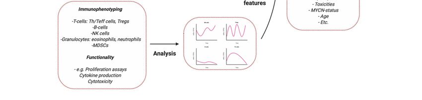
Cancers 2020, 12, 519                                                                                 2 of 20

(MDSC) accumulation, and decreased T cell cytotoxicity [7]. Infiltrating immune cells are observed
especially in low-risk NBL. The presence of tumor-infiltrating lymphocytes (TILs) was found to be
correlated with favorable clinical outcome [8]. This suggests a role of immune infiltration in regression
of NBL, which is supported by increased serum levels of granulysin, an effector molecule of cytotoxic T
cells, observed in a case study of spontaneous NBL regression [9]. Therapeutic interference to increase
immune infiltration and recognition might therefore be key to increase therapy efficiency against NBL.
      High-risk NBL is currently treated with surgery, radiotherapy, 5–8 cycles of intensive
chemotherapy, including platinum-, alkylating-, and topoisomerase agents—which is often followed
by autologous stem cell transplantation (ASCT)—and immunotherapy [1–4,10]. High expression of
GD2 (a disialoganglioside) across NBLs and low expression levels in healthy tissue has led to the
rationale of GD2 targeting immunotherapy [11]. Administration of the chimeric monoclonal antibody
(mAb) anti-GD2 (ch14.18), combined with the cytokines IL-2 and granulocyte macrophage-colony
stimulation factor (GM-CSF), and isotretinoin in patients with high-risk NBL resulted in a significant
increase 2-year event-free (EFS) and overall survival (OS) [10]. The observation of this effect, despite the
harsh immunomodulatory immune environment of NBL, shows the potential of immune interference
in NBL. However, as about 40% relapse is still observed in these patients, there is a clear medical
need to optimize (immuno)therapeutic strategies. The current immunotherapy protocol is particularly
ineffective for high-burden disease. In addition, osteomedullary metastatic disease occurs in most
patients with high-risk neuroblastoma [12]. Elucidating the mechanisms of effective anti-tumor
responses is key to find out, and act upon, what discriminates responders from non-responders.
      Several studies in multiple types of cancer report increased tumor infiltration of immune cells
upon (chemo)therapy [13–15], which could potentially predict overall therapy response and prognosis
in an early stage. Immune monitoring during therapy provides the opportunity to study biological
mechanisms of response and resistance [16]. This enables identification of biomarkers to monitor
therapy response, potentially aiding to early stratification of responders and non-responders. In the
1960s, it was reported that the correlation between prognosis and degree of lymphocyte infiltration is
also observed in NBL [17–19]. It is now known that NBL tumors are intermixed with different immune
cells, recently identified to include CD4- and CD8-T cells, natural killer (NK) cells, and γδ-T cells [20].
Interestingly, Mina et al. showed that the prognostic value of TIL levels at diagnosis is even better than
criteria currently used to stage NBL, such as MYCN amplification [8]. This illustrates the potential
role of immune cells in influencing the clinical outcome and emphasizes the need for standardized
immune monitoring during therapy in this patient group.
      This review provides an overview of predictive immune biomarkers of clinical response to
treatment, emphasizes the importance of immune monitoring during NBL treatment, and describes
its relevance for evaluation of the immune response and patient stratification by developing
new biomarkers.

2. Immune Monitoring at Diagnosis: Correlates of Outcome?

2.1. Immune Markers at the Tumor Site
      Histological analysis of human tumor types, including melanoma, ovarian-, head- and neck,
breast-, urothelial-, colorectal-, lung- hepatocellular-, and esophageal cancer showed the presence of
tumor infiltrating immune cells, such as macrophages, dendritic cells (DCs), polymorphonuclear cells,
NK cells, B cells, and T cells. Although these studies revealed a broad interpatient variability, a high
density of CD3 T cells, CD8 T cells, and CD45RO+ memory T cells was generally associated with
improved EFS and OS [21,22]. In addition, for NBL, multiple studies show that increased CD3 T cell
infiltration and proliferation is associated with favorable clinical outcome [8,23]. These data correspond
with the findings that high-risk MYCN-amplified primary metastatic NBL tumors show lower levels of
infiltrated lymphocytes, monocytes, and macrophages, and exhibit lower interferon pathway activity
Cancers 2020, 12, 519 3 of 20
and chemokine expression [23]. T cell proliferation is most likely impaired by high arginase activity in
the TME [24], resulting in low arginine levels (an essential molecule for T cell proliferation).
CD4 T cell infiltration is associated with better survival, regardless of MYCN amplification [23].
However, extensive phenotyping of these CD4 T cells, with markers such as CD25, CD127, and FoxP3 to
distinguish regulatory T cells (Treg), is lacking. Based on gene set enrichment analyses, gene expression
of IL-4 (indicative for Th2) was elevated and associated with better prognosis in tumors with high
CD4 T cells infiltration whereas no association was observed with interferon γ (IFNγ), IL-2 and tumor
necrose factor α (TNFα) (indicative for Th1) [25]. In addition, NKT cell infiltration has been reported
to be favorable for outcome, possibly by inhibition of suppressive monocytes in the TME [26].
Presence of immune cell populations with presumed regulatory properties, including tumor
associated monocytes, macrophages (TAMs), and Tregs, predict poor outcome [8,27]. Tumor infiltrating
macrophages often display an immunosuppressive M2-phenotype supporting T cell suppression,
tumor cell migration, and treatment evasion [28]. High expression of TAM-associated genes CD14,
CD16, IL-6, IL-6R, and transforming growth factor (TGFβ1) is associated with decreased 5-year EFS [26].
Additionally, monocytes isolated from the TME are able to suppress T cell proliferation in vitro [24]
and excrete multiple soluble T cell inhibitory factors, such as TGFβ and IL-10 [29,30]. Although
Tregs and MDSCs are well known for their suppressive effects on the immune system, associations
of these subsets with clinical outcome remain limited to studies investigating bone marrow (BM) or
peripheral blood (PB). One study showed that tumor-induced overexpression of high-mobility group
box 1 (HMBG1) induces a Treg phenotype. Patients with overexpression of this protein were at higher
risk for progression of disease, relapse, and death [7].
Despite these correlations in retrospective analyses, there are no prognostic markers in the TME
to steer clinical decision making. Inclusion of markers of cell differentiation, function, activation,
and exhaustion in multiple parameter analysis may help determine which factors have the strongest
associations with, and prognostic value for patient outcome. Advanced techniques, such as tissue
cytometry by time-of-flight (CyTOF), can overcome the limitations of measuring only a few markers in
common practice immune histology.
2.2. Circulatory Immune Markers
Despite advances in immune profiling and the easy accessibility of, PB; no validated circulatory
immune biomarkers exist for patients with NBL. Surprisingly, studies implementing immune
monitoring in NBL patients are limited.
2.2.1. Cytokines and Soluble Molecules in Plasma/Serum
Cytokines and chemokines are components of a complex network promoting angiogenesis and
metastasis, diminishing adaptive immunity, and changing responses to hormones and therapeutic
agents. As such, cytokines involved in cancer-related inflammation are easy to monitor and relate to
patient outcome and could be a target for therapeutic strategies.
Oliveira and colleagues reported an increase of IL-2, IL-4, IL-5, IL-6, IL-9, IL-10, IL-13, IL-17A,
IL-17F, IL-21, IL-22, interferon γ (IFNγ), and tumor necrose factor α (TNFα) in plasma of NBL
patients compared to age matched controls. One of these cytokines, IL-6, is a key growth-promoting
and anti-apoptotic inflammatory cytokine, which correlated with poor prognosis and high-risk
disease [31]. When integrating levels of IL-6 with other candidate biomarkers (serum amyloid A (SAA),
apolipoprotein (APOA1), epidermal growth factor (EGF), macrophage derived chemokine (MDC),
sCD40L, and Eotaxin), the multivariate classifier predicted active disease with a sensitivity of 81% and
specificity of 90% [32]. These data are encouraging and are awaiting validation in other patient cohorts.
Even though mRNA levels of IL-10 (a product of Tregs, NK cells and macrophages) in BM and,
PB; as well as IL-10 plasma concentrations were higher in metastatic NBL patients compared to healthy
controls, a prognostic role of IL-10 alone could not be demonstrated [29]. Low levels of soluble proteins
other than cytokines, such as human leukocyte antigens HLA-E and HLA-F, were associated withCancers 2020, 12, 519 4 of 20
worse prognosis [33]. In contrast to this, several other studies were not able to correlate markers
measured in plasma/serum to outcome.
In other cancers, the analyses of cytokine profiles rather than single markers has shown positive
prediction values at diagnosis [34–37]. The studies summarized above hopefully initiate validation
studies in NBL patients with multiparameter analyses in different patient cohorts.
2.2.2. Immune Cells in Peripheral Blood
Leukocyte counts have found to be significantly higher in NBL patients compared to controls [38].
Although no difference is observed in total lymphocyte count between healthy controls, localized,
and metastatic patients, relative numbers of multiple lymphocyte subsets do vary [31]. Semeraro et al.
measured a significant increase in the percentage of CD3−CD56+ NK cells in PB of metastatic NBL
patients compared to patients with localized tumors, which was associated with a minor response to
induction chemotherapy. The percentage of cytotoxic (CD16+) NK cells positively correlated with
clinical response to therapy [39]. These correlations could potentially be explained by NK cell mediated
cytotoxic effects on MHC-I lacking tumor cells. Activated NK cells may also upregulate MHC-I
expression on NBL cells, thereby circumventing further NK cell mediated cytotoxicity, while at the
same time increasing their susceptibility to T cell mediated cytotoxicity [40].
Morandi and colleagues found that Treg (CD4+CD25hiCD127−) and Tr1
(CD4+CD45R0+CD49b+LAG3+) subsets are decreased in NBL patients compared to controls, but no
correlation was found with prognostic factors, such as age and stage. MYCN amplification was
the only prognostic factor associated with higher levels of Treg numbers in BM and Tr1 levels
in PB [41]. In addition, CD4+ and CD8+ T cells show increased surface expression levels of the
checkpoint inhibitor CTLA-4, and PD-1 on CD4 T cells. In contrast, Semeraro et al. found increased
CD4+FoxP3+ T cells in metastatic NBL patients compared to localized tumors. The differences in the
markers used to identify specific cell subsets (in this case Treg) in different studies complicates a valid
comparison between the data from these studies and indicates the need for harmonization of immune
phenotyping protocols.
When comparing the myeloid compartment, NBL patients with localized tumors showed
higher monocyte, neutrophil and erythrocyte counts as compared to patients with metastatic disease.
When zooming in on the phenotype of myeloid cells, increased levels of the checkpoint inhibitor
programmed death ligand 2 (PD-L2) were observed in transitional (CD14+CD16+) and non-classical
monocytes (CD14-CD16+) in patients compared to controls [31]. Furthermore, increased expression of
CSF-1R, a regulator inducing MDSC expression, was observed in patients, and correlated with poor
clinical outcome [42].
In summary, it is clear that NBL patients show alterations in absolute numbers and subset
percentages of immune cells, as well as in immune proteins in the TME and in PB. An overview of the
reviewed studies can be found in Table 1. However, so far, no robust prognostic marker correlating
with survival has been identified and validated. Such immune signatures at diagnosis could aid in
therapy decision making and prognosis prediction.Cancers 2020, 12, 519 5 of 20
Table 1. Overview of neuroblastoma immune monitoring studies at diagnosis.
Flow Cytometry
Number of Unique Patient Samples
Tumor Characteristics Markers Reference
Measured Including Material
Primary Tumor
I (4x) + II (1x) + III (2x) + IV (1x) CD3, CD4, CD8, CD25, CD45RA, CCR7 Carlson et al.
8
PB 2013 [43]
Primary Tumor GD2
II (2x) + III (2x) + IV (21x) + IVs (1x) Mussai et al.
26
PB 15 MYCN amp, 11 non MYCN amp CD15, CD14, CD11b 2015 [24]
Song et al.
20 BM IV (20x) CD45, CD33, CD14, GD2, CD56
2009 [26]
Morandi et al.
41 PB 13 MYCN amp, 28 non MYCN amp CD4, CD25, CD127, CD45RO, CD49b, LAG-3
2015 [29]
Gowda et al.
5 PB High-Risk Patients HLA-DR, CD33, CD11b
2013 [44]
21 PB
7 MYCN amp, 20 non MYCN amp (14 localized, CD4, CD25, CD127, CD45RO, CD49b, LAG-3 Morandi et al.
27 BM 13 metastatic) 2016 [41]
CD8, NKp46, CD4, CD16, CD56, NKp30, DNAM-1, CD127, CD25, CD14,
PB 23 localized, 36 metastatic tumors Semeraro et al.
59 CD45, CD15, GD2, CD235a, CD9, CD81, TCRgd, NKp44, NKp80, CD3e,
BM CD158a/h, CD158b, CD158e/k, CD158i, FoxP3 2015 [39]
Immunohistochemistry
24 CD68
21 CD3 Apps et al.
Primary Tumor 7 MYCN amp, 26 non MYCN amp 2013 [45]
19 pSTAT3
Carlson et al.
8 Primary Tumor I (4x) + II (1x) + III (2x) + IV(1x) Ki67, CD3
2013 [43]
Zhang et al.
15 Primary Tumor 3 low risk, 6 intermediate risk, 6 high risk CD4, CD45
2017 [25]
Song et al. 2017
129 Primary Tumor IV (129x) CD1d, Vα24-Jα18inv, TCRα6β11
[26]
Asgharzadeh
71 Primary Tumor stage I–III (n = 29), stage IV (n = 31), stage IVS (n = 11) CD163, AIF1
et al. 2012 [27]
Mina et al.
84 Primary Tumor I (34x) + II (19x) + III (5x) + IV (20x) + IVS (6x) CD3, CD4, CD8, CD25, FOXP3, Ki67, β2m-free MHC1 heavy chain
2015 [8]Cancers 2020, 12, 519 6 of 20
Table 1. Cont.
Flow Cytometry
Number of Unique Patient Samples
Tumor Characteristics Markers Reference
Measured Including Material
Elisa
Morandi et al.
57 Plasma IV (49x) + non IV (8x) IL-10, ARG-1 (57) 2015 [29]
53 PB PB: I (8x) + II (8x) + III (6x) + IV (28x) + IVS (3x)
IL-6
18 BM BM: I (1x) + II (3x) + III (2x) + IV (11x) + IVS (1x)
Egler et al.
35 PB PB: I (5x) + II (5x) + III (3x) + IV (20x) + IVS (2x) 2008 [46]
sIL-6R
16 BM BM: I (1x) + II (2x) + III (2x) + IV (10x) + IVS (1x)
I (7x) + II (8x) + III (22x) + IV (42x) + 4S (5x) Morandi et al.
84 Primary Tumor sHLA-F, sB7H3, sHLA-E
27 MYCN amp, 57 non MYCN amp 2013 [33]
Luminex
Plasma GM-CSF, G-CSF, IFNγ, IL-1a, IL-1ra, IL-1b, IL-2, IL-3, IL-4, IL-5, IL-6, IL-7,
20 low-risk, 35 high-risk In addition, 28 HR blood samples IL-8, IL-9, IL-10, IL-12p70, IL-12p40, IL-13, IL-15, IL-17, MCP-1, MCP-3, Egler et al.
55 MDC, TNFα, TNFβ, TGFα, Eotaxin, IFNα2, IP-10, MIP-1a, MIP-1b, EGF,
from 7 patients at various timepoints during treatment 2011 [32]
PB FGF-2, FLT3L, Fractalkine, GRO, VEGF, sCD40L, sIL-2RaCancers 2020, 12, 519 7 of 20
3. Immune Monitoring during Therapy
Immune monitoring during therapy is crucial to identify potentially prognostic factors that could
be exploited to enhance immunogenicity of the tumor and predict treatment response. This section
will start by reviewing studies which monitor the immune response upon standard therapy, including
chemotherapy and monoclonal antibody therapy. Subsequently, immune monitoring in more
experimental treatment regimens, including vaccination strategies, adoptive cell-, and checkpoint
inhibition therapy will be discussed.
3.1. Chemotherapy
In general, high-risk NBL patients receive 5–8 cycles of intensive chemotherapy including platinum,
alkylating, and topoisomerase agents. In North-America, induction regimens include vincristine,
doxorubicin, cyclophosphamide, cisplatin, and etoposide, while the Society of pediatric oncology
Europe NBL group (SIOPEN) used a rapid COJEC regimen that gives eight cycles with combinations
of vincristine, carboplatin, etoposide, cyclophosphamide, and cisplatin [47].
A limited number of studies has monitored immune profiles during chemotherapy in cancer
patients. Monitoring lymphocyte levels during and after chemotherapy in hematopoietic and solid
tumors generally showed increased EFS in patients with higher lymphocyte counts at diagnosis as well
as after induction chemotherapy [42,47–49] In addition, fast monocyte recovery after chemotherapy
is predictive for EFS in patients with leukemias and lymphomas [50,51]. In line with these data,
an elevated neutrophil to lymphocyte ratio after chemotherapy, but before surgical resection of the
NBL tumor, was associated with decreased OS [52].
Upon chemotherapy treatment, Treg counts decreased, possibly due to nonspecific targeting
of Tregs by chemotherapeutic agents. More studies are warranted to determine if the effect can be
subscribed to chemotherapy-induced decrease in T cells in general, or whether specific subsets, like Tregs,
might be more susceptible to chemotherapy-induced cytotoxicity [53]. Chemotherapy generally does
not affect NK cells [54], however, the expression levels of NKp30, an NK cell receptor involved in tumor
cell killing and DC recognition, positively correlate with survival after chemotherapy [55]. Expression
levels of the immunosuppressive isoform NKp30C and the activating isoforms NKp30A and NKp30B
affect NK cell function and correlate with EFS of NBL patients after chemotherapy [39].
No studies have monitored immune markers in the TME during chemotherapy in patients.
An in vivo mouse study showed that depletion of TAMs from NBL tumors is associated with increased
chemotherapeutic efficacy without requiring T cell contribution [56]. This observation led to the author’s
suggestion to combine CSF-1R blockade with chemotherapy to potentially increase treatment efficacy.
To date, too few studies have monitored immune status during chemotherapy to identify markers
that could predict response to therapy. It seems that patients with higher numbers or faster recovery of
lymphocytes and monocytes have better EFS changes. Whether this is a reflection or a result of the
development of a healthier immunological niche should be studied, but the observation could lead to
the hypothesis that these patients might also be responding better to immune treatment options.
3.2. Monoclonal Antibody Therapy
A frequent immunotherapy protocol of high-risk NBL consists of anti-GD2 combined with
all-trans-retinoic acid (ATRA), IL-2, and GM-CSF. Even though immunotherapy increased 2-year EFS
and OS [10], relapse is still observed in 40% of patients. Elucidation of effective anti-tumor responses is
key to study what discriminates responders from non-responders.
IL-2 and GM-CSF have been added to the treatment protocol as they were observed to enhance
cytotoxicity of anti-GD2 in vitro [10,57–59]. In addition, GM-CSF also increased myeloid cell activation,
another important cell type in the anti-tumor response [57,60]. The multi-component nature of the
immunotherapy protocol makes that the observed immune effects cannot be ascribed to a specific
component of the protocol.Cancers 2020, 12, 519 8 of 20
A primary mechanism of action of anti-GD2 is the induction of antibody-dependent cellular
cytotoxicity (ADCC), which requires recognition by effector cells (mainly NK cells, monocytes,
neutrophils, and macrophages) [11,60]. Cytotoxic activity of NK-cells is mediated by CD16, whereas
cytotoxic activity of monocytes, neutrophils, and macrophages is mediated by CD32. Both receptors
recognize the Fc fragment of anti-GD2 on opsonized NBL cells and induce cytotoxic effector functions.
Complement-dependent cytotoxicity (CDC) is another mechanism of action of anti-GD2, however,
most studies focus on its implications regarding pain toxicity rather than on-tumor toxicity.
Nassin et al. monitored immune reconstitution at the start of immunotherapy containing IL-2,
GM-CSF, and anti-GD2 [61]. They showed that absolute lymphocyte counts (T., B.; and NK subsets)
are lower in the vast majority of patients as compared to age-matched controls. Patients with disease
progression, relapse or residual disease had significantly lower total leukocyte counts, as well as a lower
absolute lymphocyte-, neutrophil-, and CD16+ cell counts compared to disease-and progression-free
patients observed three months after therapy. Siebert and colleagues found that presence of human
anti-chimeric antibodies against chimeric anti-GD2 resulted in significant reduction in peripheral
anti-GD2 levels, as well as significant abrogation in ADCC and CDC [62]. However, in this study, it is
not clear if such immune responses are a disadvantage for survival of the treated patients. In addition,
the induction of the host anti-idiotype network, measured indirectly by human anti-mouse antibody
responses correlated with long term survival [63–65]
The importance of NK cells in ADCC was illustrated by Chowdhury et al.; showing that
in vitro anti-GD2 mediated lysis of the LAN1 NBL cell line upon co-culture with peripheral blood
mononuclear cells (PBMCs) from a NBL patient abrogated after NK cell depletion [66]. In addition,
cell lysis correlated with NK cell expression of CD69, an early activation marker, as well as with the
degranulation marker CD107a. Furthermore, variation in ADCC between patients was found to be
caused by genetic predispositions resulting in better cytotoxic activity of effector cells and correlations
with better survival [59,63,64]. Siebert et al. studied the level of ADCC in vitro in combination with
FCGR polymorphisms and killer cell immunoglobulin like receptor KIR/KIR ligand genotypes of 53
patients. They showed that patients with high affinity FCGRs had higher ADCC levels and better EFS
compared to patients with low affinity genotypes. In addition, a correlation was found between the
activating KIR 2DS2 genotype on ADCC and EFS. A combination of high-affinity FCGR2A,-3A and
stimulating genotypeB/x or the presence of activating KIR 2DS2 resulted in the strongest anti-NBL
cellular cytotoxicity mediated by anti-GD2 and improved EFS [67]. In addition, Tarek et al. found
that patients treated with monoclonal antibodies (moABs) lacking HLA class I ligands for their
inhibitory KIRs have significantly higher survival rates. Unlicensed NK cells mediate tumor control via
ADCC [68]. These results show that FCGR polymorphisms and KIR/KIRL genotypes could function as
biomarkers in response to immunotherapy.
The importance of NK cell mediated ADCC in anti-GD2 efficacy, together with the observation
of relatively fast NK cell recovery early after ASCT was an important rationale for immunotherapy
timing early after transplantation [69]. However, more detailed evaluation of NK cell subsets showed
that most of these NK cells are immature, cytokine releasing (CD56bright, CD16+/−) rather than
the cytotoxic (CD56dim, CD16+) NK cells known to be mainly responsible for anti-GD2 dependent
ADCC [61]. This is further substantiated by observed impaired immune recovery of CD16+ NK cells
in patients with disease progression or relapse at time of transplantation compared to those without.
These studies may suggest suboptimal timing of anti-GD2 immunotherapy early after transplantation.
Utilizing haploidentical allogeneic hematopoietic cell sources rather than autologous sources could be
explored as a transplantation source regarding NK cell recovery early after transplantation.
In addition to NK cell subset monitoring, Nassin et al. showed increased CD25 expression on CD4
T cells as compared to CD8 T cells at the start of immunotherapy [61]. Even though FoxP3 expression of
these cells is unknown, it is hypothesized that these cells could be classified as Tregs [70]. Ladenstein et
al. recently concluded from a phase III clinical trial that there is no additive effect of IL-2 administration
on outcome of high-risk NBL patients [71]. As (low dose) IL-2 administration to autoimmune patientsCancers 2020, 12, 519 9 of 20
resulted in preferential expression of Tregs [72], authors hypothesize that adjuvant IL-2 administration
could be responsible for Treg expansion during immunotherapy, diminishing the positive effects (e.g.,
NK expansion) of IL-2. Indeed, we observed a rise in Treg numbers upon every round of IL-2 to NBL
patients (unpublished). Furthermore, administration of IL-2 also results in development of eosinophils
through stimulating growth factors derived from T cells, such as IL-3, IL-5, and GM-CSF that help to
maintain eosinophils in vitro [73]. In addition, an increase in IL-5 levels during immunotherapy with
anti-GD2 and cytokines was observed, that could induce and maintain eosinophils [74]. Although their
effect is unknown, suppressive eosinophils have been reported in murine studies and could possibly
also be present in IL-2 treated NBL patients [75]. Further studies are required to confirm these effects
of IL-2 on levels of Treg and eosinophils, preferably with functional testing of the different cell subsets.
To overcome limitations of the current anti-GD2 monoclonal antibody therapy, including
monoclonality of the response, anti-idiotype responses, and memory induction, Kusher et al. reported
a strategy in which patients are vaccinated with GD2 and GD3. This results in an intrinsic, polyclonal,
multivalent antibody response through stimulation of B-cells to produce anti-GD2 and –GD3 [76].
As B-cell recovery is key for vaccination efficacy, immunomonitoring of B-cell recovery after ASCT is
key for optimal vaccination timing.
To conclude, immune monitoring studies during immunotherapy are largely lacking and studies
that are available have only monitored patients at start and end of therapy. All new phase II/III trials
and standard treatment protocols should not only asses outcome but also monitor the immune system
during therapy to get a better and faster understanding of treatment success.
3.3. Adoptive Cell Therapy
The development of adoptive cell therapy (ACT) strategies has taken flight in the last decade.
These cell products should ideally possess the capacity to expand, actively migrate through the entire
body (including to the solid tumor core and over the blood brain barrier), and induce systemic immune
memory to prevent future relapse. ACT products are generated by harvesting, ex vivo expansion,
and re-direction of immune cells to target tumor cells. Even though successes have been achieved in
several hematological malignancies [77–80], translation of these successes to solid tumors is difficult.
Target expression heterogeneity, localization to the tumor site, and overcoming the immunosuppressive,
nutrient-, and stimuli- deprived TME are thought to be the main challenges in effective adoptive cell
therapy in solid tumors [81].
To date, pre-clinical studies as well as clinical trials are exploiting T-, NK-, and NKT cells in
both autologous and allogeneic ACT strategies in NBL. Infused cells can be isolated from PB [82–85],
as well as from tumor tissue (e.g., TILs) [8,20]. In addition, isolated cells can be genetically modified to
improve recognition of tumor cells, for example through knock-in of a NBL-specific T cell receptor
(TCR) [40,86,87], a chimeric antigen receptor (CAR) [20,88–95], or a bispecific antibody [96,97] against
tumor specific targets such as GD2, PRAME, NY-ESO-1, L1-CAM, B7-H3, and mutated ALK. Very low
or even absent surface expression of major histocompatibility complex I (MHC-I) on NBL cells has led
to the focus on MHC-I unrestricted ACT-strategies, mainly exploiting NK cell- and CAR therapy (or
a combination of both). Excellent overviews of clinical trials of (adoptive cell) therapy strategies in
NBL are provided by Le and Thai [98] and Zage [99].
Even though multiple recent (pre-clinical) studies have demonstrated efficacy of various forms of
ACT in NBL, the observed clinical benefit is limited. Persistence of ACT products in general is a widely
discussed topic and is thought to be depending on balanced activation (appropriate co-stimulation,
prevention of tonic/chronic receptor signaling, and of activation-induced cell death), tolerance induction
(caused by native antigen expression and tumor immunosuppression), the cell phenotype of both
the apheresis material and the product itself, as well as by the need of antigen availability for cell
persistence [87,100,101]. The extremely immunosuppressive, nutrient- and stimuli deprived TME
of solid tumors is thought to be the main factor responsible for the discrepancies in successes with
hematological tumors [82,102].Cancers 2020, 12, 519 10 of 20
Monitoring the tumor infiltrating cell population, its phenotype, and other related factors will
provide the much-needed insight to predict therapy response and to understand what is driving
resistance and success and where can be acted upon. Sporadic monitoring after ACT resulted in the
observation that the presence of central memory and naïve T cell phenotypes in cell products has
a positive effect on persistence of the cell product in several cancer types, including NBL [8,82,103–106].
Hurtado et al. recently showed that the presence of central memory NBL TILs greatly decreases after
non-specific ex vivo rapid expansion cycles, stressing the importance of ex vivo expansion protocol
evaluation in adoptive cell strategies [20]. Moreover, lymphodepletion before T cell infusion or early
T cell infusion after ASCT (day 2) caused significantly improved expansion and persistence of the
cell product [82,89]. This indicates that the immunosuppressive status of the immune system of these
patients is an important limiting factor in effective ACT and led to the rationale to combine ACT with
checkpoint inhibitors [89,107]. Remarkably, combining PD-1 blockade by pembrolizumab with GD2
CAR T cell administration upon lymphodepletion in a phase 1 clinical trial in NBL did not show any
beneficial effect of PD-1 blockade on peripheral CAR T cell expansion [89]. A limitation of this study is
that the arm studying the effect of PD-1 blockade on non-lymphodepleted GD2 CAR T cell treated
patients was missing. Furthermore, T cell expansion and persistence was solely measured in the blood
and not at the tumor site. The fact that two out of three treated patients in this arm experienced
complete remission is encouraging and warrants more research. Controversially, immune monitoring
in the same phase 1 GD2 CAR study showed specific expansion of circulating immunosuppressive M2
macrophage-like myeloid cells (CD45+CD33+CD11b+CD163+) independent of lymphodepletion and
PD-1 blockade [89]. Inhibitory myeloid cells are correlated with poor prognosis in several cancer types,
including NBL, even though this study does not provide any data on whether these mobilized cells are
attracted to the tumor site. Induction of MDSCs was also reported in GD2−CAR T cell therapy for
sarcomas in xenograft mice, which impaired CAR T cell activity [108]. Addition of all-trans retinoic
acid (ATRA) destroyed the MDSCs and thereby improved the efficacy of the GD2-CAR T cells [108],
suggesting that patients might also benefit from ATRA to eradicate the circulating M2 macrophage-like
myeloid cells that are induced by GD2-CAR3. Thirdly, immune monitoring in this phase 1 study
indicated a correlation between circulating IL-15 levels and GD2 CAR T cell expansion [89]. Circulating
IL-15 levels could not only potentially be used as a biomarker for CAR T cell expansion, but a pre-clinical
xenograft mouse model transduced with GD2 CAR T cells overexpressing IL-15 also showed improved
expansion, enhanced anti-tumor activity and improved survival [93].
Even though immune monitoring data during ACT in NBL is scarce, the above mentioned studies
all resulted in clear rationales for research into new therapeutic strategies or biomarker development.
This clearly indicates the need for more elaborate immune monitoring during ACT.
3.4. Checkpoint Inhibitors
Another interesting strategy would be to target the immunosuppressive environment of NBL [7].
Checkpoint inhibitors (CPis), blocking CTLA-4, PD-1, PD-L1, or PD-L2, are proven to be effective
in a variety of tumors, including solid tumors [109]. The potential of these inhibitors is illustrated
by the growing list of FDA-approvals, as reviewed by Hargadon et al. [110]. Currently, most of
these approvals are in unresectable metastatic disease settings. Nevertheless, the potency of CPis to
induce a more potent anti-tumor immune response, and therefore potential induction of anti-tumor
immunological memory, makes checkpoint inhibition a very interesting candidate as an adjuvant
therapy in curative treatment settings. The utilization of CPis as a monotherapy in NBL has been
investigated in multiple pre-clinical studies [42,111–113]. These studies show no effect of CPi treatment
on systemic NBL progression in vivo. This has been supported in a phase I clinical trial in pediatric
patients with advanced solid tumors, in which one NBL patient was included [114]. More clinical trials
assessing CPis as a monotherapy in refractory NBL are currently running (NCT02304458; NCT02332668;
NCT02541604).Cancers 2020, 12, 519 11 of 20
Several rationales have been proposed for the ineffectiveness of CPi therapy in high-risk NBL.
First of all, evaluation of NBL tumors from patients with different tumor stages does not only,
as mentioned before, show inverse correlation between tumor grade and TILs [8], but also between
tumor grade and PD-L1 tumor expression [113]. Absence of TILs and PD-L1 tumor expression provides
a first rationale to CPi therapy resistance. Secondly, one of the immunomodulatory mechanisms
of NBL is the upregulation of PD-L1 in response to IFN-γ [111,113]. This highlights a potential
resistance mechanism against CPi therapy, as persistent IFN-γ production by activated T cells may
lead to a continuous cascade causing even higher upregulation of the immune checkpoints resulting
in T cell senescence. Impaired IFN pathway activity in MYCN-amplified tumors [23] may prevent
PD-L1 expression upon IFN-γ exposure and provides a third rationale for CPi therapy effectiveness in
high-risk NBL.
Next to these observations, especially studies investigating CPi-including combination therapies
revealed some insights in mechanisms explaining NBL resistance to CPi monotherapy [42,111–113].
Rigo and colleagues showed that combining CPi with temporary CD4 depletion by anti-CD4 monoclonal
antibody (mAb) treatment caused a very potent CD8 T cell dependent response causing significantly
longer tumor-free survival, complete tumor regression, and durable anti-NBL immunity in vivo [111].
Combining CPi with immune enhancer IL-21, or Treg targeting agents polyoxotungstate (POM-1) and
anti-CD25 antibody, revealed no significant effect. This indicates that CD4 CD25high Tregs, as well as
adenosine generation in Tregs are not responsible for CPi resistance in NBL. More specific studies into
effects of CD4 CD25−/low FOXP3+ Tregs or other CD4 Tregs should be performed to further unravel
the synergistic effect observed upon CD4 depletion [111].
Mao et al. reported that targeting of CSF-1Rþ+ suppressive myeloid cells in combination with
CPis caused spontaneous control of NBL in vivo [42], a mechanism confirmed in several other types
of cancers, as reviewed by Weber et al. [115]. A possible explanation for this is the observation that
T cells start to produce M-CSF upon PD-1 blockade, which can bind to CSF-1R on myeloid-derived
suppressor cells, thereby enhancing their suppressive phenotype [116]. This causes a further reduction
of IFN-regulated chemokine release (e.g., CXCL9, 10, and 11) in the TME, which are important for T
cell infiltration and could therefore potentially explain the synergistic effect of targeting CSF-1Rþ+
suppressive myeloid cells in combination with CPi.
Other combination strategies which seem promising based on preclinical studies include
combinations of CPis with whole tumor cell vaccination [113], high intensity ultrasound [117],
and anti-GD2 treatment [112]. All mentioned preclinical evidence of effective NBL targeting treatment
combinations provides a clear rationale for clinical assessment of CPi therapy as an adjuvant therapy
against NBL. Even though data obtained in these induced tumor mouse models should be interpreted
with caution, the data should be used as a rationale for focusing on immune monitoring of adjuvant
CPi therapy in coming clinical trials.
4. Discussion
The present review shows the potential of standardized immune monitoring in NBL. Despite
many efforts, high-risk NBL has a poor response to treatment and novel therapies are urgently required.
We should therefore better understand the functional kinetics of tumor- and immune cells to guide
and modulate immune therapy strategies. As such, immune monitoring may provide evidence-based
directions to optimize treatment protocols, e.g., in the recent discussion whether IL-2 should be
removed from the current treatment protocol [118].
Studies listed in this review use flow cytometry, CyTOF, immunohistochemistry, as well as
functional assays to perform immune monitoring on tissue, PBMCs or plasma/serum from NBL
patients. Monitoring PBMCs and plasma/serum cytokines is by far the easiest approach as blood is
easily available during treatment. As the immune system is a multifaceted system, it is important to
question if findings in the circulatory system are related to the TME. Monitoring peripheral blood
subsets currently have insufficient clinical value for clinical decision making. Future studies withStudies listed in this review use flow cytometry, CyTOF, immunohistochemistry, as well as
functional assays to perform immune monitoring on tissue, PBMCs or plasma/serum from NBL
patients. Monitoring PBMCs and plasma/serum cytokines is by far the easiest approach as blood is
easily available during treatment. As the immune system is a multifaceted system, it is important to
question
Cancers 2020,if12,findings
519 in the circulatory system are related to the TME. Monitoring peripheral12blood of 20
subsets currently have insufficient clinical value for clinical decision making. Future studies with
paired monitoring of PB, BM, and TME samples will increase our understanding of underlying
paired
mechanismmonitoring
and shed of PB, lightBM,on and TMEofsamples
the use biomarkers will over
increase
the our understanding
treatment course asofpredictors
underlying for
mechanism and shed
efficacy and/or toxicity. light on the use of biomarkers over the treatment course as predictors for efficacy
and/or Astoxicity.
most studies describe small patient populations, high interpatient variability, and no
As most studies
harmonization describeprotocols,
of monitoring small patient
it can populations,
be difficult to high interpatient
interpret results andvariability, and
relate it to no
recent
harmonization of monitoring protocols, it can be difficult to interpret results
literature [119]. The studies used in this review are sometimes limited to results of 3–5 patients. and relate it to recent
literature
Therefore,[119].
we proposeThe studies usedand
to define in implement
this reviewstandardized
are sometimes limitedmonitoring
immune to results of 3–5 patients.
protocols in the
Therefore, we propose to define and implement standardized immune
trial- and study design to increase patient numbers and study interpatient variability. An example monitoring protocols in theis
trial- and study
proposed in Figure design
1. Astoeach
increase patient
patient numbers
is unique, and study
immune interpatient
monitoring variability.
can facilitate Anpersonalized
future example is
proposed in Figure
treatment in NBL. 1. As each patient is unique, immune monitoring can facilitate future personalized
treatment in NBL.
Advances in organoid technology is promising in predicting the immunosuppressive capacities
Advances in organoid
as well as the sensitivity to technology is promising
(immune) therapy in predictingsetting.
in a personalized the immunosuppressive
Neuroblastoma tumor capacities
models as
well as the sensitivity to (immune) therapy in a personalized setting. Neuroblastoma
have been developed that represent the tumor better than classical cell lines [120]. This strategy has tumor models have
been developed
previously shown that represent the tumor
to be effective better thangastrointestinal
in metastatic classical cell linescancers,
[120]. This
in strategy has previously
which patient derived
shown
organoidsto be wereeffective in metastatic
instrumental gastrointestinal
in the prediction of tumorcancers,
specific inresponses,
which patient derived
stimulating organoids
personalized
were instrumental
treatment strategies in [121].
the prediction of RNA
Single-cell tumorsequencing
specific responses, stimulating
of NBL tumors can, personalized
besides studying treatment
tumor
strategies [121]. Single-cell RNA sequencing of NBL tumors can, besides
heterogeneity and driving factors behind tumor heterogeneity, help to identify new leads for studying tumor heterogeneity
and driving factors behind
immunotherapeutic strategiestumor heterogeneity, help to identify new leads for immunotherapeutic
[122].
strategies [122].
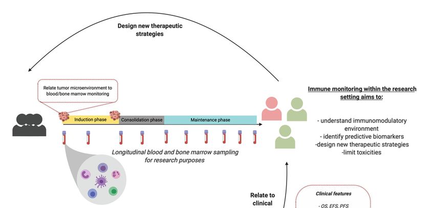
Figure 1. Immune monitoring for research purposes. Monitoring peripheral blood subsets currently
Figure
have 1. Immune
insufficient monitoring
clinical forclinical
value for research purposes.
decision Monitoring
making. Createdperipheral blood subsets currently
with BioRender.com.
have insufficient clinical value for clinical decision making. Created with BioRender.com.
5. Conclusions
5. Conclusions
In conclusion, immune monitoring before and during therapy of NBL patients could facilitate
identification of predictive biomarkers to guide patients towards effective treatment with limited
toxicities and optimal quality of life. Furthermore, understanding the immunomodulatory environment
of NBL and its response to treatment in responders and non-responders is important to facilitate design
of new therapeutic strategies improving outcome of high-risk NBL.Cancers 2020, 12, 519 13 of 20
Funding: This research was funded by Villa Joep Foundation, Grant number IWOV-Actief.51381.180034.
Conflicts of Interest: The authors declare no conflict of interest.
References
1. Matthay, K.K.; Maris, J.M.; Schleiermacher, G.; Nakagawara, A.; Mackall, C.L.; Diller, L.; Weiss, W.A.
Neuroblastoma. Nat. Rev. Dis. Prim. 2016, 2, 16078. [CrossRef]
2. Park, J.R.; Eggert, A.; Caron, H. Neuroblastoma: Biology, Prognosis, and Treatment. Hematol. Oncol. Clin. N.
Am. 2010, 24, 65–86. [CrossRef] [PubMed]
3. Maris, J.M. Recent Advances in Neuroblastoma. N. Engl. J. Med. 2010, 362, 2202–2211. [CrossRef] [PubMed]
4. Davidoff, A.M. Neuroblastoma. Semin. Pediatr. Surg. 2012, 21, 2–14. [CrossRef] [PubMed]
5. Chen, D.S.; Mellman, I. Elements of cancer immunity and the cancer-immune set point. Nature 2017, 541,
321–330. [CrossRef]
6. Bonaventura, P.; Shekarian, T.; Alcazer, V.; Valladeau-Guilemond, J.; Valsesia-Wittmann, S.; Amigorena, S.;
Caux, C.; Depil, S. Cold Tumors: A Therapeutic Challenge for Immunotherapy. Front. Immunol. 2019, 10,
168. [CrossRef] [PubMed]
7. Vanichapol, T.; Chutipongtanate, S.; Anurathapan, U.; Hongeng, S. Immune Escape Mechanisms and Future
Prospects for Immunotherapy in Neuroblastoma. Biomed. Res. Int. 2018, 2018, 1812535. [CrossRef]
8. Mina, M.; Boldrini, R.; Citti, A.; Romania, P.; D’Alicandro, V.; De Ioris, M.; Castellano, A.; Furlanello, C.;
Locatelli, F.; Fruci, D. Tumor-infiltrating T lymphocytes improve clinical outcome of therapy-resistant
neuroblastoma. Oncoimmunology 2015, 4, 1–14. [CrossRef] [PubMed]
9. Nagasawa, M.; Kawamoto, H.; Tsuji, Y.; Mizutani, S. Transient increase of serum granulysin in a stage
IVs neuroblastoma patient during spontaneous regression: Case report. Int. J. Hematol. 2005, 82, 456–457.
[CrossRef]
10. Yu, A.L.; Gilman, A.L.; Ozkaynak, M.F.; London, W.B.; Kreissman, S.G.; Chen, H.X.; Smith, M.; Anderson, B.;
Villablanca, J.G.; Matthay, K.K.; et al. Anti-GD2 Antibody with GM-CSF, Interleukin-2, and Isotretinoin for
Neuroblastoma. N. Engl. J. Med. 2010, 363, 1324–1334. [CrossRef]
11. Zeng, Y.; Fest, S.; Kunert, R.; Katinger, H.; Pistoia, V.; Michon, J.; Lewis, G.; Ladenstein, R.; Lode, H.N.
Anti-neuroblastoma effect of ch14.18 antibody produced in CHO cells is mediated by NK-cells in mice. Mol.
Immunol. 2005, 42, 1311–1319. [CrossRef] [PubMed]
12. Modak, S.; Cheung, N.-K. V Neuroblastoma: Therapeutic strategies for a clinical enigma. Cancer Treat. Rev.
2010, 36, 307–317. [CrossRef] [PubMed]
13. Kim, R.; Coppola, D.; Wang, E.; Chang, Y.D.; Kim, Y.; Anaya, D.; Kim, D.W. Prognostic value of CD8CD45RO
tumor infiltrating lymphocytes in patients with extrahepatic cholangiocarcinoma. Oncotarget 2018, 9,
23366–23372. [CrossRef] [PubMed]
14. Parra, E.R.; Villalobos, P.; Behrens, C.; Jiang, M.; Pataer, A.; Swisher, S.G.; William, W.N.; Zhang, J.; Lee, J.;
Cascone, T.; et al. Effect of neoadjuvant chemotherapy on the immune microenvironment in non-small
cell lung carcinomas as determined by multiplex immunofluorescence and image analysis approaches. J.
Immunother. Cancer 2018, 6, 48. [CrossRef]
15. Lo, C.S.; Sanii, S.; Kroeger, D.R.; Milne, K.; Talhouk, A.; Chiu, D.S.; Rahimi, K.; Shaw, P.A.; Clarke, B.A.;
Nelson, B.H. Neoadjuvant chemotherapy of ovarian cancer results in three patterns of tumor-infiltrating
lymphocyte response with distinct implications for immunotherapy. Clin. Cancer Res. 2017, 23, 925–934.
[CrossRef]
16. Hegde, P.S.; Karanikas, V.; Evers, S. The where, the when, and the how of immune monitoring for cancer
immunotherapies in the era of checkpoint inhibition. Clin. Cancer Res. 2016, 22, 1865–1874. [CrossRef]
17. Martin, R.F.; Bruce Beckwith, J. Lymphoid infiltrates in neuroblastomas: Their occurrence and prognostic
significance. J. Pediatr. Surg. 1968, 3, 161–164. [CrossRef]
18. Lauder, I. The significance of lymphocytic infiltration in neuroblastoma. Br. J. Cancer 1972, 26, 321–330.
[CrossRef]
19. Hellstrom, I.E.; Hellstrom, K.E.; Pierce, G.E.; Bill, A.H. Demonstration of cell-bound and humoral immunity
against neuroblastoma cells. Proc. Natl. Acad. Sci. USA 1968, 60, 1231–1238. [CrossRef]Cancers 2020, 12, 519 14 of 20
20. Ollé Hurtado, M.; Wolbert, J.; Fisher, J.; Flutter, B.; Stafford, S.; Barton, J.; Jain, N.; Barone, G.; Majani, Y.;
Anderson, J. Tumor infiltrating lymphocytes expanded from pediatric neuroblastoma display heterogeneity
of phenotype and function. PLoS ONE 2019, 14, e0216373. [CrossRef]
21. Fridman, W.H.; Zitvogel, L.; Sautès-Fridman, C.; Kroemer, G. The immune contexture in cancer prognosis
and treatment. Nat. Rev. Clin. Oncol. 2017, 14, 717–734. [CrossRef] [PubMed]
22. Berghoff, A.S.; Fuchs, E.; Ricken, G.; Mlecnik, B.; Bindea, G.; Spanberger, T.; Hackl, M.; Widhalm, G.;
Dieckmann, K.; Prayer, D.; et al. Density of tumor-infiltrating lymphocytes correlates with extent of brain
edema and overall survival time in patients with brain metastases. Oncoimmunology 2016, 5, e1057388.
[CrossRef] [PubMed]
23. Layer, J.P.; Kronmüller, M.T.; Quast, T.; Van Den Boorn, D.; Effern, M.; Hinze, D.; Althoff, K.; Westermann, F.;
Peifer, M.; Hartmann, G.; et al. Amplification of N-Myc is associated with a T-cell-poor microenvironment in
metastatic neuroblastoma restraining interferon pathway activity and chemokine expression. Oncoimmunology
2017, 6, 1–13. [CrossRef] [PubMed]
24. Mussai, F.; Egan, S.; Hunter, S.; Webber, H.; Fisher, J.; Wheat, R.; McConville, C.; Sbirkov, Y.; Wheeler, K.;
Bendle, G.; et al. Neuroblastoma arginase activity creates an immunosuppressive microenvironment that
impairs autologous and engineered immunity. Cancer Res. 2015, 75, 3043–3053. [CrossRef] [PubMed]
25. Zhang, P.; Wu, X.; Basu, M.; Dong, C.; Zheng, P.; Liu, Y.; Sandler, A.D. MYCN amplification is associated with
repressed cellular immunity in neuroblastoma: An in silico immunological analysis of TARGET database.
Front. Immunol. 2017, 8, 1473. [CrossRef] [PubMed]
26. Song, L.; Asgharzadeh, S.; Salo, J.; Engell, K.; Wu, H.; Sposto, R.; Ara, T.; Silverman, A.M.; Declerck, Y.A.;
Seeger, R.C.; et al. V α 24-invariant NKT cells mediate antitumor activity via killing of tumor-associated
macrophages. J. Clin. Invest. 2009, 119, 1524–1536. [CrossRef]
27. Asgharzadeh, S.; Salo, J.A.; Ji, L.; Oberthuer, A.; Fischer, M.; Berthold, F.; Hadjidaniel, M.; Liu, C.W.Y.;
Metelitsa, L.S.; Pique-Regi, R.; et al. Clinical significance of tumor-associated inflammatory cells in metastatic
neuroblastoma. J. Clin. Oncol. 2012, 30, 3525–3532. [CrossRef]
28. Qian, B.Z.; Pollard, J.W. Macrophage Diversity Enhances Tumor Progression and Metastasis. Cell 2010, 141,
39–51. [CrossRef]
29. Morandi, F.; Croce, M.; Cangemi, G.; Barco, S.; Rigo, V.; Carlini, B.; Amoroso, L.; Pistoia, V.; Ferrini, S.;
Corrias, M.V. IL-10 and ARG-1 concentrations in bone marrow and peripheral blood of metastatic
neuroblastoma patients do not associate with clinical outcome. J. Immunol. Res. 2015, 2015, 718975.
[CrossRef]
30. Scarpa, S.; Coppa, A.; Ragano-Caracciolo, M.; Mincione, G.; Giuffrida, A.; Modesti, A.; Colletta, G.
Transforming growth factor β regulates differentiation and proliferation of human neuroblastoma. Exp. Cell
Res. 1996, 229, 147–154. [CrossRef]
31. Oliveira, F.B.; Magalhães, L.M.; Passos, L.S.; Neto, J.C.A.; Dutra, Á.P.; Martins, P.R.; Salles, P.G.O.; Dutra, W.O.;
Gollob, K.J. Abstract 707: Circulating immune profile in childhood neuroblastoma displays an activated
response with simultaneous expression of checkpoint proteins by T cells and monocytes. In Proceedings of
the AACR Annual Meeting 2018, Chicago, IL, USA, 14–18 April 2018; p. 707. [CrossRef]
32. Egler, R.A.; Li, Y.; Dang, T.A.T.; Peters, T.L.; Leung, E.; Huang, S.; Russell, H.V.; Liu, H.; Man, T.K.
An integrated proteomic approach to identifying circulating biomarkers in high-risk neuroblastoma and
their potential in relapse monitoring. Proteomics Clin. Appl. 2011, 5, 532–541. [CrossRef] [PubMed]
33. Morandi, F.; Cangemi, G.; Barco, S.; Amoroso, L.; Giuliano, M.; Gigliotti, A.R.; Pistoia, V.; Corrias, M.V.
Plasma levels of soluble HLA-E and HLA-F at diagnosis may predict overall survival of neuroblastoma
patients. Biomed. Res. Int. 2013, 2013, 956878. [CrossRef] [PubMed]
34. Gogali, A.; Charalabopoulos, K.; Zampira, I.; Konstantinidis, A.K.; Tachmazoglou, F.; Daskalopoulos, G.;
Constantopoulos, S.H.; Dalavanga, Y. Soluble Adhesion Molecules E-Cadherin, Intercellular Adhesion
Molecule-1, and E-Selectin as Lung Cancer Biomarkers. Chest 2010, 138, 1173–1179. [CrossRef] [PubMed]
35. Mirabelli, P.; Incoronato, M. Usefulness of traditional serum biomarkers for management of breast cancer
patients. Biomed. Res. Int. 2013, 2013, 685641. [CrossRef]
36. Bassani-Sternberg, M.; Barnea, E.; Beer, I.; Avivi, I.; Katz, T.; Admon, A. Soluble plasma HLA peptidome as
a potential source for cancer biomarkers. Proc. Natl. Acad. Sci. USA 2010, 107, 18769. [CrossRef]Cancers 2020, 12, 519 15 of 20
37. Baron, A.T.; Cora, E.M.; Lafky, J.M.; Boardman, C.H.; Buenafe, M.C.; Rademaker, A.; Liu, D.; Fishman, D.A.;
Podratz, K.C.; Maihle, N.J. Soluble Epidermal Growth Factor Receptor (sEGFR/sErbB1) as a Potential Risk,
Screening, and Diagnostic Serum Biomarker of Epithelial Ovarian Cancer. Cancer Epidemiol. Biomarkers Prev.
2003, 12, 103–113. [PubMed]
38. Morandi, F.; Barco, S.; Stigliani, S.; Croce, M.; Persico, L.; Lagazio, C.; Scuderi, F.; Belli, M.L.; Montera, M.;
Cangemi, G.; et al. Altered erythropoiesis and decreased number of erythrocytes in children with
neuroblastoma. Oncotarget 2017, 8, 53194–53209. [CrossRef] [PubMed]
39. Semeraro, M.; Rusakiewicz, S.; Minard-colin, V.; Delahaye, N.F.; Enot, D.; Vély, F.; Marabelle, A.; Papoular, B.;
Piperoglou, C.; Ponzoni, M.; et al. Clinical impact of the NKp30 / B7-H6 axis in high-risk neuroblastoma
patients. Sci. Transl. Med. 2015, 7, 283ra55. [CrossRef]
40. Spel, L.; Boelens, J.-J.; van der Steen, D.M.; Blokland, N.J.G.; van Noesel, M.M.; Molenaar, J.J.;
Heemskerk, M.H.M.; Boes, M.; Nierkens, S. Natural killer cells facilitate PRAME-specific T-cell reactivity
against neuroblastoma. Oncotarget 2015, 6, 35770–35781. [CrossRef] [PubMed]
41. Morandi, F.; Pozzi, S.; Barco, S.; Cangemi, G.; Amoroso, L.; Carlini, B.; Pistoia, V.; Corrias, M.V.
CD4+CD25hiCD127− Treg and CD4+CD45R0+CD49b+LAG3+ Tr1 cells in bone marrow and peripheral
blood samples from children with neuroblastoma. Oncoimmunology 2016, 5, e1249553. [CrossRef]
42. Mao, Y.; Eissler, N.; Le Blanc, K.; Johnsen, J.I.; Kogner, P.; Kiessling, R. Targeting suppressive myeloid
cells potentiates checkpoint inhibitors to control spontaneous neuroblastoma. Clin. Cancer Res. 2016, 22,
3849–3859. [CrossRef] [PubMed]
43. Carlson, L.-M.; De Geer, A.; Sveinbjørnsson, B.; Orrego, A.; Martinsson, T.; Kogner, P.; Levitskaya, J.
The microenvironment of human neuroblastoma supports the activation of tumor-associated T lymphocytes.
Oncoimmunology 2013, 2, e23618. [CrossRef] [PubMed]
44. Gowda, M.; Payne, K.; Godder, K.; Manjili, M.H. HLA-DR expression on myeloid cells is a potential
prognostic factor in patients with high-risk neuroblastoma. Oncoimmunology 2013, 2, e26616. [CrossRef]
[PubMed]
45. Apps, J.R.; Hasan, F.; Campus, O.; Behjati, S.; Jacques, T.S.J.; Sebire, N.; Anderson, J. The immune environment
of paediatric solid malignancies: Evidence from an immunohistochemical study of clinical cases. Fetal Pediatr.
Pathol. 2013, 32, 298–307. [CrossRef] [PubMed]
46. Egler, R.A.; Burlingame, S.M.; Nuchtern, J.G.; Russell, H.V. Interleukin-6 and soluble interleukin-6 receptor
levels as markers of disease extent and prognosis in neuroblastoma. Clin. Cancer Res. 2008, 14, 7028–7034.
[CrossRef] [PubMed]
47. Smith, V.; Foster, J. High-Risk Neuroblastoma Treatment Review. Child 2018, 5, 114. [CrossRef]
48. Behl, D.; Porrata, L.F.; Markovic, S.N.; Letendre, L.; Pruthi, R.K.; Hook, C.C.; Tefferi, A.; Elliot, M.A.;
Kaufmann, S.H.; Mesa, R.A.; et al. Absolute lymphocyte count recovery after induction chemotherapy
predicts superior survival in acute myelogenous leukemia. Leukemia 2006, 20, 29–34. [CrossRef]
49. Siddiqui, M.; Ristow, K.; Markovic, S.N.; Witzig, T.E.; Habermann, T.M.; Colgan, J.P.; Inwards, D.J.;
White, W.L.; Ansell, S.M.; Micallef, I.N.; et al. Absolute lymphocyte count predicts overall survival in
follicular lymphomas. Br. J. Haematol. 2006, 134, 596–601. [CrossRef]
50. Thoma, M.D.; Huneke, T.J.; DeCook, L.J.; Johnson, N.D.; Wiegand, R.A.; Litzow, M.R.; Hogan, W.J.;
Porrata, L.F.; Holtan, S.G. Peripheral blood lymphocyte and monocyte recovery and survival in acute
leukemia postmyeloablative allogeneic hematopoietic stem cell transplant. Biol. Blood Marrow Transplant.
2012, 18, 600–607. [CrossRef]
51. Galvez-Silva, J.; Maher, O.M.; Park, M.; Liu, D.; Hernandez, F.; Tewari, P.; Nieto, Y. Prognostic Analysis
of Absolute Lymphocyte and Monocyte Counts after Autologous Stem Cell Transplantation in Children,
Adolescents, and Young Adults with Refractory or Relapsed Hodgkin Lymphoma. Biol. Blood Marrow
Transplant. 2017, 23, 1276–1281. [CrossRef]
52. Nayak, A.; McDowell, D.T.; Kellie, S.J.; Karpelowsky, J. Elevated Preoperative Neutrophil–Lymphocyte Ratio
is Predictive of a Poorer Prognosis for Pediatric Patients with Solid Tumors. Ann. Surg. Oncol. 2017, 23,
1276–1281. [CrossRef]
53. Tilak, T.; Sherawat, S.; Agarwala, S.; Gupta, R.; Vishnubhatla, S.; Bakhshi, S. Circulating T-regulatory cells in
neuroblastoma: A pilot prospective study. Pediatr. Hematol. Oncol. 2014, 31, 717–722. [CrossRef]You can also read


