Ischemic Stroke among the Symptoms Caused by the COVID-19 Infection - MDPI
←
→
Page content transcription
If your browser does not render page correctly, please read the page content below
Journal of
           Clinical Medicine

Review
Ischemic Stroke among the Symptoms Caused by the
COVID-19 Infection
Rafal Szelenberger 1 , Joanna Saluk-Bijak 1 and Michal Bijak 2, *
 1    Department of General Biochemistry, Faculty of Biology and Environmental Protection, University of Lodz,
      Pomorska 141/143, 90-236 Lodz, Poland; rafal.szelenberger@unilodz.eu (R.S.);
      joanna.saluk@biol.uni.lodz.pl (J.S.-B.)
 2    Biohazard Prevention Centre, Faculty of Biology and Environmental Protection, University of Lodz,
      Pomorska 141/143, 90-236 Lodz, Poland
 *    Correspondence: michal.bijak@biol.uni.lodz.pl; Tel./Fax: +48-42-635-43-36

 Received: 14 July 2020; Accepted: 17 August 2020; Published: 19 August 2020

 Abstract: The 2019 global pandemic of coronavirus disease 2019 (COVID-19) caused by the severe
 acute respiratory syndrome coronavirus 2 (SARS-CoV-2) has been declared a public health emergency
 of international concern by the World Health Organization (WHO). The WHO recognized the
 spread of COVID-19 as a pandemic on 11 March 2020. Based on statistics from 10 August 2020,
 more than 20.2 million cases of COVID-19 have been reported resulting in more than 738,000 deaths.
 This completely new coronavirus has spread worldwide in a short period, causing economic crises
 and healthcare system failures worldwide. Initially, it was thought that the main health threat
 was associated with respiratory system failures, but since then, SARS-CoV-2 has been linked to
 a broad spectrum of symptoms indicating neurological manifestations, including ischemic stroke.
 Current knowledge about SARS-CoV-2 and its complications is very limited because of its rapidly
 evolving character. However, further research is undoubtedly necessary to understand the causes of
 neurological abnormalities, including acute cerebrovascular disease. The viral infection is inextricably
 associated with the activation of the immune system and the release of pro-inflammatory factors,
 that can stimulate the host organism to defend itself. However, the body’s immune response is a
 double-edged sword that on one hand, destroys the virus but also disrupts the homeostasis leading
 to serious complications, including thrombosis. Numerous studies have linked coagulopathies with
 COVID-19, however, there is great uncertainty regarding it functions on the molecular level. In this
 review, a detailed insight into the biological processes associated with ischemic stroke in COVID-19
 patients and suggest a possible explanation for this phenomenon is provided.

 Keywords: COVID-19; SARS-CoV-2; coagulopathy; thrombosis; stroke

1. Introduction
      Coronavirus disease 2019 (COVID-19) is a new infectious disease caused by the newly identified
severe acute respiratory syndrome coronavirus 2 (SARS-CoV-2), which was classified by the World
Health Organization (WHO) as a pandemic on 11 March 2020. Belonging to the Orthocoronavirinae
subfamily in the Coronaviridae family, SARS-CoV-2 is the seventh member of all coronaviruses with the
ability to infect humans [1]. As regards its origin, there are few theories, the most probable one being
that SARS-CoV-2 has a natural, zoonotic origin. It is closely related to bat coronaviruses, pangolin
coronaviruses and SARS-CoV. The first diagnostic reports of an unusual respiratory disease appeared
in December 2019 in the city of Wuhan (Hubei province), China and were linked to a cluster of wet
markets processing bat meat and their guano [2]. However, new reports from China suggest that a
55-year-old person from Hubei may have been the first person infected on 17 November 2019. Isolation

J. Clin. Med. 2020, 9, 2688; doi:10.3390/jcm9092688                                  www.mdpi.com/journal/jcm
J. Clin. Med. 2020, 9, 2688 2 of 16
and genome sequencing of the new virus led to the discovery of a new pathogen that primarily caused
“pneumonia of an unknown etiology” [1]. However, current knowledge about this virus is very limited
and is mostly derived from previous coronaviruses. Longitudinal serological and immunological
studies are necessary to assess the efficiency of an immune response to SARS-CoV-2 [3].
Initially, SARS-CoV-2 was thought to cause fever, dyspnea, cough and fatigue via infection of the
host’s respiratory system [2]. However, the ongoing scientific effort in order to profiling of COVID-19
patients revealed that patients exhibit a broader range of atypical symptoms affecting the severity and
disease progression, including headache, nasal congestion, diarrhea, loss of taste or smell, rash and
conjunctivitis [4]. Furthermore, there is the onset of a wide range of symptoms, the presence of
comorbidities and response to existing therapies failure, which may result in mild pneumonia that
quickly develops into the acute respiratory syndrome sepsis, and even to multi-organ dysfunction
within a short period of time [5].
The vast majority of SARS-CoV-2 infections are asymptomatic at the time of testing. However,
most of infected people developed symptoms later, which enhance the virus transmission. Furthermore,
the presence of flu-like symptoms with a prolonged viral incubation period may result in wrong
diagnosis or the disease not being detected at all. Not isolated and infected individuals are a vector for
the rapid spread and advanced migration of SARS-CoV-2. An estimation of the basic reproduction
number (R0) for COVID-19 in January showed that it may be about 5.7 (with a 95% confidence interval
of 3.8 to 8.9). The low mortality threshold facilitates a host-to-host transfer, increasing the number of
cases exponentially [6].
According to the 10 August 2020 WHO data, more than 20.2 million cases of COVID-19 have been
reported resulting in more than 738,000 deaths. This number will continue to grow unless an effective
treatment or vaccine is developed.
The appearing threat associated with COVID-19 pandemic is related to the virus ability to
induce microvascular, venous and arterial thrombosis, thus exacerbating the functionality of organs.
Many clinical studies have shown an association between SARS-CoV-2 infection and hypercoagulability
diagnosed on the basis of abnormal coagulation parameters, including activated partial thromboplastin
time, prothrombin time, fibrinogen, D-dimer and C-reactive protein level. Furthermore, studies
showed that ischemic events, including venous thromboembolism, were present in 25–49% of patients
with severe viral infection. Statistics proved that patients with thrombotic complications have 5-fold
augmented mortality. What is more, autopsy series on COVID-19 non-survivors found not only
macrovascular complications, but also microvascular thrombosis. Small thrombi were found in over
80% samples of pulmonary vasculature. Several groups reported also augmented rates of ischemic
stroke in COVID-19 patients admitted to hospital [7].
All those evidence indicate that SARS-CoV-2 may contribute to a number of vascular disorders,
indicating the necessity for detailed patients diagnoses in order to avoid further complications that
significantly reduce life quality. In this review, the potential mechanism and the effect of the SARS-CoV-2
viral infection on the development of ischemic stroke in COVID-19 patients were carefully studied.
2. Thrombosis and COVID-19
Thrombosis is a pathological process associated with the blood clots formation in circulatory
system. Thrombosis may occur within the venous and arterial system and contribute to various medical
complications, including stroke, myocardial infarction or pulmonary embolism [8]. As mentioned
above, many studies confirm the presence of thrombosis in patients diagnosed with COVID-19.
Although studies do not implicate SARS-CoV-2 to have procoagulant effect itself, scientists more likely
assess COVID-19 coagulopathy with profound inflammatory response [9].
Spreading the viral infection can contribute to the formation of many inflammatory foci in the
human body in various places. The proliferation of the virus in the lungs causes diffuse interstitial
and alveolar inflammatory exudation, which leads to edema and gas exchange disorders, resulting
in hypoxia in the central nervous system (CNS). Thus increasing oxygen-free metabolism in theJ. Clin. Med. 2020, 9, 2688 3 of 16
brain cells mitochondria [10,11]. What is more, rapidly progressing inflammation, activation of
the coagulation system and an imbalance between pro- and anti-coagulant properties may lead to
the formation of disseminated intravascular coagulation (DIC) syndrome. Moreover, a systematic
disorder characterized by a widespread activation of the hemostatic system leading to excessive
blood clot formation in small vessels with simultaneous, massive consumption of blood platelets and
coagulation factors, resulting in hemorrhagic complications are observed [12,13]. The presence of DIC
was confirmed by the Tang et al. study, in which most non-survivor COVID-19 patients’ (71.4%) blood
tests showed prolonged prothrombin time and an increased D-dimer levels, which indicated the state
after activation of the plasma coagulation system [14]. Data from many studies showed a significant
decrease in the platelet count, increased fibrinogen and D-dimer levels and prolonged prothrombin
time, which was associated with severe COVID-19 infections. Thus indicating excessive activity of the
coagulation system and the risk of DIC development [10,15–17]. Ranucci et al. besides the augmented
level of fibrinogen and D-dimer levels, also presented a significant increase of IL6 and antithrombin
levels, prolonged coagulation indicator-activated partial thromboplastin time (APTT) and elevated
parameters of blood viscoelasticity [18]. Coagulation changes were also proven by Magro et al. in lung
histopathological analysis and skin biopsies, which showed generalized microvascular thrombotic
disorder [19]. Furthermore, in a study conducted by Carsana et al. a pulmonary autopsy showed that
small arterial vessel fibrin thrombus was observed in 86.8% of examined, non-survived patients [20].
3. Ischemic Stroke
3.1. Epidemiology of Stroke
Stroke is a medical condition caused by a deficit of blood flow in the brain causing neurological
dysfunctions [21]. Global epidemiologic reports ranked stroke as the second death cause globally,
with a mortality rate of approximately 5.5 million per year. Stroke survivors are at high risk of chronic
disability leading to loss of their independence, work capacity, employment and material resources [22].
A sudden loss of neurological function is caused by infarction or cerebral vessels hemorrhage, the spinal
cord or retina. Clinically, patients mostly experienced unilateral weakness, ataxia, altered speech,
numbness and/or visual loss. However, atypical symptoms like amnesia, dysphagia, dysarthria,
anosognosia, headache and confusion may occur simultaneously [23].
The term “stroke” is not commonly used in clinical practice, because of its various etiology.
The most common and generally diagnosed subtype of stroke is ischemic stroke, which constitutes
88% of all diagnosed cases. This subtype of stroke is caused by a partial or complete blockage of blood
flow in the brain, which results in cerebral ischemia. A reduction in blood circulation to 16 mL/100 g
of the brain tissue per minute may cause irreversible tissue damage within one hour. Moreover,
full occlusion and the absence of blood flow leads to the death of brain cells within 4 to 10 minutes [21].
Most commonly, ischemia is caused by local vessel injury as an effect of atherosclerosis. The formation
of plaque in the vessel lumen begins with damaged endothelium, ongoing inflammation and activation
of the coagulation system. Along with the increased severity of pathological processes, plaque forms
become thicker and fibrous. In the final step, a clot that forms may partially or completely limit the
blood flow in the vessels, or break free, forming an embolus, which is able to travel through vessels
and block the blood flow further on [21].
A cerebral hemorrhage is the next subtype of stroke, caused by the rupture of a cerebral
vessel, resulting in extravasation of blood within the brain [21]. Generally, hemorrhagic stroke is a
complication of hypertension, cerebral amyloid angiopathy, anticoagulation therapy and/or vascular
structural lesions [23]. Symptoms may vary between patients, depending on the anatomical site of the
hemorrhage [21].
The major risk factors for the stroke development are: modifiable and include hypertension,
atrial fibrillation and atrial cardiopathy, dyslipidemia, obesity, lack of physical activity, diet, untreated
co-morbidities and inflammation, alcohol consumption and smoking. Mostly, they contribute to theJ. Clin. Med. 2020, 9, 2688 4 of 16
elevation of blood pressure and the progression of atherosclerosis. Health improvement associated
with the elimination of behavioral and medical risk factors can significantly reduce the risk of stroke.
However, non-modifiable risk factors including age, sex, genetics and ethnicity can also increase the
chance of stroke development [24].
Identification of a stroke syndrome is usually easy to recognize because of visible neurologic
deficits. However, symptoms differ among various regions of the brain and types of stroke. Therefore,
neuroimaging is a gold standard method for all stroke diagnostics. The vast majority of strokes may be
recognized using FAST acronym, which means Facial droop, Arm droop, Speech disturbances and
Time. Computed tomography (CT) is the first examination that can with almost 100% certainty confirm
stroke and in over 95% accuracy assess the type of stroke. However, small-volume ischemia may not be
detected in CT because of insufficient resolution. For higher resolution, magnetic resonance imagining
(MRI) is recommended. For all acute stroke syndromes, CT angiography is recommended due to
the identification of ischemic area. The determination of occlusion and evaluation of extracranial
vertebral and carotid, aortic arch and proximal great vessels is necessary for further management.
Although patients with acute coronary syndromes have helpful diagnostic biomarkers (i.e., serum
troponin, electrocardiography), for stroke patients those tests are not available [25]. Despite the
available clinical studies evaluating the potential role of hemostatis biomarkers (i.e., von Willebrand
factor (vWF), P-selectin, fibrinogen, thrombomodulin, tissue factor, d-dimer, etc.) in ischemic stroke
patients, the value of studied biomarkers is still unproven and requires further investigation [26].
3.2. Molecular Pathophysiology of Ischemic Stroke
Ischemic stroke is a dynamic process that persists for more than 24 h. An ischemic cascade is
activated rapidly after lack of blood flow in the brain, resulting in an ionic imbalance, excitotoxicity,
blood–brain barrier dysfunction, generation of nitrosative and oxidative stress and inflammation
(Figure 1). Shortages in glucose and oxygen delivery, caused by the ischemic event, force the human body
to use alternative biochemical pathways and substrates like glycogen, fatty acids or lactate. However,
lack of oxygen leads to the reduction of adenosine triphosphate (ATP) (inducing glycolytic metabolism),
accumulation of lactate and protons and diminishment in intracellular pH. Dysfunction in the activity
of the electron transport chain in mitochondria causes a further reduction in ATP concentration and
disturbances in the functioning of ionic pumps. A loss of potassium ion concentration and an increase
in sodium, chloride and calcium ion concentration leads to the depolarization of the cell membrane of
astrocytes and neurons and to the secretion of neurotransmitters causing excitotoxicity [27]. During the
excitotoxicity process, neuronal cells are exposed to a high amount of glutamate. The augmented
concentration of glutamate may occur after neuronal depolarization, which is excessively released after
neuronal depolarization. Increased exposition of brain tissue to glutamate induces neuronal death,
mitochondria failure and apoptosis. An influx of calcium ions causes degeneration of organelles and
disrupts the integrity of cellular membrane [28]. Removal of excess calcium ions is possible through
ATP-dependent mitochondria activity. However, this involves the production of reactive oxygen
species (ROS), thus inducing the peroxidation of lipids, activation of proteases, disruption of cell
membrane integrity, dysfunction of mitochondria, stimulation of microglia and production of cytotoxic
factors. During shortages of oxygen and glucose, mitochondria switches to anaerobic ATP production,
resulting in the formation of lactic acid and hydrogen ions, which provide a substrate for the conversion
of superoxide anion into hydrogen peroxide or hydroxyl radical. Along with nitrogen oxides, oxidative
and nitrosative stress increase, thus enhancing brain tissue damage. Ongoing ischemia and associated
pathological processes cause necrotic cell death [27], which induces the release of damaged-associated
molecular patterns (DAMPs), endogenous biomolecules responsible for the activation of the innate
immune system from dead cells [29]. Ischemic stroke also triggers the inflammation of the brain tissue
as a result of oxidative and nitrosative stress and the formation of free radicals, hypoxia or necrotic
cell death [27]. The inflammatory response to ischemia causes the rapid activation of microglial
cells, which induce the infiltration of circulating inflammatory cells. Ischemic cell damage generatesJ. Clin. Med. 2020, 9, 2688 5 of 16
and releases pro-inflammatory mediators and ROS, thus promoting transendothelial migration of
circulating leukocytes and inducing the expression of adhesion molecules in endothelial brain cells.
Within hours and days, mobilized leukocytes release chemokines, cytokines and ROS, which enhance
the inflammatory response in brain tissue [30]. Circulating monocytes activated by cytokine storm and
chemotactic factors roll from the central axis to the peripheral marginal bloodstream and bind with
the endothelium surface. The rapidly repeating and overlapping processes of cytokine releasement,
monocyte migration and its binding with endothelium cause excessive cell accumulation. Trapped
monocytes undergo a transformation process into macrophages, which intensively internalize and
accumulate lipids, thus transforming into foam cells [31]. Oxidized low-density lipoproteins inhibit a
tethered macrophages chemotaxis, thus preventing them from leaving the endothelium and amplifying
the accumulation [32]. The leukocytes sequential migration causes lymphocytopenia, which contributes
to the increased risk of infection via immunodepression [28]. The ongoing pathological state results in
the expression of pro-inflammatory genes and the augmented production of pro-inflammatory factors
via the NF-κB pathway. Intra- and extracellular signaling pathways trigger the interaction among brain
tissue, endothelial cells, immune cells and hemostatic cells, thus stimulating the release of cytotoxic
molecules like matrix metalloproteinases (MMPs), which initially, causes the disruption of blood–brain
barrier (BBB) permeability, nitric oxide, which constitute an independent source of reactive nitrogen
species and DAMPs, which enhance the cells mobilization and migration. Disruption of BBB permits
the infiltration of leukocytes, neurotoxic substances, cytokines, chemokines and pathogens to enter the
brain tissue, exaggerating the infarct zone and resulting in the microvascular occlusion [27,33].
Figure 1. The brain ischemia pathway. Brain ischemia causes shortages in the oxygen supply, brain
tissue necrosis and release of cytokines and chemokines that cause an inflammatory response. Lack of
oxygen causes the dysregulation of mitochondria and induces the anaerobic production of adenosine
triphosphate (ATP), which generates the reactive oxygen species (ROS). Disorders in the concentration
of ions cause excitotoxicity, which results in cell damage and brain tissue necrosis. Necrotic cells release
damaged-associated molecular patterns (DAMPs), which induce the activation of microglia, resulting in
a massive release of cytokines and chemokines. Pro-inflammatory factors mobilize leukocytes to migrate
into the infarct zone enhancing the release of inflammatory response molecules. Cerebral endothelium
is stimulated to express the adhesion molecules on its surface and accumulate the cells, narrowing
the vessel lumen and elevating the formation of atherosclerotic plaque. The ongoing mobilization
of leukocytes results in the immunodeficiency caused by lymphocytopenia, thus increasing the risk
of infection, which complicates the stroke by increasing the activation of the immune system and its
interaction with endothelial and neural cells.J. Clin. Med. 2020, 9, 2688 6 of 16
Neuronal damage caused by brain injury may be monitored by some brain markers including
S100B protein and neuron-specific enolase (NSE). S100B belongs to the Ca2+ binding protein family and
is responsible for intracellular level of Ca2+ ions regulation. The concentration of S100B in cerebrospinal
fluid and plasma is correlated with brain damage and disease severity. Serum S100B levels are 40-fold
decreased in comparison to cerebrospinal fluid level, however, serum protein is significantly easier and
less invasive to collect and measure. Several studies concluded that serum S100B level shows strong
correlation with the volume of infarct and the size of neurological deficit [34]. NSE is an isoenzyme of
the enolase found in neuron’s cytoplasm and is considered as neuronal damage biomarker. NSE is
present in peripheral blood serum in negligible concentration and its level rise during cell death.
The study conducted by Bharosay et al. has shown that NSE serum level increases significantly due to
cerebrovascular stroke (p < 0.001) and is correlated with score and disability degree [35]. Both neuronal
damage biomarkers have a potential to be use in the determination of the reason of brain damage
(injury caused by SARS-CoV-2, or injury caused by stroke). However, there are currently no studies
that describe this association.
The contribution of viral infection in atherogenesis has been discussed for many years. Studies
showed that viral infection can be associated with endothelial dysfunction, the progression of
atherosclerosis and future cardiovascular mortality. Pathogens residing in the vascular wall induce
the response of the immune system and the endothelium dysfunction, promoting the inhibition of
vasodilatation, elevating the expression of pro-inflammatory factors and reactive oxygen species (ROS),
as well as contributing to the rupturing of plaque caused by MMP activity. Unfavorable features of an
ongoing pathological state of viral infection devastate the host organism and may contribute to severe
complications of the initial pneumonia [32].
4. The Association between COVID-19 and Ischemic Stroke
4.1. The Clinical Characteristics of COVID-19 Patients
The formation of blood clots in the cerebral vessel as a complication of SARS-CoV-2 infection,
has been reported in a significant number of research articles. In a study conducted by Mao et al.
of the 214 patients diagnosed with COVID-19 who enrolled for their study, 78 had neurological
disorders categorized into three categories: CNS, which included headache, dizziness, impaired
consciousness, ischemic stroke and cerebral hemorrhage; skeletal muscular injury defined as pain
muscle or augmented level of serum creatine kinase (higher than 200 U/I); and peripheral nervous
system (PNS), which included smell, taste or vision impairment, and/or nerve pain. CNS symptoms
were the most relevant among all the neurological manifestations in patients. Of 5 patients with
diagnosed ischemic stroke, only one survived. The authors showed that patients with CNS symptoms
had lower platelet counts, lower lymphocyte levels and augmented blood urea nitrogen levels
compared to patients without CNS symptoms. What is more, patients with severe infections had
augmented D-dimer levels [10]. Similar results were conducted by Beyrouti et al. where the clinical
characteristics of six patients were presented. The first patient, a 64-year-old man diagnosed with
COVID-19 and exhibiting symptoms like cough, fever, breathlessness, myalgia and poor appetite
was admitted to the intensive care unit due to respiratory failure. During hospitalization, the patient
developed mild left upper limb weakness and incoordination. Magnetic resonance imaging (MRI)
showed acute left posterior inferior cerebellar artery territory infarct with petechial hemorrhage and
intradural left vertebral artery occlusion. Moreover, the patient had markedly elevated D-dimer levels
(>80,000 µg/L). The patient’s deteriorating health revealed a bilateral pulmonary embolism and acute
bilateral incoordination, high homonymous hemianopia and extensive acute posterior cerebral artery
territory infarction diagnosed with MRI. The second patient was a 53-year-old woman with valvular
atrial fibrillation and confirmed COVID-19 with cough, dyspnea, acute confusion, incoordination
and drowsiness. A computed tomography (CT) scan showed acute large left cerebellar and right
parieto-occipital infarcts. At the time of the stroke, there was an onset of symptoms: the patientJ. Clin. Med. 2020, 9, 2688 7 of 16
had augmented D-dimer levels (7750 µg/L) and a prolonged prothrombin time with an international
normalized ratio (INR) of 3.6. Cardiorespiratory deterioration and disease severity contributed to the
patient’s decease. The third patient, an 85-year-old man diagnosed with COVID-19 and risk factors like
hypertension, atrial fibrillation and ischemic heart disease, developed a left posterior cerebral artery
occlusion and infarction confirmed with a CT scan. The D-dimer levels were also highly increased
(16,100 µg/L). The fourth patient, a 61-year-old man admitted to the hospital with hypertension,
a high body mass index and previous stroke history at the time of the medical interview, had acute
right striatal infarct detected by a brain MRI, and markedly elevated D-dimer levels (27,190 µg/L).
During hospitalization, the patient developed respiratory symptoms with a pulmonary embolus
confirmed with CT angiogram and was diagnosed with COVID-19. The fifth patient, an 83-year-old
man diagnosed with COVID-19, diabetes, hypertension, smoking and alcohol consumption and
ischemic heart disease, developed a thrombotic occlusion of proximal M2 branch of the right middle
cerebral artery and infarct in the right insula. Similarly to all patients, the D-dimer levels were
augmented (19,450 µg/L). The final and sixth patient, a 70-year-old man with common COVID-19
symptoms, was admitted to the hospital with dysphasia and right hemiparesis. An MRI brain test
confirmed bilateral P2 segment stenosis, thrombus in the basilar artery and multiple acute infarcts in
the left pons, right thalamus, right cerebellar hemisphere and right occipital lobe. The D-dimer levels
were 1080 µg/L and measured after intravenous thrombolysis. Based on their observations, the authors
suggest that ischemic stroke is a complication of COVID-19, and may have distinct characteristics.
However, the mechanisms of this disorder are not yet understood [16].
Oxley et al. published a case report study, in which five patients younger than 50 years of age,
diagnosed with COVID-19, developed a large-vessel stroke. The first and second patients were a
33-year-old female and 37-year-old man, respectively, displayed no risk factors for stroke in their
medical records. The female patient had mild COVID-19 symptoms like cough, headache and chills.
Medical tests showed a partial infarction of the right middle cerebral artery with a partially occlusive
thrombus in the right carotid artery at the cervical bifurcation, hemiplegia on the left side, dysarthria,
sensory deficit, homonymous hemianopia and facial droop. The male patient, recently exposed to
a SARS-CoV-2 infected family member, showed no symptoms of COVID-19. However, medical
tests confirmed ischemia in a left middle cerebral artery, and stroke symptoms such as sensory
deficit, dysarthria, hemiplegia on the right side, reduced consciousness and dysphasia. Other patients,
a 39-year-old man, a 44-year-old man and a 49-year-old man were diagnosed with ischemia in a right
posterior cerebral artery, left middle cerebral artery and right middle cerebral artery respectively.
Patients had a burden of medical records with risk factors like hypertension, hyperlipidemia, diabetes
or previous mild stroke, with various COVID-19 symptoms (from none symptoms to lethargy).
The authors suggest that vascular endothelial dysfunction and coagulopathy are a complication of the
ongoing COVID-19 disease. Furthermore, before the world pandemic, the same hospital in the same
2-week period admitted 0.73 patients on average, in comparison to five admitted patients during the
pandemic [36]. The large disproportion in the number of patients admitted suggests that neurological
manifestations, including ischemic stroke, are very serious complications of the ongoing SARS-CoV-2
virus infection and differential diagnoses should be implemented to hospitals to avoid delays in the
diagnosis of concomitant complications. Furthermore, the above-mentioned studies showed that
patients with severe infections manifested neurologic symptoms more often.
Merkler et al. published a study in which 1916 COVID-19 patients were enrolled. The study
aimed to evaluate the association and risk of acute ischemic stroke in COVID-19 patients in comparison
to influenza. Patients were enrolled on two hospitals, with median age 64 years. Patients mostly
presented mild symptoms like dyspnea, cough and fever. However, 330 patients had severe infection
and required mechanical ventilation. Of 1916 patients, 31 were diagnosed with ischemic stroke (1.6%).
Influenza was identified in 1486 patients, in which 1427 had symptoms of viral respiratory illness
like: cough, fever and hypoxia. Of 1427 patients, 48 had severe infection that required mechanical
ventilation, however, only 3 patients had ischemic stroke (0.2%). The results of studies by Merkler et al.J. Clin. Med. 2020, 9, 2688 8 of 16
were consistent across multiple analysis and indicated that COVID-19 more likely caused ischemic
stroke [37].
Klok et al. published clinical characteristics of 184 patients diagnosed with SARS-CoV-2
infection admitted to the Intensive-Care Unit (ICU), with median age of 64 years. All patients
received thromboprophylaxis (nadroparin), with increased doses over time. Among 184 patients, 31%
experienced thrombotic complication: pulmonary embolism (25 patients), venous thromboembolic
events (3 patients) and ischemic stroke (3 patients) [38].
Lodigiani et al. performed a study, in which the primary outcome of thromboembolic complications,
including acute coronary syndrome, venous thromboembolism, ischemic stroke and DIC syndrome
due to viral infection was evaluated. Results showed that among 388 patients with confirmed
COVID-19, 16 patients had venous thromboembolism, 10 patients were confirmed with pulmonary
embolism, 9 patients experienced ischemic stroke and 4 patients were diagnosed with acute coronary
syndrome [39].
Yaghi et al. have reported that cryptogenic stroke was confirmed in 32 of 3556 hospitalized
patients with positive COVID-19 test. Nearly half of the stroke patients (43.8%) were admitted because
of cerebrovascular infarction. D-dimer levels and C-reactive protein were significantly augmented
(with median values of 3913 ng/mL and 1011 ng/mL, respectively). The development of stroke with
unknown etiology may be related with hypercoagulability caused by SARS-CoV-2 infection [40].
In the study performed by Qin et al. the clinical characteristics and outcomes of COVID-19 patients
with and without history of stroke were evaluated. Authors showed that patients with a history of
stroke presented more comorbidities, more coagulation disorders and more aggressive inflammatory
response. Moreover, those patients had poorer outcomes and higher risk of severe events. Patients
with history of stroke had elevated number of neutrophils and interleukin 6 level, which may induce
the cytokine storm and augmented, harmful immune system response. However, more severe course
of the disease in patients with stroke history may not be associated with viral infection, but with
enhanced risk factors and poorer health condition [41].
4.2. Molecular Association between SARS-CoV-2 and Ischemic Stroke
Genetically, SARS-CoV-2 shows about 79% similarity with SARS-CoV, and about 50% similarity
with the middle-eastern respiratory syndrome coronavirus (MERS-CoV). Studies showed that this
new coronavirus enters the human cells by binding to the angiotensin-converting enzyme 2 (ACE2),
such as SARS-CoV [42,43]. The parallels between those two viruses are very important in laboratory
diagnostics, medical treatment, spreading prevention and clinical characteristics, because since its
discovery, the virus has proved itself to be extremely harmful and highly contagious. However,
very few studies have yielded any conclusive explanations regarding the virus properties.
ACE2 is mainly expressed in the human airway epithelia, lung parenchyma, kidney cells, heart,
testis, vascular endothelial cells, intestinal epithelial cells and brain [44]. Hamming et al. carried
out research based on immunohistochemistry testing on 15 different human tissue organs, localized
ACE2 in endothelial cells from arteries and veins in all the studied samples, including the brain [45].
These studies, published in 2004 and 2005, demonstrated that SARS-CoV was found in the brain
samples of infected patients. Interestingly, virus particles were found mostly in the neurons [46–48].
In order to find the means of virus neuroinvasion, Netland et al. performed a study on transgenic
mice, infected intranasally with SARS-CoV, and confirmed viral antigen distribution in the brain.
Thus suggesting that the virus can enter the brain via the olfactory nerve [49]. There are currently no
similar studies that could confirm SARS-CoV-2 brain infection through the olfactory system, however,
the similarity between these viruses may suggest that this new coronavirus may invade the brain in
the same way.
ACE2 is a part of the renin-angiotensin system (RAS), which is very important in the cardiovascular
functions regulation, through the degradation of angiotensin II to angiotensin1-7 . Experimental studies
have shown that angiotensin II induces myocardial hypertrophy, interstitial fibrosis, endothelialJ. Clin. Med. 2020, 9, 2688 9 of 16
dysfunction, hypertension, vasoconstriction, oxidative stress, coagulation and enhances inflammation.
The opposite role was shown in the case of angiotensin, which provides anti-inflammatory properties,
thus reducing inflammation, fibrosis, migration and infiltration of cells. SARS-CoV-2 binding with the
ACE2 receptor leads to its down-regulation, increasing harmful and pathological state development in
the host organism [50]. In the CNS, angiotensin II increases blood pressure and releases vasopressin.
Moreover, in ACE2 knockout mice models, gene deletion was correlated with the augmented level of
superoxides [51].
The severity and mortality of COVID-19 are correlated with the body’s immune response. In a
study conducted by Chen et al. most patients diagnosed with COVID-19 had fewer lymphocytes
and more c-reactive protein. Furthermore, 52% of enrolled patients had an increased level of serum
interleukin 6 (IL6) [52]. Huang et al. conducted a study, in which patients with severe infections,
admitted to the intensive care unit, had elevated levels of plasma pro-inflammatory cytokines like
IL2, IL10, IL7, granulocyte colony-stimulating factor (GSCF), interferon γ-induced protein (IP10),
monocyte chemoattractant protein-1 (MCP1), macrophage inflammatory protein-1-α (MIP1A) and
tumor necrosis factor α (TNFα). What is more, the concentration of platelet-derived growth factor
(PDGF), vascular endothelial growth factor (VEGF), IL1β, IL8, IL9 and interferon γ (IFN-γ) were
elevated in all diagnosed patients [53].
In order to better understand all molecular processes ongoing during viral infection, the molecular
mechanisms occurring in various cell types were described, maintaining the events chronology during
the infection.
4.3. Endothelial Cells
A series of processes causing harmful body response to a viral infection leading to thromboembolic
complications, begin with the endothelial cells. ACE2 receptor located in endothelium allows the
virus to connect and enter in the cells [44]. Although the adhesion of leukocytes and blood platelets to
endothelium is normally prevented, localized pro-inflammatory mediators (cytokines and chemokines),
clotting cascade factors, growth factors and nitric oxide effect the reduced barrier integrity [54].
Furthermore, experimental studies showed that TNF and IL1β, which are released from endothelial
cells during viral infection, are able to activate endothelial cells via NFκB pathway, which finally
induces the new genes expression associated with the inflammatory response, i.e., adhesion molecules
like vascular cell adhesion protein 1 (VCAM-1) and intracellular adhesion molecule 1 (ICAM-1) [55].
Furthermore, IL1 and TNF have an ability to increase tissue factor (TF) and plasminogen activator
inhibitor, increase the endothelium adhesivity for leukocytes and stimulate the secretion of PDGF.
These effects tip the balance between pro- and anti-coagulant properties towards intravascular
coagulation [56]. Ongoing inflammation in the endothelium causes changes in vascular permeability
and leads to the cells death [54]. Release of DAMPs from injured endothelial cells induces the migration
of immune system cells, whose task is to eliminate the pathogen [29].
4.4. Leukocytes and Macrophages
A very important role in the viral infection response is played by neutrophils, which are the first
cell population that migrates to the damaged area. To eliminate the threat, neutrophils are equipped
with various biological features, including chemokines, ROS and proteases (i.e., MPPs). However,
all the invasive and aggressive mechanisms responsible for the pathogens elimination also work
efficiently with host cells, which can cause damage to the inflamed tissue [29].
Mobilization of macrophages, leukocytes and neutrophils (which constitute an innate immunity
system) at the site of infection involves a massive release of cytokines and chemokines in damaged
tissue. In the case of the brain tissue, mainly TNFα, IL1β and IL6 were found to be associated with
ischemic stroke [48,57]. Cytokines and chemokines, which are released activate endothelium cell
adhesion molecules that capture macrophages, leukocytes and neutrophils. The other pro-inflammatory
molecule, IFN-γ, may increase the immune response by augmented infiltration of monocytes andJ. Clin. Med. 2020, 9, 2688 10 of 16
lymphocyte into the damaged vessel. Thus elevating the level of surface adhesion molecules and
chemokines [58]. Released IL2 possesses an ability to induce T-cell proliferation (which constitute an
adaptive immune system) and regulates their development, function and survival, and induces the
differentiation of T-helper cells [59]. T-cell development is regulated also by released IL7, which shown
the properties to stimulate the recruitment and adhesion of macrophages and monocytes to endothelial
cells, and upregulated MCP1 in the endothelium, which is responsible for the antiviral immune response,
and the migration and infiltration of monocytes and T-cells [60,61]. Chemokines have a similar effect
to MIP1A and IP10 [62,63]. Migration of neutrophils and activation of mast cells are mainly provided
by releasing IL8 and IL9, respectively [64,65]. What is more, IL9 may contribute to the augmented
production of other pro-inflammatory cytokines in airways, resulting in its hyperresponsiveness [65].
To increase the immune response, GCSF stimulates the generation of the granulocytes, mainly
neutrophils, and their release into the bloodstream. However, GCSF has also shown to inhibit the
production of TNF and IL8 in monocytes, macrophages and neutrophils, and to induce the expression
of IL10, anti-inflammatory cytokine, which enables the reduction of interaction between monocytes
and endothelial cells resulting in decreased adhesiveness [66,67]. Strengthening the inflammation as a
result of the body’s response to infection is caused by the rapid production of IL6, which is released
by microglial, leukocytes, endothelial cells and astrocytes, and is responsible for the stimulation
of production of C-reactive protein and fibrinogen. Thus increasing the risk of a thrombotic event.
Furthermore, IL6 may accelerate the migration of leukocytes as well as the production of adhesion
molecules and chemokines. Studies showed that IL6 is associated with neurovascular dysfunction,
neurodegeneration and inflammation of peripheral nerves [28].
As a result of cell death, released DAMPs activate astro- and microglia, thus amplifying the
mobilization of immune response cells [29]. The accumulation of immune cells in the vascular
wall in response to the viral infection, especially among patients with ischemic risk factors, induces
endothelial dysfunction, migration and proliferation of cells, activation of coagulation cascade and
production of fibrous plaques. TF, which is activated by cytokines is the key initiator that triggers the
coagulation cascade.
4.5. Blood Platelets and Coagulation Cascade
Blood platelets are the smallest nucleated blood morphotic elements, which are responsible for the
maintaining a hemostasis process. In addition to that, platelets are the only cytoplasmic fragments of
megakaryocytes, they are equipped with large number of receptors and biologically active compounds
that interact with vascular microenvironment. Under physiological conditions, platelets freely circulate
in bloodstream without interacting with endothelium. This property is ensured by a glycoproteins layer
and proteoglycans present between endothelium and blood, known as the glycocalyx [68]. However,
inflammatory mediators released during viral infection, such as TNFα and lipopolysaccharide (LPS),
can cause degradation of the glycocalyx, thus regulating the permeability of endothelium.
The injured endothelium expose TF, which triggers the coagulation cascade. Firstly, TF binds
with serine protease factor VIIa, which further activates factor X and factor IX, resulting in thrombin
generation in the final [69]. The positive thrombin feedback brings a blood platelet activation. Activated
platelets change their shape in order to expose their adhesion receptors and to release granular content,
pro-inflammatory cytokines and chemokines, and other activators (i.e., ADP, vWF, thromboxane A2)
that enhance thrombus formation [68]. Simultaneously, during the progression of the coagulation
cascade, factor XIIa cleaves plasma prekallikrein to form the active serine protease plasma kallikrein
that generates bradykinin. Its binding to endothelium resulting in the induction of glial activation,
enhancing an inflammation and neuronal death, which in turn enhances the secretion of DAMPs [70].
Activated blood platelets interact with leukocytes via glycoprotein P-selectin platelets and its
ligand (P-selectin glycoprotein ligand-1; PSGL-1) on leukocytes, and support their migration to
inflamed endothelium [68]. Adhesive molecules on endothelium (E-selectin, P-selectin, VCAM-1)
trap the rolling leukocytes [71]. During the accumulation of immune and hemostatic cells, thrombinJ. Clin. Med. 2020, 9, 2688 11 of 16
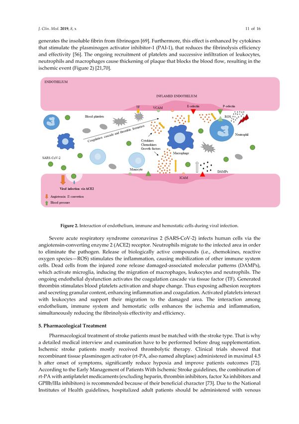
generates the insoluble fibrin from fibrinogen [69]. Furthermore, this effect is enhanced by cytokines
that stimulate the plasminogen activator inhibitor-1 (PAI-1), that reduces the fibrinolysis efficiency
and effectivity [56]. The ongoing recruitment of platelets and successive infiltration of leukocytes,
neutrophils and macrophages cause thickening of plaque that blocks the blood flow, resulting in the
ischemic event (Figure 2) [21,70].
Figure 2. Interaction of endothelium, immune and hemostatic cells during viral infection.
Severe acute respiratory syndrome coronavirus 2 (SARS-CoV-2) infects human cells via the
angiotensin-converting enzyme 2 (ACE2) receptor. Neutrophils migrate to the infected area in order to
eliminate the pathogen. Release of biologically active compounds (i.e., chemokines, reactive oxygen
species—ROS) stimulates the inflammation, causing mobilization of other immune system cells. Dead
cells from the injured zone release damaged-associated molecular patterns (DAMPs), which activate
microglia, inducing the migration of macrophages, leukocytes and neutrophils. The ongoing endothelial
dysfunction activates the coagulation cascade via tissue factor (TF). Generated thrombin stimulates
blood platelets activation and shape change. Thus exposing adhesion receptors and secreting granular
content, enhancing inflammation and coagulation. Activated platelets interact with leukocytes and
support their migration to the damaged area. The interaction among endothelium, immune system
and hemostatic cells enhances the ischemia and inflammation, simultaneously reducing the fibrinolysis
effectivity and efficiency.
5. Pharmacological Treatment
Pharmacological treatment of stroke patients must be matched with the stroke type. That is why
a detailed medical interview and examination have to be performed before drug supplementation.
Ischemic stroke patients mostly received thrombolytic therapy. Clinical trials showed that recombinant
tissue plasminogen activator (rt-PA, also named alteplase) administered in maximal 4.5 h after onset of
symptoms, significantly reduce hypoxia and improve patients outcomes [72]. According to the Early
Management of Patients With Ischemic Stroke guidelines, the combination of rt-PA with antiplatelet
medicaments (excluding heparin, thrombin inhibitors, factor Xa inhibitors and GPIIb/IIIa inhibitors)
is recommended because of their beneficial character [73]. Due to the National Institutes of Health
guidelines, hospitalized adult patients should be administered with venous thromboembolic events
(VTE) prophylaxis, if hematologic and coagulation parameters indicate the possibility of thrombotic
complications, or patients are at high risk of thromboembolic event. What is more, patients receiving
antiplatelet and anticoagulant therapies before COVID-19 diagnosis should continue the treatment.
However, available data are insufficient to recommend the use of thrombolytics and anticoagulant drugs.J. Clin. Med. 2020, 9, 2688 12 of 16
In the case of SARS-CoV-2 infection, there is no antiviral agent for COVID-19, however, several
medicaments, including Remdesivir, Chloroquine, Lopinavir, Rotinavir and other HIV Protease
Inhibitors, are evaluated as a potential antiviral drug (Table 1). Administration and selection of
anticoagulant or antiplatelet drug for COVID-19 patients should be always considered to potential
drug–drug interactions. For this reason, The University of Liverpool collated a list of drug interactions
for medical personnel [74].
Table 1. Potential antiviral drugs under evaluation for the treatment of coronavirus disease 2019
(COVID-19) [74].
Intravenous prodrug responsible for inhibiting viral replication via
Remdesivir
binding to the viral RNA polymerase.
Antimalarial drug, which inhibits the fusion of virus with host cell
membranes. In vitro studies showed that both drugs may block the viral
Chloroquine/Hydroxychloroquine transport from endosomes to endolysosomes, thus regulating the
releasement of viral genome. Chloroquine has an ability to inhibits
glycosylation of ACE2 receptor, thus interfering the viral linkage.
Lopinavir/Ritonavir inhibits the activity of proteases responsible for
Lopinavir/Ritonavir
replication of SARS-CoV-2.
6. Conclusions
Numerous studies showed that COVID-19 may cause thromboembolic complications, which lead
to many vascular disorders, including ischemic stroke. The rapidly growing number of case-reports
demonstrates the need for more detailed medical examination of patients, especially those with severe
infections. Oxygen and nutrient shortages caused by a viral infection, along with the release of cytokines
and chemokines, migration and influx of immune defense cells, their interactions with endothelium and
accumulation in the damaged area, activation of the coagulation system and generation of thrombus
result in many thromboembolic complications.
Author Contributions: R.S., J.S.-B. and M.B. conceived the figures, table and wrote the manuscript. All the
authors read and approved the final draft of the manuscript and agree to be personally accountable for their own
contributions. All authors have read and agreed to the published version of the manuscript.
Funding: This research received no external funding.
Acknowledgments: This work was supported by Grant 506/1136 from the University of Lodz
Conflicts of Interest: The authors declare no conflict of interest.
References
1. Li, H.; Liu, S.-M.; Yu, X.-H.; Tang, S.-L.; Tang, C.-K. Coronavirus disease 2019 (COVID-19): Current status
and future perspectives. Int. J. Antimicrob. Agents 2020, 55, 105951. [CrossRef]
2. Yuki, K.; Fujiogi, M.; Koutsogiannaki, S. COVID-19 pathophysiology: A review. Clin. Immunol. 2020,
215, 108427. [CrossRef]
3. Zheng, J. SARS-CoV-2: An emerging coronavirus that causes a global threat. Int. J. Biol. Sci. 2020, 16,
1678–1685. [CrossRef]
4. World Health Organization. Q&A on Coronaviruses (COVID-19). Available online: https://www.who.int/
emergencies/diseases/novel-coronavirus-2019/question-and-answers-hub/q-a-detail/q-a-coronaviruses#:
~{}:text=symptoms (accessed on 18 June 2020).
5. Tu, Y.F.; Chien, C.S.; Yarmishyn, A.A.; Lin, Y.Y.; Luo, Y.H.; Lin, Y.T.; Lai, W.Y.; Yang, D.M.; Chou, S.J.;
Yang, Y.P.; et al. A review of SARS-CoV-2 and the ongoing clinical trials. Int. J. Mol. Sci. 2020, 21, 2657.
[CrossRef]
6. Gandhi, M.; Yokoe, D.S.; Havlir, D.V. Asymptomatic transmission, the Achilles’ Heel of current strategies to
control Covid-19. N. Engl. J. Med. 2020, 382, 2158–2160. [CrossRef]J. Clin. Med. 2020, 9, 2688 13 of 16
7. McFadyen James, D.; Stevens, H.; Peter, K. The emerging threat of (Micro)Thrombosis in COVID-19 and its
therapeutic implications. Circ. Res. 2020, 127, 571–587. [CrossRef]
8. Oklu, R. Thrombosis. Cardiovasc. Diagn. Ther. 2017, 7, S131–S133. [CrossRef]
9. Connors, J.M.; Levy, J.H. COVID-19 and its implications for thrombosis and anticoagulation. Blood 2020, 135,
2033–2040. [CrossRef]
10. Mao, L.; Jin, H.; Wang, M.; Hu, Y.; Chen, S.; He, Q.; Chang, J.; Hong, C.; Zhou, Y.; Wang, D.; et al. Neurologic
manifestations of hospitalized patients with Coronavirus Disease 2019 in Wuhan, China. JAMA Neurol. 2020,
77, 683–690. [CrossRef]
11. Wu, Y.; Xu, X.; Chen, Z.; Duan, J.; Hashimoto, K.; Yang, L.; Liu, C.; Yang, C. Nervous system involvement
after infection with COVID-19 and other coronaviruses. Brain Behav. Immun. 2020, 87, 18–22. [CrossRef]
12. van Gorp, E.C.M.; Suharti, C.; ten Cate, H.; Dolmans, W.M.V.; van der Meer, J.W.M.; ten Cate, J.W.;
Brandjes, D.P.M. Review: Infectious diseases and coagulation disorders. J. Infect. Dis. 1999, 180, 176–186.
[CrossRef]
13. Venugopal, A. Disseminated intravascular coagulation. Indian J. Anaesth. 2014, 58, 603–608. [CrossRef]
14. Tang, N.; Li, D.; Wang, X.; Sun, Z. Abnormal coagulation parameters are associated with poor prognosis in
patients with novel coronavirus pneumonia. J. Thromb. Haemost. 2020, 18, 844–847. [CrossRef]
15. Lippi, G.; Plebani, M.; Henry, B.M. Thrombocytopenia is associated with severe coronavirus disease 2019
(COVID-19) infections: A meta-analysis. Clin. Chim. Acta Int. J. Clin. Chem. 2020, 506, 145–148. [CrossRef]
16. Beyrouti, R.; Adams, M.E.; Benjamin, L.; Cohen, H.; Farmer, S.F.; Goh, Y.Y.; Humphries, F.; Jäger, H.R.;
Losseff, N.A.; Perry, R.J.; et al. Characteristics of ischaemic stroke associated with COVID-19. J. Neurol.
Neurosurg. Psychiatry 2020, 91. [CrossRef]
17. Wright, F.L.; Vogler, T.O.; Moore, E.E.; Moore, H.B.; Wohlauer, M.V.; Urban, S.; Nydam, T.L.; Moore, P.K.;
McIntyre, R.C. Fibrinolysis shutdown correlates to thromboembolic events in severe COVID-19 infection.
J. Am. Coll. Surg. 2020. [CrossRef]
18. Ranucci, M.; Ballotta, A.; Di Dedda, U.; Bayshnikova, E.; Dei Poli, M.; Resta, M.; Falco, M.; Albano, G.;
Menicanti, L. The procoagulant pattern of patients with COVID-19 acute respiratory distress syndrome.
J. Thromb. Haemost. 2020, 18, 1747–1751. [CrossRef]
19. Magro, C.; Mulvey, J.J.; Berlin, D.; Nuovo, G.; Salvatore, S.; Harp, J.; Baxter-Stoltzfus, A.; Laurence, J.
Complement associated microvascular injury and thrombosis in the pathogenesis of severe COVID-19
infection: A report of five cases. Transl. Res. 2020, 220, 1–13. [CrossRef]
20. Carsana, L.; Sonzogni, A.; Nasr, A.; Rossi, R.; Pellegrinelli, A.; Zerbi, P.; Rech, R.; Colombo, R.; Antinori, S.;
Corbellino, M.; et al. Pulmonary post-mortem findings in a large series of COVID-19 cases from Northern
Italy. MedRxiv 2020. [CrossRef]
21. Frizzell, J.P. Acute stroke: Pathophysiology, diagnosis, and treatment. AACN Clin. Issues 2005, 16, 421–440.
[CrossRef]
22. Donkor, E.S. Stroke in the 21(st) Century: A snapshot of the burden, epidemiology, and quality of life.
Stroke Res. Treat. 2018, 2018, 3238165. [CrossRef] [PubMed]
23. Hankey, G.J. Stroke. Lancet 2017, 389, 641–654. [CrossRef]
24. Boehme, A.K.; Esenwa, C.; Elkind, M.S.V. Stroke risk factors, genetics, and prevention. Circ. Res. 2017, 120,
472–495. [CrossRef] [PubMed]
25. Musuka, T.D.; Wilton, S.B.; Traboulsi, M.; Hill, M.D. Diagnosis and management of acute ischemic stroke:
Speed is critical. CMAJ Can. Med. Assoc. J./J. De L’association Med. Can. 2015, 187, 887–893. [CrossRef]
26. Donkel, S.J.; Benaddi, B.; Dippel, D.W.J.; Ten Cate, H.; de Maat, M.P.M. Prognostic hemostasis biomarkers in
acute ischemic stroke. Arterioscler. Thromb. Vasc. Biol. 2019, 39, 360–372. [CrossRef]
27. Kanyal, N. The science of ischemic stroke: Pathophysiology and pharmacological treatment. Int. J. Pharma
Res. Rev. 2015, 4, 65–84.
28. Pawluk, H.; Woźniak, A.; Grześk, G.; Kołodziejska, R.; Kozakiewicz, M.; Kopkowska, E.; Grzechowiak, E.;
Kozera, G. The role of selected pro-inflammatory cytokines in pathogenesis of ischemic stroke.
Clin. Interv. Aging 2020, 15, 469–484. [CrossRef]
29. Strecker, J.-K.; Schmidt, A.; Schäbitz, W.-R.; Minnerup, J. Neutrophil granulocytes in cerebral
ischemia—Evolution from killers to key players. Neurochem. Int. 2017, 107, 117–126. [CrossRef]
30. Jin, R.; Yang, G.; Li, G. Inflammatory mechanisms in ischemic stroke: Role of inflammatory cells. J. Leukoc. Biol.
2010, 87, 779–789. [CrossRef]J. Clin. Med. 2020, 9, 2688 14 of 16
31. Bobryshev, Y.V. Monocyte recruitment and foam cell formation in atherosclerosis. Micron 2006, 37, 208–222.
[CrossRef]
32. Lindsberg Perttu, J.; Grau Armin, J. Inflammation and infections as risk factors for ischemic stroke. Stroke
2003, 34, 2518–2532. [CrossRef] [PubMed]
33. Kawabori, M.; Yenari, M.A. Inflammatory responses in brain ischemia. Curr. Med. Chem. 2015, 22, 1258–1277.
[CrossRef] [PubMed]
34. Rezaei, O.; Pakdaman, H.; Gharehgozli, K.; Simani, L.; Vahedian-Azimi, A.; Asaadi, S.; Sahraei, Z.;
Hajiesmaeili, M. S100 B: A new concept in neurocritical care. Iran. J. Neurol. 2017, 16, 83–89. [PubMed]
35. Bharosay, A.; Bharosay, V.V.; Varma, M.; Saxena, K.; Sodani, A.; Saxena, R. Correlation of Brain Biomarker
Neuron Specific Enolase (NSE) with degree of disability and neurological worsening in cerebrovascular
stroke. Indian J. Clin. Biochem. IJCB 2012, 27, 186–190. [CrossRef] [PubMed]
36. Oxley, T.J.; Mocco, J.; Majidi, S.; Kellner, C.P.; Shoirah, H.; Singh, I.P.; De Leacy, R.A.; Shigematsu, T.;
Ladner, T.R.; Yaeger, K.A.; et al. Large-vessel stroke as a presenting feature of Covid-19 in the young. N. Engl.
J. Med. 2020, 382. [CrossRef] [PubMed]
37. Merkler, A.E.; Parikh, N.S.; Mir, S.; Gupta, A.; Kamel, H.; Lin, E.; Lantos, J.; Schenck, E.J.; Goyal, P.;
Bruce, S.S.; et al. Risk of ischemic stroke in patients with Coronavirus Disease 2019 (COVID-19) vs Patients
with influenza. JAMA Neurol. 2020. [CrossRef] [PubMed]
38. Klok, F.A.; Kruip, M.J.H.A.; van der Meer, N.J.M.; Arbous, M.S.; Gommers, D.A.M.P.J.; Kant, K.M.;
Kaptein, F.H.J.; van Paassen, J.; Stals, M.A.M.; Huisman, M.V.; et al. Incidence of thrombotic complications in
critically ill ICU patients with COVID-19. Thromb. Res. 2020, 191, 145–147. [CrossRef] [PubMed]
39. Lodigiani, C.; Iapichino, G.; Carenzo, L.; Cecconi, M.; Ferrazzi, P.; Sebastian, T.; Kucher, N.; Studt, J.-D.;
Sacco, C.; Alexia, B.; et al. Venous and arterial thromboembolic complications in COVID-19 patients admitted
to an academic hospital in Milan, Italy. Thromb. Res. 2020, 191, 9–14. [CrossRef]
40. Yaghi, S.; Ishida, K.; Torres, J.; Mac Grory, B.; Raz, E.; Humbert, K.; Henninger, N.; Trivedi, T.; Lillemoe, K.;
Alam, S.; et al. SARS-CoV-2 and stroke in a New York Healthcare System. Stroke 2020, 51, 2002–2011.
[CrossRef]
41. Qin, C.; Zhou, L.; Hu, Z.; Yang, S.; Zhang, S.; Chen, M.; Yu, H.; Tian, D.-S.; Wang, W. Clinical characteristics
and outcomes of COVID-19 patients with a history of stroke in Wuhan, China. Stroke 2020, 51, 2219–2223.
[CrossRef]
42. Corman, V.M.; Lienau, J.; Witzenrath, M. Coronaviruses as the cause of respiratory infections. Internist 2019,
60, 1136–1145. [CrossRef] [PubMed]
43. Mousavizadeh, L.; Ghasemi, S. Genotype and phenotype of COVID-19: Their roles in pathogenesis.
J. Microbiol. Immunol. Infect. 2020, in press. [CrossRef] [PubMed]
44. Li, Y.-C.; Bai, W.-Z.; Hashikawa, T. The neuroinvasive potential of SARS-CoV2 may play a role in the
respiratory failure of COVID-19 patients. J. Med. Virol. 2020, 92, 552–555. [CrossRef] [PubMed]
45. Hamming, I.; Timens, W.; Bulthuis, M.L.C.; Lely, A.T.; Navis, G.J.; van Goor, H. Tissue distribution of ACE2
protein, the functional receptor for SARS coronavirus. A first step in understanding SARS pathogenesis.
J. Pathol. 2004, 203, 631–637. [CrossRef] [PubMed]
46. Ding, Y.; He, L.; Zhang, Q.; Huang, Z.; Che, X.; Hou, J.; Wang, H.; Shen, H.; Qiu, L.; Li, Z.; et al. Organ
distribution of severe acute respiratory syndrome (SARS) associated coronavirus (SARS-CoV) in SARS
patients: Implications for pathogenesis and virus transmission pathways. J. Pathol. 2004, 203, 622–630.
[CrossRef] [PubMed]
47. Gu, J.; Gong, E.; Zhang, B.; Zheng, J.; Gao, Z.; Zhong, Y.; Zou, W.; Zhan, J.; Wang, S.; Xie, Z.; et al. Multiple
organ infection and the pathogenesis of SARS. J. Exp. Med. 2005, 202, 415–424. [CrossRef] [PubMed]
48. Xu, J.; Zhong, S.; Liu, J.; Li, L.; Li, Y.; Wu, X.; Li, Z.; Deng, P.; Zhang, J.; Zhong, N.; et al. Detection of severe
acute respiratory syndrome coronavirus in the brain: Potential role of the chemokine MiG in pathogenesis.
Clin. Infect. Dis. 2005, 41, 1089–1096. [CrossRef]
49. Netland, J.; Meyerholz, D.K.; Moore, S.; Cassell, M.; Perlman, S. Severe acute respiratory syndrome
coronavirus infection causes neuronal death in the absence of encephalitis in mice transgenic for human
ACE2. J. Virol. 2008, 82, 7264–7275. [CrossRef]
50. Verdecchia, P.; Cavallini, C.; Spanevello, A.; Angeli, F. The pivotal link between ACE2 deficiency and
SARS-CoV-2 infection. Eur. J. Intern. Med. 2020, 76, 14–20. [CrossRef]You can also read