G9a histone methyltransferase plays a dominant role in euchromatic histone H3 lysine 9 methylation and is essential for early embryogenesis
←
→
Page content transcription
If your browser does not render page correctly, please read the page content below
G9a histone methyltransferase plays a
dominant role in euchromatic histone
H3 lysine 9 methylation and is essential
for early embryogenesis
Makoto Tachibana,1 Kenji Sugimoto,2 Masami Nozaki,3 Jun Ueda,1 Tsutomu Ohta,4 Misao Ohki,4
Mikiko Fukuda,1 Naoki Takeda,5,7 Hiroyuki Niida,5,8 Hiroyuki Kato,6 and Yoichi Shinkai1,9
1
 Department of Cell Biology, Institute for Virus Research, Kyoto University, Shogoin Kawara-cho, Kyoto 606-8507, Japan;
2
 Laboratory of Applied Molecular Biology, Department of Applied Biochemistry, Osaka Prefecture University, Osaka
599-8501, Japan; 3Research Institute for Microbial Disease, Osaka University, Osaka 565-0871, Japan; 4Medical Genomics
Center, National Cancer Center Research Institute, Tokyo 104-0045, Japan; 5Department of Oncology, Nippon Roche
Research Center, Kamakura 247-0063, Japan; 6National Institute of Infectious Diseases, Musashimurayama,
Tokyo 208-0011, Japan

Covalent modification of histone tails is crucial for transcriptional regulation, mitotic chromosomal
condensation, and heterochromatin formation. Histone H3 lysine 9 (H3-K9) methylation catalyzed by the
Suv39h family proteins is essential for establishing the architecture of pericentric heterochromatin. We
recently identified a mammalian histone methyltransferase (HMTase), G9a, which has strong HMTase activity
towards H3-K9 in vitro. To investigate the in vivo functions of G9a, we generated G9a-deficient mice and
embryonic stem (ES) cells. We found that H3-K9 methylation was drastically decreased in G9a-deficient
embryos, which displayed severe growth retardation and early lethality. G9a-deficient ES cells also exhibited
reduced H3-K9 methylation compared to wild-type cells, indicating that G9a is a dominant H3-K9 HMTase in
vivo. Importantly, the loss of G9a abolished methylated H3-K9 mostly in euchromatic regions. Finally, G9a
exerted a transcriptionally suppressive function that depended on its HMTase activity. Our results indicate
that euchromatic H3-K9 methylation regulated by G9a is essential for early embryogenesis and is involved in
the transcriptional repression of developmental genes.
[Key Words: Euchromatin; heterochromatin; histone H3-K9 methylation; G9a HMTase; mammalian
development; transcriptional regulation]
Received March 5, 2002; revised version accepted May 22, 2002.

In all eukaryotes, the covalent modification of histone                   also phosphorylated during different cellular processes.
N-terminal tails plays an important role in the regula-                   Phosphorylation of serine 10 in histone H3 (H3-S10) is
tion of transcription, mitosis, and heterochromatin for-                  required for proper chromosome condensation and seg-
mation. The “histone code” hypothesis (Strahl and Allis                   regation (Wei et al. 1999). The phosphorylation of H3-
2000; Jenuwein and Allis 2001) predicts that different                    S10 also correlates with transcriptional activation of im-
modifications of specific amino acids in histones or their                mediate-early genes upon mitogen stimulation (Mahad-
combinations are translated into functionally distinct ef-                evan et al. 1991). In addition to acetylation and
fects on nuclear processes. For example, transcription-                   phosphorylation, methylation of histone tails at differ-
ally active euchromatic regions are often associated with                 ent residues has been implicated in transcriptional regu-
hyperacetylated histones, while silent heterochromatic                    lation (Jenuwein and Allis 2001; Zhang and Reinberg
regions associate with hypoacetylated forms (Grunstein                    2001). It has been shown that the histone methyltrans-
1997). Besides acetylation, the histone H3 N terminus is                  ferase (HMTase) responsible for methylation of Arg 2,
                                                                          Arg 17, and Arg 26 in H3 (Ma et al. 2001; Xu et al. 2001)
                                                                          and Arg 3 in H4 (Strahl et al. 2001; Wang et al. 2001b)
Present addresses: 7Center for Animal Resources and Development, Ku-      plays an important role in the transcriptional activation
mamoto University, Kumamoto 860-0811, Japan; 8Department of Bio-
chemistry, Nagoya City University Medical School, Nagoya 467-8601,
                                                                          of certain genes. Methylation of Lys 4 in H3 (H3-K4)
Japan.                                                                    localizes to heterochromatin boundaries and transcrip-
9
 Corresponding author.                                                    tionally active loci (Litt et al. 2001; Noma et al. 2001). In
E-MAIL yshinkai@virus.kyoto-u.ac.jp; FAX 81-75-751-3991.
Article and publication are at http://www.genesdev.org/cgi/doi/10.1101/   contrast, current evidence suggests that H3 Lys 9 (H3-
gad.989402.                                                               K9) methylation is responsible for the creation of tran-

    GENES & DEVELOPMENT 16:1779–1791 © 2002 by Cold Spring Harbor Laboratory Press ISSN 0890-9369/02 $5.00; www.genesdev.org      1779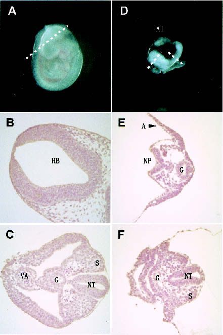
Tachibana et al.

scriptionally repressive heterochromatin (Rea et al.         absence of G9a protein (Fig. 1D, −/− lane; data not
2000). Pericentric heterochromatin contains enriched         shown). Therefore, it was likely that our G9a targeting
HP1 proteins from specific interaction between methyl-       results in a null mutation.
ated H3-K9 and the chromodomain of HP1 (Bannister et            Western blot analysis of wild-type cells detected two
al. 2001; Lachner et al. 2001). Clr4 in yeast (Nakayama et   distinct sizes (∼165 and 140 kD) of G9a proteins (Fig. 1D,
al. 2001) and Suv39h in mammals (Aagaard et al. 1999;        +/+ lane), neither of which was consistent with the pre-
O’Carroll et al. 2000), which are counterparts of Dro-       viously reported size of human G9a (112 kD) (Milner and
sophila melanogaster Su(var)3-9 protein, are major het-      Campbell 1993). Because of this discrepancy, we
erochromatic H3-K9 HMTases and play a dominant role          searched the EST and genomic DNA databases and iden-
in pericentric heterochromatin formation. The pericen-       tified partial DNA sequences that potentially encode
tric heterochromatin architecture also plays crucial roles   larger G9a molecules containing additional N-terminal
in chromosomal segregation as well as in establishing        peptides. Using this DNA sequence information, we
transcriptional repression. According to this notion, the    then successfully isolated cDNAs corresponding to these
loss of Suv39h HMTases abolished H3-K9 methylation           two isoforms, designated as G9a-S (GenBank accession
at pericentric heterochromatin, inducing chromosomal         no. AB077209) and G9a-L (GenBank accession no.
instability (Peters et al. 2001).                            AB077210). When these two cDNAs were introduced
   We previously provided evidence that G9a, a mamma-        into the G9a−/− ES cells, ␣-G9a antibodies clearly de-
lian HMTase, is a candidate for H3-K9 methylation in         tected proteins that corresponded in size to those de-
nonheterochromatic loci (Tachibana et al. 2001). In vitro    tected in wild-type cells (Fig. 1D, lanes −/−S and −/−L). It
experiments showed that G9a had a 10- to 20-fold stron-      was also revealed that human G9a exists as these two
ger HMTase activity toward H3-K9 compared to                 splicing variants (data not shown). A similar conclusion
Suv39h1, and also that it could methylate Lys 27 of H3       was recently reached by others (Brown et al. 2001).
(H3-K27) as well as K9, whereas the targeting site on H3
for Suv39h1 was solely Lys 9. Localization analysis of
                                                             Lethality and severe growth/developmental defect of
G9a in nuclei suggested that this HMTase might target
                                                             G9a-deficient embryos
sites at transcriptionally active euchromatin rather than
repressive pericentric heterochromatin. To investigate       Mouse lines derived from two independent G9a+/− ES
the in vivo function(s) of G9a, we generated G9a-defi-       cell lines (#36 and #64) were used for phenotypic analy-
cient mice and ES cells. Here, we show that G9a is es-       ses. G9a+/− mice were indistinguishable from their wild-
sential for early embryonic development and plays a          type littermates. The G9a+/− mice were intercrossed and
dominant role in H3-K9 methylation of euchromatin.           the offspring were genotyped by Southern blot hybrid-
Moreover, G9a can exert transcriptional repression in        ization. Among over 200 liveborn offspring analyzed
vitro, a function that depends on its HMTase activity.       from each line, no homozygous mutant animals were
Our data suggest that the euchromatic H3-K9 methyl-          found, strongly suggesting embryonic lethality (data not
ation regulated by G9a is involved in the transcriptional    shown). To determine the time of lethality, embryos of
silencing of developmentally regulated genes.                heterozygous mating were isolated at days 8.5 (E8.5) to
                                                             E12.5 of gestation. We occasionally found G9a−/−-geno-
                                                             typed yolk sacs without an embryo proper or completely
Results                                                      resorbed embryos at E9.5–E12.5, indicating that the
                                                             G9a−/− embryos likely died within this time frame
Targeted disruption of G9a gene in mouse germ line
                                                             (Table 1). Body weights of live G9a−/− embryos at E8.5
G9a genes were mapped within the major histocompat-          were already mildly reduced, and no further growth was
ibility complex class III region in mouse and human          observed at E9.5–E12.5 (Table 2). Since the targeted ES
(Brown et al. 2001). The murine G9a gene was disrupted       cell line, TT2, is an F1 hybrid of C57BL/6 and CBA,
by homologous recombination in embryonic stem (ES)           G9a+/− mice were backcrossed sequentially with
cells using a conventional targeting approach that re-       C57BL/6 to minimize mixed genetic background influ-
places the glutamic acid stretch, adjacent cysteine-rich     ences, and offspring of F9 or later generations were used
region, and ankyrin repeats with the Pgk-neomycin gene       in further studies.
(Fig. 1A). Successfully targeted ES cell clones were iso-       The gross morphology and histological analysis of
lated and used to generate chimeric mice that transmit-      typical G9a−/− embryos at E9.5 are shown in Figure 2A–
ted the mutated allele of G9a through the germ line. ES      F. The allantois (Al) extended into the exocoelom, mak-
cells homozygous for the G9a mutation (G9a−/−) were          ing contact with the chorion. However, the fusion was
also obtained from the heterozygous mutant (G9a+/−)          incomplete and the allantois could be readily detached.
cells by high G418 selection (Fig. 1B). No transcripts       G9a−/− embryos at E9.5 developed to only the 5–6 somite
were detected with a probe corresponding to the deleted      stage (Fig. 2D) as opposed to 21–25 somites in wild-type
ankyrin repeats, but several truncated and very faint        siblings (Fig. 2A). The neural groove was unfused at the
transcripts were detectable with the G9a SET-domain          anterior region and was closed at the tail region. Cardio-
probe in the G9a−/− ES cells (asterisks in Fig. 1C). West-   genesis was initiated but the primitive blood vessels,
ern blot analysis with specific antibodies against the de-   which extended from the cephalic region to the most
leted and undeleted region of C terminus confirmed the       caudal region of these embryos, contained no primitive

1780       GENES & DEVELOPMENT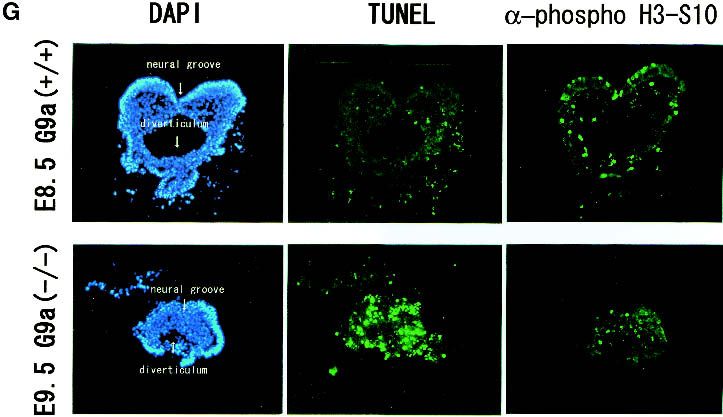
G9a, euchromatic H3-K9 HMTase

Figure 1. Targeting and genotyping of G9a-mutant mice. (A) Schematic representation of the mouse G9a gene locus, the G9a
targeting construct, and the targeted allele. Exons are numbered and indicated by black boxes. Two splicing variants of the murine G9a
(mG9a-S and mG9a-L) are presented at the top. The G9a targeting construct was designed to delete exons 5 to HindIII site of 21, which
correspond to the glutamic acid stretch to ankyrin (ANK) repeats. R, EcoRI; D, DraI (not unique in the 14-kb EcoRI fragment); N, NotI;
H, HindIII; B, BamHI. (B) Southern blot of EcoRI-digested DNA isolated from wild-type, G9a+/−, and G9a−/− ES cells. (C) Northern blot
analyses of G9a−/− ES cells. ANK and SET probes correspond to the region encoding for the ankyrin-repeats and the SET-domain,
respectively. (D) Western blot analyses of G9a expression in wild-type and G9a−/− ES cells and G9a−/− ES cells stably expressing cDNA
for the G9a-S or G9a-L isoform.

nucleated red blood cells. Gut (G) diverticulum was well              G9a−/− embryos resembled those of E8.0–E8.5 wild-type,
defined in the fore and hind gut regions in embryos.                  and no organ-specific abnormalities were recognized.
These morphological characteristics observed in E9.5

                                                                      Table 2. Growth arrest of the G9a-mutant embryos
Table 1. Embryonic lethality of the G9a-mutant mice
                                                                                                            Genotype
                             Genotype
                                                                      Embryonic day          +/+               +/−               −/−
Embryonic day       +/+      +/−        −/−         Resorption
                                                                      E8.5               0.7 ± 0.4 (9)     0.7 ± 0.1 (8)    0.4 ± 0.1 (7)
E8.5                 9       25          10              0            E9.5               3.9 ± 0.4 (8)     3.4 ± 0.7 (11)   0.4 ± 0.1 (7a)
E9.5                11       19      8 (7a + 1b)         3            E10.5             19.7 ± 2.1 (8)    17.7 ± 1.7 (6)    0.5 ± 0.2 (3)
E10.5               10       21      6 (4a + 2b)         6            E11.5             53.0 ± 4.2 (9)    56.1 ± 3.0 (3)       0.6 (1)
E11.5               11       21      4 (1a + 3b)         6
E12.5                7       15           1b             4            Values represent average wet weight (mg) of whole embryo with
                                                                      yolk sac. (n) Represents the examined sample number.
a                                                                     a
Severely growth-retarded embryo.                                       Among seven embryos, three embryos were male and the oth-
b
No embryo proper and only yolk sack remained.                         ers were female, which were equally growth-retarded.

                                                                                                   GENES & DEVELOPMENT                 1781
1782
Tachibana et al.
GENES & DEVELOPMENT
Figure 2. G9a−/− embryos were delayed in their development.
(A) The wild-type littermate at E9.5. (D) The lateral view of
G9a−/− embryo at E9.5. The approximate levels of the section
presented in B, C, E, and F are indicated as dashed lines. Allan-
tois (Al) develops normally. (B,C) Section through hindbrain
(HB in B) to tail region (C). (E,F) Section through forebrain to tail
region. S, somites; NT, neural tube; G, gut; VA, vitelline artery;
A, amnion; NP, neural plate. (G) TUNEL analysis and ␣-phos-
pho H3-S10 staining of E9.5 G9a−/− (bottom) and the E8.5 wild-
type embryos (top) on transversal sections. (H) Growth charac-
teristics of G9a−/− PEFs. Here, 104 cells of PEFs derived from individual E9.0 embryos were cultured, and population doublings (PDL) were monitored. (I) Measurement of DNA
content of G9a−/− PEFs. The wild-type and G9a−/− PEFs prepared from E9.5 embryos were cultured for 16 h and the DNA content was subsequently visualized by PI staining.
Calculated populations (%) at each cell cycle stage are represented below. (J) Growth defect of the G9a-mutant ES cells in differentiation conditions. Here, 105 cells of each ES
cell line were cultured in ES medium (upper) or RA (1 µM), no LIF-containing differentiation medium (lower). Vertical bars represent the cell numbers after 4 d of culture. Typical
data were picked up from triplicate independent reproducible examinations.G9a, euchromatic H3-K9 HMTase
Several mitotic cells were observed as well as wild-type Loss of H3-K9 methylation leads to accumulation of
embryos. acetylated H3-K9 and methylated H3-K4
To further examine the cause of G9a−/− embryonic
growth arrest, we performed immunohistochemical To address the issue of how G9a exerts its HMTase ac-
analysis for apoptotic cell death with TdT-mediated tivity in vivo, we examined the covalent modification
dUTP nick end labeling (TUNEL) on sections of E9.5 status of H3 N-terminal tails in G9a−/− cells. As shown
G9a−/− and E8.5 wild-type embryos. Typical transverse in Figure 3A, Western blot analysis using ␣-dimethyl H3-
sections are shown in Figure 2G. Massive TUNEL-posi- K9 antibodies demonstrated that dimethylated H3-K9 in
tive cells were readily detected in G9a−/− embryos, the G9a−/− embryos at E9.5 was drastically decreased. A
whereas very few cells were positive in wild-type em- reduction of dimethyl H3-K9 was also confirmed in two
bryos. Since phosphorylation of H3-S10 is tightly corre- G9a−/− ES cell lines (Fig. 3B; data not shown). The defect
lated with chromosome condensation during mitosis, we was completely rescued by the expression of exogenous
also examined the population of mitotic-stage cells in G9a protein (Fig. 3B, right lane). No significant alteration
G9a−/− embryos. As seen in wild-type embryos, in which of dimethyl H3-K9 status was detected between G9a+/+
small populations of nuclei were brightly stained, some and G9a+/− in embryos and ES cells (data not shown).
mitotic cells also existed in G9a−/− embryos, and this Estimation by serial dilution analysis indicated that the
population was not dissimilar to those in wild-type em- amount of dimethyl H3-K9 in G9a−/− ES cells was com-
bryos. The feature of a drastic accumulation of apoptotic parable to only one-eighth of that in wild-type ES cells
cells and the constant existence of mitotic cells in (Fig. 3C), which is much greater than the 50% loss of
G9a−/− embryos was commonly observed in all sections H3-K9 methylation in Suv39h mutant cells (Maison et
examined. al. 2002).
To determine whether the growth defect of G9a−/− em- It has been shown that H3-K9 methylation can influ-
bryos was due to extra-embryonic defects, we examined ence other modifications of the neighboring residues in
the growth potential of G9a−/− primary embryonic fibro- H3 tails, such as phosphorylation and acetylation. Meth-
blasts (PEFs) in vitro. Similar to the embryonic pheno- ylated H3-K9 can inhibit the phosphorylation of H3-S10
type, PEFs prepared from the G9a−/−embryos at E9.0 by Ipl1/aurora (Rea et al. 2000). In addition, S10 phos-
showed severe growth defects, whereas the G9a+/− PEFs phorylation facilitates the acetylation of Lys 14 in H3
were indistinguishable from wild-type PEFs (Fig. 2H), (H3-K14) by the histone acetyltransferase GCN5
suggesting that the G9a−/− embryo proper had intrinsic (Cheung et al. 2000). Thus, we investigated the acetyl
growth defects. Next, we examined the DNA content of status of H3-K9 and H3-K14, and the phosphorylation
the PEFs cultured for 16 h in vitro. As illustrated in Fig- status of H3-S10 in G9a−/− ES cells (Fig. 3D). H3-S10
ure 2I, a significant increase in cells with lower DNA phosphorylation and H3-K14 acetylation were indistin-
contents compared to those of typical G1 phase was ob- guishable in wild-type and G9a−/− ES cells. In contrast,
served in G9a−/− PEFs, whereas G1 to G2/M ratios were H3-K9 acetylation was increased about twofold in
indistinguishable between G9a−/− and wild-type. Similar G9a−/− ES cells, suggesting the existence of competition
results were obtained from five independently prepared between methylation and acetylation at H3-K9 in vivo.
lines of G9a−/− PEFs. Therefore, these in vivo and in vitro In addition to these modifications, methylated H3-K4
data suggest that the G9a−/− embryonic growth defect is, has been shown to localize to transcriptionally active
at least in part, due to apoptotic cell death but not cell chromatin (Litt et al. 2001; Noma et al. 2001) and sup-
cycle arrest. press H3-K9 methylation in vitro (Wang et al. 2001a). As
We used the G9a−/− ES cells for further analyses of G9a shown in Figure 3D, we found a two- to threefold in-
function. As shown in Figure 2J, no obvious growth crease in H3-K4 methylation, a result that further indi-
defects were observed for G9a−/− ES cells during their cates the presence of a functional competition between
maintenance in cell culture (Fig. 2J, upper panel). To H3-K4 and H3-K9 methylation in vivo.
investigate whether G9a has an important function(s) To date, the catalytically defined set of H3-K9 HMTases
in more differentiated cells, G9a−/− ES cells were in- in mammals consists of Suv39h (1 and 2) (O’Carroll et al.
duced to differentiate by being cultured with all-trans 2000; Rea et al. 2000), ESET (Schultz et al. 2002; Yang et
retinoic acid (RA) in the absence of leukemia inhibitory al. 2002), and G9a. The dominant contribution of G9a to
factor (LIF). With this treatment, G9a−/− ES cells ex- H3-K9 methylation in vivo predicts that total HMTase
hibited a distinct growth defect (Fig. 2J, bottom panel). activity toward H3-K9 might be reduced in G9a−/− cells.
These data indicate that, while G9a function may be In addition, the recombinant G9a HMTase domain could
dispensable for the survival of undifferentiated cells (e.g., transfer methyl groups H3-K27 as well as H3-K9 in vitro
epiblasts), it is crucial for more differentiated somatic (Fig. 3E; Tachibana et al. 2001). Thus, we investigated
cells. Exogenous introduction of G9a-S or G9a-L mol- the HMTase activities toward H3-K9 and H3-K27 in
ecules restored normal growth potential to G9a−/− ES G9a−/− ES cells. Surprisingly, nuclear extracts from mu-
cells during differentiation. This feature was confirmed tant cells retained HMTase activity toward both H3-K9
by triplicate independent examination. The limited and H3-K27. The activity in G9a−/− ES cells was compa-
growth defect of differentiating G9a−/− ES cells accords rable to levels observed in wild-type cells (Fig. 3F). These
well with that of G9a−/− somatic cells during embryo- data suggest that, while many proteins exhibit HMTase
genesis. activities toward H3-K9 and H3-K27 in vitro, only a sub-
GENES & DEVELOPMENT 1783Tachibana et al.
Figure 3. (A–D) Alteration of G9a-dependent covalent H3 modifications detected by Western blot analyses. (A) Purified and pre-
calibrated histones with ␣-H3 (upper panel) from the G9a+/− and G9a−/− embryos were stained with ␣-dimethyl H3-K9 antibodies. (B)
␣-dimethyl H3-K9 staining of H3 from wild-type and G9a−/− ES cells and G9a−/− ES cells expressing exogenous G9a-L. (C) Calibration
of the dimethyl H3-K9 content in G9a−/− ES cells. The ratio of H3 content between wild-type and mutant were precalibrated and are
represented below. (D) Other covalent modification statuses of H3 between wild-type and G9a−/− ES cells. All the presented data
shown in Fig. 2A–D were reproducible at least twice. (E) HMTase activity of G9a-HMTase domain. Amino acid sequences of GST-
fused recombinant H3 N terminus protein as substrates are at the top. Recombinant G9a-HMTase domain could add methyl groups
to H3-K9 and H3-K27. (F) H3 HMTase activity in wild-type and G9a−/− ES cells. HMTase activity and substrate specificity of nuclear
extracts of wild-type and G9a−/− ES cells were indistinguishable. HMTase activities of the nuclear extracts toward Lys 4, Lys 9, and
Lys 27 were calibrated as Nx–NT.
set may function in vivo (e.g., Suv39h and G9a). It is ited a nuclear-specific pattern that was broad in inter-
likely that other lysine methyltransferases are targeted phase nuclei but almost excluded from nucleoli. Impor-
to proteins other than histones in vivo (Jenuwein 2001). tantly, the intensity of EGFP-G9a signals was reduced
significantly at pericentric heterochromatin regions,
which were DAPI-densely stained and accumulated with
Global loss of euchromatic H3-K9 methylation in
HP1 molecules (arrows). The localization profiles were
G9a−/− cells
indistinguishable between G9a-S and G9a-L molecules
Previously we showed that a truncated human G9a mol- (data not shown). These observations again suggest that
ecule (Milner and Campbell 1993) displayed a nuclear the localization profiles of G9a and pericentric hetero-
localization profile that was quite different from that of chromatin-associated Suv39h were quite distinct and
Suv39h1 (Tachibana et al. 2001). We therefore reinvesti- mutually exclusive.
gated the cellular localization profiles of fluorescent pro- To further investigate the contributions of G9a to
tein (EGFP)-tagged full-length G9a-S and G9a-L in mu- functionally different chromatic regions, we performed
rine fibroblasts with DsRed-tagged human HP1 mol- immunohistochemical analyses of methylated H3-K9 in
ecules, which were shown to localize in pericentric wild-type and G9a−/− ES cells. Dimethylated H3-K9 was
heterochromatin and be involved in SUV39H1 complex broadly detected in wild-type nuclei, indicating that
(Fig. 4A; Aagaard et al. 1999). EGFP-G9a proteins exhib- methylated H3-K9 might exist in many loci, including
1784 GENES & DEVELOPMENTG9a, euchromatic H3-K9 HMTase
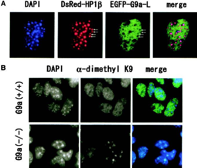
Figure 4. (A) Nuclear localization profiles of transiently expressed EGFP-tagged mouse G9a molecules (G9a-L) and DsRed-tagged
human HP-1 molecules in the murine fibroblast cell line C3H10T 1/2 . EGFP-G9a-L molecules exist broadly in interphase nuclei and
mostly excluded from HP-1-enriched pericentric heterochromatin (arrows). (B) Immunohistochemical analyses of G9a−/− ES cells
with ␣-dimethyl H3-K9. Nuclei of G9a−/− ES cells contained dimethyl H3-K9 only in DAPI-dense regions, whereas these were broadly
distributed to both euchromatic and heterochromatic loci in wild-type nuclei.
eu- and heterochromatic regions (Fig. 4B, upper panels). fused with full-length mG9a-L (PM-G9a-L) or an
However, the G9a−/− cell nucleus contained only large HMTase domain of G9a (residues 969V-stop) (PM-HMT)
speckles of methylated H3-K9, which were completely into HeLa S2 cells (Fig. 5A,B). We also expressed a GAL4-
overlapping with the DAPI-dense loci (Fig. 4B, bottom DBD fused with a dead HMTase full-length mG9a-S
panels). This result fits well with the observation that (PM-G9a-S⌬NHLC, deletion of 1165NHLC1168 of the
ectopically expressed G9a molecules were excluded from HMTase domain) or a dead HMTase domain alone (PM-
HP1-enriched pericentric heterochromatin. It is highly ⌬NHLC). Transcription of the reporter gene was signifi-
likely that methylation of H3-K9 in the pericentric het- cantly inhibited by PM-G9aL and PM-HMT, but not by
erochromatin of G9a−/− cells is catalyzed mainly by PM-G9a-S⌬NHLC or PM-⌬NHLC (Fig. 5C). A second
Suv39h HMTases (Peters et al. 2001). Considering that G9a truncated mutant with crippled HMTase function
H3 in G9a−/− ES cells possessed only one-eighth the (1162R to H substitution) also failed to repress reporter
amount of dimethylated H3-K9 compared to that of gene transcription (data not shown). These results sug-
wild-type cells, we conclude that G9a is the major in gest that G9a functions as a negative regulator of tran-
vivo H3-K9 HMTase that directs methylation of euchro- scription through its HMTase activity. To evaluate the
matic regions. involvement of histone deacetylases (HDACs) in the ob-
served transcriptional repression, we performed the ex-
periments in the presence of trichostatin A (TSA), an
G9a exerts a transcriptionally suppressive function
inhibitor of HDACs (Fig. 5D). Acetylation of total H3-K9
dependent on its HMTase activity
was substantially enhanced by the TSA treatment, but
To assess whether G9a-mediated histone methylation the transcriptional repression was unaffected. Together,
functions in transcriptional regulation, we performed re- these data indicate that the G9a HMTase-mediated re-
porter gene assays using pGL3-G5pol, which contains a pression of gene transcription occurs in a manner inde-
luciferase gene driven by a DNA pol  promoter proxi- pendent of the HDAC pathway.
mal to GAL4-binding sites (Sekimata et al. 2001). We To further investigate the G9a function in transcrip-
transiently introduced pGL3-G5pol and a construct ex- tional regulation, we performed oligonucleotide micro-
pressing a GAL4 DNA-binding domain (GAL4-DBD) array analyses using G9a+/+, G9a−/− ES cells, and G9a−/−
GENES & DEVELOPMENT 1785Tachibana et al. Figure 5. HMTase-dependent transcriptional repression mediated by G9a. (A) Schematic representation of the used plasmids for in vitro luciferase assays. (B) Protein expression analysis of the GAL4-DBD-fused constructs described in A. Fusion molecules were detected by ␣-GAL4-DBD. (C) Full-length G9a (PM-G9a-L) and a G9a HMTase domain (PM-HMT) suppress transcription, but dead HMTase full-length G9a (PM-G9a-S⌬NHLC ) and a G9a dead HMTase domain (PM-⌬NHLC) cannot. All the transient luciferase assays were performed multiple times and the results presented were reproduced. (D) The G9a-mediated suppression was not relieved in the presence of TSA (100 ng/mL). Upper left panel shows an accumulation of H3-K9 acetylation by the TSA treatment. ES cells expressing exogenous G9a (T. Ohta, M. (2–3+mock1 and 2). Nucleotide sequencing of the RT- Tachibana and Y. Shinkai, unpubl.). We identified some PCR products demonstrated that Mage-a2, Mage-a6, and candidate genes transcription-regulated by G9a, and one Mage-a8 were expressed in G9a−/− ES cells (data not of them was a Mage-a gene(s). Human MAGE genes have shown). To further evaluate the Mage-a genes as direct been isolated as tumor-specific antigen genes (Boon et al. targets of G9a, we performed a chromatin immunopre- 1994). Eight murine Mage-a genes (Mage-a1 to Mage-a8, cipitation (ChIP) analysis. As illustrated in Figure 6B, highly homologous each other) have been identified, dimethylated H3-K9 was enriched in chromatin contain- with some of them being expressed not only in tumor ing the Mage-a2 promoter sequences in G9a+/+ (TT2) and cell lines but also in testis and blastocysts (De Plaen et severely decreased in G9a−/− ES cells (22–10) (about one- al. 1999). The function of Mage-a genes is currently un- fourth of TT2, Fig. 6B, lanes 5 and 10). In contrast with known. As shown in Figure 6A, the expression of Mage-a H3-K9 methylation, dimethylated H3-K4 was enriched gene(s) was induced in G9a−/− ES cells (2–3 and 22–10) in the Mage-a2 promoter region of 22–10 (at least 10-fold and suppressed by expression of exogenous G9a (2–3+L enrichment in 22–10 compared to TT2, Fig. 6B, lanes 3 and +S) but not by a drug-selectable molecule alone and 11). Exogenous expression of G9a in G9a−/− ES cells 1786 GENES & DEVELOPMENT
G9a, euchromatic H3-K9 HMTase

Figure 6. Characterization of Mage-a genes. (A) RNA expression of Mage-a genes. Six micrograms of total RNA prepared from G9a+/+
(TT2), G9a+/− (#36), G9a−/− (2–3 and 22–10), 2–3-expressing exogenous G9a-L (2–3-L), G9a-S (2–3-S), or a drug-selectable molecule alone
(2–3+mock1 and 2) were separated, blotted to a nylon membrane, and hybridized with a 32P-labeled Mage-a8 cDNA. Expression of
Mage-a2, Mage-a6, and Mage-a8 genes was determined in G9a−/− ES cells by nucleotide sequencing of the RT-PCR products. The lower
panel shows 28S rRNA stained with ethidium bromide in the same gel before blotting. (B) ChIP analyses on the Mage-a2 promoter
region. Formaldehyde cross-linked chromatin from TT2, 22–10, and 15–3 (2–3-S) ES cells was immunoprecipitated without (−) or with
␣-dimethyl H3-K9 (K9), ␣-dimethyl H3-K4 (K4), or ␣-acetyl-H3-K9+14 (right panel). An equal amount of Mage-a2 promoter sequence
in TT2, 22–10, and 15–3 nucleosomal preparations was determined by PCR from 1.4% of the input ChIP/ PCR reaction (lanes 12–14).
The linearity of this PCR was confirmed by serial dilution of the TT2 input DNA (lanes 12,15–17). The product size of the Mage-a2
PCR is 242 bp. Similar results were obtained with multiple independent experiments.

(15–3) reverted the status of H3-K9 and H3-K4 dimeth-                 chromosomes (Xi) in females (Boggs et al. 2002; Heard et
ylation to the G9a+/+ ES cell level. Furthermore, acety-              al. 2001). This modification occurs by an Suv39h-HP1-
lation of H3-K9+K14 at this region was increased signifi-             independent pathway, because Suv39h-mutant female
cantly in G9a−/− ES cells (Fig. 6B, right panel). These data          cells still possess methylated H3-K9-rich Xi (Peters et al.
strongly suggest that Mage-a2 gene is silenced by G9a-                2002). We considered the possibility that H3-K9 methyl-
mediated H3-K9 methylation in ES cells.                               ation by G9a is responsible for determining Xi formation.
                                                                      If this model is correct, female G9a−/− mice would pos-
                                                                      sess two active X chromosomes, which is known to in-
Discussion                                                            duce female-specific lethality at a very early develop-
                                                                      mental stage (Marahrens et al. 1997). However, the de-
Embryonic lethality in G9a-mutant mice                                velopmentally retarded phenotypes of G9a−/− male and
The early embryonic lethality of G9a−/− mice (E9.5–                   female embryos at E9.5 were indistinguishable, as shown
E12.5) is distinct from Suv39h1/2 double mutant mice,                 in Table 2. Thus, this possibility is still open to discus-
which are born at sub-Mendelian ratios (Peters et al.                 sion, but defective Xi formation cannot explain male em-
2001). This finding suggests that Suv39h and G9a                      bryonic lethality in G9a-deficient mice.
HMTase contribute nonoverlapping roles to embryonic
growth/development. The developmental arrest of G9a−/−
                                                                      Histone methylation of euchromatin
embryos at ∼E8.0 coincides with the dramatic reorgani-
zation of somatic tissues, which is accompanied by                    The broad methylation patterns of H3-K9 in the nucleus
widespread alterations in gene expression profiles and                suggest that its functions are not restricted to pericentric
chromatin organization. Thus, it is probable that G9a-                heterochromatin organization in mammals. In G9a-defi-
mediated methylation of euchromatin is a key compo-                   cient cells, the broad methylation of chromatin was
nent of the mechanism that regulates gene expression                  abolished. In contrast, Suv39h1/2 double mutant mice
during this stage of embryogenesis. Apoptotic cells were              retain broader methylation of H3-K9, but they lose
increased drastically in growth-arrested G9a−/− embryos,              methylation at pericentric heterochromatic regions (Pe-
and this seemed to be the dominant cause of embryonic                 ters et al. 2001). Taken together, these findings strongly
growth retardation. Interestingly, they still possessed               suggest that G9a and Suv39h HMTases have nonover-
significant populations of mitotic cells. G9a−/− ES cells             lapping functions and target distinct chromosomal loci.
also displayed growth defects under conditions of cellu-              Comparisons of Suv39h and G9a protein sequences re-
lar differentiation, but not during routine maintenance.              veal high similarities in the HMTase enzymatic regions.
These facts suggest that G9a is dispensable for simple                However, other regions are quite divergent (e.g., chromo-
proliferating processes but is necessary for some impor-              domain for Suv39h and ankyrin-repeats for G9a). Thus, it
tant events during embryonic development or differen-                 is likely that the unique molecular domains of Suv39h
tiation.                                                              and G9a are responsible for targeting their HMTase func-
   Recent studies have shown that methylation marking                 tions to pericentric heterochromatin and euchromatin,
on H3-K9 is specifically targeted to the inactivated X                respectively.

                                                                                                   GENES & DEVELOPMENT            1787
Tachibana et al.
The HP1 protein is also involved in transcriptional can be translated into different biological outputs in a
silencing and modulation of chromatin architecture manner dependent upon their chromosomal locations.
(Jones et al. 2000). The chromodomain of HP1 exhibits
high affinity for methylated H3-K9 (Bannister et al. 2001;
Lachner et al. 2001), which recruits HP1 to pericentric Materials and methods
heterochromatin regions following Suv39h-mediated
H3-K9 methylation. We speculate that there are two pos- Generation and genotyping of the G9a-mutant embryos and
sible functions of G9a-mediated H3-K9 methylation at ES cells
euchromatin: (1) G9a creates a local heterochromatic ar- A partial cDNA clone for the mouse G9a gene was isolated from
chitecture in euchromatic regions via interactions with a Uni-ZAP XR mouse testis library (Stratagene) using a radiola-
methylated H3-K9 and subpopulations of the HP1 pro- beled human counterpart cDNA (Milner and Campbell 1993) as
tein. Indeed, recent studies indicate that HP1 might play a probe. The isolated mouse G9a cDNA fragment was used as a
a crucial role in creating local heterochromatic domains probe to screen a C57 Black/6 mouse genomic library (Strata-
to establish a silent state (Matsuda et al. 2001; Ogawa et gene). To make a G9a targeting construct, a 4.5-kb genomic
al. 2002); (2) Since G9a also methylates H3-K27 in vitro, fragment, a region from the HindIII site in exon 21 to the BamHI
H3 molecules carrying doubly methylated lysine resi- site downstream of exon 27, was inserted into the modified SalI
site of pLNTK, and then a 0.75-kb DraI-BamHI genomic frag-
dues may recruit distinct factors that further influence
ment located in the intron between exon 4 and 5 was further
chromatin structure and gene regulation. subcloned. The G9a targeting construct potentially replaces ex-
ons 5-part21 of mouse G9a with the Pgk-neomycin gene.
Next, 1 × 10⁷ ES cells, TT2 line (Yagi et al. 1993) were trans-
fected with 20 µg of NotI-linearized G9a-targeting construct and
G9a functions as transcriptional repression
selected in ES cell medium containing 0.25 mg/mL G418 and
Existing data support the notion that H3-K9 methylation 1.5 µg/mL ganciclovir. Homologous recombinant cells (#36 and
may contribute to transcriptional repression (Firestein et #64) were identified by Southern blot analysis of EcoRI-digested
al. 2000; Litt et al. 2001; Nakayama et al. 2001; Nielsen DNA probed with an exon 28-containing 1.1-kb BamHI geno-
et al. 2001; Noma et al. 2001; Vandel et al. 2001). Data of mic DNA fragment and injected into the morula stage of ICR
mouse embryos. Established chimeric male mice derived from
our reporter gene assays imply that G9a exerts a tran-
both clones successfully generated F1 offspring carrying the mu-
scriptional repression function in vivo and it is depen- tated G9a allele.
dent on its HMTase activity. These data strongly suggest PCR genotyping of the G9a-mutant mice was carried out us-
that one important function of G9a is gene silencing me- ing primers external to the short arms of the targeting vector
diated by H3-K9 methylation in euchromatic regions. (G3, 5⬘-GGGCCAGCTCATTCCTCCACTC-3⬘; mG9a127, 5⬘-
The expression profiles of Mage-a2, Mage-a6, and Mage- GCAGATGTGATGGCTTGGGGTAG-3⬘). G9a+/− mice were
a8 genes and the dimethylation status of H3-K9 in chro- sequentially backcrossed with the C57BL/6 strain mice, and
matin containing the Mage-a2 promoter sequences in offspring of F9 or later generation animals were used for further
G9a+/+ and G9a−/− ES cells clearly support this notion. studies. Two independent lines of the G9a homozygous recom-
To further elucidate the physiological function(s) of binant (G9a−/−) ES cells were isolated from line #36 line selected
in G418 (3.0 mg/mL)-containing medium.
HMTase G9a, a crucial next step will be the identifica-
tion and characterization of entire target genes.
In contrast to H3-K9 methylation, transcriptionally ES cell culture and transfection
active chromatic regions are associated with methylated Undifferentiated ES cells were maintained in 10% fetal calf se-
H3-K4 (Litt et al. 2001; Noma et al. 2001). Furthermore, rum and LIF (500 µg/mL)-containing medium. For the growth
preexisting H3-K4 methylation inhibits methylation of assays of the retinoic-acid (RA)-induced differentiated cells,
H3-K9 in vitro and vice versa (Wang et al. 2001a). A cells were cultured in the presence of 1 µM all-trans-RA with-
significant increase in methylated H3-K4 in G9a−/− cells out LIF. Full-length murine G9a cDNA was isolated from a
was observed (Figs. 3D and 6B), suggesting that G9a-me- murine thymus cDNA library and subcloned into the expres-
sion vector pCAGGS and introduced into ES cells using LIPO-
diated H3-K9 methylation negatively regulates H3-K4
FECTAMINE 2000 reagent (GIBCO) according to the manual.
methylation in vivo. These findings leave open two pos-
sibilities for the mechanisms by which G9a exerts its
transcriptionally suppressive function: (1) up-regulation Generation of ␣-G9a monoclonal antibody
of H3-K9 methylation, or (2) down-regulation of H3-K4 GST-mG9a C-terminal proteins described previously (Tachi-
methylation. This type of cross-regulation also exists be- bana et al. 2001) were used to immunize Armenian hamsters.
tween H3-K9 methylation and H3-S10 phosphorylation Monoclonal antibodies were generated by fusion of the immu-
(Rea et al. 2000), and phosphorylation of H3-S10 links to nized spleen cells to the myeloma cell line NS1.
transcriptional activation (Mahadevan et al. 1991). In
contrast to Suv39h1/2 double mutant mice (Peters et al. Histological analysis
2001), no accumulation of H3-S10 phosphorylation was Embryos were fixed in Bouin fixative, dehydrated through
observed in G9a−/− cells. This finding also indicates that graded ethanol, embedded in paraffin wax and sectioned. Sec-
euchromatic H3-K9 methylation is functionally differ- tions were stained with hematoxylin-eosin as described (Nozaki
ent from that of heterochromatin. Future studies will et al. 1999).
address whether the histone code of H3-K9 methylation For TUNEL and immunohistochemical analysis with sec-
1788 GENES & DEVELOPMENTG9a, euchromatic H3-K9 HMTase
tions, embryos were fixed in 4% paraformaldehyde in phos- presence of 100 ng/mL trichostatin A (TSA) for the last 12 h of
phate-buffered saline (PBS) at 4°C for 2 h, and embedded and incubation. For the expression of a GAL4-DBD fused to G9a-L
frozen in OSC compound. TUNEL analysis was performed with or a G9a HMTase domain, a full-length or truncated G9a-L
a detection kit (Boehringer-Mannheim). For phosphorylated H3- cDNA was subcloned into the appropriate site(s) of pM (Clon-
S10 staining of embryos, ␣- phospho H3-S10 (Cell Signaling) and tech) to obtain PM-G9a-L, PM-HMT (GAL4-DBD-G9a969V-end),
biotinylated goat antibody against rabbit IgG (Vector) as second- or PM-⌬NHLC (G9a1165NHLC1168 deletion of PM-HMT). In an
ary antibody were used. The antibody complex was visualized in vitro assay, the ⌬NHLC molecule showed no HMTase activ-
by avidin-labeled fluorescein (Vector). ity. Protein expression of these GAL4-DBD fusion molecules
was confirmed by Western blot analysis with ␣-GAL4-DBD
(RK5C1, Santa Cruz).
Measurement of DNA content
Primary embryonic fibroblasts (PEFs) were cultured on cover
glasses for 16 h. PBS-washed cells were fixed in 70% ethanol for Northern blot analysis
5 min and stained with propidium iodide (PI) (25 µg/mL) and Six micrograms of total RNAs were separated by 1% agarose-
RNase (200 µg/mL). The relative intensity of PI fluorescence formaldehyde gel electrophoresis, transblotted to a nylon mem-
was measured by laser scanning cytometer (LSC101; Olympus). brane, and probed with 32P-labeled Mage-a8 cDNA.
Protein blot analysis for histone tail modifications Chromatin immunoprecipitation
Acid-extracted histones were prepared from E9.5 embryos and The chromatin immunoprecipitation (ChIP) analyses were done
ES cells as described (Cheung et al. 2000). Histones were re- as described with some modifications (Luo et al. 1998). First,
solved by SDS-PAGE (15%; 30:0.8) gels and transferred to nitro- 1 × 10⁷/mL ES cells (in PBS containing 10% FCS) was cross-
cellulose membranes for Western blotting. The amount of his- linked with 1% formaldehyde for 10 min at 37°C. After quench-
tone H3 was precalibrated with ␣-H3 antibody (Cell Signaling) ing of the cross-linking reaction with 125 mM glycine, the fixed
and with ponceau-S (Sigma) staining. Covalent modification cells were washed with PBS containing protease inhibitors (1
status of H3 tails was analyzed with ␣-dimethH3-K9 (Upstate), mM PMSF, 1 µg/mL aprotinin, and 1 µg/mL pepstatin A). The
␣-acetylH3-K9 (Cell Signaling), ␣-phosH3-S10 (described above), cells were suspended in SDS lysis buffer (1% SDS, 10 mM
␣-acetylH3-K14 (Upstate), and ␣-dimethylH3-K4 (Upstate) anti- EDTA, 50 mM Tris-HCl at pH 8.1) (1 × 10⁷/0.2 mL) and soni-
bodies. cated to average fragment size of 200–1000 bp. Solubilized chro-
matin was clarified by centrifugation for 10 min at 13,000 rpm
HMTase assay of nuclear extract of G9a-mutant ES cells at 4°C and diluted 10-fold in ChIP dilution buffer (1% Triton
X-100, 1 mM EDTA, 150 mM NaCl, 15 mM Tris-HCl at pH 8.1).
Nuclear extracts were prepared from wild-type and G9a-mutant The diluted chromatin of 5 × 10⁶ cells was incubated with ␣-di-
ES cells as described (Andrews and Faller 1991) and subse- methyl H3-K9 and ␣-dimethyl H3-K4 antibodies for 12–16 h at
quently used for HMTase assays as described (Tachibana et al. 4°C. Immune complexes were bound to protein A sepharose
2001). Briefly, 40 µL of reaction mixture containing 100 µg of beads preblocked with salmon sperm DNA and BSA for 1 h
the nuclear extracts, 20 µg of recombinant H3-N terminus pro- at 4°C. The beads were washed once each with low-salt wash
tein, and 125 nCi S-adenosyl-[methyl-14C]-L-methionine in buffer (0.1% SDS, 1% Triton-X100, 2 mM EDTA, 150 mM
methylase activity buffer (50 mM Tris at pH 8.5, 20 mM KCl, 10 NaCl, 20 mM Tris-HCl at pH 8.1), high-salt wash buffer (500
mM MgCl2, 10 mM -mercaptoethanol, 250 mM sucrose) was mM NaCl wash buffer), LiCl wash buffer (0.25 M LiCl, 1%
incubated for 60 min at 37°C. The reaction products were sepa- NP-40, 1% deoxycholate, 1 mM EDTA, 10 mM Tris-HCl at
rated by 15% SDS-PAGE and visualized by CBB staining. Gels pH 8.1), and twice with TE. Immune complexes bound to
were dried and quantification of methyl-14C was performed us- Protein A beads were treated with 100 µg/mL Proteinase K for
ing a BAS-5000-Mac imaging analyzer (Fuji Film). 2 h at 56°C, extracted once with phenol/chloroform, and the
DNA was precipitated with ethanol plus glycogen as carrier.
Immunofluorescence analysis Precipitated DNA was resuspended in 60 µL of water. DNA was
analyzed by PCR using specific primer pairs to Mage-a2 pro-
ES cells grown on glass coverslips were fixed with 2% paraform- moter sequences (Mage-a2A, 5⬘-TTGGTGGACAGGGAAGC
aldehyde, treated with 0.1% Triton X-100, and incubated with TAGGGGA-3⬘; Mage-a2B, 5⬘-CGCTCCAGAACAAAATGGC
the antibodies described above. Primary antibodies were probed GCAGA-3⬘). The product size of Mage-a2A/2B PCR is 242 bp.
by Cy3-conjugated anti-rabbit IgG or FITC-conjugated anti-
mouse IgG antibodies (Jackson Immunoresearch Laboratories).
Nuclei were counterstained with DAPI, and observed under
Acknowledgments
fluorescence microscopy (Eclipse E600, Nikon). Images were ac-
quired using MetaMorph software (Universal Imaging). We thank Dr. J. Miyazaki (Osaka University) for pCAGGS plas-
mid and Dr. M. Sekimata (Fukushima Medical University) for
pGL3-G5pol plasmid. We also thank Drs. N. Yoshida (Kyoto
Luciferase assay
University), S. Mori, and Y. Yokota (Fukui Medical University)
Hela S2 cells were plated on 24-well plates (5 × 10⁴/well), cul- for technical assistance with embryo preparation. We thank
tured overnight, and transfected with 50 ng of pGL3-G5pol Drs. T. Matsumoto (Japanese Foundation for Cancer Research),
(Sekimata et al. 2001) and 200–400 ng of the indicated expres- H. Hirata, and R. Kageyama (Kyoto University) for help with
sion plasmids. Transfection was carried out using a TransIT- immunohistochemical techniques, Dr. T. Shimura (Kyoto Uni-
LT1 lipofection reagent (Mirus). After 48-h incubation, cell ly- versity) for help with cell cycle analysis, and Dr. E.M. Oltz
sates were prepared and luciferase activity was measured using (Vanderbilt University) for critically reading this manuscript.
a Bright-Glu luciferase assay system (Promega). For the HDAC This work was supported by a Grant-in Aid from the Ministry of
inhibition experiment, the transfected cells were cultured in the Education, Science, Technology, and Culture of Japan.
GENES & DEVELOPMENT 1789Tachibana et al.
The publication costs of this article were defrayed in part by histone deacetylase to repress transcription. Cell 92: 463–
payment of page charges. This article must therefore be hereby 473.
marked “advertisement” in accordance with 18 USC section Ma, H., Baumann, C.T., Li, H., Strahl, B.D., Rice, R., Jelinek,
1734 solely to indicate this fact. M.A., Aswad, D.W., Allis, C.D., Hager, G.L., and Stallcup,
M.R. 2001. Hormone-dependent, CARM1-directed, arginine-
specific methylation of histone H3 on a steroid-regulated
promoter. Curr. Biol. 11: 1981–1985.
References
Mahadevan, L.C., Willis, A.C., and Barratt, M.J. 1991. Rapid
Aagaard, L., Laible, G., Selenko, P., Schmid, M., Dorn, R., histone H3 phosphorylation in response to growth factors,
Schotta, G., Kuhfittig, S., Wolf, A., Lebersorger, A., Singh, phorbol esters, okadaic acid, and protein synthesis inhibi-
P.B., et al. 1999. Functional mammalian homologues of the tors. Cell 65: 775–783.
Drosophila PEV-modifier Su(var)3–9 encode centromere-as- Maison, C., Bailly, D., Peters, A.H., Quivy, J.P., Roche, D., Tad-
sociated proteins which complex with the heterochromatin dei, A., Lachner, M., Jenuwein, T., and Almouzni, G. 2002.
component M31. EMBO J. 18: 1923–1938. Higher-order structure in pericentric heterochromatin in-
Andrews, N.C. and Faller, D.V. 1991. A rapid micropreparation volves a distinct pattern of histone modification and an RNA
technique for extraction of DNA-binding proteins from lim- component. Nat. Genet. 30: 329–334.
iting numbers of mammalian cells. Nucleic Acids Res. Marahrens, Y., Panning, B., Dausman, J., Strauss, W., and Jae-
19: 2499. nisch, R. 1997. Xist-deficient mice are defective in dosage
Bannister, A.J., Zegerman, P., Partridge, J.F., Miska, E.A., Thom- compensation but not spermatogenesis. Genes & Dev.
as, J.O., Allshire, R.C., and Kouzarides, T. 2001. Selective 11: 156–166.
recognition of methylated lysine 9 on histone H3 by the HP1 Matsuda, E., Agata, Y., Sugai, M., Katakai, T., Gonda, H., and
chromo domain. Nature 410: 120–124. Shimizu, A. 2001. Targeting of Kruppel-associated box-con-
Boggs, B.A., Cheung, P., Heard, E., Spector, D.L., Chinault, A.C., taining zinc finger proteins to centromeric heterochromatin.
and Allis, C.D. 2002. Differentially methylated forms of his- Implication for the gene silencing mechanisms. J. Biol.
tone H3 show unique association patterns with inactive hu- Chem. 276: 14222–14229.
man X chromosomes. Nat. Genet. 30: 73–76. Milner, C.M. and Campbell, R.D. 1993. The G9a gene in
Boon, T., Cerottini, J.C., Van den Eynde, B., van der Bruggen, P., the human major histocompatibility complex encodes a
and Van Pel, A. 1994. Tumor antigens recognized by T lym- novel protein containing ankyrin-like repeats. Biochem. J.
phocytes. Annu. Rev. Immunol. 12: 337–365. 290: 811–818.
Brown, S.E., Campbell, R.D., and Sanderson, C.M. 2001. Novel Nakayama, J., Rice, J.C., Strahl, B.D., Allis, C.D., and Grewal,
NG36/G9a gene products encoded within the human and S.I. 2001. Role of histone H3 lysine 9 methylation in
mouse MHC class III regions. Mamm. Genome 12: 916– epigenetic control of heterochromatin assembly. Science
924. 292: 110–113.
Cheung, P., Tanner, K.G., Cheung, W.L., Sassone-Corsi, P., Nielsen, S.J., Schneider, R., Bauer, U.M., Bannister, A.J., Morri-
Denu, J.M., and Allis, C.D. 2000. Synergistic coupling of son, A., O’Carroll, D., Firestein, R., Cleary, M., Jenuwein, T.,
histone H3 phosphorylation and acetylation in response to Herrera, R.E., et al. 2001. Rb targets histone H3 methylation
epidermal growth factor stimulation. Mol. Cell 5: 905–915. and HP1 to promoters. Nature 412: 561–565.
De Plaen, E., De Backer, O., Arnaud, D., Bonjean, B., Chomez, Noma, K., Allis, C.D., and Grewal, S.I. 2001. Transitions in
P., Martelange, V., Avner, P., Baldacci, P., Babinet, C., et al. distinct histone H3 methylation patterns at the heterochro-
1999. A new family of mouse genes homologous to the hu- matin domain boundaries. Science 293: 1150–1155.
man MAGE genes. Genomics 55: 176–184. Nozaki, M., Ohishi, K., Yamada, N., Kinoshita, T., Nagy, A.,
Firestein, R., Cui, X., Huie, P., and Cleary, M.L. 2000. Set do- and Takeda, J. 1999. Developmental abnormalities of glyco-
main-dependent regulation of transcriptional silencing and sylphosphatidylinositol-anchor-deficient embryos revealed
growth control by SUV39H1, a mammalian ortholog of Dro- by Cre/loxP system. Lab Invest. 79: 293–299.
sophila Su(var)3–9. Mol. Cell Biol. 20: 4900–4909. O’Carroll, D., Scherthan, H., Peters, A.H., Opravil, S., Haynes,
Grunstein, M. 1997. Histone acetylation in chromatin structure A.R., Laible, G., Rea, S., Schmid, M., Lebersorger, A., Jer-
and transcription. Nature 389: 349–352. ratsch, M., et al. 2000. Isolation and characterization of
Heard, E., Rougeulle, C., Arnaud, D., Avner, P., Allis, C.D., and Suv39h2, a second histone H3 methyltransferase gene that
Spector, D.L. 2001. Methylation of histone H3 at Lys-9 is an displays testis-specific expression. Mol. Cell. Biol. 20: 9423–
early mark on the X chromosome during X inactivation. Cell 9433.
107: 727–738. Ogawa, H., Ishiguro, K., Gaubatz, S., Livingston, D.M., and Na-
Jenuwein, T. 2001. Re-SET-ting heterochromatin by histone katani, Y. 2002. A complex with chromatin modifiers that
methyltransferases. Trends Cell Biol. 11: 266–273. occupies E2F- and Myc-responsive genes in G0 cells. Science
Jenuwein, T. and Allis, C.D. 2001. Translating the histone code. 296: 1132–1136.
Science 293: 1074–1080. Peters, A.H., O’Carroll, D., Scherthan, H., Mechtler, K., Sauer,
Jones, D.O., Cowell, I.G., and Singh, P.B. 2000. Mammalian S., Schofer, C., Weipoltshammer, K., Pagani, M., Lachner,
chromodomain proteins: Their role in genome organisation M., Kohlmaier, A., et al. 2001. Loss of the suv39h histone
and expression. BioEssays 22: 124–137. methyltransferases impairs mammalian heterochromatin
Lachner, M., O’Carroll, D., Rea, S., Mechtler, K., and Jenuwein, and genome stability. Cell 107: 323–337.
T. 2001. Methylation of histone H3 lysine 9 creates a binding Peters, A.H., Mermoud, J.E., O’Carroll, D., Pagani, M.,
site for HP1 proteins. Nature 410: 116–120. Schweizer, D., Brockdorff, N., and Jenuwein, T. 2002. His-
Litt, M.D., Simpson, M., Gaszner, M., Allis, C.D., and Felsen- tone H3 lysine 9 methylation is an epigenetic imprint of
feld, G. 2001. Correlation between histone lysine methyl- facultative heterochromatin. Nat. Genet. 30: 77–80.
ation and developmental changes at the chicken beta-globin Rea, S., Eisenhaber, F., O’Carroll, D., Strahl, B.D., Sun, Z.W.,
locus. Science 293: 2453–2455. Schmid, M., Opravil, S., Mechtler, K., Ponting, C.P.,
Luo, R.X., Postigo, A.A., and Dean, D.C. 1998. Rb interacts with Allis, C.D., et al. 2000. Regulation of chromatin structure
1790 GENES & DEVELOPMENTG9a, euchromatic H3-K9 HMTase
by site-specific histone H3 methyltransferases. Nature
406: 593–599.
Schultz, D.C., Ayyanathan, K., Negorev, D., Maul, G.G., and
Rauscher 3rd., F.J. 2002. SETDB1: A novel KAP-1-associated
histone H3, lysine 9-specific methyltransferase that contrib-
utes to HP1-mediated silencing of euchromatic genes by
KRAB zinc-finger proteins. Genes & Dev. 16: 919–932.
Sekimata, M., Takahashi, A., Murakami-Sekimata, A., and
Homma, Y. 2001. Involvement of a novel zinc finger pro-
tein, MIZF, in transcriptional repression by interacting
with a methyl-CpG-binding protein, MBD2. J. Biol. Chem.
276: 42632–42638.
Strahl, B.D. and Allis, C.D. 2000. The language of covalent his-
tone modifications. Nature 403: 41–45.
Strahl, B.D., Briggs, S.D., Brame, C.J., Caldwell, J.A., Koh, S.S.,
Ma, H., Cook, R.G., Shabanowitz, J., Hunt, D.F., Stallcup,
M.R., et al. 2001. Methylation of histone H4 at arginine 3
occurs in vivo and is mediated by the nuclear receptor coac-
tivator PRMT1. Curr. Biol. 11: 996–1000.
Tachibana, M., Sugimoto, K., Fukushima, T., and Shinkai, Y.
2001. Set domain-containing protein, G9a, is a novel lysine-
preferring mammalian histone methyltransferase with hy-
peractivity and specific selectivity to lysines 9 and 27 of
histone H3. J. Biol. Chem. 276: 25309–25317.
Vandel, L., Nicolas, E., Vaute, O., Ferreira, R., Ait-Si-Ali, S., and
Trouche, D. 2001. Transcriptional repression by the retino-
blastoma protein through the recruitment of a histone meth-
yltransferase. Mol. Cell. Biol. 21: 6484–6494.
Wang, H., Cao, R., Xia, L., Erdjument-Bromage, H., Borchers, C.,
Tempst, P., and Zhang, Y. 2001a. Purification and functional
characterization of a histone H3-lysine 4-specific methyl-
transferase. Mol. Cell 8: 1207–1217.
Wang, H., Huang, Z.Q., Xia, L., Feng, Q., Erdjument-Bromage,
H., Strahl, B.D., Briggs, S.D., Allis, C.D., Wong, J., Tempst,
P., et al. 2001b. Methylation of histone H4 at arginine 3
facilitating transcriptional activation by nuclear hormone
receptor. Science 293: 853–857.
Wei, Y., Yu, L., Bowen, J., Gorovsky, M.A., and Allis, C.D. 1999.
Phosphorylation of histone H3 is required for proper chro-
mosome condensation and segregation. Cell 97: 99–109.
Xu, W., Chen, H., Du, K., Asahara, H., Tini, M., Emerson, B.M.,
Montminy, M., and Evans, R.M. 2001. A transcrip-
tional switch mediated by cofactor methylation. Science
294: 2507–2511.
Yagi, T., Tokunaga, T., Furuta, Y., Nada, S., Yoshida, M.,
Tsukada, T., Saga, Y., Takeda, N., Ikawa, Y., and Aizawa, S.
1993. A novel ES cell line, TT2, with high germline-differ-
entiating potency. Anal. Biochem. 214: 70–76.
Yang, L., Xia, L., Wu, D.Y., Wang, H., Chansky, H.A., Schubach,
W.H., Hickstein, D.D., and Zhang, Y. 2002. Molecular clon-
ing of ESET, a novel histone H3-specific methyltransferase
that interacts with ERG transcription factor. Oncogene
21: 148–152.
Zhang, Y. and Reinberg, D. 2001. Transcription regulation by
histone methylation: Interplay between different covalent
modifications of the core histone tails. Genes & Dev.
15: 2343–2360.
GENES & DEVELOPMENT 1791You can also read