Developmental Differences in Neocortex Neurogenesis and Maturation Between the Altricial Dwarf Rabbit and Precocial Guinea Pig
←
→
Page content transcription
If your browser does not render page correctly, please read the page content below
ORIGINAL RESEARCH
                                                                                                                                                  published: 31 May 2021
                                                                                                                                         doi: 10.3389/fnana.2021.678385

                                            Developmental Differences in
                                            Neocortex Neurogenesis and
                                            Maturation Between the Altricial
                                            Dwarf Rabbit and Precocial Guinea
                                            Pig
                                            Mirjam Kalusa 1 , Maren D. Heinrich 1 , Christine Sauerland 1 , Markus Morawski 2 and
                                            Simone A. Fietz 1*
                                            1
                                             Faculty of Veterinary Medicine, Institute of Veterinary Anatomy, Histology and Embryology, University of Leipzig, Leipzig,
                                            Germany, 2 Medical Faculty, Paul Flechsig Institute of Brain Research, University of Leipzig, Leipzig, Germany

                                            Mammals are born on a precocial–altricial continuum. Altricial species produce helpless
                                            neonates with closed distant organs incapable of locomotion, whereas precocial species
                                            give birth to well-developed young that possess sophisticated sensory and locomotor
                                            capabilities. Previous studies suggest that distinct patterns of cortex development differ
                                            between precocial and altricial species. This study compares patterns of neocortex
                                            neurogenesis and maturation in the precocial guinea pig and altricial dwarf rabbit, both
                                            belonging to the taxon of Glires. We show that the principal order of neurodevelopmental
                                            events is preserved in the neocortex of both species. Moreover, we show that
                                            neurogenesis starts at a later postconceptional day and takes longer in absolute
                                            gestational days in the precocial than the altricial neocortex. Intriguingly, our data indicate
                            Edited by:      that the dwarf rabbit neocortex contains a higher abundance of highly proliferative
              Richard S. Nowakowski,        basal progenitors than the guinea pig, which might underlie its higher encephalization
Florida State University, United States
                                            quotient, demonstrating that the amount of neuron production is determined by complex
                        Reviewed by:
                     Barbara L. Finlay,     regulation of multiple factors. Furthermore, we show that the guinea pig neocortex
      Cornell University, United States     exhibits a higher maturation status at birth, thus providing evidence for the notions that
                         Ivan V. Zaletel,
                                            precocial species might have acquired the morphological machinery required to attain
        University of Belgrade, Serbia
                                            their high functional state at birth and that brain expansion in the precocial newborn
                 *Correspondence:
                     Simone A. Fietz        is mainly due to prenatally initiating processes of gliogenesis and neuron differentiation
   simone.fietz@vetmed.uni-leipzig.de       instead of increased neurogenesis. Together, this study reveals important insights into the
                                            timing and cellular differences that regulate mammalian brain growth and maturation and
           Received: 09 March 2021
            Accepted: 06 May 2021           provides a better understanding of the evolution of mammalian altriciality and presociality.
            Published: 31 May 2021
                                            Keywords: precocial, altricial, cortex development, neurogenesis, neuron maturation, dwarf rabbit, guinea pig
                            Citation:
Kalusa M, Heinrich MD, Sauerland C,
     Morawski M and Fietz SA (2021)
         Developmental Differences in
                                            INTRODUCTION
         Neocortex Neurogenesis and
Maturation Between the Altricial Dwarf
                                            The neocortex is a highly complex and organized structure of the mammalian brain, which has
    Rabbit and Precocial Guinea Pig.        undergone considerable expansion and specialization during evolution. It consists of six horizontal
        Front. Neuroanat. 15:678385.        neuronal layers with two major types of neurons: glutamatergic projection neurons (∼70–85%),
    doi: 10.3389/fnana.2021.678385          born in the dorsal telencephalon and GABAergic interneurons (∼15–30%), originating from the

Frontiers in Neuroanatomy | www.frontiersin.org                                     1                                              May 2021 | Volume 15 | Article 678385
Kalusa et al.                                                                                             Altricial and Precocial Neocortex Development

ventral telencephalon (Hendry et al., 1987; Letinic et al., 2002;           presence of external support and supply structures, i.e., involving
Marin and Rubenstein, 2003; Wonders and Anderson, 2006;                     glial cells for the provision of nutrients and oxygen, the formation
Han and Sestan, 2013; Hansen et al., 2013). Projection neurons              of blood–brain barrier, the maintenance of extracellular (ion)
primarily arise during embryonic and fetal development and                  milieu and the myelination of extensions (Moe et al., 2005;
originate from two major classes of neural progenitor cells                 Wolpert et al., 2007; Hammond, 2008).
(NPCs): apical progenitors (APs) and basal progenitors (BPs)                    Mammals are born on a precocial-altricial continuum. The
(Fietz and Huttner, 2011; Florio and Huttner, 2014; Molnar et al.,          offspring of mammalian species at the altricial end of the altricial–
2014; Dehay et al., 2015; Cardenas and Borrell, 2020; Kalebic               precocial continuum are generally hairless, have closed distant
and Huttner, 2020). APs are the primary NPCs whose cell body                organs, are incapable of locomotion, and depend on heat and
resides in the ventricular zone (VZ), the germinal zone that                food from their mother at birth and only become independent
lines the lateral ventricle. They possess apical cell polarity and          late in life. In contrast, the offspring of mammalian species at
a radially oriented basal process and characteristically express            the precocial end of the altricial–precocial continuum generally
the marker protein Pax6 (Supplementary Figure 1) (Rakic, 1972;              possess a well-developed coat and functional sensory organs,
Aaku-Saraste et al., 1997; Chenn et al., 1998; Götz et al., 1998;           are capable of locomotion, require little warmth and food from
Miyata et al., 2001; Gal et al., 2006; Kosodo et al., 2008; Marthiens       the mother at birth, and are relatively independent at an early
and Ffrench-Constant, 2009). APs divide at the ventricular                  age (Dieterlen, 1963; Martin and Maclarnon, 1985; Derrickson,
surface. Before the onset of neurogenesis, APs mostly undergo               1992; Werneburg et al., 2016). Previous data suggest that the
symmetric proliferative divisions, thereby laterally expanding the          pattern of prenatal neurogenesis and brain maturation differs
VZ (Rakic, 1995). With the onset of neurogenesis, most APs start            between precocial and altricial species (Maslova and Ozirskaia,
dividing asymmetrically, thereby generating BPs that accumulate             1979; Dudine and Gozzo, 1983; Brunjes, 1984; Gozzo et al.,
in the subventricular zone (SVZ), the germinal zone basal to the            1985; Pintor et al., 1986; Tessitore and Brunjes, 1988; Tombol,
VZ (Haubensak et al., 2004; Miyata et al., 2004; Noctor et al.,             1988; Brunjes et al., 1989; Lossi et al., 2002; Charvet and
2004). BPs lack apical cell polarity and consist of two major               Striedter, 2011). Specifically, the higher cognitive, sensory, and
subtypes: basal intermediate progenitors and basal radial glia              locomotor capabilities of precocial— in contrast to altricial—
(Supplementary Figure 1) (Fietz et al., 2010; Hansen et al., 2010;          species at birth might indicate higher prenatal neurogenesis
Reillo et al., 2011). Basal intermediate progenitors retract their          and brain maturation (Glatzle et al., 2017). However, a detailed
apical and basal processes before M-phase and characteristically            direct comparison of brain development between precocial and
express the marker protein Tbr2 (Englund et al., 2005; Attardo              altricial species lacks until now. It is, therefore, the aim of this
et al., 2008). They represent the major BP cell type in rats                project to compare patterns of brain development, specifically
and mice, in which they mainly undergo symmetric neurogenic                 neocortex neurogenesis and maturation, in phylogenetically
(consumptive) division, thus displaying limited proliferative               closely related precocial and altricial species; i.e., the domestic
potential (Supplementary Figure 1A) (Attardo et al., 2008).                 guinea pig (Dunkin Hartley strain) of the order Rodentia
Basal radial glia represent the major BP cell type in mammals               and the domestic rabbit (colored dwarf rabbit) of the order
that exhibit a high degree of neocortex expansion, e.g., primates           Lagomorpha, both belonging to the taxon of Glires (Fox, 1980)
including macaque and human. Besides lacking an apical                      and diverging from a common ancestor ∼73 million years ago
domain, basal radial glia share major features with APs, including          (Supplementary Figure 2) (Upham et al., 2019).
the expression of Pax6 and a radially oriented process throughout               The domestic rabbit (Oryctolagus cuniculus f. domestica) is an
the cell cycle, and can undergo repeated cell division (Fietz               altricial species belonging to the family Leporidae of the order
et al., 2010; Hansen et al., 2010; Reillo et al., 2011; Betizeau            Lagomorpha (Fox, 1980; Varga, 2014). Colored dwarf rabbits
et al., 2013). In contrast to rats and mice, a major proportion             have an adult body weight of 1,000–1,500 g (Thormann, 2012)
of primate basal intermediate progenitors is characterized by               and a gestation period of 30–32 days, after which five to eight
sustained expression of Pax6 and a high proliferate potential               young with a birth weight of 40–100 g are born (Varga, 2014).
(Supplementary Figure 1B). Together, this results in a more                 The domestic guinea pig (Cavia porcellus f. domestica) is a
expanded SVZ and a higher neuronal output in primates,                      precocial species belonging to the family Caviidae of the order
particularly in human (Fietz et al., 2010; Hansen et al., 2010;             Rodentia (Hückinghaus, 1961; Wagner and Manning, 1976; Frye
Betizeau et al., 2013; Florio and Huttner, 2014; Gertz et al., 2014;        and Hedges, 1995; Harkness et al., 2002; Suckow et al., 2012).
Ostrem et al., 2017; Cardenas and Borrell, 2020).                           Dunkin-Hartley guinea pigs have an adult body weight of 995–
   Newborn neurons of the developing dorsal telencephalon                   1,442 g (McDougall et al., 2009). After a gestation period of 63–
migrate radially into the CP in a birth date-dependent inside–              70 days, mostly three young with a birth weight of 52–131 g
out manner. Later-born neurons form the more superficial layers             (Peaker and Taylor, 1996; Kapoor and Matthews, 2005; Rocca
and migrate past earlier-born neurons that form the deeper layers           and Wehner, 2009). Both species are herbivores, live in a similar
(Angevine and Sidman, 1961; Rakic, 1974, 1988). As they mature,             habitat with hierarchical social behavior (King, 1956; Kunkel and
they gain morphological and electrophysiological characteristics            Kunkel, 1964; Wagner and Manning, 1976; Sachser, 1998; Asher
required to attain their functional state enabling an animal’s              et al., 2004; Varga, 2014) and possess a lissencephalic brain (Zilles
cognitive, sensory, and locomotor abilities. This involves the              et al., 2013).
extension of an axon and dendrites and the establishment of                     Our data demonstrate that the basic order of neuro-
appropriate input and output connectivity and requires the                  and gliogenesis as well as dendrite and axon formation and

Frontiers in Neuroanatomy | www.frontiersin.org                         2                                        May 2021 | Volume 15 | Article 678385
Kalusa et al.                                                                                                Altricial and Precocial Neocortex Development

myelination of extensions are preserved in the neocortex of                     (Sauerland et al., 2018). In brief, fixed hemispheres were
both species analyzed. Furthermore, we show that neurogenesis                   cryoprotected in 30% sucrose in PBS at room temperature
starts at a later postconceptional time point and lasts longer                  until they sank to the bottom, embedded in Tissue-Tek (Sakura
in the neocortex of the guinea pig than the dwarf rabbit                        Finetek, AJ Alphen aan den Rijn, Netherlands), and stored at
and that the developing dwarf rabbit neocortex seems to be                      −20◦ C. Complete telencephalon was cut coronally at 30 µm
characterized by a higher abundance of highly proliferative BPs                 using a cryostat. Sections at a medium position concerning the
when compared with that of the guinea pig, thus indicating that                 rostrocaudal axis were heated for 1–1.5 h at 90–98◦ C in 0.01 M
the amount of neuron production is determined by complex                        citrate buffer (pH 6), permeabilized with 0.3% Triton X-100
regulation of multiple factors including the duration of the                    in PBS and quenched with 0.1 M glycine. Primary antibodies
neurogenesis period and the absolute and relative number of                     were incubated overnight at 4◦ C, and secondary antibodies were
distinct NPCs. Furthermore, our findings show that the newborn                  incubated for 1 h at room temperature. The following primary
guinea pig neocortex exhibits a higher maturation status than the               antibodies were used: Tbr1 (1:100, rabbit, Millipore, Darmstadt,
dwarf rabbit, suggesting that precocial species have acquired the               Germany, AB10554), Pax6 (1:100, rabbit, Biolegend, London,
morphological machinery required to attain their high functional                United Kingdom, 901301), Tbr2 (1:100, sheep, R&D Systems,
state at birth and that brain expansion observed in precocial                   Abingdon, United Kingdom, AF6166), Hu C/D (1:100, rabbit,
newborn might be largely due to prenatally initiating processes                 Abcam, Amsterdam, Netherlands, ab184267), neurofilament
of gliogenesis and neuron maturation. Together, our findings                    H (1:250, rabbit, Abcam, Amsterdam, Netherlands, ab8135),
provide new insights into the timing and cellular differences that              MAP2 (1:100, chicken, Abcam, Amsterdam, Netherlands,
regulate mammalian brain growth and maturation and a greater                    ab5392), MBP (1:100, rat, Abcam, Amsterdam, Netherlands,
understanding of the evolutionary mechanisms involved in the                    ab7349), GFAP (1:250, rabbit, antibodies.com, Cambridge,
process of speciation within the altricial–precocial spectrum.                  United Kingdom, A85419), and synaptophysin (1:500, mouse,
                                                                                Invitrogen, Darmstadt, Germany, MA1-213). Donkey secondary
MATERIALS AND METHODS                                                           antibodies coupled to Alexa rb555 (A31572) (1:500, life
                                                                                technologies, Darmstadt, Germany) and Alexa chicken 488
Brain Samples                                                                   (A11039), m488 (A21202), rat 488 (A21208), sh647 (A21448)
Developing brain tissue from 18 guinea pigs and 19 dwarf                        (1:500, Invitrogen, Darmstadt, Germany) were used. All sections
rabbits was used in the study. Guinea pigs were obtained from                   were counterstained with DAPI (1:500, Sigma, Taufkirchen,
Charles River, housed and mated at the MEZ (Medizinisch-                        Germany), mounted in Mowiol (Merck Biosciences, Darmstadt,
Experimentelles Zentrum, Faculty of Medicine, University of                     Germany), coverslipped, and kept at 4◦ C.
Leipzig). Time-pregnant dwarf rabbits were obtained from the
Tierarztpraxis Dr. Falko Pötzsch (Eilenburg/Wurzen, Germany)                    Image Acquisition and Analysis
and private breeding facilities (Blankenfelder Zwerge, Nina                     Fluorescence images were acquired using a Leica SP8 confocal
Pülmer, Blankenfelde-Mahlow; Christine Sauerland, Leipzig).                     laser-scanning microscope, using a 40×/1.1 objective. Images
The age of the guinea pigs ranged from prenatal day 15 to 60                    were acquired as single optical sections. All images were
and is stated as days post conception (p.c.) (d15 p.c., n = 2; d20              processed using Fiji 2 and Photoshop CS6 software (Adobe). The
p.c., n = 2; d25 p.c., n = 2; d31 p.c., n = 3; d40 p.c., n = 3;                 VZ, SVZ, intermediate zone (IZ)/subplate (SP), and cortical plate
d50 p.c., n = 3; d60 p.c., n = 3). The age of the dwarf rabbits                 (CP) were identified based on their cytoarchitecture as described
ranged from prenatal (PE) day 15 to postnatal (PO) day 30 (PE15,                previously (Sauerland et al., 2018). In brief, the VZ was identified
n = 3; PE20, n = 3; PE25, n = 3; PE30, n = 2; PO5, n = 2;                       as a densely packed cell layer that lines the lateral ventricle and
PO10, n = 2; PO20, n = 2; PO30, n = 2). The time of birth was                   whose nuclei exhibit radial morphology. The SVZ was identified
estimated to occur at day 30 p.c. in the dwarf rabbit (Varga, 2014),            as a cell layer adjacent to the VZ that exhibits a looser and
and all age information is stated as days p.c. Pregnant dams and                sparser cell arrangement than the VZ. The intermediate zone
pups were killed by intraperitoneal injection with pentobarbital                (IZ)/subplate (SP) was identified as a cell layer between the SVZ
(1 mL/kg). All experiments were performed following German                      and the cortical plate (CP) that exhibit a very low cell density.
animal welfare legislation and were approved by Landesdirektion                 The CP was identified as a densely packed cell layer adjacent to
Leipzig (T 50/14, 48/16, 11/19).                                                the IZ/SP.
   Animals were carefully dissected. For d15–25 p.c., complete                     Quantification of cells was performed using Fiji software
embryos/fetuses were fixed immediately in 4% paraformaldehyde                   using a Multiclass Cell Counter plug-in (Schindelin et al., 2012).
(PFA) for at least 4 days. For d30–60 p.c., fetuses and pups were               Positive nuclei for the parameters indicated were counted in
carefully dissected, brains were fixed in 4% PFA for 7 days at 4◦ C.            rectangular sectors of the cortex spanning its entire thickness
After fixation, all brains were weighted, washed in phosphate-                  (Hu C/D) or the thickness of the VZ or SVZ (Pax6, Tbr2).
buffered saline (PBS), and stored in pH 7.4 buffered 0.02% PBS                  They are expressed as the number of cells per 100 µm
azide at 4◦ C.                                                                  ventricular surface. In addition, the fluorescence signal of
                                                                                single channels was counted using grayscale color and an
Immunocytochemistry                                                             adjusted threshold. The same rater performed all quantifications
Brain samples were               processed        and subjected to an           on images from the dorsal-lateral telencephalon. The radial
immunohistochemistry              protocol         described   previously       thickness of the cortical plate and the length of the ventricular

Frontiers in Neuroanatomy | www.frontiersin.org                             3                                       May 2021 | Volume 15 | Article 678385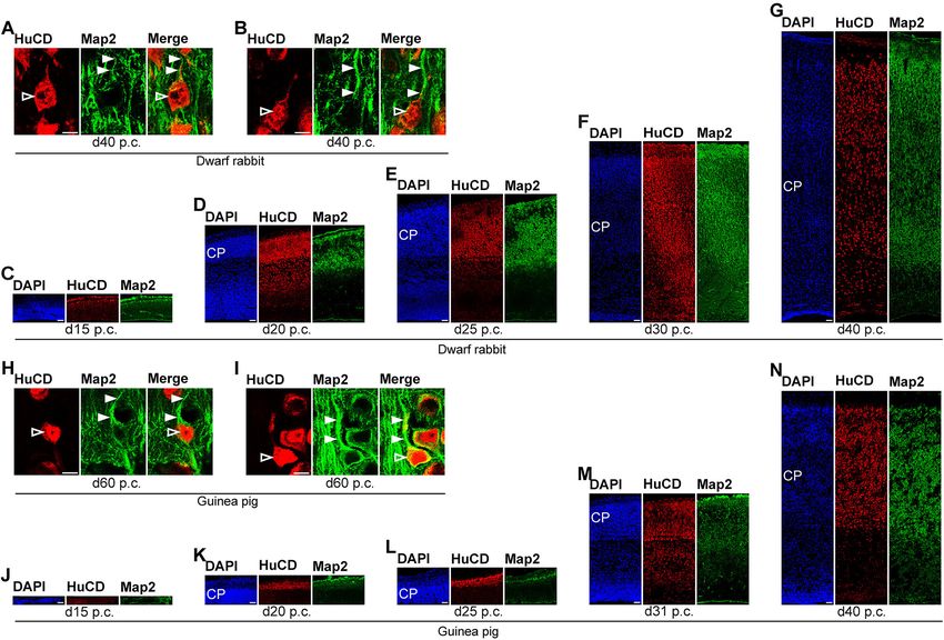
Kalusa et al.                                                                                                         Altricial and Precocial Neocortex Development

surface were determined by tracing it in Fiji software. Data were                   d40 p.c. (dwarf rabbit) and d50 p.c. (guinea pig), d50 p.c. (dwarf
further analyzed using Prism software (GraphPad Software, San                       rabbit), and d60 p.c. (guinea pig).
Diego, USA).                                                                           To extrapolate the timing of neurogenesis, the development
   NPC cell counts (i.e., Pax6+/Tbr2+, Pax6+/Tbr2–, and                             of Pax6+ and Tbr2+ NPCs in the VZ and SVZ was plotted in
Pax6-/Tbr2+ NPCs in the VZ and SVZ) and CP thickness                                Prism (GraphPad Software) assuming Gaussian distribution. As
were compared between the dwarf rabbit and guinea                                   a measure of goodness of fit, R2 was calculated. Encephalization
pig at corresponding cortical neurogenesis stages using                             quotient (EQ) was calculated according to Boddy et al. (2012):
two-tailed unpaired Student’s t-test, p-values below 0.05                           EQ = brain mass/(0.56 × body mass ∧ 0.746). Index of Neural
were considered significant. Corresponding developmental                            Development (IND) was calculated as adapted from (Portmann,
stages were determined according to Workman et al. (2013)                           1990) and (Grand, 1992): IND = developmental brain mass/adult
(www.translatingtime.org) and were as follows: d15 p.c. (dwarf                      brain mass. Data on the developmental brain weight of both
rabbit) and d25 p.c. (guinea pig), d20 (dwarf rabbit) and d 31 p.c.                 species were obtained in this study. For dwarf rabbits, adult brain
(guinea pig), d30 p.c. (dwarf rabbit) and d40 p.c. (guinea pig),                    and body weight data were obtained from the literature (Latimer

 FIGURE 1 | Pax6 and Tbr2 expression in the germinal zones of the developing dwarf rabbit neocortex. (A–H) Double immunofluorescence for Pax6 (red) and Tbr2
 (green) and DAPI staining (blue) on 30-µm cryosections of day 15 to 60 post-conception (p.c.) dwarf rabbit neocortex. The merge images show combined
 immunofluorescence for Pax6 and Tbr2. (A) The complete cortical wall is shown. (B–H) The top margin of the image corresponds to the transition zone
 SVZ/intermediate zone (B,C) or the intermediate zone (D–H). The dashed line indicates the border between VZ and SVZ. Scale bars, 50 µm. VZ, ventricular zone;
 SVZ, subventricular zone.

Frontiers in Neuroanatomy | www.frontiersin.org                                 4                                            May 2021 | Volume 15 | Article 678385
Kalusa et al. Altricial and Precocial Neocortex Development
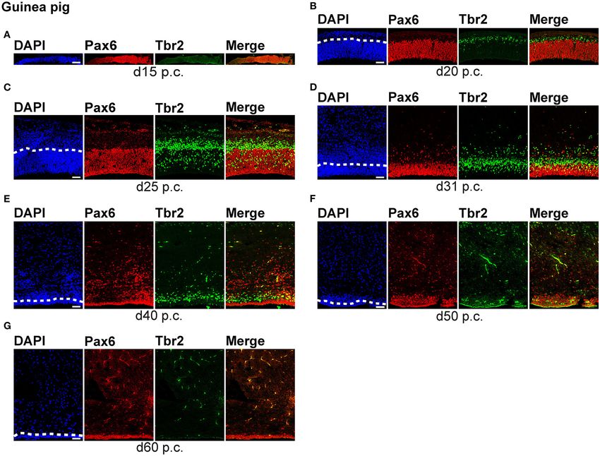
FIGURE 2 | Pax6 and Tbr2 expression in the germinal zones of the developing guinea pig neocortex. (A–G) Double immunofluorescence for Pax6 (red) and Tbr2
(green) and DAPI staining (blue) on 30-µm cryosections of day 15 to 60 post-conception (p.c.) guinea pig neocortex. The merge images show combined
immunofluorescence for Pax6 and Tbr2. (A–C) The complete cortical wall is shown. (D–G) The top margin of the image corresponds to the transition zone
SVZ/intermediate zone (D) or the intermediate zone (E–G). Scale bars, 50 µm. The dashed line indicates the border between VZ and SVZ. VZ, ventricular zone; SVZ,
subventricular zone.
and Sawin, 1955). For guinea pigs, data of adult brain weight were RESULTS
obtained in this study, data of adult body weight were obtained
from the literature (Dobbing and Sands, 1970). Developmental Differences in
The phylogenetic tree was constructed using the MammalTree Neurogenesis Between the Dwarf Rabbit
service from vertlife.org. The following species were chosen and Guinea Pig Neocortex
from the provided list: Rattus norvegicus, Mus musculus, To compare specific aspects of neocortex neurogenesis between
Hydrochoerus hydrochaeris, Cavia porcellus, Oryctolagus altricial and precocial mammalian species, we first focused
cuniculus, Lepus europeus, Homo sapiens, Macaca mulatta, our analysis on the characterization of the distinct NPCs,
Callithrix jacchus, Sus scrofa, Equus caballus, Felis silvestris. specifically on their occurrence and abundance, and analyzed
The tool first trims the phylogenetic data to a subset, cortical sections of the dwarf rabbit and guinea pig from
then samples the tree from a chosen pseudo-posterior different developmental stages by immunohistochemistry
distribution and provides the tree for downloading. The for the expression of Pax6 and Tbr2, both known to be
pruned tree was plotted with FigTree1.4.4 (http://tree.bio.ed. characteristically expressed by distinct NPC subtypes (Figures 1,
ac.uk/) and adapted in Illustrator CS6 software (Adobe, San 2, Supplementary Figure 1) (Götz et al., 1998; Englund et al.,
Jose, California). 2005; Fietz et al., 2010; Hansen et al., 2010; Reillo et al.,
2011).
Frontiers in Neuroanatomy | www.frontiersin.org 5 May 2021 | Volume 15 | Article 678385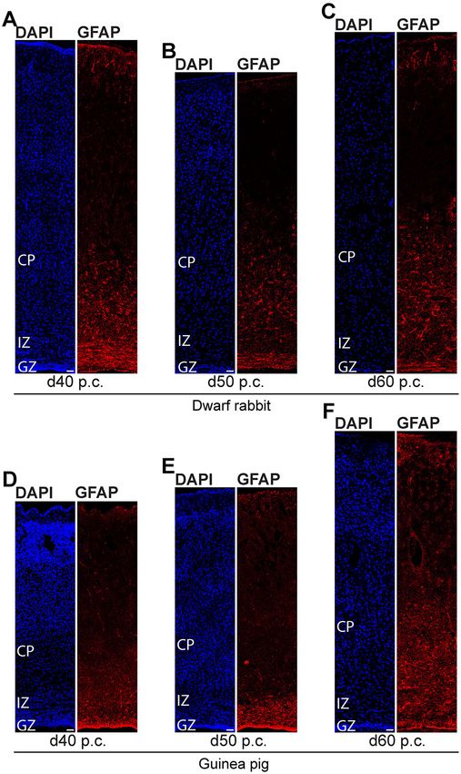
Kalusa et al. Altricial and Precocial Neocortex Development
We first concentrated on APs as the primary NPCs. As
observed in most mammalian species (Götz et al., 1998;
Englund et al., 2005; Osumi et al., 2008; Fietz et al., 2010;
Hansen et al., 2010; Reillo et al., 2011; Romer et al., 2018;
Sauerland et al., 2018), NPCs of the dwarf rabbit and guinea
pig VZ were Pax6+ and largely Tbr2– (Figures 1, 2, 3A,B).
The highest abundance of Pax6+ NPCs in the VZ of the
dwarf rabbit was observed at day 15 p.c. (Figures 1A, 3A).
Indeed, extrapolation of the Pax6+ NPCs abundance in the
dwarf rabbit VZ during early embryonic development revealed
their maximum value to be generated around day 15 p.c.
(Figures 4A,C). At day 15 p.c., the guinea pig VZ constitutes
a very thin layer indicating that the generation of APs in the
guinea pig neocortex might begin at a later postconceptional
day when compared with that of the dwarf rabbit (Figure 2A).
Similarly, the highest abundance of Pax6+ NPCs in the guinea
pig VZ was detected at an important later time point after
conception, i.e., at day 25 p.c. (Figures 2C, 3B). Interestingly,
this shift in development has, if at all, only little impact on
the maximum number of VZ NPCs generated per stage, as
this number appears to be largely the same between the dwarf
rabbit and guinea pig (Figures 3A,B). Once their maximum
value is generated, the number of Pax6+ APs progressively
declines in the further time course in both species analyzed
(Figures 3A,B).
We next focused our analysis on BPs, which generate the
majority of neurons in the developing neocortex (Haubensak
et al., 2004; Miyata et al., 2004; Noctor et al., 2004; Hansen et al.,
2010; Fietz and Huttner, 2011; Lui et al., 2011; Betizeau
et al., 2013). To define the timing of the main period of
cortical neurogenesis, we exploited previous findings that Tbr2+
NPCs are committed to neuronal fate and Tbr2 expression
in progenitor compartments rises and falls with cortical plate
neurogenesis (Englund et al., 2005; Hevner, 2019) and used the
occurrence and abundance of Tbr2+ NPCs as a proxy for the
estimation of the onset and end of cortical neurogenesis. The first
occurrence of Tbr2+ NPCs in the guinea pig SVZ was observed at
day 20 p.c. (Figure 2B). This coincides with the first observation
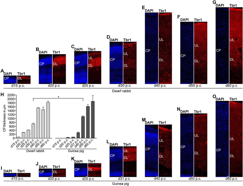
FIGURE 3 | Quantification of Pax6+/Tbr2–, Pax6+/Tbr2+, and Pax6–/Tbr2+ of deep layers, mainly containing Tbr1+ neurons, in the guinea
NPCs in the VZ and SVZ of the developing guinea pig and dwarf rabbit
neocortex. (A–D) Pax6+/Tbr2– (red), Pax6+/Tbr2+ (red/green), and
pig neocortex (Figures 5H,J). In contrast, in the dwarf rabbit
Pax6–/Tbr2+ (green) NPCs in the VZ (A,B) and SVZ (C,D) of day 15 to 60 neocortex, Tbr2+ NPCs—together with Tbr1+ neurons—are
post-conception (p.c.) dwarf rabbit (A,C) and guinea pig (B,D) neocortex, already present at day 15 p.c. (Figures 1A, 5A,H). Extrapolation
expressed as number of cells per 100 µm ventricular surface. Y-axis for (B) is of the Tbr2+ NPCs abundance in the dwarf rabbit germinal
shown in (A), y-axis for (D) is shown in (C). Color legend is shown in (A). For zones during early embryonic development revealed that they
dwarf rabbits, the cortical wall corresponding to a total ventricular surface of
4.56–8.26 µm was analyzed. Data represent mean ± SD and are from two
arise immediately before, i.e., at day 10 p.c. (Figures 4A,C,E,G).
(d30 p.c., d35 p.c., d40 p.c., d50 p.c., d60 p.c.) or three (d15 p.c., d20 p.c., This indicates that the period of cortical neurogenesis starts
d25 p.c.) brains each. The cortical wall corresponding to a total ventricular at an earlier postconceptional day in the dwarf rabbit (day
surface of 2.70–13.68 µm was analyzed for the guinea pig. Data represent ∼10 p.c.) compared with the guinea pig (day ∼20 p.c.). The
mean ± SD and are from two (d15 p.c., d20 p.c., d25 p.c.) or three (d31 p.c.,
highest abundance of Tbr2+ NPCs (Figures 3C,D) was detected
d40 p.c., d50 p.c., d60 p.c.) brains each. Asterisks indicate statistically
significant differences in Pax6+/Tbr2+ NPCs, Pax6+/Tbr2–, and
around the onset of the formation of upper layers, which
Pax6–/Tbr2+ NPCs between corresponding neurogenesis stages of the dwarf mainly contain Tbr1– neurons (Figure 5), in the neocortex of
rabbit and guinea pig, ***p < 0.001; **p < 0.01; *p < 0.05. Corresponding both species analyzed. After their peak, the number of Tbr2+
cortical neurogenesis stages were determined according to Workman et al. NPCs decreases to minimal detectable levels until day 35 p.c.
(2013) (www.translatingtime.org). For details, see the Materials and Methods
in the dwarf rabbit neocortex, indicating that neurogenesis is
section. Cell counts on a yellow background are significantly higher in the
dwarf rabbit when compared with the guinea pig. likely to end between day 30 and 35 p.c. (Figures 1E, 3C).
In the guinea pig neocortex, the number of Tbr2+ NPCs
Frontiers in Neuroanatomy | www.frontiersin.org 6 May 2021 | Volume 15 | Article 678385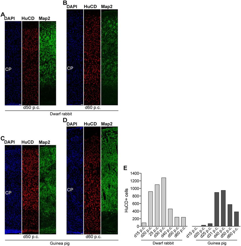
Kalusa et al.                                                                                                              Altricial and Precocial Neocortex Development

 FIGURE 4 | Extrapolation of NPCs abundance in the VZ and SVZ of the developing guinea pig and dwarf rabbit neocortex. (A,B) Pax6+/Tbr2– (red circles),
 Pax6–/Tbr2+ (green triangles), and Pax6+/Tbr2+ (blue rectangles) NPCs in the VZ of day 15 to 60 post conception (p.c.) dwarf rabbit and guinea pig neocortex,
 expressed as number of cells per 100 µm ventricular surface. Color legend is shown in (A). (C,D) Pax6+/Tbr2– (red circles) and Tbr2+ (Pax6–/Tbr2+, Pax6+/Tbr2+,
 green rectangles) NPCs in the VZ of day 15 to 60 post conception (p.c.) dwarf rabbit and guinea pig neocortex, expressed as number of cells per 100 µm ventricular
 surface. Color legend is shown in (C). (E,F) Pax6+/Tbr2– (red circles), Pax6–/Tbr2+ (green triangles), and Pax6+/Tbr2+ (blue rectangles) NPCs in the SVZ of day 15
 to 60 post conception (p.c.) dwarf rabbit and guinea pig neocortex, expressed as number of cells per 100 µm ventricular surface. Color legend is shown in (A). (G,H)
 Pax6+/Tbr2– (red circles) and Tbr2+ (Pax6–/Tbr2+, Pax6+/Tbr2+, green rectangles) NPCs in the SVZ of day 15 to 60 post conception (p.c.) dwarf rabbit and guinea
 pig neocortex, expressed as number of cells per 100 µm ventricular surface. Color legend is shown in (C). (A–H) Data were obtained as in Figure 3. Development of
 NPCs in the VZ and SVZ was extrapolated based on Gaussian distribution. For details, see Materials and Methods section. (A) red line, R2 = 0.8964; green line, R2 =
 0.6911; blue line, R2 = 0.9572; (B) red line, R2 = 0.8849; green line, R2 = 0.6944; blue line, R2 = 0.7487; (C) red line, R2 = 0.8964; green line, R2 = 0.9424; (D) red
 line, R2 = 0.8893; green line, R2 = 0.5212; (E) red line, R2 = 0.5694; green line, R2 = 0.7891; blue line, R2 = 0.9844; (F) red line, R2 = 0.9426; green line, R2 =
 0.9190; blue line, R2 = 0.8249; (G) red line, R2 = 0.5694; green line, R2 = 0.9868; (H) red line, R2 = 0.9398; green line, R2 = 0.7716.

Frontiers in Neuroanatomy | www.frontiersin.org                                    7                                              May 2021 | Volume 15 | Article 678385
Kalusa et al.                                                                                                              Altricial and Precocial Neocortex Development

 FIGURE 5 | Tbr1 expression in the developing dwarf rabbit and guinea pig neocortex. (A–G) Immunofluorescence for Tbr1 (red) and DAPI staining (blue) on 30-µm
 cryosections of day 15 to 60 post-conception (p.c.) dwarf rabbit neocortex. (H) Quantification of CP thickness of d15–60 p.c. dwarf rabbit (light gray) and guinea pig
 (dark gray) neocortex. Data represent mean ± SD and are from two brains each. The asterisk indicates a statistically significant difference in CP thickness between
 corresponding neurogenesis stages of the dwarf rabbit and guinea pig, *p < 0.05. (I–O) Immunofluorescence for Tbr1 (red) and DAPI staining (blue) on 30-µm
 cryosections of day 15–60 p.c. guinea pig neocortex. (A–G,I–O) Scale bars, 50 µm. CP, cortical plate. DL, deep layer. UL, upper layer.

reaches minimum values, not before day 50 p.c., suggesting                             corresponding cortical neurogenesis stages, i.e., dwarf rabbit d20
neurogenesis to end between day 40 and 50 p.c. (Figures 2F,                            and guinea pig d31 were compared (Figures 3C,D). Similarly,
3D), which is slightly later when compared with a previous                             the number of Pax6+/Tbr2+ NPCs in the VZ, which resemble
study (Hatakeyama et al., 2017). Extrapolation of the Tbr2+                            newborn BPs (Hevner, 2019), is significantly higher in the
NPCs abundance in the SVZ of both species revealed their                               dwarf rabbit at day 15 and 20 p.c. when compared with that
minimum value to be generated around 30 p.c. in the dwarf                              in the guinea pig SVZ at day 25 and 31 p.c., respectively
rabbit and 48 p.c. in the guinea pig (Figures 4G,H). Together,                         (Figures 3A,B). Moreover, the maximum number of SVZ NPCs,
this suggests that the total length of the cortical neurogenic                         i.e., the sum of Pax6+/Tbr2–, Pax6+/Tbr2+, Pax6–/Tbr2+ SVZ
period is longer in the guinea pig (∼28 d) than in the dwarf                           NPCs, generated at the respective peak stages of neurogenesis is
rabbit (∼20 days).                                                                     higher in the dwarf rabbit (i.e., at day 20 p.c.) when compared
   Interestingly, we observed a marked difference in the BP                            with that of the guinea pig (i.e., at day 31 p.c.) (Figures 3C,D).
subtype composition between the developing dwarf rabbit and                            Given that a higher abundance of NPCs generated would result
guinea pig neocortex during the respective main period of                              in higher neuronal output (Rakic, 2009; Fietz and Huttner,
neurogenesis (Figures 3C,D). Specifically, the proportion of                           2011; Florio and Huttner, 2014; Dehay et al., 2015; Cardenas
Tbr2+ NPCs that also express Pax6 is significantly higher                              and Borrell, 2020), we found the number of neurons, identified
in the dwarf rabbit SVZ than in the guinea pig SVZ when                                by immunofluorescence for the pan-neuronal marker Hu C/D

Frontiers in Neuroanatomy | www.frontiersin.org                                    8                                              May 2021 | Volume 15 | Article 678385
Kalusa et al. Altricial and Precocial Neocortex Development
FIGURE 6 | Hu C/D and Map2 expression in the developing dwarf rabbit and guinea pig neocortex. (A–N) Double immunofluorescence for Hu C/D (red, A–N) and
Map2 (green, A–N) and DAPI staining (blue, C–G,J–N) on 30-µm cryosections of day 15 to 40 post-conception (p.c.) dwarf rabbit (A–G) and guinea pig (J–N)
neocortex. Images in (A,B,H,I) show neurons with Hu C/D+ soma (open arrowhead) and extending Map2+ dendrites (solid arrowhead) in higher magnification of d40
p.c. dwarf rabbit (A,B) neocortex and d60 p.c. guinea pig (H,I) neocortex. Merge images in (A,B,H,I) show combined immunofluorescence of Hu C/D and Map2. CP,
cortical plate. Scale bars, 10 µm (A,B,H,I) or 50 µm (C–G,J–N).
(Figures 6, 7) (Barami et al., 1995; Gao and Keene, 1996; Okano Map2, a marker for neuronal dendrites (Figures 6, 7) (Bernhardt
and Darnell, 1997; Wakamatsu and Weston, 1997), generated and Matus, 1984; Chen et al., 1992; Dehmelt and Halpain, 2005).
at the respective end of cortical neurogenesis to be higher in In both species analyzed, Map2+ structures extend from and
the developing wall of the dwarf rabbit (i.e., at day 30 p.c.) partly localize with Hu C/D+ somata (Figures 6A,B,H,I) and
when compared with that of the guinea pig (i.e., at day 50 were mainly detected in the developing CP (Figures 6, 7). Their
p.c.) (Figure 7E). To investigate whether these developmental first appearance coincides with the first detection of neurons in
differences impact adult brain size, we obtained the absolute the developing dwarf rabbit and guinea pig neocortex, which is
brain mass. We calculated the encephalization quotient as a in line with the notion that dendrite formation constitutes one of
between-species measure for relative brain mass for both species. the initial steps in the process of neuron maturation (Bernhardt
Indeed, this revealed that the dwarf rabbit is characterized by a and Matus, 1984; Bernhardt et al., 1985; Marin-Padilla, 1992;
higher absolute brain mass and exhibits a higher encephalization Whitford et al., 2002; Jan and Jan, 2003). The process of axon
quotient when compared with that of the guinea pig (Table 1). formation was examined by double-immunofluorescence for the
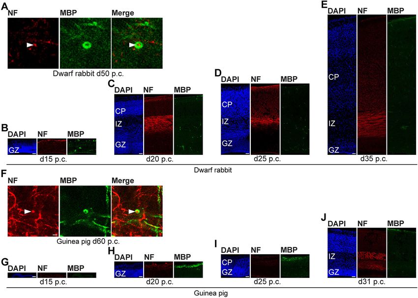
axonal marker neurofilament H (Figures 8, 9) (Shaw and Weber,
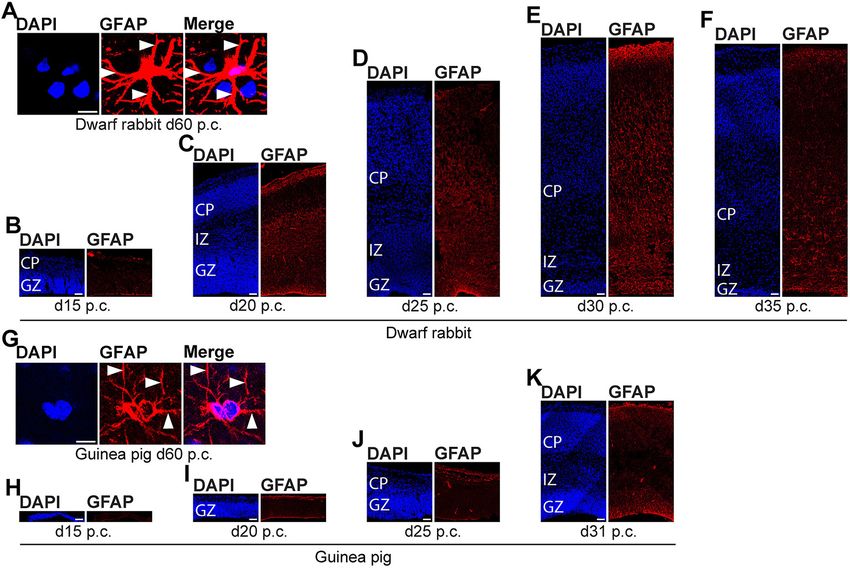
Developmental Differences in Gliogenesis 1982; Carden et al., 1987; Lariviere and Julien, 2004; Lyck et al.,
and Neuron Maturation Between the Dwarf 2008) and myelin basic protein (MBP), a marker for myelination
Rabbit and Guinea Pig Neocortex in the central nervous system (Foran and Peterson, 1992; Zecevic
In the next step, we focused our analysis on the spatial and et al., 1998; Lyck et al., 2008). In both species analyzed, an
temporal characterization of distinct parameters of neuron intense and widespread appearance of axonal structures was
maturation. We analyzed cortical sections of the dwarf rabbit first detected in the intermediate zone (IZ) ∼10 days after
and guinea pig by double-immunofluorescence for Hu C/D and the respective onset of cortical neurogenesis; i.e., at day 20
Frontiers in Neuroanatomy | www.frontiersin.org 9 May 2021 | Volume 15 | Article 678385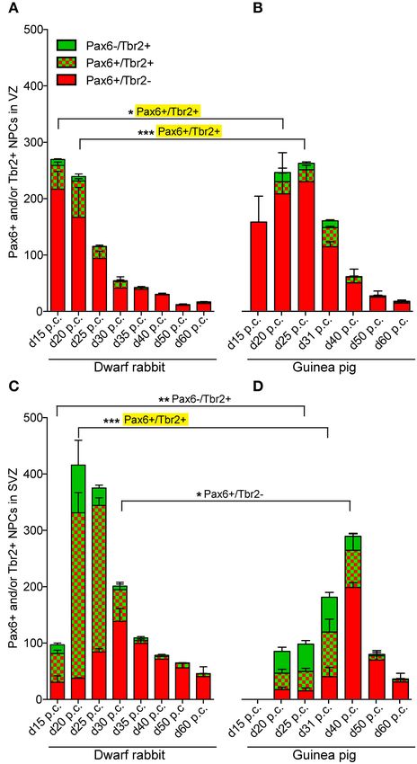
Kalusa et al.                                                                                                                Altricial and Precocial Neocortex Development

 FIGURE 7 | Hu C/D and Map2 expression in the developing dwarf rabbit and guinea pig neocortex. (A–D) Double immunofluorescence for Hu C/D (red) and Map2
 (green) and DAPI staining (blue) on 30-µm cryosections of day 50 and 60 post-conception (p.c.) dwarf rabbit (A,B) and guinea pig (C,D) neocortex. Scale bars,
 50 µm. CP, cortical plate. (E) Quantification of Hu C/D+ cells in d15–50 p.c. dwarf rabbit (light gray) and d20–60 p.c. guinea pig (dark gray) cortical wall, expressed as
 the number of cells per 100 µm ventricular surface. The cortical wall corresponding to a total ventricular surface of 3.86–6.02 µm (guinea pig) and 3.08–6.05 µm
 (dwarf rabbit) was analyzed. Data are from one brain each.

p.c. in the dwarf rabbit and day 31 p.c. in the guinea pig                               of cortical neurogenesis. Similarly, widespread myelination in
(Figures 8C,J). Intriguingly, these axonal extensions were mostly                        the CP was not detected until day 40 p.c. in the dwarf rabbit
oriented parallel to the ventricular surface (Figures 8C–E,J, 9D),                       (Figure 9A) and day 50 p.c. in the guinea pig (Figure 9E).
thus, supporting previous studies that have identified the IZ to                         Once axonal processes are generated in high abundance, we
be populated by interneurons that migrate tangentially from the                          observed a widespread formation of synapses, as identified by the
ventral into the dorsal telencephalon during early embryonic                             presynaptic marker synaptophysin (Wiedenmann and Franke,
development (DeDiego et al., 1994; De Carlos et al., 1996; Lavdas                        1985; Gil-Loyzaga and Pujol, 1988; Ichikawa et al., 1991), in
et al., 1999; Letinic et al., 2002; Marin and Rubenstein, 2003;                          the day 40 p.c. dwarf rabbit and day 50 p.c. guinea pig CP
Wonders and Anderson, 2006; Hansen et al., 2013). In the CP,                             (Supplementary Figure 3).
axonal processes, which were mainly oriented radially to the                                We further investigated the formation of astrocytes using
ventricular surface, were already detected as early as day 20 p.c.                       immunohistochemistry for GFAP that resembles the hallmark
in the dwarf rabbit (Figure 8C) and day 31 p.c. in the guinea                            intermediate filament protein in astrocytes (Figures 10, 11)
pig (Figure 8J); however, they only become numerous with day                             (Bignami and Dahl, 1973; Kalman and Hajos, 1989; Eng et al.,
40 p.c. in the dwarf rabbit (Figure 9A) and day 50 p.c. in the                           2000; Hol and Pekny, 2015). In both species analyzed, GFAP+
guinea pig (Figure 9E), and thus after the end of the main period                        structures were already detected during mid-neurogenesis, i.e.,

Frontiers in Neuroanatomy | www.frontiersin.org                                     10                                              May 2021 | Volume 15 | Article 678385
Kalusa et al.                                                                                                              Altricial and Precocial Neocortex Development

TABLE 1 | Adult brain mass, EQ, and IND of the dwarf rabbit and guinea pig.

Species                            Adult brain mass (g)                          EQ                                                   IND (%)

                                                                                                           Day 30 p.c.                                Day 60 p.c.

Dwarf rabbit                                9.6                                 0.72                       10.53 (time of birth)                      60.88
Guinea pig                                  4.3                                 0.46                       4.85                                       67.82 (time of birth)

The respective time of birth is indicated in brackets. For details, see the Materials and Methods section. EQ, encephalization quotient; IND, index of neural development;
p.c., post-conception.

 FIGURE 8 | Neurofilament H and MBP expression in the developing dwarf rabbit and guinea pig neocortex. (A–J) Double immunofluorescence for neurofilament H
 (NF, red, A–J) and MBP (green, A–J) and DAPI staining (blue, B–E,G–J) on 30-µm cryosections of day 15–35 post-conception (p.c.) dwarf rabbit neocortex and
 d15–31 p.c. guinea pig neocortex. Images in (A,F) show NF+ extension (solid arrowhead) surrounded by MBP+ myelin sheath in higher magnification of d50 p.c.
 dwarf rabbit neocortex (A) and d60 p.c. guinea pig neocortex (F). Merge images in (A,F) show combined immunofluorescence of NF and MBP. CP, cortical plate. GZ,
 germinal zone. IZ, intermediate zone. Scale bars, 2 µm (A,F) or 50 µm (B–E,G–J).

at day 20 p.c. in the dwarf rabbit neocortex and day 31 p.c. in                         (Figure 11E). Again, this suggests that major aspects of astrocyte
the guinea pig neocortex (Figures 10C,K). As GFAP expression                            formation occur after the end of the main period of cortical
was mainly observed in the germinal zones close to the lateral                          neurogenesis in both species analyzed.
ventricle (Figures 10C,K), our data suggest GFAP be expressed                              Together, our data show that the basic order of neuro- and
by distinct NPCs, e.g., radial glial cells, in the developing                           gliogenesis and neuron maturation is preserved in the dwarf
dwarf rabbit and guinea pig neocortex, which is in line with                            rabbit and guinea pig neocortex. In absolute postconceptional
observations in other mammalian species (Noctor et al., 2002;                           days, the onset of cortical neurogenesis starts at a later time point
Kriegstein and Alvarez-Buylla, 2009; Fietz et al., 2010, 2020;                          in the guinea pig compared with the dwarf rabbit (Figure 12).
Kelava et al., 2012). Mature GFAP+ astrocytes, which exhibit                            However, when expressed concerning the gestation length, the
a typical star-shaped appearance (Figures 10A,G), were first                            onset of cortical neurogenesis occurs at a similar time point in
detected at high numbers at day 40 p.c., in the dwarf rabbit                            both species analyzed, i.e., at 33% of gestation corresponding to
(Figure 11A) and day 50 p.c., in the guinea pig neocortex                               the end of the first trimester of gestation. In both species, the

Frontiers in Neuroanatomy | www.frontiersin.org                                    11                                              May 2021 | Volume 15 | Article 678385
Kalusa et al. Altricial and Precocial Neocortex Development FIGURE 9 | Neurofilament H and MBP expression in the developing dwarf rabbit and guinea pig neocortex. (A–F) Double immunofluorescence for neurofilament H (NF, red) and MBP (green) and DAPI staining (blue) on 30-µm cryosections of day 40–60 post-conception (p.c.) dwarf rabbit (A–C) and guinea pig (D–F) neocortex. CP, cortical plate. Scale bars, 50 µm. main period of cortical neurogenesis ends before birth, lasting at the time of birth (Figure 12). This indicates that—in contrast until the beginning of the third trimester in the guinea pig to the guinea pig neocortex—a greater proportion of growth neocortex until the end of gestation in the dwarf rabbit neocortex and maturation in the dwarf rabbit neocortex will occur during (Figure 12). Concerning neuron maturation, dendrite formation postnatal development. To test this, we calculated the index of tends to start during early-mid neurogenesis. In contrast, neural development (IND) as the ratio between developmental major aspects of axon formation and myelination together and adult brain mass at the time of birth for both species with astrogenesis seem to occur once neurogenesis is largely analyzed. This shows that the guinea pig achieves an IND > terminated in the neocortex of both species analyzed (Figure 12). 50% at the time of birth, and thus major brain growth in the Taken the time of birth into account, both species exhibit a guinea pig occurs during prenatal development. In contrast, in different cortical growth and maturation status at birth. While the dwarf rabbit exhibiting an IND of ∼10% at the time of the neocortex of the guinea pig contains neurons that seemingly birth, the overwhelming majority of brain growth is achieved exhibit well-developed dendrites and myelinated axons as well during postnatal development (Table 1). Interestingly, similar as astrocytes, the dwarf rabbit neocortex seems to lack neurons to the guinea pig, the dwarf rabbit achieves an IND > 50% exhibiting well-developed and myelinated axons and astrocytes at day 60 p.c. (Table 1). Moreover, at day 60 p.c., the dwarf Frontiers in Neuroanatomy | www.frontiersin.org 12 May 2021 | Volume 15 | Article 678385
Kalusa et al.                                                                                                        Altricial and Precocial Neocortex Development

 FIGURE 10 | GFAP expression in the developing dwarf rabbit neocortex and guinea pig neocortex. (A–K) Immunofluorescence for GFAP (red) and DAPI staining (blue)
 on 30-µm cryosections of day 15–35 post-conception (p.c.) dwarf rabbit (A–F) and d15-31 p.c. guinea pig (G–K) neocortex. Images in (A,G) show GFAP+ astrocyte
 with soma and extending processes (solid arrowhead) in higher magnification of d60 p.c. dwarf rabbit (A) and d60 p.c. guinea pig (F) neocortex. Merge images in
 (A,G) show combined fluorescence of GFAP and DAPI. CP, cortical plate. GZ, germinal zone. IZ, intermediate zone. Scale bars, 10 µm (A,G) and 50 µm (B–F,H–K).

rabbit neocortex is characterized by neurons with structurally                      formation and myelination to be similar between the altricial
well-developed and myelinated axons and astrocytes as has been                      dwarf rabbit and precocial guinea pig neocortex.
observed for the guinea pig of the same postconceptional day                            Intriguingly, our data show that, in absolute postconceptional
(Figures 9C,F, 11C,F).                                                              days, the onset of cortical neurogenesis is shifted between the
                                                                                    two species analyzed, starting ∼10 days later in the precocial
                                                                                    guinea pig than in the altricial dwarf rabbit. This time shift
                                                                                    corresponds to the translating time between equivalent post
DISCUSSION                                                                          conception dates of rabbit and guinea pig (Workman et al., 2013;
                                                                                    Finlay and Huang, 2020). Moreover, we show that the period
This study compares distinct patterns of neocortex development,                     of cortical neurogenesis takes longer in absolute gestational
specifically neurogenesis, gliogenesis, and neuron maturation,                      days in the precocial guinea pig compared with that of the
between the precocial guinea pig and the altricial dwarf rabbit.                    altricial dwarf rabbit. Thus, our findings are in line with previous
By using two mammalian species that show a relatively close                         studies suggesting precocial mammals exhibit a delayed onset
phylogenetic and genetic relatedness and several similarities                       and protracted duration of cortical neurogenesis when compared
concerning adult and birth body weight and biology (e.g., food                      with phylogenetically closely related altricial mammals (Brunjes
uptake and digestion, habitation and social behavior) (King,                        et al., 1989; Brunjes, 1990; Workman et al., 2013; Finlay and
1956; Kunkel and Kunkel, 1964; Sachser, 1998; Asher et al.,                         Huang, 2020). In this regard, it is interesting to note that other
2004; McDougall et al., 2009; Thormann, 2012; Varga, 2014;                          neurodevelopmental processes, including the transformation of
Upham et al., 2019; Lundwall et al., 2020), we attempted to                         the neural plate into the neural tube as well as hippocampal
minimize the confounding factors that potentially affect brain                      neurogenesis, the generation of olfactory mitral cells, and that
development. Our data demonstrate that the basic order of                           of retinal rods and cones seem to start at a later time point
distinct cortical neurodevelopmental events is preserved in the                     after conception and to last for a longer absolute duration in
altricial dwarf rabbit and precocial guinea pig. Specifically, we                   the precocial guinea pig when compared with the altricial rabbit
found neurogenesis, e.g., including the sequential generation of                    (DeSesso, 1996; Schnorr and Kressin, 2006; Workman et al.,
DL and UL neurogenesis, to precede gliogenesis, as observed                         2013). Moreover, developmental milestones of the formation of
in the neocortex of other mammalian species (Lee et al., 2000;                      primitive structures, including the primitive streak and somites,
Sauvageot and Stiles, 2002; Kriegstein and Alvarez-Buylla, 2009).                   and other organs such as liver and lung, initiate at a substantially
Moreover, we found the principal order of dendrite and axon                         later time point after conception in the precocial guinea pig than

Frontiers in Neuroanatomy | www.frontiersin.org                                13                                           May 2021 | Volume 15 | Article 678385
Kalusa et al. Altricial and Precocial Neocortex Development
FIGURE 12 | Comparison of specific neurodevelopmental events between the
dwarf rabbit and guinea pig neocortex. Onset and duration of the period of
neurogenesis, dendrite formation, axon formation, myelination, and astrocyte
formation in the dwarf rabbit and guinea pig neocortex between day 10 and 60
post-conception (p.c.). Data for the period of neurogenesis are based on the
development of Tbr2+ NPCs in the SVZ (Figure 4); data for dendrite
formation, axon formation, myelination, and astrocyte formation are based on
immunofluorescence staining (Figures 6–11). For details, see the Results
section. Data for dwarf rabbits are shown in solid black lines, data for guinea
pigs are shown in black dashed lines. Red arrows indicate the respective time
of birth (partus) of the dwarf rabbit (solid line) and guinea pig (dashed line). IZ,
intermediate zone. CP, cortical plate.
at the end of the first trimester of gestation, in the dwarf rabbit
and guinea pig neocortex. Further evidence for this comes from
recent studies revealing the lengths of neurogenesis and gestation
to be tightly related (Lewitus et al., 2014; Glatzle et al., 2017;
Stepien et al., 2020). Moreover, previously published findings
and unpublished data from our laboratory show that within
the order of carnivores, consisting of species with similar life
history patterns at birth, species exhibiting a longer gestation
FIGURE 11 | GFAP expression in the developing dwarf rabbit neocortex and
period, i.e., cat, seem to be characterized by later onset and
guinea pig neocortex. Immunofluorescence for GFAP (red) and DAPI staining
(blue) on 30-µm cryosections of day 40–60 post-conception (d p.c.) dwarf
more protracted period of cortical neurogenesis and other
rabbit (A–C) neocortex and guinea pig (D–F) neocortex. CP, cortical plate. GZ, distinct neurodevelopmental processes when compared with
germinal zone. IZ, intermediate zone. Scale bars, 50 µm. species with shorter gestation period such as ferret (Fietz et al.,
2010; Reillo et al., 2011; Reillo and Borrell, 2012; Workman
et al., 2013). Understanding the factors responsible for these
temporal differences and the potential mutual regulation of brain
in the altricial rabbit (DeSesso, 1996). Together this indicates growth and that of other somatic organ systems during gestation
that the time-shifted onset and prolonged period of development would lend essential insights into the evolutionary mechanism
is not specific to the nervous system of the precocial guinea involved in the process of speciation within the altricial–precocial
pig but instead a more common feature of its embryonic and spectrum. In this regard, it would be interesting to compare the
fetal development involving many different tissues and organs. neural developmental data obtained in the dwarf rabbit in this
Given that precocial species, in general, are characterized by study to a precocial species of the same order, i.e., the European
a longer gestation period than closely related altricial species, hare (Lepus europaeus).
it is tempting to speculate that the onset and duration of Previous studies have linked a lengthening of the neurogenic
distinct developmental processes, e.g., cortical neurogenesis, are period to higher neuron production and cortex expansion (Florio
primarily linked to gestation length (rather than life history and Huttner, 2014; Lewitus et al., 2014; Cardenas and Borrell,
patterns at birth, i.e., altriciality and precociality) (Dieterlen, 2020; Finlay and Huang, 2020; Stepien et al., 2020). However,
1963; Martin and Maclarnon, 1985; Derrickson, 1992; Finlay and our data reveal that the neuronal output generated during peak
Uchiyama, 2017; Scheiber et al., 2017). This is supported by stages of cortical neurogenesis and the absolute and relative
our findings, showing that the onset of neurogenesis occurs at a adult brain mass is lower in the guinea pig than in the dwarf
similar time point, when expressed as percentage of gestation, i.e. rabbit. Interestingly, we found a marked difference in the BP
Frontiers in Neuroanatomy | www.frontiersin.org 14 May 2021 | Volume 15 | Article 678385Kalusa et al. Altricial and Precocial Neocortex Development
subtype composition between the developing dwarf rabbit and being motorically competent (Varga, 2014). Further studies using
guinea pig neocortex at peak stages of neurogenesis. Specifically, an additional set of immunohistochemical markers of neuronal
we found the proportion of Tbr2+ BPs that also express Pax6 maturation as well as electrophysiological techniques, i.e., patch
to be markedly higher in the dwarf rabbit than in the guinea clamp, are needed to precisely evaluate the process of neuron and
pig neocortex. On the assumption that sustainment of Pax6 glia maturation and to demonstrate whether the existing neurons
expression in BPs is linked to higher cell proliferation (Betizeau in the pre- and neonatal guinea pig and early postnatal dwarf
et al., 2013; Wong et al., 2015), our data suggest that the rabbit neocortex are indeed genuine, and thus functional mature.
developing dwarf rabbit neocortex contains a higher abundance In conclusion, this study provides comprehensive data on
of highly proliferative BPs, which might enable the dwarf rabbit distinct patterns of brain development between the precocial
to achieve a higher neuron production and brain size when guinea pig and the altricial dwarf rabbit, which may serve
compared with that of the guinea pig, thereby counterbalancing as empirical reference data in future studies. While the basic
its shorter neurogenesis length. Indeed, the more rapid and order of cortical neuro- and gliogenesis and neuron maturation
higher expansion of the BP cell pool, which is not accompanied is the same during early development, their specific timing
by an equivalent decrease in the AP cell number, supports markedly differs concerning the postconceptional age and the
the notion of self-amplifying NPCs being present in higher time of birth between them. Moreover, our data provide evidence
abundance in the dwarf rabbit SVZ when compared with that for the notion that a complex regulation of multiple factors
of the guinea pig. Together, our data provide evidence for the determines the amount of neuron production in the developing
notion that neuron production in the developing neocortex is neocortex of the guinea pig and dwarf rabbit. Together, these
determined by complex regulation of multiple factors, including data expand our current understanding of the timing and cellular
the duration of the neurogenesis period, the absolute number of differences that regulate patterns of mammalian brain growth
NPCs, and the relative abundance of each NPC type (Florio and and maturation and provides a better understanding of the
Huttner, 2014; Lewitus et al., 2014; Cardenas and Borrell, 2020). evolution of mammalian altriciality and presociality.
Therefore, further studies that use a larger number of animals
and address the mode and rate of cell division of distinct NPC DATA AVAILABILITY STATEMENT
subtypes are important to directly evaluate their contribution to
neocortex development in the dwarf rabbit and guinea pig. The raw data supporting the conclusions of this article will be
Strikingly, we observed that the IND of the precocial guinea made available by the authors, without undue reservation.
pig is considerably higher at the time of birth when compared
with that of the altricial dwarf rabbit, which is in line with
ETHICS STATEMENT
previously published data showing precocial species to give birth
to larger-brained offspring after controlling for body size (Barton The animal study was reviewed and approved by
and Capellini, 2011). In this respect, our data indicate that Landesdirektion Leipzig.
the guinea pig neocortex exhibits a higher maturation status,
containing more neurons with well-developed dendrites and
myelinated axons and astrocytes, than the dwarf rabbit, in which
AUTHOR CONTRIBUTIONS
major steps of neuron maturation, i.e., axon generation and MK and SF conceived and designed the experiments and
myelination, and astrogenesis mainly set in after birth. Thus, analyzed the data. MK, MH, and CS performed the experiments.
our data provide evidence for the notion that brain expansion MK, MM, and SF discussed the data and wrote the article. All
in the precocial newborn is largely due to prenatally initiating authors reviewed and approved the manuscript.
processes of gliogenesis and neuron maturation, instead of
increased neurogenesis as previously discussed (Glatzle et al.,
2017). Moreover, they are in line with previously published data ACKNOWLEDGMENTS
showing that the precocial index, which characterizes the point
of neural maturation at birth, is higher in the guinea pig (0.841) We thank Gabriele Lindner, Kevin Richter, Jasmina Schmitt,
than in the rabbit (0.537) and confirm the expected contrast of Elisabeth Pötzsch, Megan Glatzle, Hannah Bender, and Thomas
the position of birth concerning specific neural milestones for Grochow for their technical support, and members of the MEZ,
the precocial vs. closely related altricial mammals (Workman Faculty of Medicine, University of Leipzig and the Tierarztpraxis
et al., 2013; Finlay and Uchiyama, 2017). Given that the offspring Dr. Falko Pötzsch for animal colony management. Moreover, we
of the precocial guinea pig, in contrast to that of the dwarf thank members of the BioImaging Core Facility of the University
rabbit, is born with advanced cognitive, sensory, and locomotor of Leipzig for outstanding support. Finally, the authors
abilities, our findings indicate that its offspring has acquired the acknowledge support from the German Research Foundation
morphological machinery required to attain its high functional (DFG) and Universität Leipzig within Open Access Publishing.
state at birth. Interestingly, in the dwarf rabbit, the cortical
maturation status and the ratio between developmental and adult SUPPLEMENTARY MATERIAL
brain mass largely catch up to those of the guinea pig during
early postnatal development, achieving an IND > 50%, until day The Supplementary Material for this article can be found
60 p.c., a stage which marks the approximate end of weaning in online at: https://www.frontiersin.org/articles/10.3389/fnana.
the dwarf rabbit with its pups having open ears and eyes and 2021.678385/full#supplementary-material
Frontiers in Neuroanatomy | www.frontiersin.org 15 May 2021 | Volume 15 | Article 678385You can also read