Connectivity between nidopallium caudolateral and visual pathways in color perception of zebra finches - Nature
←
→
Page content transcription
If your browser does not render page correctly, please read the page content below
www.nature.com/scientificreports
OPEN Connectivity between nidopallium
caudolateral and visual pathways
in color perception of zebra finches
Yi‑Tse Hsiao1,9, Ta‑Ching Chen2,3,9, Pin‑Huan Yu4, Ding‑Siang Huang2, Fung‑Rong Hu2,
Cheng‑Ming Chuong5 & Fang‑Chia Chang1,6,7,8*
Researchers demonstrated an elegant ability for red discrimination in zebra finches. It is interested
to understand whether red activates exhibit much stronger response than other colors in neural
network levels. To reveal the question, local field potentials (LFPs) was recorded and analyzed in two
visual pathways, the thalamofugal and the tectofugal pathways, of zebra finches. Human studies
demonstrate visual associated telencephalons communicate with higher order brain areas such as
prefrontal cortex. The present study determined whether a comparable transmission occurs in zebra
finches. Telencephalic regions of the thalamofugal (the visual Wulst) and the tectofugal pathway
(the entopallium) with their higher order telencephalon, nidopallium caudolateral (NCL) were
simultaneously recorded. LFPs of relay nuclei (the nucleus rotundus, ROT) of tectofugal pathway were
also acquired. We demonstrated that LFP powers in the tectofugal pathway were higher than those
in the thalamofugal pathway when illuminating blue lights. In addition, the LFP synchronization
was stronger between the entopallium and NCL. LFPs also revealed a higher Granger causality from
the direction of entopallium to NCL and from ROT to entopallium. These results suggest that zebra
finches’ tectofugal pathway predominately processing color information from ROT to NCL, relayed by
entopallium, and blue could trigger the strongest response.
Like humans, birds heavily rely on color vison to gather information and perception1. Color discrimination
affects their food seeking and mate choosing abilities2,3. The central visual pathways in brain exhibit similar
organizations between mammals and birds. In humans, there are two major pathways—the geniculocortical and
extra-geniculocortical pathways, which process visual information. Similarly, the thalamofugal and the tectofugal
pathways in birds are respectively corresponding to the geniculocortical and extra-geniculocortical pathways1,4.
Unlike humans, who depend mainly on the geniculocortical pathway to process visual information5,6, birds
with laterally placed eyes have the most prominent tectofugal pathway (corresponding to extra-geniculocortical
pathway in mammals)7,8. Lesions of this pathway cause deficits on color discrimination tasks9.
However, with estimating approximately 10,000 species of birds, merely the eye position (laterally placed eyes
or medially placed eyes) resulted in profound differences in how their brains process visual i nformation1. Even
in the species with similar eye positions, there still are species-specific variations for avian brains. The zebra
finches’ (Taeniopygia guttata) retina contains an extra type of cone cells, ultraviolet cones, in addition to the
short-wavelength (S)-cones, medium-wavelength (M)-cones, and long-wavelength (L)-cones10, suggesting that
‘blue’, ‘red’, and ‘green’ play important roles for zebra finches. Researchers found that the color of seed feeders
attracted different species of garden birds, implying their different color preferences11. Red is the best known for
its function than other colors for zebra finches. The capability of red color discrimination seems critical for both
male and female zebra finches to decide on their m ates3,12–14. Recently, researchers also demonstrated an elegant
ability of red discrimination in zebra fi nches15. Most of the literatures focus on the correlation between red and
social behaviors3,12–15. But, it is also interesting to investigate the neural network level of whether red activates
1
Department of Veterinary Medicine, School of Veterinary Medicine, National Taiwan University, Taipei,
Taiwan. 2Department of Ophthalmology, College of Medicine, National Taiwan University, Taipei,
Taiwan. 3Research Center for Developmental Biology and Regenerative Medicine, National Taiwan University,
Taipei, Taiwan. 4Institute of Veterinary Clinical Science, School of Veterinary Medicine, National Taiwan
University, Taipei, Taiwan. 5Department of Pathology, University of Southern California, Los Angeles, CA,
USA. 6Graduate Institute of Brain and Mind Sciences, College of Medicine, National Taiwan University, Taipei,
Taiwan. 7Graduate Institute of Acupuncture Science, College of Chinese Medicine, China Medical University,
Taichung, Taiwan. 8Department of Medicine, College of Medicine, China Medical University, Taichung,
Taiwan. 9These authors contributed equally: Yi-Tse Hsiao and Ta-Ching Chen. *email: fchang@ntu.edu.tw
Scientific Reports | (2020) 10:19382 | https://doi.org/10.1038/s41598-020-76542-z 1
Vol.:(0123456789)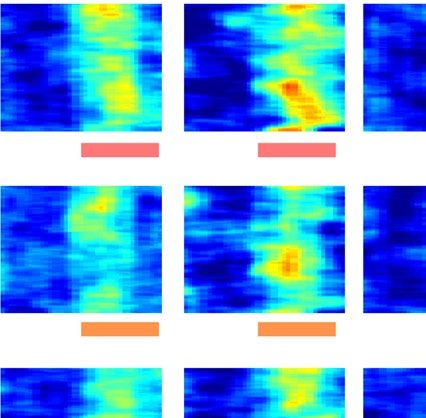
www.nature.com/scientificreports/
much strong responses than other colors, and whether the zebra finches are indeed sensitive to other colors than
red. A report demonstrates that zebra finches have green, blue, and UV light-dependent magnetic compass, sug-
avigation16. The present study recorded local field potentials (LFPs) in
gesting that blue light is critical for their n
the zebra finches’ brain regions and tried to elucidates the brain response when they were stimulated by various
colors. Next, we introduced the brain regions that were recorded in our experiments.
The visual information from the retina transmits to both the optic tectum and the geniculatis lateralis pars
dorsalis (GLd) of the thalamus in birds. The neurons in the optic tectum project to the nucleus rotundus (ROT),
which then send the signals to the entopallium (ENTO). This optic tectum-ROT-ENTO projection pathway is
referred to as the tectofugal pathway17,18. Hodos et al. conducted a series of lesion experiments to determine the
functions of ROT and ENTO. Deconstruction of ROT or ENTO cause deficits in the pattern d iscrimination19,20,
19,20 4,9
visual intensity , and color discrimination . Reports further elucidate that the tectofugal pathway processes
motion signal, color perception, luminance changes, and in-depth vision signals, and is also sensitive to looming
(approaching) objects4,21. It seems that the tectofugal pathway is in charge of some simple visual signals4. On the
other hand, the signal from GLd transmits to the visual Wulst (VW), a telencephalic region that is comparable
with mammalian visual cortex22. This pathway is called the thalamofugal pathway. The thalamofugal pathway is
important for birds, which relies on binocular vision to hunt preys (e.g., owl) in long distance and requires pre-
cise perception of three-dimensional s timuli23,24. However, the functions of VW in owl are not comparable with
zebra finches25,26, since zebra finches are laterally eyed bird that results in only marginal binocular interaction
for both of their eyes27. Seed or fruit-eating birds use frontal binocular vision only when they peck foods from
where is close to their beaks. Zebra finches are granivores (seed predators) with laterally placed eyes and mostly
depend on the monocular v ision25. A previous report showed that male zebra finches prefer to use right eye to
observe females and show off their flanks during the early ritual of courtship28. The color preference for zebra
finches is still an unsolved question and most studies employed behavioral tests to determine. In this study, we
elucidated what kinds of color spectra trigger the largest brain responses in aforementioned visual pathways.
For this purpose, we implanted electrodes into the nuclei (ROT, ENTO and VW) relayed in the tectofugal and
thalamofugal pathways in the left hemisphere, then shined a series of colors to the right eye, and acquired the
LFPs from these nuclei. The above mentioned ENTO and VW are parts of telencephalon and are also belong
to the tectofugal pathway and thalamofugal pathway, r espectively4,8. Moreover, the avian telencephalon divides
into two parts, the Wulst (in the rostral region of the brain) and dorsal ventricular ridge (in the caudal region
of the brain)4,8. In addition, dorsal ventricular ridge comprises a sub-region called nidopallium, and ENTO
transmits visual information to the nidopallium for further cognitive p rocess8. Primate or human studies reveal
that the visual associated telencephalon communicates with higher order of brain areas, such as the prefrontal
cortex29–31. In pigeons, the nidopallium caudolateral (NCL) is equivalent to the prefrontal cortex in m ammals32–34.
NCL in pigeon executes functions such as planning and decision-making33,34. In chickens, NCL also involve in
imprinting behavior35. Recently, a study indicates that the location and trajectory of NCL are species-specific;
the patterns of dopaminergic innervations are strikingly different between pigeons/chickens and zebra fi nches32.
These findings imply that the zebra finch’s NCL may modulate different functions than those of pigeons/chickens.
The function of NCL in zebra finch is not well understood. It may involve in regulation of arousal or courtship
behavior36. The present study also explored the potential role of NCL in zebra finches when processing color
information. Because the tectofugal pathway possesses the ability in color i nformation4,9, we hypothesized that
telencephalons of visual pathway communicate with higher order brain areas in zebra finches and ENTO has
stronger communication with NCL than VW, which is similar to humans29–31, when stimulated by colors. We
initially expected a prominent response in ENTO when zebra finches see colors. We also hypothesized that zebra
finches are more sensitive to red because of the mate-selection3,12–14, but blue and green may be also important
for the navigation purpose16.
In addition to revealing the optimal projection pathway which responds to certain color(s), we ask if this
pathway communicates with a higher order of brain region for cognitive processing when zebra finches perceive
the particular color(s). To address this question, we implanted additional electrode to the NCL, which resembles
mammalian prefrontal cortex, to acquire LFPs. LFPs are electrical potentials generated by neurons in a local
brain region and can determine the brain activities from the network to systemic level37–41. By analyzing the
amplitude (or power) of LFPs from a single area, the synchronization between connected brain areas, and the
directional connectivity between paired brain areas, we can pave the way for neurophysiological investigations
of color information processing in the aspects of activities (relevant to power), communication (relevant to syn-
chronization), and the leading direction (relevant to directional connectivity) between (or within) the tectofugal
pathway and the NCL or between the thalamofugal pathway and NCL. In addition, the degree of sensitivity for
different colors in brain level was also revealed in the experiments.
Results
Tectofugal pathway mainly mediates brain responses to colors. We are interested in the brain
structures to interpret the meaning of color stimulation. We recorded the LFPs from the relay nuclei of the
thalamofugal and tectofugal pathways in brains. To study the effects of different colors on activating thalam-
ofugal and tectofugal pathways, we used red, green, and blue (RGB) codes from 0 to 1 with 0.25 increment to
create 15 colors (for details, Table S1; see "Materials and Methods" section). Fifteen colors were flashed to the
subjects’ right eye (Figs. 1, S1–S3) and simultaneously recorded the LFPs at the relay nuclei of the thalamofugal
and tectofugal pathways, owing to the fact that male zebra finches usually use right eye to observe females dur-
ing courtship28. We averaged the LFPs across the same color stimuli with the same background color (black
background color (Figs. 1 and S1)). Although the subjects were recorded under general anesthesia, the evoked
potentials still emerged between 0 and 500 ms after color flashing when black background was used as baseline
Scientific Reports | (2020) 10:19382 | https://doi.org/10.1038/s41598-020-76542-z 2
Vol:.(1234567890)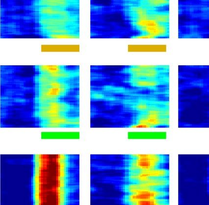
www.nature.com/scientificreports/
LFP
ROT
VW
Amplifer ENTO
NCL
NCL
VW
ENTO
ROT
Colors display on a screen
NCL
ENTO VW
ROT
No.060.51
0
-0.5
-1
No.070.51
0
-0.5
-1
No.130.51
0
-0.5
Z-score
-1
No.140.51
0
-0.5
-1
No.150.51
0
-0.5
-1
1
0.5
0
-0.5
-1
-1 -0.6 0 0.6 1 -1 -0.6 0 0.6 1 -1 -0.6 0 0.6 1 -1 -0.6 0 0.6 1
Time (Sec)
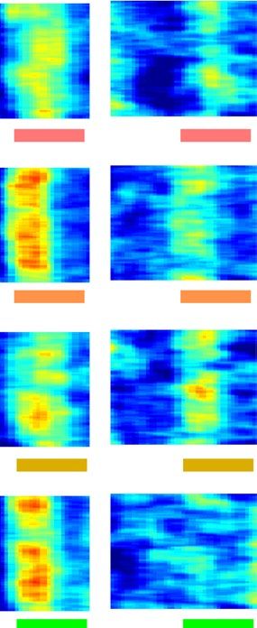
Figure 1. The averaged LFP traces in the brain regions after stimulated by 15 colors with black color as baseline
between each color. The top illustration displays the experimental protocols. The stimulation trials were the
unit for analysis. The LFP amplitudes of y-axis were Z-scored and depicted as the means ± SEMs. The zero at
the x-axis is the stimulation time point. The evoked potentials were emerged about 500 ms after the stimuli and
LFPs possessed their own waveforms in each of these four regions. In this figure we represent 5 color stimuli, the
detailed LFP traces after stimulated by 15 colors have been shown in Fig. S1.
Scientific Reports | (2020) 10:19382 | https://doi.org/10.1038/s41598-020-76542-z 3
Vol.:(0123456789)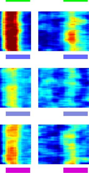
www.nature.com/scientificreports/
(Fig. 1). The LFP traces of evoked potentials were dissimilar among the four recording targets. As displayed in
Fig. 1, the evoked potentials in NCL were slower and had indistinct negative potentials (the valley of NCL peaked
after 250 ms) when compared with those of ENTO (the valley peaked at approximately 250 ms), VW (the valley
peaked at approximately 250 ms), and ROT (nearly no valley occurred). Therefore, the LFPs demonstrated no
contamination from volume conduction. Volume conduction is an electric current transmitting between nearby
brain areas and usually shows no phase delay in LFPs between sources and the nearby areas, as indicated that the
recorded waveforms are similar between different brain areas and may mask the actual LFP signals. The different
shape and response time of evoked potentials among regions in our LFP recordings suggest that volume conduc-
tion did not cause artificial synchronization and the acquired LFPs were real signals. Therefore, it is worth to
further analyze the functional connectivity. It has been demonstrated that gamma oscillations are triggered by
vision through the tectofugal pathway in pigeons42; therefore, we investigated the changes of LFP powers in dif-
ferent spectra after receiving the different 15-color stimuli before further analyzing the functional connectivity.
The LFP spectrograms stimulated by 15 different colors depicted that the strongest responses were elicited after
receiving blue to green lights (Figs. 2A, S2, color No. 6: RGB color (0, 1, 1), and color No. 7: RGB color (0.25, 1,
0.75)). However, if the white background color was used as the baseline, no distinct response was found among
these color stimuli (Fig. S3). The spectrograms of color stimuli displayed a predominant power between middle
and high frequencies from the onset of the trigger to half a second after the trigger (Fig. 2A dashed line box). The
powers between the middle and high frequency bands were increased (as the arrows indicated in Fig. 2A); there-
fore, we referred to rat’s frequency bands and divided the responsive spectrograms into three frequency bands:
the low frequency (< 20 Hz, including the delta, theta and alpha waves), the middle frequency (20–60 Hz, mainly
containing slow gamma waves), and the high frequency (60–100 Hz, mainly containing fast gamma waves)43.
Bar graphs of average powers across 0–500 ms after stimuli depicted that the subjects have low responses to
the reds (colors No.14 or No.15) when comparing to colors No. 6 or No. 7 in the ROT and ENTO (Fig. 2B–D
analyzed by one-way repeated measures ANOVA, Bonferroni-adjusted significance tests for pairwise compari-
sons; see supplementary Table S2, for detailed means ± SEMs, F values, p values, and corrected p values). NCL
also showed strong response to blue (Fig. 2B–D, analyzed by one-way repeated measures ANOVA, Bonferroni-
adjusted significance tests for pairwise comparisons; see supplementary Table S2, for detailed means ± SEMs, F
values, p values, and corrected p values). These results suggested that subjects were sensitive to the illumination
of blue (color No. 6, 7) colors when comparing with red (color No. 14, 15) colors. This phenomenon was only
observed in VW in middle frequency when flashed with No. 14 color (Fig. 2B–D, analyzed by one-way repeated
measures ANOVA; see supplementary Table S2 for detailed means ± SEMs, F values, p values, and corrected p
values). The overall Z-scored powers in ENTO are significantly higher than those of ROT and VW (Fig. 2E–G:
analyzed by one-way ANOVA compared between total of 15 colors among 4 areas, Bonferroni-adjusted sig-
nificance tests for pairwise comparisons. See supplementary Table S3A for detailed means ± SEMs, F values, p
values, and corrected p values). The overall Z-scored power in NCL are also strong (Fig. 2E–G, supplementary
Table S3A). These results suggested that the NCL-ROT pathway plays the major role in processing color infor-
mation. We further created 7 rainbow colors with the same radiation power (Table S1), since the previous 15
colors created by the color codes did not control the radiation power. After adjusting these rainbow colors to an
identical radiation power, the colors were a little bit pale (Fig. 3A; color index on the left; also see "Materials and
Methods" section, Table S1). The radiation-power-controlled rainbow colors still triggered evoked potentials
(Fig. S4, LFP traces). In addition, the rainbow colors still generated strongest power in ENTO and significantly
stronger than VW (Fig. S5A, analyzed by one-way repeated measure ANOVA compared between the total rain-
bow colors among 4 areas, Bonferroni-adjusted significance tests for pairwise comparisons. See supplementary
Table S3C for detailed means ± SEMs, F values, p values, and corrected p values). Since there were some miss-
ing values from VW recording because of the broken recording wires and one-way repeated measure ANOVA
excludes the missing trials, we analyzed the statistical differences by one-way ANOVA. The statistical analysis
still demonstrates significant higher power in ENTO than VW (see supplementary Table S3B for detail statistic
values). The blue colors triggered the highest potentials, especially in ENTO and NCL (Figs. 3B–D, and S5B, ana-
lyzed by one-way repeated measures ANOVA, Bonferroni-adjusted significance tests for pairwise comparisons;
see supplementary Tables S4 and S3D for detailed means ± SEMs, F values, p values, and corrected p values). The
powers in ROT and VW are still relatively low (Fig. 3A–D), which is consistent with the findings using 15-color
stimuli. We were interested in knowing whether blue trigged highest response in ENTO than VW, therefore, we
analyzed z-scored power after the blue stimulation. The data elucidate a significant stronger power in ENTO
than VW (Fig. S5B, One-way repeated ANOVA, see supplementary Table S3D for detailed means ± SEMs, F
values, p values, and corrected p values).
Phase synchronization between the NCL and the tectofugal pathway is stronger. We analyzed
the phase synchronizations between ROT and NCL (ROT-NCL), ENTO and NCL (ENTO-NCL), and VW and
NCL (VW-NCL) by the weighted phase lag index (WPLI) to determine whether the higher-level cognitive-
related brain region, the NCL, involves in processing color stimuli. The WPLI is an index which is insensi-
tive to volume conduction44 and suitable to analyze the levels of synchronization within a small size region
of brain. We calculated WPLI within 500 ms after different color stimuli (Fig. 4A–C) and we also determined
WPLI within 500 ms by all of rainbow color stimuli (Fig. 4D–F). The phase synchronization of the ROT-NCL,
ENTO-NCL, and VW-NCL were not significantly higher compared with other colors when stimulated with blue
(Fig. 4A–C; analyzed by one-way repeated measures ANOVA compared then Bonferroni-adjusted significance
tests for pairwise comparisons. See Table S5 for detailed statistic values). However, the ENTO-NCL pathway
showed significant higher WPLI in all colors when compared with those of VW-NCL, indicating more promi-
nent increases between NCL and relay nuclei in the tectofugal pathway than that between NCL and VW of
Scientific Reports | (2020) 10:19382 | https://doi.org/10.1038/s41598-020-76542-z 4
Vol:.(1234567890)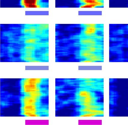
www.nature.com/scientificreports/
A. NCL ROT ENTO VW B. NCL
ROT
0.4
Low frequency band
Zscored Power
NCL ENTO
ENTO VW 0.3
VW
ROT
* **
# ## * #
0.2 ##
0.1
No.06 80 ** **
## ##
40 0
No.07 80 C. -0.1 Middle frequency band
0.4 0.4
Zscored Power
40
0.3 *
* * ##
* # #
#* *
No.13 80 0.2
# #
40
0.2 0.1
** **
No.14 80 0
## ##
40
-0.1
D. 0.4 High frequency band
No.15 80
Zscored Power
0 0.3
40
* #
* * *# #
0.2 ##
80
0.1
40
** **
-0.2 0
## ##
-0.5 0 0.5 -0.5 0 0.5 -0.5 0 0.5 -0.5 0 0.5
-0.1
Time (Sec)
06 07 13 14 15
E. F. G.
Low frequency Middle frequency High frequency
0.3 0.3 *#
0.3
*#
* * *# *# NCL
ROT
ENTO
0.2 0.2 0.2 VW
0.1 0.1 0.1
0 0 0
Figure 2. The averaged spectrograms in the brain regions after stimulated by 15 colors with black color as baseline
between each color. Here we selected results from only red stimuli and blue stimuli for comparing the brain responses
to red and blue. The stimulation trial as the unit to be analyzed. (See Fig. S2 for the complete 15-color-stimuli
spectrograms.) (A) The Z-scored spectrograms of red and blue stimuli. Frequencies (Hz) have shown on the y-axis
and stimulation time point is marked as zero at the x-axis. Dashed line box marks the time between 0 and 500 ms.
The magnitude of power is color-coded and the power scales are plotted under each column. Although the animals
were recorded under anesthesia, the color No. 7 evoked the strongest power and the high energy revealed in different
frequency bands (depicted by arrows). (B,C,D) are the mean of powers between 0 and 500 ms (from the dashed line
boxes in (A)) of low, middle, high frequency bands, respectively. The values were depicted as means ± SEMs. * denotes
the p value < 0.05 when compared to color No. 6; # denotes the p value < 0.05 when compared to color No.7. (One-way
repeated measurement ANOVA, then Bonferroni post hoc comparison). (E,F,G) display the mean of powers between
0 and 500 ms without differentiating the 15-color-stimuli. The values were depicted as means ± SEMs. *Denotes the p
value < 0.05 when compared to the ROT; #denotes the p value < 0.05 when compared to the VW (one-way ANOVA,
then Bonferroni post hoc comparison).
Scientific Reports | (2020) 10:19382 | https://doi.org/10.1038/s41598-020-76542-z 5
Vol.:(0123456789)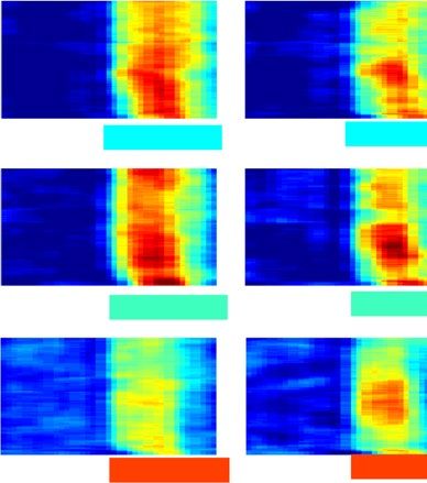
www.nature.com/scientificreports/
A. NCL ROT ENTO VW
NCL
ENTO VW
ROT
NCL
ROT
B. 0.6 ENTO
Red 80 Low frequency band VW
Zscored Power
40 0.4
*** *** *** *** ***
0.2
Orange 80
40 0
-0.1
C. 0.6 Middle frequency band
Yellow 80
Zscored Power
40 0.4
*** *** *** **** **** ***
0.2
Green 80
40 0
-0.1
D. 0.6 High frequency band
Blue 80
Zscored Power
40 0.4
*** *** *** *** *** * *
0.2
Indigo 80
40 0
-0.1
Red Orange YellowGreen Blue Indigo Violet
Violet 80
40
-0.5 0 0.5 -0.5 0 0.5 -0.5 0 0.5 -0.5 0 0.5
Time (Sec)
-0.2 0 0.2 0.4
Zscored Power
Figure 3. The averaged spectrograms in the brain regions after stimulated by rainbow colors with black color as
baseline between each color. (A) The Z-scored spectrograms of baseline and rainbow color stimuli. Frequencies
(Hz) have shown on the y-axis and stimulation time point is marked as zero at the x-axis. The magnitude of
power is color-coded and the power scales are plotted under each column. We further confirmed the results
from the 15-color-stimulation by using the rainbow colors with similar radiation intensity. (B,C,D) are averaged
powers within the time period (0–500 ms) of low, middle, and high frequency bands. The values were depicted
as means ± SEMs. *Denotes the p value < 0.05 when compared to blue. (one-way repeated measurement
ANOVA, then Bonferroni post hoc comparison; stimulation trials as the unit to be analyzed).
the thalamofugal pathway (Fig. 4D–F; one-way ANOVA, Bonferroni-adjusted significance tests for pairwise
comparisons. See Table S6A for detailed statistic values). One-way repeated measures ANOVA were also used
and demonstrated higher ENTO-NCL synchronization than VW-NCL in middle and high frequency (Fig. S5C,
Table S6B for detailed statistic values). Again, blue was extracted and further analyzed (Fig. S5D, Table S6C for
detailed statistic values; One-way repeated measures ANOVA). The results demonstrated significant stronger
phase synchronization in ENTO-NCL than that of VW-NCL in the high frequency. These results imply that
the color signal transmission was mediated by the tectofugal pathway to NCL. To further confirm the transmis-
sion pathway and connectivity, we injected the non-trans-synaptic retrograded tracer, fluorogold, into NCL to
confirm the anatomical afferent projections to NCL (Fig. S6A–C). The brain histology showed that the bilateral
ENTOs exhibit prominent fluorescent signals after injecting fluorogold bilaterally to NCL. Projections from the
striatum were also noticed. In contrast, there was a mild retrograded signal in the caudal part of VWs (Fig. S6B).
To determine the hemispheric integration of these pathways, we injected fluorogold into the left NCL and incu-
bated for additional two weeks. We found that most of the signals were noticed in the mesopallium, and there
was few fluorogold signal in the left cranial part of VW (Fig. S6C). This retrograde staining result implies that
the afferent projection to NCL is predominant from ENTO, but not from VW.
Scientific Reports | (2020) 10:19382 | https://doi.org/10.1038/s41598-020-76542-z 6
Vol:.(1234567890)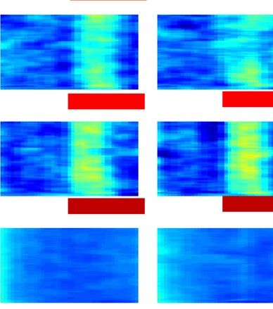
www.nature.com/scientificreports/
NCL
ENTOVW
ROT
A. Low frequency band D. Low frequency band
ROT-NCL NCL NCL NCL
ENTO-NCL ENTO
0.15 VW-NCL 0.1 ROT VW
Debiased WPLI
*
Debiased WPLI
0.05
0 0
-0.05
-0.1
Middle frequency band Middle frequency band
* *
B. E. 0.1
0.15
0.05
Debiased WPLI
Debiased WPLI
0 0
-0.05
-0.1
High frequency band
High frequency band
F. 0.1 *
C. 0.15 * *
Debiased WPLI
0.05
Debiased WPLI
0 0
-0.05
-0.1
e
w
go
en
et
ng
ue
d
lo
ol
Re
di
re
ra
Bl
l
Vi
Ye
In
G
O
Figure 4. Phase synchronization between the NCL and other 3 brain regions after stimulated by rainbow
colors with black color as baseline between each color. (A,B,C) are the means of WPLIs within the time
period (0–500 ms) for the 3 frequency bands. The stimuli of rainbow colors are depicted under the panels. The
values were depicted as means ± SEMs. (D,E,F) display the means of WPLIs between 0 and 500 ms without
differentiating the rainbow-color-stimuli. *Denotes the p value < 0.05. (one-way ANOVA, then Bonferroni post
hoc comparison; stimulation trials as the unit to be analysis).
Color information is processed in the direction from tectofugal pathway to NCL. We further
assessed the functional connectivity between the two pathways and NCL to reveal the directional interactions
when the subjects were stimulated by different colors (Fig. 5A). The overall Granger causality for both directions
are significantly higher in the ENTO ⇔ NCL (Fig. 5B,C, Table S7A for detailed statistic values and Bonferroni
post-hoc tests of one-way ANOVA). One-way repeated ANOVA demonstrated the significant strongest Granger
causality in the ENTO ⇔ NCL as well if we pooled all the results of all rainbow colors (Fig. S7A, Table S7B
for detailed statistic values). This phenomenon is still significant if we only analyzed blue stimuli (Fig. S7B,
Table S7C for detailed statistic values). The increase of Granger causality was observed in a direction from ENTO
Scientific Reports | (2020) 10:19382 | https://doi.org/10.1038/s41598-020-76542-z 7
Vol.:(0123456789)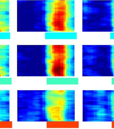
www.nature.com/scientificreports/
NCL NCL NCL NCL
ENTO VW
ROT VW
ENTO
A. ROT ROT-NCL
ROT NCL ENTO NCL VW NCL ENTO-NCL
VW-NCL
0.02
0.04
0.02
Red
B.
Direction to NCL
0
0
0
* *
0.02
0.04
0.02
*
Orange 0.02
0.04 0
0.02 0
0.02 0
G.C.
Yellow 0.01
0
0
0
0.02
0.04
0.02
G.C.
Green 0
0
0
0
C. Direction from NCL
0.02
0.04
0.02
Blue * *
0.02 *
0.02 0
0.04 0
0.02 0
G.C.
Indigo
0.01
0
0
0.02 0
0.02
0.04
Violet
0
0
0
0
-1 0 1 -1 0 1 -1 0 1
Time (Sec)
NCL NCL
D. Direction from NCL * Direction to NCL
*
*
0.024
0.02
0.016
G.C.
0.012
0.008
e
e
w
w
go
go
en
et
ng
en
et
ng
ue
d
ue
d
llo
llo
ol
Re
ol
Re
di
re
di
re
ra
ra
Bl
Bl
Vi
Ye
Vi
Ye
In
G
In
G
O
O
Figure 5. Granger causality between the NCL and other 3 brain regions after stimulated by rainbow colors
with black color as baseline between each color. (A) The dynamics of Granger causality between the NCL
and other 3 regions. The data in the y-axis are the value of Granger causality before and after stimulating
with rainbow colors. The means ± SEMs are displayed by solid lines and shades, where blue lines are Granger
causality direction from each 3 regions to the NCL and red lines are the opposite direction. Please note that
it must sample a period of time for calculating the Granger causality. We took every 0.5 s with 0.05 s moving
steps to measure the dynamics (see the Method section); therefore, the time resolution was not as precise as the
LFP traces and resulted in the peak G.C.s before 0 s. And also, the scale for ENTO-NCL is different for others
(B,C) show the mean G.C. between 0 and 500 ms without differentiating the rainbow-color-stimuli. The values
were depicted as means ± SEMs. *Denotes the p value < 0.05. (one-way ANOVA, then Bonferroni post hoc
comparison; stimulation trials as the unit to be analysis). (D) represents the mean of G.C. between 0 and 500 ms
after the stimulation of rainbow colors. The values were depicted as means ± SEMs. *Denotes the p value < 0.05.
(paired t test).
Scientific Reports | (2020) 10:19382 | https://doi.org/10.1038/s41598-020-76542-z 8
Vol:.(1234567890)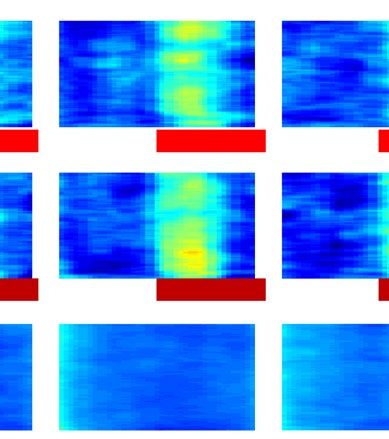
www.nature.com/scientificreports/
to NCL during blue stimuli (Fig. 5D, two-tailed paired t-test, see Table S8B for detailed degree of freedom, t
values and p values). Similar finding could also be found after indigo (Fig. 5D, Table S8B) and violet stimuli
(Fig. 5D, Table S8B), but not after other color stimuli. These results suggest that the LFPs recorded from ENTO
are leading the LFPs in NCL, because the direction from ENTO to NCL demonstrated the highest Granger
causality and implied the color information is processed from the tectofugal pathway to NCL. Finally, it is also
interesting to determine whether ROT transfers information to ENTO, because ROT is a relay nucleus of tec-
tofugal pathway. Our data demonstrated that the direction from ROT to ENTO had a higher Granger causality
than that of the direction from ENTO to ROT after the stimulation of rainbow colors (Fig. S7C, Table S7D for
detailed statistic values) or blue color (Fig. S7D, Table S7E for detailed statistic values).
Discussion
ENTO and VW are respectively the telencephalic areas of tectofugal pathway and thalamofugal pathway. How-
ever, it is still unclear whether higher order avian brain areas, such as NCL, communicate with them or not. We
hypothesized that the telencephalons of visual pathways communicate with higher order brain areas in zebra
finches and the ENTO has stronger communication with the NCL than VW, when stimulated by colors. We
initially were expecting prominent responses in ENTO when zebra finches see colors. The present study demon-
strated that the ENTO generates stronger power than those of ROT and VW when the right eye was stimulated by
colors. In addition, the NCL also represented strong power. When we further analyzed the functional connectiv-
ity between ENTO and NCL, we found that color stimuli enhanced synchronization between ENTO and NCL
in the direction from ENTO to NCL. Interestingly, the synchronization between VW and NCL were negatively
synchronized, suggesting these two areas communicated poorly with each other37. Moreover, blue is the color
that evoked the strongest power in the direction from ENTO to NCL; the Granger causality also demonstrated
the LFPs acquired from ROT leaded the consequent LFPs of ENTO. However, the different colors did not result
in different phase synchronization between NCL and other three regions. We were also interested in learning
what colors zebra finches are sensitive to. Indeed, we hypothesized that their visual pathway is sensitive to red,
but we cannot ignore the fact that blue is important for finding sky or water and green may be important for
searching plants. Surprisingly our results partially support that blue is relatively important to zebra finches. These
results also raise some interesting questions about the physiological or ecological advantages of “blue” and also
the physiological functions of the tectofugal pathway, the thalamofugal pathway, and NCL for zebra finches. The
LFP experiments are mainly done in rodent models; therefore, we firstly applied findings from rodents to explain
our current results. Then, we discussed some potential physiological meanings for zebra finches.
The LFP spectrograms. We employed the results of LFPs to determine our hypothesis. The LFP is a sum-
mation of various potentials such as membrane potential and action potentials. The synaptic potential is often
the main source of LFP38. The LFP contains multi-dimensional information including frequency, amplitude,
phase, and time. Therefore, the present study analyzed several aspects of the LFPs. In the spectrograms, we
noticed that blue evoked a strongest power within 0–500 ms after the stimuli, especially in ENTO and NCL.
Strong LFP power (or amplitude) usually suggests that the target areas are in a very active status38. In addi-
tion, the color stimuli evoked approximately three frequency bands. The LFP frequency is an unique way for
communicating between brain regions and each frequency band may encode different information and has its
own physiological functions39. We analyzed the frequency bands to explore the potential communication of
color information between ENTO and NCL. Our data demonstrated that the middle and high frequency bands
revealed the power differences between ENTO and VW. In general, low frequency oscillations are propagated
farther, given that the cell membrane is a low pass filter37. In the other hand, high frequency oscillations provide
a more temporally organized transmission than low frequency oscillations40,41. We cannot conclude that the
color information is not transmitted between VW and NCL, since we still acquired significant potentials in VW.
However, the evidence from phase synchronization and Granger causality support that the ENTO-NCL is more
sensitive to colors.
Phase synchronization and directional connectivity. The phase of LFP also encodes information
for propagating between brain regions37. Synchronizations help the communication between different brain
regions45. Although we found no color preference of phase synchronization, the ENTO-NCL still revealed
strong positive synchronization, whereas VW-NCL showed weak or negative synchronization. Even though the
blue evoked the highest activities in the ENTO and NCL, the WPLI cannot reflect the difference of amplitudes
because WPLI only measures the phases of LFPs. We postulated that the phase synchronization encoded infor-
mation of stimuli, but not colors. In the other word, the communications between ENTO and NCL increased,
which paved a way for color information but the color itself was encoded by amplitudes. The weak communica-
tion between VW-NCL implys that the tectofugal pathway is the main path in response to color stimulation.
The directional connectivity also suggests a strong information transmits from ENTO to NCL when zebra finch
received blue stimulation.
Sensitivity to blue stimulation. We demonstrated that the male zebra finch is sensitive to blue lights. The
retina studies have shown that several species of birds are sensitive to both yellow and UV lights, but some spe-
cies show their maximal sensitivity at the green s pectrum46. Therefore, the spectral sensitivity in birds may vary
among species. Bennett et. al. reported that UV vision dominantly contributes to the mate-selection in zebra
finches47. Our data could not rule out the possibility of sensitivity for UV light in the zebra finch, but we believe
our results shed some light on zebra finches’ color preference. Additionally, a recent study also demonstrates the
zebra finches’ retina is sensitive to blue as well48.
Scientific Reports | (2020) 10:19382 | https://doi.org/10.1038/s41598-020-76542-z 9
Vol.:(0123456789)www.nature.com/scientificreports/
Our results also showed that the color-evoked potentials were significantly detected when we used black as
baseline, rather than white color, between different colors. It is possible that white baseline, which contains all
spectra of the visible lights, may saturate the visual response and let the visual pathways do not respond to the
subsequent colors. There is also a behavior report indicates that the contrast of the background affects color
discrimination in zebra finches49. Although the background used in the present study are not parallel to the
back or white baseline, it is likely that the responses to colors are modulated by recent or adjacent colors. To
minimize the confounding factor, we stimulated the subjects with colors in random orders. The intensities of
white or other colors may also affect the brain activities. A study regarding the spectral sensitivity in avian retina
reports that increasing intensity of certain color shifts the maximum electroretinal potential toward the shorter
wavelengths46. Therefore, we manually adjusted the intensities of different colors to be the same and acquired
their evoked potentials. The limitation of this study is the light source, since we did not use a light with narrow
wavelength band such as laser. We simply used a laptop, which generates colors based on a human’s vision system
(i.e. trichromatic colors). Even though this study did not stimulate the visual pathway with a precisely narrow
wavelength band and no invisible light (for humans) was generated by the laptop, the results still shed light on
their brain activities when stimulated by trichromatic colors. We expected more complicated responses will be
observed when stimulated by mixing the UV light with trichromatic colors47.
In addition, the color perception in zebra finches is a complicated issue. Our report raises several new ques-
tions needed to be discuss. For instances, what the limitation is for using a monitor which generates RGB colors
to stimulate the animals with tetrachromatic vision. Do colors influence the zebra finches’ perceptive brightness
and cause the strongest evoked LFPs from blue? Although the functions of the avian brain are species-specific,
we reviewed some studies from other bird species and tried to reveal the potential answers of whether the violet
cones (or ultraviolet cones in finches48) interact with the S-, M-, and L-cones in the tetrachromatic vision of bird.
For humans, the purple from the monitor stimulates both red- and blue-sensitive cone cells and humans inter-
pret it as purple50. The purple from the laptop monitor is a mixing of red and blue, not a pure short wavelength
light. Therefore, we should roughly interpret the rainbow colors as: red, red + green (which generate orange and
yellow), green, blue, blue + red (which generate indigo and violet) (Table S2 for the detail combination of RGB).
Recently, a study from hummingbirds constructs an avian tetrahedral color s pace51 and proves their ability to
discriminate UV. Our study just implied that the stimulation on the zebra finch S-cones evoked a strong response,
but still cannot rule out the possibility that UV can generate strongest visual responses in their brain. On the
other hand, despite we adjusted the rainbow colors to have the same radiation power, it is still unclear how blue
color generates strongest responses in the brain. In human, we feel yellow is brighter than red, green, blue, even
they are displayed by the same radiation power. We proposed that the mechanism of feeling different brightness
among colors in human is not the cause of strong blue response in the zebra finch. The sensitivity spectrum of
human’s green- and red-sensitive cone cells is highly overlapped52. Therefore, yellow is able to stimulate more
cones than other colors. But the sensitivities of zebra finches’ cone cells are evenly distributed across spectrum48.
Thus, we think the strong blue response is not a phenomenon of cross-reaction between different types of cone
cells. Indeed, study from the oil droplets of zebra finches implies that zebra finches have higher cone spectral
sensitivities for blue and UV than red and g reen48. Our data obtained from the brain activities further support
this result from the retina’s study.
Although present study did not explain why the brain of zebra finch is more active to blue, their nature habitat
may hint the potential reasons. Zebra finch is a diurnal animal and lives in relative arid areas53. We hypothesized
that water and sky are key factors for zebra finch surviving, so they need to be spotted as fast as possible. During
the experiment we also observed that zebra finches became quite and standstill if we turn off the room light.
We think, as the room light was off, zebra finches were searching for sky for flying toward. Besides, researchers
discover that zebra finches have blue and UV light-dependent magnetic compass, suggesting that blue light is
critical for their navigation16.
Tectofugal pathway, thalamofugal pathway and NCL. Our findings indicated that the color-evoked
potentials were stronger in the tectofugal pathway than the thalamofugal pathway. Moreover, the synchroniza-
tion between the relayed nuclei of the tectofugal pathway and NCL was also stronger. These results are similar
to some studies using pigeons as subjects, in which lesion of ROTs impairs the color discrimination9 and some
OT54. In addition to ROT, about 30% of tectal units are able to respond to
color sensitive units are also found in R
certain wavelengths17. Although we discovered that ENTO (a downstream of ROT) represents stronger activities
than those of ROT, we cannot exclude the roles of ROT for processing color information. In our result, we dem-
onstrated that NCL responses were highly correlated with ENTO but the correlation is relatively low between the
NCL and ROT. We think ROT did not reveal as strong response as ENTO because ENTO needs more intensive
communication with NCL. This hypothesis is supported by the synchronization and directional connectivity
results of NCL-ENTO. With regard to the thalamofugal pathway, color-sensitive units have been discovered in
the ventral lateral geniculate nucleus, which consists of inputs from both retinas and V W55. Bredenkotter and
Bischof used 1-ms flash to evoke and record the field potentials of VW and ENTO, and found the amplitudes
recorded from the contralateral hemispheres are similar56. A lesion study demonstrates that the VW in zebra
finch involves in spatial information processing and ENTO analyzes the pattern of o bjects57. The zebra finches’
VW even perceive vision mediated by earth magnetic field orientation58. It is still unclear whether the VW
modulates other cognitive functions related to vision, but researchers demonstrate the important role of VW
in imprinting for c hicken59. These pieces of evidence may support our hypothesis that the tectofugal pathway is
much critical than the thalamofugal pathway in regard to the color information processing, since colors are also
important cues for discriminating objects. It would be also of interest to simultaneously record from the ventral
Scientific Reports | (2020) 10:19382 | https://doi.org/10.1038/s41598-020-76542-z 10
Vol:.(1234567890)www.nature.com/scientificreports/
lateral geniculate nucleus. However, because of the limitations of channel and skull space, we selected only to
record the relay nucleus (ROT) of tectofugal pathway.
Conclusions
Our result suggests that the communication between nidopallium caudolateral and tectofugal pathways is crucial
for color discrimination. Moreover, ENTO and NCL are more active when the eyes are stimulated by blue and
the visual information was transmitted from the direction of ENTO to NCL.
Materials and methods
Animals. In the experiments, the male zebra finches (n = 9, 5 to 8-month-old) were acquired from the com-
mercial bird breeders (San-Xing Bird Store, Taipei, Taiwan). The birds were housed in home cages individually.
The temperature of the environment temperature was controlled at 23 ± 1 °C, and the light–dark cycle was main-
tained under nature light (AM 7:00 light, PM 7:00 dark; summer). Food and water were available ad libitum.
All procedures performed in this study were approved by the National Taiwan University Animal Care and Use
Committee, approval ID: NTU-106-EL-026. All methods described in this paper were performed in accordance
with the guidelines and regulations of National Taiwan University Animal Care and Use Committee.
Surgery and electrophysiological data collection. After at least a 7-day accommodation in their
home cages, the zebra finches were randomly selected for the recordings of evoke potentials. These finches stayed
in an induction box, which provided pure oxygen for 10 min to raise their blood oxygen levels. Subsequently,
they were intraperitoneally administrated butophanol (2 µg/g) and midazolam (2 µg/g) for analgesia and muscle
relaxing. Anesthesia was induced by 2% isoflurane mixed with oxygen. Once they lost their reflex of deep pain,
the subjects were fixed on the stereotaxic instrument and maintained anesthesia with 1.5% isoflurane60. Four
tetrodes, consisting of two twisted 0.05 mm stainless steel wires (California Fine Wire, Grover Beach, CA) in
each one, were implanted into the NCL (AP, 1.0 mm; ML, − 4.5 mm; DV, 4.0 mm relative to y point), the ENTO
(AP, 3.0 mm; ML, − 3.6 mm; DV, 3.3 mm relative to y point), and the ROT (AP, 2.6 mm; ML, − 2.0 mm; DV,
5.0 mm relative to y point), and the VW (AP, 5.5 mm; ML, − 2.0 mm; DV, 1.5 mm relative to y point) in the left
hemisphere. The coordinates were selected based on A stereotaxic atlas of the brain of the zebra finch by Nixdorf-
Bergweiler and Bischof61. The tetrodes were connected to an interface board which linked to a head stage and
tether to the preamplifier of OmniPlex A system (omniplex version 1.2.0, https://plexon.com/products/omnip
lex-software, Plexon, Dallas, TX, USA). One grounding screw was anchored on the rostral part of right frontal
skull. During the recording, the grounding electrode was used as a reference and the LFP signals were ampli-
fied, digitalized, and recorded by a 16 channels OmniPlex A system. The raw LFP signals were bandpass filtered
between 0 and 500, amplification time was set at 2500, and the sampling rate was 40 kHz. These digitalized data
then down sampled to 2000 Hz for storage. Some reports demonstrate that zebra finches use their right eye to
choose their mates which have colorful beaks and f eathers28. To test if the left visual pathways of zebra finches are
sensitive to certain colors, we flashed 15 colors or 7 rainbow colors to the right eyes of birds. The eyelids of the
right eyes were opened and fixed with #6-0 surgical sutures. The color flashing time stamps were also integrated
and stored in OmniPlex A system by commercial recording software OmniPlex (version 1.2.0, Plexon). For
detailed color flashing method, see the section of color flashing. All of the post-recording data were analyzed by
custom written code in MATLAB R2016b (MathWorks, Natick, MA, USA).
Color flashing. Fifteen colors were generated using MATLAB code: jet(15), which makes RGB codes from
blue to green to red. (The RGB color codes are: (0, 0, 0.75), (0, 0, 1), (0, 0.25, 1), (0, 0.5, 1), (0, 0.75, 1), (0, 1, 1),
(0.25, 1, 0.75), (0.5, 1, 0.5), (0.75, 1, 0.25), (1, 1, 0), (1, 0.75, 0), (1, 0.5, 0), (1, 0.25, 0), (1, 0, 0), and (0.75, 0, 0)).
We further created 7 rainbow colors with the same radiation power (Table S1), because the 15 colors did not
generate the same radiation power. The RGB color codes of the rainbow colors are: red: (1, 0.47, 0.47), orange:
(1, 0.58, 0), yellow: (0.86, 0.68, 0), green: (0, 1, 0), blue: (0.4, 0.4, 1), indigo: (0.51, 0.54, 0.87), and violet: (0.85, 0,
0.85). The intensity of radiation powers (Table S1) were confirmed by light meter (model: MR-16, RAINBOW-
LIGHT, Taiwan). For the comparison between these colors and black/white colors, four kinds of color sequences,
which are black order (BO), black random (BR), white order (WO), and white random (WR), were used in the
experiment. The color sequences in the BO were used above mentioned sequence of color codes (either 7 or 15
colors) and inserted black color between colors. The pattern of BR is to rule out the confounding factor caused
by the order of colors. We randomly picked 15 (or 7) colors without regular sequence and inserted black color
between colors. The color sequences of WO and WR are similar to BO and BR, except that we inserted white
color between colors. These colors were flashed on a laptop (model: SVP132A1CP, SONY) placed in front of the
subjects’ right eyes (the monitor was approximately 30 cm away from the subject). Each color, including black or
white colors, emerged for 2 s and then switched to another color in a sequence (order or random) as described
earlier. Each subject received a total of 80-time stimuli of color sequences (20 trials in each BO, BR, WO, and
WR). The pilot tests had indicated that zebra finches sobered up when isoflurane was below 1%; thus, we main-
tained the concentration of isoflurane between 1 and 1.5% during the whole experiment. At the end of the exper-
iment, we euthanized the subject with intraperitoneal administration of Zoletil (Tiletamine:Zolazepam = 1:1
Virbac, Carros, France). Their brains were collected and soaked in 10% formalin to confirm the location of
implanted electrodes.
LFP analysis. We extracted the time stamps of every color-changing point and extended the time ± 1 s. The
local field potentials in the ± 1-s periods were used for further analysis as described below.
Scientific Reports | (2020) 10:19382 | https://doi.org/10.1038/s41598-020-76542-z 11
Vol.:(0123456789)www.nature.com/scientificreports/
Evoked potentials (LFPs) and power estimation. The LFPs were Z-scored and averaged across the same color
for measuring the evoked potentials, and the negative Z-scored power means it is below the average power.
The power spectrograms were analyzed with the multitaper method from the open-source MATLAB toolbox
Chronux62. Because the spectrograms need a period of samples and step a short time to create a dynamic of
spectrum as function of time. We used 0.5-s windows with 0.05-s overlapping steps, set the time-bandwidth
product at 3, and set the number of tapers at 5. We also extracted and averaged the values of stimuli between 0
and 500 ms for testing the statistic differences among stimuli or brain regions.
Phase synchronization across regions. The levels of synchronization between the ROT-NCL, ENTO-NCL, and
VW-NCL were evaluated with a debiased estimator of the squared weighted phase lag index (WPLI)44. The codes
can be download from the open source tool box, Fieldtrip https://www.fieldtriptoolbox.org/download.php63.
The WPLI analyzes an imaginary component of the spectrum across two LFPs, because it not only relates to the
phase synchronization between two LFPs but is also insensitive to noise or contamination from volume conduc-
tion. Compared with classical coherence, this estimator analyzes the phase synchronization across brain regions
and minimizes the effect of volume conduction contamination and sample size bias44. The WPLI normalized the
two LFP with perfect synchronization to 1 and completed out of phase to -1. In order to access the WPLI in a
similar manner with multitaper power estimation, we also broke the ± 1-s periods into 0.5-s windows with 0.05-s
step and estimated the WPLIs. We extracted and averaged the values of each stimulation during 0–500 ms for
testing the statistic differences among stimuli or brain regions.
Granger causality. The function connectivity between the ROT⇔NCL, ENTO⇔NCL, and VW⇔NCL were
accessed by using Granger causality. We adapted the open-source MATLAB toolbox developed by Barnett and
Seth64. It is also due to the input samples must contain a period of time to generated a Granger causality dynamic
over time, which is similar to the method described in the power spectrogram and the WPLI, we calculated
the Granger causality for the time domain every 0.5 s with 0.05-s steps for measuring the dynamics of Granger
causality. We extracted and averaged the values of each stimulation during 0–500 ms for testing the statistic dif-
ferences among stimuli or brain regions.
Histology and retrograde tracing of the afferents to the NCL. For tracing the afferent projections
to NCL, a retrograde tracer, 4% fluorogold (Sigma Chemical), was microinjected into NCL in two birds. One
subject was bilaterally (the NCL; AP, 1.0 mm; ML, ± 4.5 mm; DV, 4.0 mm relative to y point) injected with
fluorogold using microinjection syringe pump at a speed of 1 µl/10 min. We administered 0.33 µl of fluorogold at
each site and waited for 1 min, then moved the tip of the needle up for 100 µm and administered another 0.33 µl
again and waited another 1 min, and repeated the procedure once again. Two weeks after injections, this bird
was euthanized by isoflurane. The brain was removed and dissected into a 30-µm coronal section by a cryostat
microtome. Some brain slices were stained with DAPI. We used an ultraviolet filter in the inverted microscope
(IX83; Olympus, Tokyo, Japan) to detect the fluorescent reaction of fluorogold. Since the fluorescent reaction in
VW was not high in this bird (Fig. S6B), we double confirmed this result with anther finch, which was microin-
jected with 4% fluorogold to the left NCL and waited for 4 weeks to let fluorogold travel a longer distance. The
brain slices for confirming the locations of the implanted electrodes were also dissected into a 30-µm coronal
section by the cryostat microtome. The pictures in Fig. S6E–H were taken under stereo microscope without
staining. The coordinates were adopted from A stereotaxic atlas of the brain of the zebra finch61.
Results and statistics
All results in the figures are depicted as the means ± SEMs. The results of statistical analyses were done by SPSS
(Version: 10.0.7, IBM, New York, USA). The stimulation trials were used as unit to be analyzed. We initially used
one-way ANVOA to compared the differences between brain areas. In addition, we excluded the miss values of
VW in one bird and retested differences between brain areas with one-way repeated measures ANOVA. Bon-
ferroni post hoc comparison was used if the ANOVA test indicates a significant difference. For measuring the
difference between the two causality directions, we used two-tailed paired t-tests.
Received: 15 May 2020; Accepted: 29 October 2020
References
1. Zeigler, H. P. & Bischof, H. J. Vision, Brain, and Behavior in Birds (MIT Press, Cambridge, 1993).
2. Collins, S. A., Hubbard, C. & Houtman, A. M. Female mate choice in the zebra finch—the effect of male beak colour and male
song. Behav. Ecol. Sociobiol. 35, 21–25 (1994).
3. Burley, N. & Coopersmith, C. Bill color preferences of zebra finches. Ethology 76, 133–151 (1987).
4. Wylie, D. R., Gutierrez-Ibanez, C., Pakan, J. M. & Iwaniuk, A. N. The optic tectum of birds: mapping our way to understanding
visual processing. Can. J. Exp. Psychol. 63, 328–338 (2009).
5. Gandhi, N. J. & Katnani, H. A. Motor functions of the superior colliculus. Annu. Rev. Neurosci. 34, 205–231 (2011).
6. Remington, L. A. Clinical Anatomy and Physiology of the Visual System 3rd edn. (Elsevier/Butterworth-Heinemann, Amstredam,
2012).
7. Karten, H. J., Hodos, W., Nauta, W. J. & Revzin, A. M. Neural connections of the “visual wulst” of the avian telencephalon. Experi-
mental studies in the piegon (Columba livia) and owl (Speotyto cunicularia). J. Comp. Neurol.. 150, 253–278 (1973).
8. Clark, W. J. & Colombo, M. The functional architecture, receptive field characteristics, and representation of objects in the visual
network of the pigeon brain. Prog. Neurobiol., 101781 (2020).
9. Hodos, W. Color discrimination deficits after lesions of the nucleus rotundus in pigeons. Brain Behav. Evol. 2, 185–200 (1969).
Scientific Reports | (2020) 10:19382 | https://doi.org/10.1038/s41598-020-76542-z 12
Vol:.(1234567890)You can also read