Fibrosis in Arrhythmogenic Cardiomyopathy: The Phantom Thread in the Fibro-Adipose Tissue
←
→
Page content transcription
If your browser does not render page correctly, please read the page content below
REVIEW
                                                                                                                                                    published: 03 April 2020
                                                                                                                                            doi: 10.3389/fphys.2020.00279

                                                Fibrosis in Arrhythmogenic
                                                Cardiomyopathy: The Phantom
                                                Thread in the Fibro-Adipose Tissue
                                                Angela Serena Maione 1* , Chiara Assunta Pilato 1 , Michela Casella 2 , Alessio Gasperetti 2,3 ,
                                                Ilaria Stadiotti 1 , Giulio Pompilio 1,4 and Elena Sommariva 1
                                                1
                                                  Vascular Biology and Regenerative Medicine Unit, Centro Cardiologico Monzino IRCCS, Milan, Italy, 2 Heart Rhythm Center,
                                                Centro Cardiologico Monzino IRCCS, Milan, Italy, 3 University Heart Center, Zurich University Hospital, Zurich, Switzerland,
                                                4
                                                  Department of Clinical Sciences and Community Health, University of Milan, Milan, Italy

                                                Arrhythmogenic cardiomyopathy (ACM) is an inherited heart disorder, predisposing to
                                                malignant ventricular arrhythmias leading to sudden cardiac death, particularly in young
                                                and athletic patients. Pathological features include a progressive loss of myocardium
                                                with fibrous or fibro-fatty substitution. During the last few decades, different clinical
                                                aspects of ACM have been well investigated but still little is known about the molecular
                                                mechanisms that underlie ACM pathogenesis, leading to these phenotypes. In about
                                                50% of ACM patients, a genetic mutation, predominantly in genes that encode
                            Edited by:          for desmosomal proteins, has been identified. However, the mutation-associated
                      Martina Calore,           mechanisms, causing the observed cardiac phenotype are not always clear. Until now,
    Maastricht University, Netherlands
                                                the attention has been principally focused on the study of molecular mechanisms that
                        Reviewed by:
                                                lead to a prominent myocardium adipose substitution, an uncommon marker for a
                 Michelle S. Parvatiyar,
Florida State University, United States         cardiac disease, thus often recognized as hallmark of ACM. Nonetheless, based on
                   Raffaella Lombardi,          Task Force Criteria for the diagnosis of ACM, cardiomyocytes death associated with
  University of Naples Federico II, Italy
                                                fibrous replacement of the ventricular free wall must be considered the main tissue
                 *Correspondence:
               Angela Serena Maione             feature in ACM patients. For this reason, it urges to investigate ACM cardiac fibrosis.
               angela.maione@ccfm.it            In this review, we give an overview on the cellular effectors, possible triggers, and
                                                molecular mechanisms that could be responsible for the ventricular fibrotic remodeling
                   Specialty section:
         This article was submitted to          in ACM patients.
          Striated Muscle Physiology,
                                                Keywords: arrhythmogenic cardiomyopathy, cardiac fibrosis, cardiac extracellular matrix, scar formation, cellular
               a section of the journal
                                                effectors
                Frontiers in Physiology
         Received: 02 January 2020
          Accepted: 12 March 2020               INTRODUCTION
           Published: 03 April 2020
                               Citation:        Arrhythmogenic cardiomyopathy (ACM) is a rare genetic cardiac disease, with an incidence
     Maione AS, Pilato CA, Casella M,           estimated in 1:5000 (Corrado et al., 2017), which affects predominantly the right ventricle (RV),
   Gasperetti A, Stadiotti I, Pompilio G        although left or biventricular forms have been also described. In about 50% of ACM patients,
     and Sommariva E (2020) Fibrosis
                                                a genetic mutation can be identified, mostly in genes coding for cardiac desmosomes. Non-
  in Arrhythmogenic Cardiomyopathy:
                   The Phantom Thread
                                                desmosomal forms of ACM also exist. The mode of inheritance is generally autosomic dominant,
           in the Fibro-Adipose Tissue.         even if recessive syndromic forms are also described (Stadiotti et al., 2019). However, these different
                 Front. Physiol. 11:279.        genetic determinants lead to a similar disease phenotype. All forms of ACM are characterized by
      doi: 10.3389/fphys.2020.00279             incomplete penetrance and variable expressivity even in carriers of the same causative mutation.

Frontiers in Physiology | www.frontiersin.org                                          1                                                April 2020 | Volume 11 | Article 279
Maione et al. Arrhythmogenic Cardiomyopathy and Fibrosis
This characteristic likely means that different factors, such as presence and extent of fibrotic replacement. For these reasons,
genetic background, or environmental determinants, contribute magnetic resonance imaging (MRI) is currently increasingly
to define the clinical phenotype. From an anatomo-pathological recommended for a definitive diagnosis (de Boer et al., 2019).
point of view, ACM hearts show a progressive loss of MRI allows to asses ventricular volumes, systolic function, and
myocardium, inflammatory infiltrates, and fibrous or fibro- regional wall motion that are included in diagnostic criteria
fatty replacement. Such tissue heterogeneity predisposes to re- (Marcus et al., 1982, 2010). Moreover, MRI can detect adipose
entrant electrical activity that is known to support ventricular tissue, and, thanks to gadolinium delayed contrast enhancement
arrhythmias, cause, in the worst-case scenario, of sudden cardiac acquisition, MRI is the gold-standard exam to characterize
death. Specifically, cardiac fibrosis is originally a protective myocardial tissue in terms of fibrosis, fatty infiltration, and
mechanism against injury, but its uncontrolled progression fibrofatty scar (Kim et al., 1999, 2000; Tandri et al., 2002, 2005).
may lead to excessive collagen deposition and myocardial scar Invasive tissue characterization to confirm ACM diagnosis can
formation. The fibrotic molecular mechanisms are known for be obtained by an endomyocardial biopsy (EMB). In this setting,
cardiac diseases but those specific for ACM still need to be an extensive application of EMB has been limited by the low
investigated in order to uncover therapeutic targets to improve sensitivity of biopsies usually obtained from the interventricular
ACM clinical management. septum, not frequently involved by the disease. In the last few
years, EMB is performed after a complete and detailed 3D
electroanatomic mapping (EAM) of the ventricular chamber.
CLINICAL ASPECTS OF FIBROSIS IN EAM is used to detect bipolar and/or unipolar low potential
ACM PATIENTS areas, which, in ACM, mostly correspond to fibrotic or fibro-
adipogenic scar tissue (Santangeli et al., 2012; Casella et al., 2015).
Arrhythmogenic cardiomyopathy is a rare cardiac pathology Thus, a preliminary EAM allows to directly identify fibrotic
characterized by cardiomyocytes (CM) death and replacement substitution areas and perform EMB in the portion of RV wall
of myocardium with fibrotic or fibro-fatty tissue. The fibrotic in the immediate adjacency of the scar (Casella et al., 2017).
and fibro-fatty substitution, regardless of the ventricular However, EAM and EAM-guided EMB are not yet recognized in
district, progresses from epicardium to endocardium provoking task force guidelines (Santangeli et al., 2011).
structural and functional myocardial alterations (Lin et al., 2018).
Generally, segmental or irregular fibro-fatty tissue distribution
can be observed among patches of CM (Hoorntje et al., 2017). PRO-FIBROTIC TRIGGERS
In most cases, the region of the heart typically interested by
pathological changes is the RV where abnormal myocardium The compromised heart of ACM patients undergoes a functional
remodeling is localized in the so-called “triangle of dysplasia,” worsening when subject to intense physical exercise. The
composed by RV inflow tract, RV outflow tract, and RV apex. practice of physical activity at a competitive level represents
In particular, during the first stages of the disease, the basal one of the major triggers for life-threatening arrhythmias
inferior RV region is usually compromised, while RV apex and sudden death in the ACM setting and therefore it
involvement occurs in advanced phases of ACM progression (Te is highly discouraged for ACM patients (Cerrone, 2018).
Riele et al., 2013). Fibrosis in the ventricular septum is rare Endurance athletes become symptomatic at an earlier age,
(Hoorntje et al., 2017). more likely develop an overt phenotype, and show more
Although ACM has long been defined as a pathology of the frequently ventricular arrhythmias and heart failure (James
RV, left ventricle (LV) involvement has also been reported either et al., 2013). Therefore, an athletic lifestyle affects disease
in advanced stages of the RV disease or in peculiar LV-dominant penetrance. Furthermore, it is associated with the activation of
forms. Particularly, in the LV, myocardial remodeling mainly the sympathetic nervous system, mechanical and oxidative stress,
affects the posterolateral area and the original concept of “triangle which may prime ACM pathogenesis.
of dysplasia,” has evolved to a new scenery of a “quadrangle of It has been reported that sympathetic dysinnervation
ACM” (Te Riele et al., 2013). It has been shown that the LV characterize both the left (Wichter et al., 1994; Paul et al., 2011)
involvement is different based on the genetic defect. Specifically, and the RVs (Todica et al., 2018) of ACM patients: the areas
more fibrosis is located in the LV free wall of the ACM hearts affected by myocardial replacement show reduced reuptake of
mutated for phospholamban (PLN) than those mutated in norepinephrine leading to chronic stimulation of adrenergic
desmosomes (Sepehrkhouy et al., 2017). Among desmosomal receptors, which in turn has been related to cardiac fibrosis. The
gene mutations, desmoplakin (DSP) or desmoglein 2 (DSG2) are norepinephrine treatment induces cardiac fibrosis promoting a
often associated with LV-forms (Norman et al., 2005; Pilichou series of events, such as CM death and collagen and transforming
et al., 2006; Sen-Chowdhry et al., 2008). Mechanisms of regional- growth factor beta 1 (TGFβ1) gene expression in rats’ ventricular
differences are still to be investigated. endocardium (Bhambi and Eghbali, 1991; Castaldi et al., 2014).
The presence of fibro-fatty substitution in ACM hearts TGFβ1 overexpression, in turn, could promote an increase of
could be evidenced through different diagnostic tools. β-adrenergic expression, further enhancing interstitial fibrosis
Echocardiography is the standard imaging technique used (Iizuka et al., 1994; Mak et al., 2000; Rosenkranz et al., 2002).
to evaluate structural and functional abnormalities of the RV Moreover, the β-adrenergic system could regulate the
chamber, although it provides limited information on the extracellular matrix (ECM) protein turnover: norepinephrine
Frontiers in Physiology | www.frontiersin.org 2 April 2020 | Volume 11 | Article 279Maione et al. Arrhythmogenic Cardiomyopathy and Fibrosis
could increase the expression of metalloproteinase-2 the generation of active TGFβ and regulate ECM protein
(MMP-2) and decrease the expression of tissue inhibitors expression and degradation acting on synthesis and activity
of metalloproteinases 1 and 2 (TIMP-1/2) during cardiac of MMPs (Barcellos-Hoff and Dix, 1996; Siwik et al., 2001;
remodeling (Briest et al., 2001; Meier et al., 2007). Jacob-Ferreira and Schulz, 2013).
High-level sport activity also implies an excessive effort of Although numerous pieces of evidence concur to a role
the heart muscle. of oxidative stress in fibrosis, its implication in ACM fibrotic
Excessive heart stimulation can cause mechanical stretch remodeling still to be investigated. Indeed, only one report
of fibers. It has been reported that athletes with a history described increased ROS levels in an ACM cell model
of endurance sport have increased levels of plasmatic TGFβ1 (Kim et al., 2013).
and develop myocardial fibrosis in contrast to novice athletes An important independent fibrosis cofactor in ACM hearts is
(Heinemeier et al., 2003; Czarkowska-Paczek et al., 2006). While inflammation. ACM hearts are characterized by progressive CM
the physiological adaptation to strength training causes a pressure death that is replaced by non-contractile fibrotic tissue according
load and resulting eccentric hypertrophy, endurance exercise to a reparative mechanism against myocardial loss (Valente et al.,
causes a volume load and ventricular dilation mostly affecting 1998; Rusciano et al., 2019).
the RV (Morganroth et al., 1975; Wilson et al., 1985; La Cardiac fibroblasts and CM are in contact through soluble
Gerche et al., 2012). Interestingly, a positive loop promoting factors and cell–cell interactions. CM death may represent
fibrosis is described: changes in ECM composition during cardiac the initial phase in the remodeling process, by initiating
fibrosis alter the mechanical tissue properties increasing its an inflammatory response, myofibroblast activation, and
rigidity. Tissue stiffness further promotes the differentiation myocardial scar formation (Frangogiannis, 2008; Kakkar and
of myofibroblasts that produce and release collagen. Collagen Lee, 2010; Suthahar et al., 2017). Moreover, during inflammation,
deposition, in turn, increases stiffness of the tissue (Hinz, 2009). inflammatory cytokines IL-6, TNFα, and IL-1β are upregulated
Independently of ACM, exercise training is a known source of and involved in promoting cardiac fibroblast proliferation and
fibrotic cardiac remodeling. A rat model of intensive training activation (Plenz et al., 1998; Ferrari, 1999; Turner et al., 2007;
is characterized by increased cardiac mass, diffuse interstitial Bujak and Frangogiannis, 2009).
collagen deposition, and increased levels of TGFβ1, fibronectin- Transgenic mice with cardiac restricted overexpression of
1, and MMP-2. Intriguingly, detraining can revert the cardiac TNFα exhibit increased collagen synthesis and deposition, MMP-
remodeling observed to control levels (Benito et al., 2011). 2 and MMP-9 activity and TGFβ expression (Sivasubramanian
In ACM patients, due to (Wang et al., 2018) the genetically et al., 2001). Furthermore, it has been demonstrated that
determined fragility of desmosomes, the mechanical stretch the suppression of the IL-1 signaling ameliorates the adverse
of CM during endurance exercise may favor cell injury fibrotic remodeling in association with a reduced inflammation
and accentuate the development of the disease. Moreover, (Bujak et al., 2008). The presence of inflammatory cell patches,
a mechanotransduction mechanism (the Hippo pathway), mostly macrophages, neutrophils, and T-lymphocytes, in the
translating mechanical stimuli into activation of fibro-adipogenic ventricular wall affected by CM death, has been reported in
signals, is known to participate in ACM pathogenesis (Dupont ACM heart along with a high plasmatic level of pro-inflammatory
et al., 2011; Chen et al., 2014). Cardiac overload reduction cytokines (Campian et al., 2010; Asimaki et al., 2011; Campuzano
therapies have been proposed based on ACM animal model et al., 2012). It has been observed that NFκB signaling is
findings (Fabritz et al., 2011). Detraining has only a limited effect activated in ACM mouse and cell models characterized by
on arrhythmias reduction (Wang et al., 2018). different causative desmosomal gene variants. The inhibition
Excessive training is also associated with oxidative stress of NFκB signaling is able to rescue, in vitro, different
increase (Fabritz et al., 2011; Wang et al., 2018). Uncontrolled ACM phenotypic features as distribution of plakoglobin (PG),
reactive oxygen species (ROS) balance causes cell necrosis and Cx43, and GSK3β, apoptotic rate, and inflammatory cytokines
apoptosis due to ROS oxidizing effects on proteins, lipids, and production. In vivo, the pharmacological inhibition of NFκB
DNA, and prompting of pathway modifications (Moris et al., signaling improves contractile function, reduces the amount of
2017). ROS are involved in the development and progression ventricular myocardial necrosis and fibrosis and the number of
of cardiovascular diseases, such as cardiac hypertrophy, heart apoptotic cells, and normalizes the ECG abnormalities (Chelko
failure, and hypertension (Rani et al., 2016; Siasos et al., 2018). et al., 2019). This evidence hints to a primary role of inflammation
Moreover, oxidative stress is linked to cardiac fibrotic remodeling in ACM. In a translational prospect, targeting inflammation
by regulating fibroblast function and ECM composition. TGFβ1 could improve different aspects of ACM pathogenesis.
and ROS positively affect each other during myofibroblast Arrhythmogenic cardiomyopathy most frequently occurs
differentiation. Particularly, TGFβ increases oxidative stress by in men, with more severe clinical complications compared
inducing ROS production by mitochondria and decreasing the to women (Bauce et al., 2008). ACM affected women are
activity of antioxidant enzymes (Purnomo et al., 2013; Liu and characterized by low serum levels of estradiol and raised
Desai, 2015). In particular, TGFβ1 acts: (1) on mitochondrial ROS cardiovascular events underling the cardioprotective role of this
production by inducing the expression of NAD(P)H Oxidases4; hormone. In contrast, a high level of testosterone has been found
(2) reducing the concentration of glutathione. Both these events in the ACM male serum, in line with previous data describing the
typically occur in fibrotic disease (Cucoranu et al., 2005; Liu involvement of testosterone in arrhythmia induction (Ayaz and
and Gaston Pravia, 2010). On the other hand, ROS promote Howlett, 2015; Akdis et al., 2017; Stadiotti et al., 2019).
Frontiers in Physiology | www.frontiersin.org 3 April 2020 | Volume 11 | Article 279Maione et al. Arrhythmogenic Cardiomyopathy and Fibrosis
Interestingly, the development of cardiac fibrosis has Following cardiac injury, ECM degradation occurs and
also been linked to gender-associated differences. During promotes inflammatory cell infiltration and fibroblast
cardiac fibrosis collagen type I and III deposition is proliferation. The following fibroblasts to myofibroblasts
higher in men compared to women (Kararigas et al., 2014; differentiation represents the event responsible for consistent
Regitz-Zagrosek and Kararigas, 2017). novel ECM deposition during scar formation.
The molecular mechanisms underlying the cardioprotective Alterations in ECM composition and turnover are involved
role of estrogens have not been fully clarified (Piro et al., 2010). in different cardiac diseases characterized by adverse remodeling
It is known that female hormones inhibit cardiac fibroblast with loss of myocardium integrity (Swynghedauw, 1999; Aggeli
proliferation and their capability to synthesize and deposit et al., 2012; Santulli et al., 2012; Cipolletta et al., 2015). Patients
collagen (Dubey et al., 1998). affected by idiopathic dilated cardiomyopathy are characterized
Notably, the estradiol differentially acts on collagen expression by an excessive deposition of collagen type III fibers that
in cardiac fibroblasts in a gender-dependent manner. Indeed, are poorly cross-linked and lead to cell slippage, ventricular
an estradiol treatment decreases collagen I and III expression dilatation, and altered diastolic compliance (Gunja-Smith et al.,
in female derived cardiac fibroblasts via estradiol receptor α, 1996). Furthermore, altered expression of TIMP and MMP
while in men cardiac fibroblasts, the activation of estradiol levels have been found in the explanted hearts of these patients
receptor β induces the upregulation of collagen synthesis while increased plasma concentrations have been associated
(Mahmoodzadeh et al., 2010). with systolic dysfunction during hypertrophic cardiomyopathy
Moreover, the estradiol could regulate ECM turnover by (Brilla et al., 1994; Tyagi et al., 1996; Thomas et al., 1998;
affecting the expression of MMP-2, which in turn is associated Noji et al., 2004).
with altered ventricular remodeling in different cardiovascular The molecular basis of ECM organization and remodeling in
pathologies (Dworatzek et al., 2019). ACM is still under-investigated. Recently few papers identified a
The anti-fibrotic effects of estradiol have also been reported signature of ACM cardiac cell microRNAs, known to be involved
in a mouse model of heart failure where the treatment reduces in ECM turnover and mechanosensing (Rainer et al., 2018;
the expression of TGFβ1 and profibrotic genes, like collagen Puzzi et al., 2019).
I, and therefore suppresses cardiac fibrosis (Iorga et al., 2016).
One report demonstrated the role of sex hormones on different
ACM phenotypes in an ACM CM model (Akdis et al., 2017). CELLULAR EFFECTORS
Nevertheless, further investigations are needed in order to link
the sex hormones involvement to ACM associated fibrosis. Cardiac injury represents a trigger for the activation of
immune cells that in turn stimulate fibroblasts proliferation and
differentiation in myofibroblasts. During physiological cardiac
CARDIAC EXTRACELLULAR MATRIX repair, after the wound closure, myofibroblasts apoptosis occurs
REGULATION with consequent resolution of the process. On the contrary,
during pathological conditions, myofibroblast secretory activity
The excessive deposition of fibrous connective tissue leads results extended, inducing the switch from reparative process to
to the formation of a myocardial scar which contributes to fibrotic scar formation (Tomasek et al., 2002; Santiago et al., 2010;
the dysregulation of cardiac electrical properties and thus to Stempien-Otero et al., 2016; Murtha et al., 2017).
arrhythmic events. To date, the cellular source of myofibroblasts is still not fully
Cardiac ECM is a well-organized network composed of defined. The most reliable hypothesis is that resident cardiac
support proteins that create a solid substrate in which myocytes fibroblasts are activated during damage, as following pressure
and non-contractile cells such as fibroblasts, leukocytes, and overload, with consequent differentiation into myofibroblasts.
endothelial cells are placed (Aggeli et al., 2012). Notably, it has been reported that ventricular resident Tcf21
The cardiac ECM supporting fibers are predominantly positive fibroblasts are a source of myofibroblasts involved in
composed of collagen type I (which forms thick fibers cardiac fibrosis after myocardial infarction (Moore-Morris et al.,
that ensure tensile strength), collagen type III (which forms 2014; Furtado et al., 2016; Kanisicak et al., 2016).
thin fibers that ensure elasticity) and in a minor fraction In this context, it is known that epicardial cells undergo
by collagen type IV, V, and VI. Moreover, cardiac ECM epithelial-to-mesenchymal transition (EMT) to generated
contains glycosaminoglycans, glycoproteins, and proteoglycans fibroblasts that could populate the cardiac injury area promoting
(Frangogiannis, 2012). The ECM also plays a non-structural fibrotic remodeling (Russell et al., 2011; Ruiz-Villalba et al.,
function supplying growth factors, cytokines, and proteases 2013). Notably, typical pro-fibrotic factors such as TGFβ can
necessary for cardiac function, cardiac cell destiny, and induce the EMT of the epicardial cells after cardiac injury
homeostatic regulation (Rienks et al., 2014). (Zeisberg et al., 2007).
Extracellular matrix deposition is mostly associated with Recently, a subset of resident adult cardiac stem cells
fibroblasts activation. Different proteinases such as matrix MMPs characterized by the expression of PW1 has been identified as
and TIMPs overall act to a fine regulated homeostatic balance responsible for fibrosis after myocardial infarction. The amount
between synthesis and degradation (Kassiri and Khokha, 2005; of PW1 positive cells is increased in the ischemic damaged
Spinale et al., 2016). area. PW1 cells are characterized by the high expression of
Frontiers in Physiology | www.frontiersin.org 4 April 2020 | Volume 11 | Article 279Maione et al. Arrhythmogenic Cardiomyopathy and Fibrosis
profibrotic genes and the ability to differentiate into fibroblasts contributing to scar formation, have been found in the infarcted
(Yaniz-Galende et al., 2017). cardiac area of EGFP bone marrow chimeric mice. Bone marrow
However, other studies indicate that cardiac fibroblasts could cells may represent the fibroblast population in the initial phase
derive from resident cardiac mesenchymal cells (C-MSC). In the of the remodeling process but are not involved in the persistent
injured mouse heart, as during myocardial infarction, C-MSC fibrotic deposition (van Amerongen et al., 2008).
resident population (not recruited from the bone marrow) In addition, fibrocytes could be a further circulating
express stem cell and fibroblast markers like collagen type I and source of cardiac fibroblasts as CD34/CD45 positive cells that
DDR2, suggesting their involvement in scar formation (Carlson expressed fibroblast markers and have been identified in a
et al., 2011). C-MSC have been involved as major player of ACM model of fibrotic ischemia/reperfusion cardiomyopathy (Abe
adipogenesis (Sommariva et al., 2016; Pilato et al., 2018). A C- et al., 2001; Haudek et al., 2006; Mollmann et al., 2006;
MSC population isolated based on PDGFRα and Sca1 could be Krenning et al., 2010).
responsible for fibrofatty scar formation in ACM patients. In
human and mouse hearts, the fibro-adipogenic progenitors (FAP)
population have been implicated in the fibro-fatty substitution MOLECULAR MECHANISMS
in ACM. Indeed, they were characterized as bi-potential cells,
most with fibrous commitment, and a small percentage with fat The most well-known pro-fibrotic cytokine involved in cardiac
genes expression. In particular, the cardiac FAP limited deletion fibrosis is TGFβ (Lloyd-Jones et al., 2009; Borthwick et al.,
of DSP leads to an increase interstitial fibrosis with a high TGFβ1 2013). It participates to tissue remodeling by: (1) promoting
level in mice ventricular myocardium (Lombardi et al., 2016; fibroblasts expansion and conversion into myofibroblasts; (2)
Sommariva et al., 2017). inducing the production and deposition of ECM; and (3)
Moreover, the possible origin of cardiac fibroblasts from preventing matrix degradation by increasing the expression of
non-cardiac departments is still a matter of debate. It has TIMP (Bujak and Frangogiannis, 2007).
been reported that bone marrow-derived cells could generate Specifically, binding of TGFβ to its receptors is the starting
fibroblasts that are in turn involved in cardiac scar formation point for the activation of downstream signaling cascade that
after myocardial infarction (van Amerongen et al., 2008). involves different mediators of the canonical (SMADs proteins)
Indeed, EGFP positive cells, that are able to produce collagen I or non-canonical (ERK, JNK, and p38 MAPK) pathways.
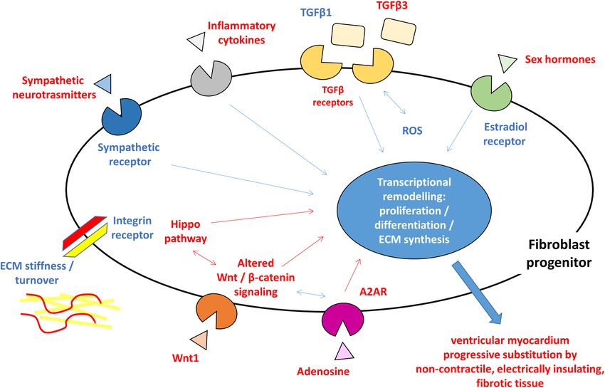
FIGURE 1 | Schematic figure highlighting the hypothesized pro-fibrotic process in ACM. The presence of different triggers (sympathetic nervous system activity,
extracellular matrix ECM component, reactive oxygen species ROS, inflammatory cytokines, and sex hormones) and the activation of molecular pathways (Hippo,
Wnt/β-catenin, and TGFβ) lead to transcriptional rearrangement for excessive proliferation and myofibroblasts differentiation of fibroblast progenitors. These changes
ultimately result in ventricular myocardium progressive substitution by non-contractile, electrically insulating, fibrotic tissue. In blue, what is known about pro-fibrotic
mechanisms in general and hypothesized in ACM, in red what is reported for ACM pathogenesis. ACM: arrhythmogenic cardiomyopathy; A2AR: adenosine 2A
receptor; ECM: extracellular matrix; ROS: reactive oxygen species; TGFβ: transforming growth factor β.
Frontiers in Physiology | www.frontiersin.org 5 April 2020 | Volume 11 | Article 279Maione et al. Arrhythmogenic Cardiomyopathy and Fibrosis Although ACM is commonly defined as a “desmosomal It is important to emphasize that adipogenesis and disease” being the majority of the patients mutated in fibrogenesis are differentiation programs well regulated desmosomal genes, additional mutations have been identified by independent pathways. TGFβ1 induces myofibroblast in genes that encode for non-desmosomal proteins (Moccia differentiation reducing in parallel the expression of PPARγ, et al., 2019). One of those in TGFB3, responsible for the the mast regulator of adipogenic differentiation (Vallee et al., ARVD1 form (Beffagna et al., 2005). In ACM patients, mutations 2017). On the contrary, PPARγ acts preventing myofibroblasts in TGFB3 are linked both to an increase in cardiac fibrotic differentiation and collagen deposition. remodeling and to the regulation of desmosomal gene expression One further molecular mechanism involved in ACM (Beffagna et al., 2005; Tamargo, 2012). Interestingly, also pathogenesis as well as in myofibroblast differentiation is the existence of a possible desmosomal protein-dependent the Hippo pathway that acts by regulating YAP/TAZ shuttling TGFβ expression has been reported. Particularly, it has been between nucleus and cytoplasm. Specifically, in the ACM context, demonstrated that plakophilin 2 (PKP2) and DSP control the altered PG distribution induces the retention of YAP into the the activity of TGFβ1/p38 MAPK pathway both in vitro and cytoplasm with activation of Hippo pathway and suppression in vivo. Indeed, in CM with a loss of PKP2, an increase in of canonical Wnt-related gene expression (Wada et al., 2011; TGFβ1 signaling is observed with consequent fibrotic genes Zhou and Zhao, 2018). Furthermore, during myofibroblasts expression, like collagen and fibronectin (Li et al., 2011). differentiation, the YAP/TAZ nuclear localization is associated Moreover, DSP expression is lost following PKP2 knockdown. with Wnt activation and TGFβ1 increase level with consequent Since the restoration of DSP expression rescues the activation SMAD phosphorylation in fibrotic tissues (Liu et al., 2015, 2018; of TGFβ1/p38 signaling, DSP acts upstream TGFβ1/p38 and Piersma et al., 2015). downstream PKP2 (Dubash et al., 2016). Recently, the activation of the adenosine 2A receptor (A2AR) Conversely, TGFβ1 treatment induces both an increase of has been reported to contribute to the progression of fibrosis DSP I and II expression and a reduction of DSP degradation in in an ACM animal model (Cerrone et al., 2018). The binding bronchial epithelia (Yoshida et al., 1992). of adenosine to A2AR stimulates expression of TGFβ, CTGF, Overall, these observations demonstrate that TGFβ could and matrix production (Shaikh et al., 2016). Moreover, A2AR modulate the expression of junctional proteins leading to the activation interacts with the Wnt pathway (Shaikh et al., 2016; modification of cellular phenotype and promoting the formation Zhang et al., 2017). of fibroblasts. In this context, it is important to underline that TGFβ promotes EMT, which is a process characterized by cell– cell contact changes (Zeisberg et al., 2007). CONCLUSION It has been hypothesized that, in ACM, desmosome mutations cause PG translocation from intercalated discs to the nucleus The ACM specific cardiac remodeling is characterized by the where it competes with β-catenin for the binding to TCF/LEF progressive substitution of ventricular myocardium of patients transcription factors based on the high structural homology by non-contractile fibrotic or adipose tissue. While adipogenesis (Garcia-Gras et al., 2006; Miravet et al., 2016). The abnormal has been extensively studied in this pathological context, fibrosis, PG translocation causes the altered canonical activation of a cardiac phenotype common to most cardiac diseases, remains Wnt/β-catenin signaling pathway promoting the pathological under-investigated. fibro-adipose myocardial tissue substitution (Garcia-Gras et al., Myocardial fibrosis is a clinical feature shared by several 2006; Moccia et al., 2019). heart diseases such as ischemic cardiomyopathy, dilated Intriguingly, it has been reported that TGFβ influences cardiomyopathy, hypertrophic cardiomyopathy, hypertensive Wnt/β-catenin signaling in a positive manner (Dzialo et al., heart disease, and heart failure. Ventricular fibrosis may 2018). TGFβ acts on canonical Wnt pathway in cardiac fibroblasts develop different modality depending on disease progression by: (1) inducing Wnt proteins release; (2) decreasing the and typically result in the formation of substrate vulnerable expression of Wnt pathway inhibitors; and (3) inhibiting GSK- to arrhythmic events. The cardiac fibroblast activation and 3β leading to the translocation of active β-catenin from the differentiation into myofibroblasts and the resulting scar cytosol to the nucleus (Akhmetshina et al., 2012; Lal et al., 2014; formation commonly occur following a cardiac injury. This Blyszczuk et al., 2017). event represents a reparative process but during a pathological On the other hand, the action of Wnt1 ligand, overexpressed cardiac condition, it becomes a persistent status that leads to in ventricular epicardium after cardiac damage, causes the altered myocardial structure and function. As described in activation of Wnt pathway, with consequent differentiation other cardiac diseases, the presence of fibroblasts and fibroblast of epicardial fibroblasts into myofibroblasts with collagen progenitors, the excessive collagen deposition, and the following synthesis (Duan et al., 2012). The presence of Wnt modification of mechanical stiffness may improve the tissue ligands, in combination with decreased expression of discontinuity occurring in the ACM hearts. Therefore, most of Wnt pathway inhibitors, contributes to nuclear β-catenin what is known about fibrotic processes and is summarized in localization in human fibroblasts during the fibrotic this review is iterated from studies in other settings. However, process while loss of β-catenin in cardiac fibroblasts it is expected that triggering agents, cellular effectors, and reduced ECM gene expression and collagen deposition mechanisms are comparable to what previously described. (Xiang et al., 2017). Responsible cells are likely cardiac fibroblasts, either from Frontiers in Physiology | www.frontiersin.org 6 April 2020 | Volume 11 | Article 279
Maione et al.                                                                                                                        Arrhythmogenic Cardiomyopathy and Fibrosis

FAP progenitors or C-MSC. ACM key pathogenic mechanisms                                        AUTHOR CONTRIBUTIONS
such as Wnt and Hippo are playing direct roles, with
the support of TGFβ-mediated mechanisms, which prompts                                         All authors contributed to the writing of the paragraphs and to
fibrosis as an alternative to adipogenesis. The whole process is                               the critical review of them.
possibly triggered by genetically driven myocardial damage, and
inflammation, oxidative stress, mechanical and neuro-hormonal
signaling are magnifying factors (Figure 1), thus representing                                 FUNDING
possible targets for therapies.
   Nevertheless, ACM specific fibrosis remains a scientific                                    This study was supported by the Transnational Research Projects
gap of knowledge to be filled with further studies, in                                         on Cardiovascular Diseases (ACM-HFJTC2016_FP-40-021) and
order to clarify specific pathways as target for novel specific                                the Italian Ministry of Health (RC 2019 – ID 2754330 to Centro
therapeutic actions.                                                                           Cardiologico Monzino-IRCCS).

REFERENCES                                                                                     Briest, W., Holzl, A., Rassler, B., Deten, A., Leicht, M., Baba, H. A., et al.
                                                                                                  (2001). Cardiac remodeling after long term norepinephrine treatment in rats.
Abe, R., Donnelly, S. C., Peng, T., Bucala, R., and Metz, C. N. (2001).                           Cardiovasc. Res. 52, 265–273. doi: 10.1016/s0008-6363(01)00398-4
   Peripheral blood fibrocytes: differentiation pathway and migration to                       Brilla, C. G., Zhou, G., Matsubara, L., and Weber, K. T. (1994). Collagen
   wound sites. J. Immunol. 166, 7556–7562. doi: 10.4049/jimmunol.166.12.                         metabolism in cultured adult rat cardiac fibroblasts: response to angiotensin II
   7556                                                                                           and aldosterone. J. Mol. Cell Cardiol. 26, 809–820. doi: 10.1006/jmcc.1994.1098
Aggeli, C., Pietri, P., Felekos, I., Rautopoulos, L., Toutouzas, K., Tsiamis, E., et al.       Bujak, M., Dobaczewski, M., Chatila, K., Mendoza, L. H., Li, N., Reddy, A., et al.
   (2012). Myocardial structure and matrix metalloproteinases. Curr. Top. Med.                    (2008). Interleukin-1 receptor type I signaling critically regulates infarct healing
   Chem. 12, 1113–1131. doi: 10.2174/1568026611208011113                                          and cardiac remodeling. Am. J. Pathol. 173, 57–67. doi: 10.2353/ajpath.2008.
Akdis, D., Saguner, A. M., Shah, K., Wei, C., Medeiros-Domingo, A.,                               070974
   von Eckardstein, A., et al. (2017). Sex hormones affect outcome in                          Bujak, M., and Frangogiannis, N. G. (2007). The role of TGF-beta signaling in
   arrhythmogenic right ventricular cardiomyopathy/dysplasia: from a                              myocardial infarction and cardiac remodeling. Cardiovasc. Res. 74, 184–195.
   stem cell derived cardiomyocyte-based model to clinical biomarkers of                          doi: 10.1016/j.cardiores.2006.10.002
   disease outcome. Eur. Heart J. 38, 1498–1508. doi: 10.1093/eurheartj/                       Bujak, M., and Frangogiannis, N. G. (2009). The role of IL-1 in the pathogenesis
   ehx011                                                                                         of heart disease. Arch. Immunol. Ther. Exp. (Warsz) 57, 165–176. doi: 10.1007/
Akhmetshina, A., Palumbo, K., Dees, C., Bergmann, C., Venalis, P., Zerr, P.,                      s00005-009-0024-y
   et al. (2012). Activation of canonical Wnt signalling is required for TGF-beta-             Campian, M. E., Verberne, H. J., Hardziyenka, M., de Groot, E. A., van
   mediated fibrosis. Nat. Commun. 3:735. doi: 10.1038/ncomms1734                                 Moerkerken, A. F., van Eck-Smit, B. L., et al. (2010). Assessment
Asimaki, A., Tandri, H., Duffy, E. R., Winterfield, J. R., Mackey-Bojack, S.,                     of inflammation in patients with arrhythmogenic right ventricular
   Picken, M. M., et al. (2011). Altered desmosomal proteins in granulomatous                     cardiomyopathy/dysplasia. Eur. J. Nucl. Med. Mol. Imaging 37, 2079–2085.
   myocarditis and potential pathogenic links to arrhythmogenic right ventricular                 doi: 10.1007/s00259-010-1525-y
   cardiomyopathy. Circ. Arrhythm Electrophysiol. 4, 743–752. doi: 10.1161/                    Campuzano, O., Alcalde, M., Iglesias, A., Barahona-Dussault, C., Sarquella-
   CIRCEP.111.964890                                                                              Brugada, G., Benito, B., et al. (2012). Arrhythmogenic right ventricular
Ayaz, O., and Howlett, S. E. (2015). Testosterone modulates cardiac contraction                   cardiomyopathy: severe structural alterations are associated with inflammation.
   and calcium homeostasis: cellular and molecular mechanisms. Biol. Sex Differ.                  J. Clin. Pathol. 65, 1077–1083. doi: 10.1136/jclinpath-2012-201022
   6:9. doi: 10.1186/s13293-015-0027-9                                                         Carlson, S., Trial, J., Soeller, C., and Entman, M. L. (2011). Cardiac mesenchymal
Barcellos-Hoff, M. H., and Dix, T. A. (1996). Redox-mediated activation of latent                 stem cells contribute to scar formation after myocardial infarction. Cardiovasc.
   transforming growth factor-beta 1. Mol. Endocrinol. 10, 1077–1083. doi: 10.                    Res. 2011, 99–107. doi: 10.1093/cvr/cvr061
   1210/me.10.9.1077                                                                           Casella, M., Dello Russo, A., Vettor, G., Lumia, G., Catto, V., Sommariva, E., et al.
Bauce, B., Frigo, G., Marcus, F. I., Basso, C., Rampazzo, A., Maddalena, F., et al.               (2017). Electroanatomical mapping systems and intracardiac echo integration
   (2008). Comparison of clinical features of arrhythmogenic right ventricular                    for guided endomyocardial biopsy. Expert Rev. Med. Dev. 14, 609–619. doi:
   cardiomyopathy in men versus women. Am. J. Cardiol. 102, 1252–1257. doi:                       10.1080/17434440.2017.1351875
   10.1016/j.amjcard.2008.06.054                                                               Casella, M., Pizzamiglio, F., Dello Russo, A., Carbucicchio, C., Al-Mohani,
Beffagna, G., Occhi, G., Nava, A., Vitiello, L., Ditadi, A., Basso, C., et al.                    G., Russo, E., et al. (2015). Feasibility of combined unipolar and bipolar
   (2005). Regulatory mutations in transforming growth factor-beta3 gene cause                    voltage maps to improve sensitivity of endomyocardial biopsy. Circ. Arrhythm
   arrhythmogenic right ventricular cardiomyopathy type 1. Cardiovasc. Res. 65,                   Electrophysiol. 8, 625–632. doi: 10.1161/CIRCEP.114.002216
   366–373. doi: 10.1016/j.cardiores.2004.10.005                                               Castaldi, A., Zaglia, T., Di Mauro, V., Carullo, P., Viggiani, G., Borile, G., et al.
Benito, B., Gay-Jordi, G., Serrano-Mollar, A., Guasch, E., Shi, Y., Tardif, J. C.,                (2014). MicroRNA-133 modulates the beta1-adrenergic receptor transduction
   et al. (2011). Cardiac arrhythmogenic remodeling in a rat model of long-term                   cascade. Circ Res. 115, 273–283. doi: 10.1161/CIRCRESAHA.115.303252
   intensive exercise training. Circulation 123, 13–22. doi: 10.1161/circulationaha.           Cerrone, M. (2018). Exercise: a risky subject in arrhythmogenic cardiomyopathy.
   110.938282                                                                                     J. Am. Heart Assoc. 7:e009611.
Bhambi, B., and Eghbali, M. (1991). Effect of norepinephrine on myocardial                     Cerrone, M., van Opbergen, C. J. M., Malkani, K., Irrera, N., Zhang, M., Van Veen,
   collagen gene expression and response of cardiac fibroblasts after                             T. A. B., et al. (2018). Blockade of the adenosine 2A receptor mitigates the
   norepinephrine treatment. Am. J. Pathol. 139, 1131–1142.                                       cardiomyopathy induced by loss of plakophilin-2 expression. Front. Physiol.
Blyszczuk, P., Muller-Edenborn, B., Valenta, T., Osto, E., Stellato, M., Behnke,                  9:1750. doi: 10.3389/fphys.2018.01750
   S., et al. (2017). Transforming growth factor-beta-dependent Wnt secretion                  Chelko, S. P., Asimaki, A., Lowenthal, J., Bueno-Beti, C., Bedja, D., Scalco,
   controls myofibroblast formation and myocardial fibrosis progression in                        A., et al. (2019). Therapeutic modulation of the immune response in
   experimental autoimmune myocarditis. Eur. Heart J. 38, 1413–1425.                              arrhythmogenic cardiomyopathy. Circulation 140, 1491–1505. doi: 10.1161/
Borthwick, L. A., Wynn, T. A., and Fisher, A. J. (2013). Cytokine mediated tissue                 CIRCULATIONAHA.119.040676
   fibrosis. Biochim. Biophys. Acta 1832, 1049–1060. doi: 10.1016/j.bbadis.2012.               Chen, S. N., Gurha, P., Lombardi, R., Ruggiero, A., Willerson, J. T., and Marian,
   09.014                                                                                         A. J. (2014). The hippo pathway is activated and is a causal mechanism for

Frontiers in Physiology | www.frontiersin.org                                              7                                                    April 2020 | Volume 11 | Article 279
Maione et al. Arrhythmogenic Cardiomyopathy and Fibrosis adipogenesis in arrhythmogenic cardiomyopathy. Circ. Res. 114, 454–468. doi: cardiomyopathy in mice. Proc. Natl. Acad. Sci. U.S.A. 103, 18284–18289. doi: 10.1161/CIRCRESAHA.114.302810 10.1073/pnas.0608799103 Cipolletta, E., Rusciano, M. R., Maione, A. S., Santulli, G., Sorriento, D., Del Heinemeier, K., Langberg, H., and Kjaer, M. (2003). Exercise-induced changes Giudice, C., et al. (2015). Targeting the CaMKII/ERK interaction in the heart in circulating levels of transforming growth factor-beta-1 in humans: prevents cardiac hypertrophy. PLoS ONE 10:e0130477. doi: 10.1371/journal. methodological considerations. Eur. J. Appl. Physiol. 90, 171–177. doi: 10.1007/ pone.0130477 s00421-003-0881-8 Corrado, D., Link, M. S., and Calkins, H. (2017). Arrhythmogenic right ventricular Hinz, B. (2009). Tissue stiffness, latent TGF-beta1 activation, and mechanical signal cardiomyopathy. N. Engl. J. Med. 376, 61–72. transduction: implications for the pathogenesis and treatment of fibrosis. Curr. Cucoranu, I., Clempus, R., Dikalova, A., Phelan, P. J., Ariyan, S., Dikalov, S., Rheumatol. Rep. 11, 120–126. doi: 10.1007/s11926-009-0017-1 et al. (2005). NAD(P)H oxidase 4 mediates transforming growth factor-beta1- Hoorntje, E. T., Te Rijdt, W. P., James, C. A., Pilichou, K., Basso, C., Judge, induced differentiation of cardiac fibroblasts into myofibroblasts. Circ Res. 97, D. P., et al. (2017). Arrhythmogenic cardiomyopathy: pathology, genetics, and 900–907. doi: 10.1161/01.res.0000187457.24338.3d concepts in pathogenesis. Cardiovasc. Res. 113, 1521–1531. doi: 10.1093/cvr/ Czarkowska-Paczek, B., Bartlomiejczyk, I., and Przybylski, J. (2006). cvx150 The serum levels of growth factors: PDGF, TGF-beta and VEGF are Iizuka, K., Sano, H., Kawaguchi, H., and Kitabatake, A. (1994). Transforming increased after strenuous physical exercise. J. Physiol. Pharmacol. 57, growth factor beta-1 modulates the number of beta-adrenergic receptors in 189–197. cardiac fibroblasts. J. Mol. Cell Cardiol. 26, 435–440. de Boer, R. A., De Keulenaer, G., Bauersachs, J., Brutsaert, D., Cleland, J. G., Diez, J., Iorga, A., Li, J., Sharma, S., Umar, S., Bopassa, J. C., Nadadur, R. D., et al. (2016). et al. (2019). Towards better definition, quantification and treatment of fibrosis Rescue of pressure overload-induced heart failure by estrogen therapy. J. Am. in heart failure. A scientific roadmap by the committee of translational research Heart Assoc. 5:e002482. of the heart failure association (HFA) of the european society of cardiology. Eur. Jacob-Ferreira, A. L., and Schulz, R. (2013). Activation of intracellular matrix J. Heart Fail 21, 272–285. doi: 10.1002/ejhf.1406 metalloproteinase-2 by reactive oxygen-nitrogen species: consequences and Duan, J., Gherghe, C., Liu, D., Hamlett, E., Srikantha, L., Rodgers, L., et al. therapeutic strategies in the heart. Arch. Biochem. Biophys. 540, 82–93. doi: (2012). Wnt1/betacatenin injury response activates the epicardium and cardiac 10.1016/j.abb.2013.09.019 fibroblasts to promote cardiac repair. EMBO J. 31, 429–442. doi: 10.1038/emboj. James, C. A., Bhonsale, A., Tichnell, C., Murray, B., Russell, S. D., Tandri, 2011.418 H., et al. (2013). Exercise increases age-related penetrance and arrhythmic Dubash, A. D., Kam, C. Y., Aguado, B. A., Patel, D. M., Delmar, M., Shea, L. D., risk in arrhythmogenic right ventricular dysplasia/cardiomyopathy-associated et al. (2016). Plakophilin-2 loss promotes TGF-beta1/p38 MAPK-dependent desmosomal mutation carriers. J. Am. Coll. Cardiol. 62, 1290–1297. doi: 10. fibrotic gene expression in cardiomyocytes. J. Cell Biol. 212, 425–438. doi: 1016/j.jacc.2013.06.033 10.1083/jcb.201507018 Kakkar, R., and Lee, R. T. (2010). Intramyocardial fibroblast myocyte Dubey, R. K., Gillespie, D. G., Jackson, E. K., and Keller, P. J. (1998). 17Beta- communication. Circ. Res. 106, 47–57. doi: 10.1161/CIRCRESAHA.109.207456 estradiol, its metabolites, and progesterone inhibit cardiac fibroblast Kanisicak, O., Khalil, H., Ivey, M. J., Karch, J., Maliken, B. D., Correll, R. N., et al. growth. Hypertension 31(1 Pt 2), 522–528. doi: 10.1161/01.hyp.31. (2016). Genetic lineage tracing defines myofibroblast origin and function in the 1.522 injured heart. Nat. Commun. 7:12260. doi: 10.1038/ncomms12260 Dupont, S., Morsut, L., Aragona, M., Enzo, E., Giulitti, S., Cordenonsi, M., et al. Kararigas, G., Dworatzek, E., Petrov, G., Summer, H., Schulze, T. M., Baczko, I., (2011). Role of YAP/TAZ in mechanotransduction. Nature 474, 179–183. doi: et al. (2014). Sex-dependent regulation of fibrosis and inflammation in human 10.1038/nature10137 left ventricular remodelling under pressure overload. Eur. J. Heart Fail 2014, Dworatzek, E., Mahmoodzadeh, S., Schriever, C., Kusumoto, K., Kramer, L., Santos, 1160–1167. doi: 10.1002/ejhf.171 G., et al. (2019). Sex-specific regulation of collagen I and III expression by Kassiri, Z., and Khokha, R. (2005). Myocardial extra-cellular matrix and its 17beta-Estradiol in cardiac fibroblasts: role of estrogen receptors. Cardiovasc. regulation by metalloproteinases and their inhibitors. Thromb. Haemost 93, Res. 115, 315–327. doi: 10.1093/cvr/cvy185 212–219. doi: 10.1160/th04-08-0522 Dzialo, E., Tkacz, K., and Blyszczuk, P. (2018). Crosstalk between the TGF-beta Kim, C., Wong, J., Wen, J., Wang, S., Wang, C., Spiering, S., et al. (2013). Studying and WNT signalling pathways during cardiac fibrogenesis. Acta Biochim. Pol. arrhythmogenic right ventricular dysplasia with patient-specific iPSCs. Nature 65, 341–349. doi: 10.18388/abp.2018_2635 494, 105–110. doi: 10.1038/nature11799 Fabritz, L., Hoogendijk, M. G., Scicluna, B. P., van Amersfoorth, S. C., Fortmueller, Kim, R. J., Fieno, D. S., Parrish, T. B., Harris, K., Chen, E. L., Simonetti, O., L., Wolf, S., et al. (2011). Load-reducing therapy prevents development et al. (1999). Relationship of MRI delayed contrast enhancement to irreversible of arrhythmogenic right ventricular cardiomyopathy in plakoglobin- injury, infarct age, and contractile function. Circulation 100, 1992–2002. doi: deficient mice. J. Am. Coll. Cardiol. 57, 740–750. doi: 10.1016/j.jacc.2010. 10.1161/01.cir.100.19.1992 09.046 Kim, R. J., Wu, E., Rafael, A., Chen, E. L., Parker, M. A., Simonetti, O., et al. Ferrari, R. (1999). The role of TNF in cardiovascular disease. Pharmacol. Res. 40, (2000). The use of contrast-enhanced magnetic resonance imaging to identify 97–105. doi: 10.1006/phrs.1998.0463 reversible myocardial dysfunction. N. Engl. J. Med. 343, 1445–1453. doi: 10. Frangogiannis, N. G. (2008). The immune system and cardiac repair. Pharmacol. 1056/nejm200011163432003 Res. 58, 88–111. doi: 10.1016/j.phrs.2008.06.007 Krenning, G., Zeisberg, E. M., and Kalluri, R. (2010). The origin of fibroblasts and Frangogiannis, N. G. (2012). Matricellular proteins in cardiac adaptation and mechanism of cardiac fibrosis. J. Cell Physiol. 2010, 631–637. doi: 10.1002/jcp. disease. Physiol. Rev. 92, 635–688. doi: 10.1152/physrev.00008.2011 22322 Furtado, M. B., Costa, M. W., and Rosenthal, N. A. (2016). The cardiac fibroblast: La Gerche, A., Burns, A. T., Mooney, D. J., Inder, W. J., Taylor, A. J., Bogaert, origin, identity and role in homeostasis and disease. Differentiation 92, 93–101. J., et al. (2012). Exercise-induced right ventricular dysfunction and structural doi: 10.1016/j.diff.2016.06.004 remodelling in endurance athletes. Eur. Heart J. 33, 998–1006. doi: 10.1093/ Garcia-Gras, E., Lombardi, R., Giocondo, M. J., Willerson, J. T., Schneider, eurheartj/ehr397 M. D., Khoury, D. S., et al. (2006). Suppression of canonical Wnt/beta-catenin Lal, H., Ahmad, F., Zhou, J., Yu, J. E., Vagnozzi, R. J., Guo, Y., et al. (2014). Cardiac signaling by nuclear plakoglobin recapitulates phenotype of arrhythmogenic fibroblast glycogen synthase kinase-3beta regulates ventricular remodeling right ventricular cardiomyopathy. J. Clin. Invest. 116, 2012–2021. doi: 10.1172/ and dysfunction in ischemic heart. Circulation 130, 419–430. doi: 10.1161/ jci27751 CIRCULATIONAHA.113.008364 Gunja-Smith, Z., Morales, A. R., Romanelli, R., and Woessner, J. F. Jr. Li, D., Liu, Y., Maruyama, M., Zhu, W., Chen, H., Zhang, W., et al. (2011). (1996). Remodeling of human myocardial collagen in idiopathic dilated Restrictive loss of plakoglobin in cardiomyocytes leads to arrhythmogenic cardiomyopathy. Role of metalloproteinases and pyridinoline cross-links. Am. cardiomyopathy. Hum. Mol. Genet. 20, 4582–4596. doi: 10.1093/hmg/ddr392 J. Pathol. 148, 1639–1648. Lin, C. Y., Lin, Y. J., Li, C. H., Chung, F. P., Lo, M. T., Lin, C., et al. Haudek, S. B., Xia, Y., Huebener, P., Lee, J. M., Carlson, S., Crawford, J. R., (2018). Heterogeneous distribution of substrates between the endocardium et al. (2006). Bone marrow-derived fibroblast precursors mediate ischemic and epicardium promotes ventricular fibrillation in arrhythmogenic right Frontiers in Physiology | www.frontiersin.org 8 April 2020 | Volume 11 | Article 279
Maione et al.                                                                                                                             Arrhythmogenic Cardiomyopathy and Fibrosis

   ventricular dysplasia/cardiomyopathy. Europace 20, 501–511. doi: 10.1093/                       Noji, Y., Shimizu, M., Ino, H., Higashikata, T., Yamaguchi, M., Nohara, A.,
   europace/euw393                                                                                     et al. (2004). Increased circulating matrix metalloproteinase-2 in patients with
Liu, F., Lagares, D., Choi, K. M., Stopfer, L., Marinkovic, A., Vrbanac, V., et al.                    hypertrophic cardiomyopathy with systolic dysfunction. Circ. J. 68, 355–360.
   (2015). Mechanosignaling through YAP and TAZ drives fibroblast activation                           doi: 10.1253/circj.68.355
   and fibrosis. Am. J. Physiol. Lung Cell Mol. Physiol. 308, L344–L357. doi: 10.                  Norman, M., Simpson, M., Mogensen, J., Shaw, A., Hughes, S., Syrris, P., et al.
   1152/ajplung.00300.2014                                                                             (2005). Novel mutation in desmoplakin causes arrhythmogenic left ventricular
Liu, R. M., and Desai, L. P. (2015). Reciprocal regulation of TGF-beta and reactive                    cardiomyopathy. Circulation 112, 636–642. doi: 10.1161/circulationaha.104.
   oxygen species: a perverse cycle for fibrosis. Redox Biol. 6, 565–577. doi: 10.                     532234
   1016/j.redox.2015.09.009                                                                        Paul, M., Wichter, T., Kies, P., Gerss, J., Wollmann, C., Rahbar, K.,
Liu, R. M., and Gaston Pravia, K. A. (2010). Oxidative stress and glutathione in                       et al. (2011). Cardiac sympathetic dysfunction in genotyped patients with
   TGF-beta-mediated fibrogenesis. Free Radic. Biol. Med. 48, 1–15. doi: 10.1016/                      arrhythmogenic right ventricular cardiomyopathy and risk of recurrent
   j.freeradbiomed.2009.09.026                                                                         ventricular tachyarrhythmias. J. Nucl. Med. 52, 1559–1565. doi: 10.2967/
Liu, X., Long, X., Liu, W., Zhao, Y., Hayashi, T., Yamato, M., et al. (2018). Type I                   jnumed.111.088997
   collagen induces mesenchymal cell differentiation into myofibroblasts through                   Piersma, B., Bank, R. A., and Boersema, M. (2015). Signaling in fibrosis: TGF-beta,
   YAP-induced TGF-beta1 activation. Biochimie 150, 110–130. doi: 10.1016/j.                           WNT, and YAP/TAZ converge. Front. Med. (Lausanne) 2:59. doi: 10.3389/fmed.
   biochi.2018.05.005                                                                                  2015.00059
Lloyd-Jones, D., Adams, R., Carnethon, M., De Simone, G., Ferguson, T. B., Flegal,                 Pilato, C. A., Stadiotti, I., Maione, A. S., Saverio, V., Catto, V., Tundo, F.,
   K., et al. (2009). Heart disease and stroke statistics–2009 update: a report from                   et al. (2018). Isolation and characterization of cardiac mesenchymal stromal
   the American Heart Association Statistics Committee and Stroke Statistics                           cells from endomyocardial bioptic samples of arrhythmogenic cardiomyopathy
   Subcommittee. Circulation 119, e21–e181.                                                            patients. J. Vis. Exp. 28:57263.
Lombardi, R., Chen, S. N., Ruggiero, A., Gurha, P., Czernuszewicz, G. Z., Willerson,               Pilichou, K., Nava, A., Basso, C., Beffagna, G., Bauce, B., Lorenzon, A.,
   J. T., et al. (2016). Cardiac fibro-adipocyte progenitors express desmosome                         et al. (2006). Mutations in desmoglein-2 gene are associated with
   proteins and preferentially differentiate to adipocytes upon deletion of the                        arrhythmogenic right ventricular cardiomyopathy. Circulation 113,
   desmoplakin gene. Circ. Res. 119, 41–54. doi: 10.1161/CIRCRESAHA.115.                               1171–1179.
   308136                                                                                          Piro, M., Della Bona, R., Abbate, A., Biasucci, L. M., and Crea, F. (2010). Sex-related
Mahmoodzadeh, S., Dworatzek, E., Fritschka, S., Pham, T. H., and Regitz-Zagrosek,                      differences in myocardial remodeling. J. Am. Coll. Cardiol. 55, 1057–1065.
   V. (2010). 17beta-Estradiol inhibits matrix metalloproteinase-2 transcription                       doi: 10.1016/j.jacc.2009.09.065
   via MAP kinase in fibroblasts. Cardiovasc. Res. 85, 719–728. doi: 10.1093/cvr/                  Plenz, G., Song, Z. F., Reichenberg, S., Tjan, T. D., Robenek, H., and Deng, M. C.
   cvp350                                                                                              (1998). Left-ventricular expression of interleukin-6 messenger-RNA higher in
Mak, J. C., Rousell, J., Haddad, E. B., and Barnes, P. J. (2000). Transforming                         idiopathic dilated than in ischemic cardiomyopathy. Thorac. Cardiovasc. Surg.
   growth factor-beta1 inhibits beta2-adrenoceptor gene transcription. Naunyn                          46, 213–216. doi: 10.1055/s-2007-1010227
   Schmiedebergs Arch. Pharmacol. 362, 520–525. doi: 10.1007/s002100000321                         Purnomo, Y., Piccart, Y., Coenen, T., Prihadi, J. S., and Lijnen, P. J. (2013).
Marcus, F. I., Fontaine, G. H., Guiraudon, G., Frank, R., Laurenceau, J. L.,                           Oxidative stress and transforming growth factor-beta1-induced cardiac fibrosis.
   Malergue, C., et al. (1982). Right ventricular dysplasia: a report of 24 adult cases.               Cardiovasc. Hematol. Disord. Drug Targets 13, 165–172. doi: 10.2174/
   Circulation 65, 384–398. doi: 10.1161/01.cir.65.2.384                                               1871529x11313020010
Marcus, F. I., McKenna, W. J., Sherrill, D., Basso, C., Bauce, B., Bluemke,                        Puzzi, L., Borin, D., Gurha, P., Lombardi, R., Martinelli, V., Weiss, M., et al.
   D. A., et al. (2010). Diagnosis of arrhythmogenic right ventricular                                 (2019). Knock down of plakophillin 2 dysregulates adhesion pathway through
   cardiomyopathy/dysplasia: proposed modification of the Task Force Criteria.                         upregulation of mir200b and alters the mechanical properties in cardiac cells.
   Eur. Heart J. 31, 806–814. doi: 10.1093/eurheartj/ehq025                                            Cells 8:E1639.
Meier, H., Bullinger, J., Deten, A., Marx, G., Rabald, S., Zimmer, H. G., et al.                   Rainer, J., Meraviglia, V., Blankenburg, H., Piubelli, C., Pramstaller, P. P., Paolin,
   (2007). Tissue inhibitor of matrix metalloproteinase-1 in norepinephrine-                           A., et al. (2018). The arrhythmogenic cardiomyopathy-specific coding and non-
   induced remodeling of the mouse heart. Cell Physiol. Biochem. 20, 825–836.                          coding transcriptome in human cardiac stromal cells. BMC Genomics 19:491.
   doi: 10.1159/000110442                                                                              doi: 10.1186/s12864-018-4876-6
Miravet, S., Piedra, J., Miro, F., Itarte, E., Garcia, de Herreros, A., et al. (2016).             Rani, V., Deep, G., Singh, R. K., Palle, K., and Yadav, U. C. (2016). Oxidative stress
   The transcriptional factor Tcf-4 contains different binding sites for beta-                         and metabolic disorders: pathogenesis and therapeutic strategies. Life Sci. 148,
   catenin and plakoglobin. J. Biol. Chem. 291:18854. doi: 10.1074/jbc.a116.11                         183–193. doi: 10.1016/j.lfs.2016.02.002
   0248                                                                                            Regitz-Zagrosek, V., and Kararigas, G. (2017). Mechanistic pathways of sex
Moccia, F., Lodola, F., Stadiotti, I., Pilato, C. A., Bellin, M., Carugo, S., et al. (2019).           differences in cardiovascular disease. Physiol. Rev. 97, 1–37. doi: 10.1152/
   Calcium as a key player in arrhythmogenic cardiomyopathy: adhesion disorder                         physrev.00021.2015
   or intracellular alteration? Int. J. Mol. Sci. 20:3986. doi: 10.3390/ijms2016                   Rienks, M., Papageorgiou, A. P., Frangogiannis, N. G., and Heymans, S. (2014).
   3986                                                                                                Myocardial extracellular matrix: an ever-changing and diverse entity. Circ. Res.
Mollmann, H., Nef, H. M., Kostin, S., von Kalle, C., Pilz, I., Weber, M., et al. (2006).               114, 872–888. doi: 10.1161/CIRCRESAHA.114.302533
   Bone marrow-derived cells contribute to infarct remodelling. Cardiovasc. Res.                   Rosenkranz, S., Flesch, M., Amann, K., Haeuseler, C., Kilter, H., Seeland, U., et al.
   71, 661–671. doi: 10.1016/j.cardiores.2006.06.013                                                   (2002). Alterations of beta-adrenergic signaling and cardiac hypertrophy in
Moore-Morris, T., Guimaraes-Camboa, N., Banerjee, I., Zambon, A. C., Kisseleva,                        transgenic mice overexpressing TGF-beta(1). Am. J. Physiol. Heart Circ. Physiol.
   T., Velayoudon, A., et al. (2014). Resident fibroblast lineages mediate pressure                    283, H1253–H1262.
   overload-induced cardiac fibrosis. J. Clin. Invest. 124, 2921–2934. doi: 10.1172/               Ruiz-Villalba, A., Ziogas, A., Ehrbar, M., and Perez-Pomares, J. M. (2013).
   JCI74783                                                                                            Characterization of epicardial-derived cardiac interstitial cells: differentiation
Morganroth, J., Maron, B. J., Henry, W. L., and Epstein, S. E. (1975). Comparative                     and mobilization of heart fibroblast progenitors. PLoS ONE 8:e53694. doi: 10.
   left ventricular dimensions in trained athletes. Ann. Intern. Med. 82, 521–524.                     1371/journal.pone.0053694
Moris, D., Spartalis, M., Spartalis, E., Karachaliou, G. S., Karaolanis, G. I.,                    Rusciano, M. R., Sommariva, E., Douin-Echinard, V., Ciccarelli, M., Poggio, P., and
   Tsourouflis, G., et al. (2017). The role of reactive oxygen species in the                          Maione, A. S. (2019). CaMKII activity in the inflammatory response of cardiac
   pathophysiology of cardiovascular diseases and the clinical significance of                         diseases. Int. J. Mol. Sci. 20:E4374.
   myocardial redox. Ann. Transl. Med. 5:326. doi: 10.21037/atm.2017.06.27                         Russell, J. L., Goetsch, S. C., Gaiano, N. R., Hill, J. A., Olson, E. N., and Schneider,
Murtha, L. A., Schuliga, M. J., Mabotuwana, N. S., Hardy, S. A., Waters, D. W.,                        J. W. (2011). A dynamic notch injury response activates epicardium and
   Burgess, J. K., et al. (2017). The processes and mechanisms of cardiac and                          contributes to fibrosis repair. Circ. Res. 108, 51–59. doi: 10.1161/CIRCRESAHA.
   pulmonary fibrosis. Front. Physiol. 8:777. doi: 10.3389/fphys.2017.00777                            110.233262

Frontiers in Physiology | www.frontiersin.org                                                  9                                                     April 2020 | Volume 11 | Article 279
You can also read