Cocaine Abusers Have an Overexpression of -Synuclein in Dopamine Neurons
←
→
Page content transcription
If your browser does not render page correctly, please read the page content below
2564 • The Journal of Neuroscience, April 1, 2003 • 23(7):2564 –2571
Cocaine Abusers Have an Overexpression of ␣-Synuclein in
Dopamine Neurons
Deborah C. Mash,1,2 Qinjie Ouyang,1 John Pablo,1 Margaret Basile,1 Sari Izenwasser,1 Abraham Lieberman,1 and
Richard J. Perrin3
Departments of 1Neurology and 2Molecular and Cellular Pharmacology, University of Miami School of Medicine, Miami, Florida 33101, and 3Department of
Cell and Structural Biology, University of Illinois at Urbana-Champaign, Urbana, Illinois 61801
␣-Synuclein is a presynaptic protein that has been implicated as a possible causative agent in the pathogenesis of Parkinson’s disease. The
native protein is a major component of nigral Lewy bodies in Parkinson’s disease, and full-length ␣-synuclein accumulates in Lewy
neurites. Here we present evidence that ␣-synuclein levels are elevated in midbrain dopamine (DA) neurons of chronic cocaine abusers.
Western blot and immunoautoradiographic studies were conducted on postmortem neuropathological specimens from cocaine users
and age-matched drug-free control subjects. The results demonstrated that ␣-synuclein levels in the DA cell groups of the substantia
nigra/ventral tegmental complex were elevated threefold in chronic cocaine users compared with normal age-matched subjects. The
increased protein levels in chronic cocaine users were accompanied by changes in the expression of ␣-synuclein mRNA in the substantia
nigra and ventral tegmental area. Although ␣-synuclein expression is prominent in the hippocampus, there was no increase in protein
expression in this brain region. The levels of -synuclein, a possible negative regulator of ␣-synuclein, also were not affected by cocaine
exposure. ␣-Synuclein protein levels were increased in the ventral tegmental area, but not the substantia nigra, in victims of excited
cocaine delirium who experienced paranoia, marked agitation, and hyperthermia before death. The overexpression of ␣-synuclein may
occur as a protective response to changes in DA turnover and increased oxidative stress resulting from cocaine abuse. However, the
accumulation of ␣-synuclein protein with long-term cocaine abuse may put addicts at increased risk for developing the motor abnor-
malities of Parkinson’s disease.
Key words: cocaine; postmortem; brain; synucleins; DA; delirium
Introduction ␣-synuclein occur in several animal models (Betarbet et al., 2000;
The synucleins are a family of soluble presynaptic proteins that Feany and Bender, 2000; Kowall et al., 2000; Masliah et al., 2000).
are abundant in neurons and include ␣-synuclein, -synuclein, The widespread ability of ␣-synuclein to be toxic in various cells
and ␥-synuclein (for review, see Clayton and George, 1998; Lave- and conditions suggests that some cells may not tolerate this gene
dan, 1998, 1999). Although the functions of the synucleins are product. In Drosophila melanogaster, ␣-synuclein overexpression
poorly understood, it has been suggested that synucleins are im- resulted in degeneration of dopaminergic (DAergic) neurons and
portant regulatory elements of synaptic vesicle transport pro- motor deficits (Feany and Bender, 2000). These studies suggest
cesses (Jenco et al., 1998; Maroteaux and Scheller, 1999). An that altered ␣-synuclein function can trigger the neurodegenera-
overexpression of human ␣-synuclein has been implicated in the tion of dopamine (DA) neurons.
etiology of two neurodegenerative diseases: Parkinson’s disease The mesolimbic DAergic system is an important pathway me-
(Polymeropoulos et al., 1997; Kruger et al., 1998) and Alzhei- diating reinforcement and addiction to psychostimulants (Self
mer’s disease (Ueda et al., 1993). The native protein is a major and Nestler, 1998). Cocaine potentiates DAergic neurotransmis-
component of nigral Lewy bodies in sporadic Parkinson’s disease sion by binding to the DA transporter and blocking neurotrans-
(Spillantini et al., 1997, 1998). The pathological hallmark of Alz- mitter uptake, leading to marked elevations in synaptic DA (for
heimer’s disease involves widespread deposition of -amyloid, review, see Giros and Caron, 1993). Long-term cocaine abuse
which leads to the death of hippocampal and cortical neurons. A leads to neuroadaptive changes in the signaling proteins that reg-
non-amyloidogenic fragment of ␣-synuclein is an integral con- ulate DA homeostasis. DA transporter binding sites are upregu-
stituent of amyloid plaques (Ueda et al., 1993), and this fragment lated in vitro in the postmortem brain of cocaine addicts (Little et
facilitates the aggregation of the 42 amino acid -amyloid peptide al., 1993, 1998; Staley et al., 1994, 1995; Mash et al., 2002) and in
(Jensen et al., 1997; Paik et al., 1998). Pathological inclusions of vivo in acutely abstinent cocaine-dependent individuals (Malison
et al., 1995, 1998). The direct binding and functional coupling of
Received July 29, 2002; revised Dec. 23, 2002; accepted Jan. 13, 2003.
␣-synuclein to the DA transporter has been shown to increase DA
This study was supported by National Institutes of Health Grant DA-06227 and the National Parkinson Founda- uptake and accelerate DA-induced apoptosis (Lee et al., 2001).
tion, Miami, FL. We thank the members of the Clayton lab for helpful discussions, Dr. Euijung Jo for providing Because ␣-synuclein binds to the DA transporter and affects its
recombinant ␣-synuclein protein, and Dr. Matthew Farrer for providing anti-␣-synuclein antibody that was devel- activity, alterations in ␣-synuclein expression may occur as a
oped in the laboratory of Dr. John Hardy.
Correspondence should be addressed to Dr. Deborah C. Mash, Department of Neurology (D4-5), 1501 Northwest
neuroadaptive response to chronic cocaine exposure.
Ninth Avenue, Miami, FL 33136. E-mail: dmash@newssun.med.miami.edu. The issue of cocaine-induced DA neurotoxicity has not been
Copyright © 2003 Society for Neuroscience 0270-6474/03/232564-08$15.00/0 resolved (Bartzokis et al., 1999). Subclinical parkinsonian-likeMash et al. • Cocaine Triggers Abnormal ␣-Synuclein Expression J. Neurosci., April 1, 2003 • 23(7):2564 –2571 • 2565
motor abnormalities that persist over a 3 month period of absti- 50 g/ml lima bean trypsin inhibitor) and centrifuged at 12,000 ⫻ g for
nence have been reported in cocaine-dependent subjects (Bauer, 10 min at 4°C (Langston et al., 1998). The supernatants were collected
1996). Cocaine-dependent subjects exhibited impaired perfor- and the proteins were measured by the bicinchoninic acid assay (Pierce
mance on tests of motor system functioning and showed signifi- Chemical, Rockford, IL). Protein extracts (0.01– 0.2 g) were processed
cant resting hand tremor that did not remit during a 3 month on SDS-PAGE on 15% separating and 4% stacking gel and transferred to
period of verified abstinence. The possibility of long-lasting and Immobilon-P nitrocellulose (Millipore, Bedford, MA). Blots were
possibly permanent brain changes is supported by brain imaging blocked in 100% methanol for 30 sec and then incubated for 2 hr at room
studies showing structural as well as functional abnormalities temperature with either anti-␣-synuclein (1:1000) or anti--synuclein
(1:1000) antibodies (Chemicon International) diluted with PBS contain-
that persist after 4 –9 months of abstinence (Pascual-Leone et al.,
ing 1% nonfat dry milk and 0.04% Tween 20, followed by 30 min in a
1991; Volkow et al., 1992; Bartzokis et al., 1996). We report here
donkey anti-rabbit horseradish peroxidase-conjugated secondary anti-
that ␣-synuclein is overexpressed in midbrain DA neurons from
body (Amersham Biosciences) diluted 1:10,000. Blots were stripped and
cocaine abusers. The overexpression of ␣-synuclein is a neuroad- reprobed with a monoclonal anti-␣-tubulin (1:1000) in PBS containing
aptive response to cocaine exposure that may put cocaine addicts 1% nonfat dry milk and 0.04% Tween 20 to confirm that equal amounts
at risk for degenerative changes in DA neurons, including the of protein were loaded for each case. Proteins were visualized by Super-
motor abnormalities of Parkinson’s disease. Signal West Pico Chemiluminescent Substrate (Pierce Chemical). Expo-
sures with maximal signal yet below the photographic saturation point
Materials and Methods were quantitatively analyzed by densitometry and compared with dilu-
All chemicals were obtained from Sigma (St. Louis, MO). Iodine and C 14 tional standards of recombinant ␣-synuclein protein (gift from Dr. Eui-
standards and Hyperfilm were purchased from Amersham Biosciences jung Jo, Centre for Neurodegenerative Diseases, University of Toronto).
(Piscataway, NJ). Optical densities were determined using IMAGE (version 1.44, NIH
Neuropathological tissue specimens. Postmortem neuropathological Shareware) and expressed as arbitrary units.
specimens were obtained during routine autopsy from age-matched In situ hybridization in human brain tissue. Full-length human
drug-free control subjects. Medicolegal investigations of the deaths were ␣-synuclein cDNA containing the entire coding region and 290 bp of the
conducted by forensic pathologists. The circumstances of death and tox- 3⬘ untranslated region was cloned into pGEM-3 vector. The integrity of
icology were reviewed carefully before classifying a cocaine intoxication the construct was confirmed by sequencing. A 400 base riboprobe of the
case with or without preterminal excited delirium (ED) (Ruttenber et al., 3⬘ end of ␣-synuclein cDNA (352–752 bp) was determined to be specific
1997). ED victims exhibited an acute onset of bizarre and violent behav- for the ␣-synuclein target gene transcript (National Center for Biotech-
ior, which was characterized by one or more of the following: aggression, nical Information Database; BLAST Search). The 35S-labeled RNA probe
combativeness, hyperactivity, extreme paranoia, demonstration of unex- was prepared using a ScriptEase Probe Kit (Novagen, Madison, WI). In
pected strength, or incoherent shouting (Wetli and Fishbain, 1985; Wetli situ hybridization was done by a modification of the method of Kholo-
et al., 1996). The syndrome of fatal ED is defined as accidental cocaine dilov et al. (1999). Slide-mounted brain tissue sections were fixed by
toxicity in subjects who exhibited bizarre and violent behavior (as de- immersion in 4% paraformaldehyde–PBS, pH 7.4, for 5 min, rinsed with
scribed above) followed by sudden death (Ruttenber et al., 1997). All PBS, and incubated in triethanolamine acetic anhydride solution for 10
cocaine cases (n ⫽ 21) were evaluated for common drugs of abuse and
min. The sections were defatted in a series of graded ethanol washes and
alcohol, and positive urine screens were confirmed by quantitative anal-
then chloroform and were dried at 37°C for 1–2 hr. Sections were prehy-
ysis of blood. Blood cocaine was quantified using gas–liquid chromatog-
bridized at 40°C for 2 hr with 1:1 formamide/prehybridization mix. Hy-
raphy with a nitrogen detector. Drug-free age-matched control subjects
(n ⫽ 13) were selected from accidental or cardiac sudden deaths with bridization was performed in a 1:1 formamide/hybridization mix at 55°C
negative urine screens for all common drugs and where there was no overnight. Labeled riboprobe was added to a final activity of ⬃3000
history of licit or illicit drug use before death. cpm/l of the 1:1 formamide/hybridization solution. Sections were
Immunoautoradiography and Western blotting. Serial coronal sections washed in 2⫻ saline sodium citrate (SSC) and then treated with RNase at
(30 m) from fresh-frozen blocks of human brain were cut on a cryostat, 37°C for 45 min. Sections were then washed in 2⫻ SSC for 60 min at
thaw-mounted on subbed slides, and dried under reduced pressure at 40°C, followed by immersion in 4 l of 0.1⫻ SSC containing 0.05% so-
4°C. Adjacent sections were stained for Nissl substance to delineate cy- dium pyrophosphate and 14 mM 2-mercaptoethanol at 40°C for 3 hr with
toarchitecture. Slide-mounted sections of the midbrain were postfixed in gentle stirring, dehydrated in ethanol, vacuum-dried, and apposed to
4% paraformaldehyde, 20% ethanol, 20% ethylene glycol, 10% glycerol, Hyperfilm at ⫺80°C for 2 weeks together with brain paste and 14C stan-
and 0.32 M sucrose in PBS, pH 7.4, and then blocked for 2 hr in 0.3% dards (Amersham Biosciences). The specificity of riboprobe hybridiza-
Tween 20, 3% bovine serum albumin, 3% goat serum, and 0.05% NaN3 tion was confirmed by comparing sections hybridized with antisense
in PBS, pH 7.4, and incubated overnight at 4°C with a polyclonal anti-␣- riboprobes with two control conditions: serial sections hybridized with
synuclein-peptide (amino acid 111–131) antibody (AB5038; Chemicon sense strand probes and antisense-hybridized sections that were predi-
International, Temecula, CA) diluted 1:1000. After a 1 hr incubation with gested with RNase A. In both of these conditions, there was no detectable
125
I-goat anti-rabbit IgG (PerkinElmer Life Sciences, Boston, MA) sec- hybridization signal.
ondary antibody, the sections were rinsed eight times for 10 min each and Data analysis. Slide-mounted sections of the midbrain were apposed
dried. The slide-mounted tissue was apposed to Hyperfilm along with with radioactive standards to Hyperfilm for the times indicated. Films
[ 125I] standard for 7 d at ⫺80°C. To establish antigen specificity for the
were scanned using a Howtek Scanmaster 3 at 400 dots per inch using
␣-synuclein antibody, controls included no primary antibody or an ir-
a transparency illuminator. After background subtraction, two-
relevant IgG. In some initial experiments, sections from control subjects
dimensional maps were created to allow specific radioactivity levels to be
and cocaine users were labeled with purified IgG1 isotype raised against
amino acid residues 98 –115 of human ␣-synuclein (Dr. Matthew Farrer, superimposed on the sections. The midbrain sections were compared
Mayo Clinic, Jacksonville, FL) that verified a comparable pattern of ex- with adjacent Nissl-stained sections along with a delineated map of ob-
pression and distribution of the protein. All sections were immunola- servable tracts and nuclei at a resolution of 150 ⫻ 150 ⫻ 500 m. The
beled at the same time and in the same batch of chemicals to minimize region-of-interest measurements were exported in Brain (version 3.0) to
variability. PICT files readable by the Canvas program (version 3.5). The resulting
Brain samples from the substantia nigra and hippocampus (100 mg tagged image file format for RGB color files was converted to pseudo-
tissue punch) were sonicated in lysis buffer containing protease inhibi- color format in specific activity units. ANOVA followed by Dunnett’s
tors (25 mM Tris, pH 7.4, with 300 g/ml phenylmethanesulfonyl fluo- post hoc comparisons for significance at p ⬍ 0.05 or better was performed
ride, 2 g/ml leupeptin, 16 g/ml benzamidine, 2 g/ml pepstatin A, and using Prism (Graphpad, San Diego, CA).2566 • J. Neurosci., April 1, 2003 • 23(7):2564 –2571 Mash et al. • Cocaine Triggers Abnormal ␣-Synuclein Expression
Results
Cocaine-related fatalities were identified and classified as part of
an ongoing case-control study of the toxicology reports, scene
descriptions, supplemental background information, and au-
topsy findings (Escobedo et al., 1991; Ruttenber et al., 1997). The
cocaine users were selected for the present study on the basis of
evidence of a number of surrogate variables of chronicity, includ-
ing the review of previous arrest records, hospital and previous
substance abuse treatment admissions, and pathological signs
determined at autopsy (e.g., perforation of the nasal septum, nee-
dle track marks, cardiomegaly, and “crack” lung). Most of the
cocaine cases had informant reports of binge cocaine use in the
days immediately before death. Review of hospital records and
interviews with next-of-kin informants were done to confirm
pattern and history of illicit drug use. The cocaine users (n ⫽ 13;
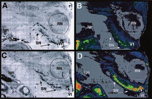
12 male/1 female), ED victims (n ⫽ 8; 8 male), and control sub- Figure 1. Cocaine-induced ␣-synuclein upregulation in substantia nigra/ventral tegmental
jects (n ⫽ 13; 11 male/2 female) were not significantly different complex. Nissl-stained sections from a representative control subject ( A) and cocaine user (C)
on demographic characteristics. Their mean (⫾SEM) ages were show the location of the substantia nigra (SN ) and ventral tegmental area (Vt). B, D,
36.6 ⫾ 2.2, 32.3 ⫾ 1.9, and 33.1 ⫾ 2.6, respectively. The postmor- ␣-Synuclein immunoautoradiography in adjacent sections from a control subject ( B) and co-
tem intervals did not differ significantly across groups (cocaine caine user ( D). Pseudocolor codes represent a rainbow scale (red ⫽ highest densities; yellow to
users ⫽ 12.7 ⫾ 1.70; ED ⫽ 13.1 ⫾ 1.1; control subjects ⫽ 15.9 ⫾ green ⫽ intermediate densities; blue to purple ⫽ low densities). RN, Red nucleus; lgn, lateral
1.5). Excited cocaine delirium cases have been included in this geniculate nucleus.
study as a comparison group. This psychiatric syndrome com-
prises delirium with marked agitation, respiratory depression,
hyperthermia, and sudden death (Wetli et al., 1996; Ruttenber et
al., 1997). The mode of death and agonal state are important
variables when investigating postmortem human brain (Wester
et al., 1985). All of the cocaine deaths were sudden because of
cocaine intoxication. ED victims survived longer, but all of the
cases included in this study had cocaine measured in blood, sug-
gesting that these subjects died only a few hours after their co-
caine binge (Ruttenber et al., 1997). Four of the control cases
were homicide victims of gunshot wounds, one was a blunt
trauma death, and the remaining eight cases died from cardiac
sudden death. Thus, all of the cocaine users and control subjects
died suddenly.
Cocaine and benzoylecgonine (BE) were detected in blood
and urine at the time of death for all cocaine intoxication cases
and ED victims. No other illicit drugs were detected in urine
screens, suggesting that none of the subjects had recent poly-drug Figure 2. Densitometric measurements of ␣-synuclein immunolabeling in cocaine users
and control subjects. ␣-Synuclein levels were increased in the substantia nigra and ventral
abuse. Two of the cocaine cases and three of the ED victims had
tegmental area in the cocaine users (COC; n ⫽ 13) but not in victims of excited delirium (ED; n ⫽
alcohol in postmortem blood at low levels (blood alcohol con- 8). No change in ␣-synuclein was observed in the hippocampus. Significant differences from
centration ⬍0.08%). The concentrations of cocaine and its main control values: *p ⬍ 0.05; **p ⬍ 0.01.
metabolite BE were measured in blood samples obtained at au-
topsy. The average (mean ⫾ SEM) blood levels of cocaine and BE
were 3.7 ⫾ 0.6 and 4.8 ⫾ 1.0 mg/l in the cocaine users. The ED cocaine users with and without preterminal excited delirium.
victims had lower levels of cocaine (1.9 ⫾ 0.7 mg/l) and compa- Densitometric measurements demonstrated a threefold elevation
rable levels of BE (4.4 ⫾ 1.2 mg/l) in blood. In cases that had in the substantia nigra and ventral tegmental area of the cocaine
alcohol measured in blood, cocaethylene levels were 0.2 ⫾ 0.1 for users as compared with drug-free age-matched control subjects
cocaine users and 0.3 ⫾ 0.1 for ED victims. None of the control ( p ⬍ 0.01) (Fig. 2). Interestingly, in the brains of ED victims there
cases tested positive for any neuroactive drug or metabolite. was a significant but smaller increase in ␣-synuclein immunola-
None of the cases selected for this study tested positive for opiates beling in the ventral tegmental area ( p ⬍ 0.05) (Fig. 2). However,
in blood or in urine toxicology screens. unlike in other cocaine users, the densities of ␣-synuclein immu-
nolabeling in the substantia nigra in ED victims were not elevated
Regulation of ␣-synuclein levels by cocaine exposure but were comparable with the levels measured in brains of the
The effect of cocaine exposure on ␣-synuclein protein expression age-matched drug-free control subjects.
was examined by immunoautoradiography with an anti-␣- There was a trend toward reduced ␣-synuclein labeling for
synuclein antibody. The substantia nigra was faintly labeled in some ED cases, but the average protein levels were not signifi-
drug-free control subjects (Fig. 1 B). In contrast, immunolabeling cantly different from control subjects, in agreement with the re-
was intense over the substantia nigra and ventral tegmental area sults of Western immunoblot analysis (Fig. 3). Victims of fatal ED
(VTA) in cocaine users (Fig. 1 D). Quantitative region-of-interest failed to demonstrate an increase in ␣-synuclein protein, al-
measurements of ␣-synuclein immunolabeling were taken to as- though these cases had comparable premorbid histories of co-
sess the regulatory effects of cocaine on protein expression in caine abuse and were positive at autopsy for cocaine and benzo-Mash et al. • Cocaine Triggers Abnormal ␣-Synuclein Expression J. Neurosci., April 1, 2003 • 23(7):2564 –2571 • 2567
Figure 4. Densitometric analysis of ␣-synuclein blots in substantia nigra and hippocampus.
Results demonstrate upregulation of ␣-synuclein protein in cocaine users (COC) but not excited
delirium (ED) cases in the substantia nigra. There was no change in ␣-synuclein protein expres-
sion in the hippocampus. Significant differences from control values: **p ⬍ 0.01.
Figure 3. Western blotting of ␣-synuclein protein. A, Dilutional standards of recombinant
␣-synuclein protein (0.01– 0.2 g). Serial dilution of human ␣-synuclein was loaded across
the lanes and probed by Western blotting with enhanced chemiluminescence. B, Standard expression within the human hippocampus with cocaine expo-
curve generated from the densitometric values obtained from computer analysis of digitized sure (Fig. 4).
film from immunoblot. A linear relationship between optical density and the amount of recom- Immunoblot analysis with a -synuclein-specific antibody
binant ␣-synuclein protein was observed with a correlation coefficient of 0.98. C, Representa- was done in human midbrain from cocaine users and control
tive blots with an antibody against ␣-synuclein show 19 kDa bands in the human substantia
subjects. -Synuclein is the 34 amino acid non-amyloidogenic
nigra from control subjects, cocaine users (COC), and excited delirium (ED) victims. D, Represen-
tative blots with an antibody against -synuclein show 14 kDa bands in the human substantia
homolog of ␣-synuclein. A single band was observed for
nigra and illustrate no change in -synuclein protein expression with cocaine exposure. -synuclein at the expected molecular mass of 14 kDa in cocaine
users with and without preterminal excited delirium and in con-
trol subjects (Fig. 3D). The upregulation of ␣-synuclein in the
substantia nigra (Fig. 3C) contrasted with the lack of an increase
ylecgonine in both blood and urine. Specific ␣-synuclein in -synuclein (Fig. 3D) and -tubulin (50 kDa; data not shown).
immunolabeling was seen in the hippocampus over the pyrami- Densitometric analysis of -synuclein immunoblots gave values
dal layer of the CA1 sector (data not shown). Region-of-interest for cocaine users and ED victims that were not different from
measurements gave comparable values across cocaine users, ED control subjects (data not shown). These results demonstrate that
victims, and age-matched drug-free control subjects (Fig. 2). the cocaine-induced upregulation of ␣-synuclein was not accom-
There was no increase seen in the entorhinal cortex or adjacent panied by changes in -synuclein.
deep layers of the neocortex with cocaine exposure (data not
shown). There was no labeling under control conditions, using Expression of ␣-synuclein mRNA in DA neurons
either no primary antibody or IgG isotype. A specific hybridization signal was observed in young control
subjects for the ␣-synuclein gene in the DA cells of the midbrain
␣- and -synuclein-immunoreactive proteins in (Fig. 5A). In the midbrain, the label was clearly localized to the
cocaine users substantia nigra. Increased expression was seen in the ventral
␣-Synuclein expression was examined by Western immunoblot tegmental area in cocaine users as compared with control subjects
analysis with the anti-human ␣-synuclein antibody in the sub- (Fig. 5B). Within the pars compacta, the ventral tier exhibited the
stantia nigra from cocaine users, ED victims, and control subjects strongest hybridization signal in cocaine users (Fig. 5B). In-
and compared with dilutional standards of recombinant creased expression was confirmed by image analysis, which re-
␣-synuclein (Fig. 3). A single band was observed at the expected vealed a significant increase in the substantia nigra and ventral
molecular mass of 19 kDa for all cases (Fig. 3C). Denser tegmental area in cocaine users as compared with control subjects
␣-synuclein-positive bands were consistently observed in cocaine ( p ⬍ 0.01) (Fig. 6). In contrast to these findings, ␣-synuclein
users as compared with control subjects. There was a significant mRNA measured over the DA cells of the substantia nigra/ventral
increase in protein expression observed for the cocaine users tegmental area complex was not increased in ED victims as com-
( p ⬍ 0.01) (Fig. 4). The amount of ␣-synuclein (nanograms per pared with cocaine users (Fig. 6).
microgram of total protein) measured in the substantia nigra was
48.1 ⫾ 2.8 in cocaine users, 11.3 ⫾ 1.8 for ED victims, and 14.4 ⫾ Discussion
2.0 in control subjects. These results demonstrate that the protein We have investigated the effect of cocaine abuse on the expression
levels were the same for ED victims as compared with control of ␣-synuclein protein and mRNA in postmortem human brain.
subjects. The marked increase in the heat-soluble fraction of These findings provide the first demonstration of adaptations in
␣-synuclein protein determined by Western immunoblot analy- ␣-synuclein expression with cocaine exposure in midbrain DA
sis was comparable in magnitude with the regional densitometric neurons. In cocaine users, ␣-synuclein mRNA was elevated in the
analysis of total protein levels measured in slide-mounted mid- substantia nigra and ventral tegmental area compared with age-
brain sections from cocaine users. In keeping with immunoauto- matched drug-free control subjects. The functional relevance of
radiographic analysis, there was no change in ␣-synuclein protein this increase was confirmed by robust increases in the levels of2568 • J. Neurosci., April 1, 2003 • 23(7):2564 –2571 Mash et al. • Cocaine Triggers Abnormal ␣-Synuclein Expression
levels of ␣-synuclein protein were measured in the ventral teg-
mental area in ED victims. The failure of ␣-synuclein to upregu-
late in the substantia nigra in these subjects is in keeping with the
lack of an increase in DA transporter function and binding sites
(Mash et al., 2002). ED victims exhibit profound neuropsychiat-
ric complications and hyperthermia before death, suggesting that
there may be a different pattern of ␣-synuclein regulation in this
subgroup of chronic cocaine users.
Previous high resolution in situ hybridization histochemical
studies have demonstrated that ␣-synuclein is expressed in
melanin-containing neurons of the human substantia nigra (So-
lano et al., 2000). Non-melanized cells in the substantia nigra do
not contain ␣-synuclein. Thus, the increased levels of immuno-
reactivity in the substantia nigra and ventral tegmental area mea-
sured in cocaine users likely reflect increases in neuronal expres-
sion of the protein in DA neurons. ␣-Synuclein levels were
unchanged in the hippocampus or adjacent temporal neocortex,
brain regions that have relatively high protein expression in nor-
mal subjects (Murphy et al., 2000). Because the increase in
␣-synuclein with cocaine exposure was confined to DA cell
groups and was not observed over the hippocampus or neocor-
tex, the effect of cocaine on ␣-synuclein expression appears to be
specific for DA-containing cells of the midbrain.
Lewy bodies are aggregates of ␣-synuclein, ubiquitin, neuro-
filaments, and other proteins (Spillantini et al., 1998). In the
course of neurodegeneration in Parkinson’s disease, susceptible
regions and vulnerable nerve cell populations become progres-
sively impaired because of the extensive presence of Lewy neu-
rites and Lewy bodies (Neystat et al., 1999; Del Tredici et al.,
2002). Brain regions in which Lewy bodies have been reported in
Parkinson’s disease (substantia nigra and locus coeruleus) and
diffuse Lewy body disease (hippocampus and deep layers of the
entorhinal and neocortex) (Forno, 1996; McKeith et al., 1996)
Figure 5. Expression of ␣-synuclein in substantia nigra and ventral tegmental area. A shows express the ␣-synuclein gene. In cocaine users, none of the ex-
a representative age-matched control subject (white, male, age 34), and B shows a cocaine user
tranigral regions expressing ␣-synuclein had elevations in the
who died suddenly (black, male, age 31). Film autoradiograms of midbrain sections reveal
intense expression for ␣-synuclein mRNA, particularly in the ventral tier of the substantia nigra
levels of the protein. This observation suggests that the substantia
pars compacta. Hipp, Hippocampus; RN, red nucleus; SN, substantia nigra; Vt, ventral tegmental nigra may be an induction site in brain for cocaine-induced in-
area. creases in ␣-synuclein. However, Lewy bodies are also found in
monoaminergic neurons of the coeruleus/subcoeruleus complex
and caudal raphe nuclei (Forno, 1996; Del Tredici et al., 2002).
Because cocaine also blocks the reuptake of norepinephrine and
serotonin, it will be important to determine in future studies
whether cocaine affects ␣-synuclein expression in brainstem
monoamine neurons.
The synucleins are enriched in presynaptic terminals, as
shown by combined immunocytochemistry and subcellular frac-
tionation studies in the songbird, rat, and human brain (for re-
view, see Clayton and George, 1998). A contribution of brain
␣-synuclein to regulation or support of synaptic plasticity is sug-
gested by early studies implicating synelfin in the canary song-
learning process (George et al., 1995). Synelfin is a 143 amino acid
homolog of the human ␣-synuclein protein that is highly ex-
pressed during critical periods of song plasticity in birds (for
review, see Clayton and George, 1998, 1999). Whether the change
in gene expression is a cause or consequence of synaptic reorga-
Figure 6. Quantitative analysis of ␣-synuclein mRNA expression. Measurements were nization is unknown. However, evidence exists for changes in
made in the substantia nigra and ventral tegmental area of cocaine users (COC) and excited synuclein gene expression and protein localization after synaptic
delirium (ED) victims. Significant differences from control values: **p ⬍ 0.01. activity or metabolic stress (Maroteaux et al., 1988; Maroteaux
and Scheller, 1991; Clayton and George, 1998). Synuclein mRNA
is upregulated in the substantia nigra when nigrostriatal neurons
␣-synuclein protein. Victims of excited cocaine delirium failed to are developing target contacts, sprouting, and forming synapses
show an upregulation of ␣-synuclein protein or mRNA levels in (Hsu et al., 1998). Graybiel (Canales and Graybiel, 2000) has
the substantia nigra. Significant albeit smaller increases in the suggested that different neural circuits become activated in re-Mash et al. • Cocaine Triggers Abnormal ␣-Synuclein Expression J. Neurosci., April 1, 2003 • 23(7):2564 –2571 • 2569
sponse to cocaine as a result of repeated administrations and sion may reflect a progressive change in ␣-synuclein levels that
involve DA and glutamate as key co-players in regulating basal occurs in the nigral/VTA complex depending on the duration
ganglia loops that affect both locomotion and stereotypy. Thus, and intensity of cocaine misuse. Because disruption of the syn-
changes in the expression of ␣-synuclein protein may be an adap- thesis, function, or possible aggregation of ␣-synuclein protein is
tive response to cocaine in reward-related neurons of the nigral/ predicted to increase DA release from DA neurons (Abeliovich et
ventral tegmental area complex. al., 2002), the lack of a compensatory increase in ␣-synuclein in
Cocaine inhibits the activity of the DA transporter (Ritz et al., the substantia nigra may augment DA release in particular striatal
1987; Madras et al., 1989; Reith and Selmeci, 1992) and increases regions during a cocaine “binge” in ED subjects. Further studies
vesicular DA uptake (Brown et al., 2001). Repeated exposure to are needed to link coordinated regulation of the DA transporter
cocaine may shift the normal balance of DA signaling through or other presynaptic proteins with ␣-synuclein expression to the
modifications of vesicular release and recycling of the neuro- progression of habitual drug-seeking and the occurrence of co-
transmitter. These changes are likely linked to altered DA uptake caine delirium. The adaptive change in ␣-synclein in DA neurons
function, which is upregulated in human cocaine addicts (Mash from human cocaine users is another example of the extreme
et al., 2002). The regulatory effects of cocaine on DA transporter neuronal plasticity that occurs in response to altered DA ho-
binding site densities have been studied in vitro in the postmor- meostasis with long-term cocaine abuse.
tem brain of cocaine addicts and in vivo in acutely abstinent Overexpression of wild-type or mutant forms of ␣-synuclein
cocaine-dependent individuals. Some of the previous studies in cultured human DA neurons leads to apoptosis, an effect that
(Little et al., 1993, 1998; Staley et al., 1994, 1995), but not all is blocked by the addition of a tyrosine hydroxylase inhibitor (Xu
(Hurd and Herkenham, 1993; Wilson et al., 1996), have reported et al., 2002). These observations suggest that it is the combination
increased numbers of DA transporters using radiolabeled cocaine of ␣-synuclein and DA that causes cell death. The pattern of
congeners. One possible explanation for conflicting results across cocaine-induced increases in ␣-synuclein expression described
studies is a loss of DA nerve terminals in more advanced and here suggests that increased protein expression is part of a neu-
severely dependent cocaine users (Wilson et al., 1996). ronal response to chronic cocaine exposure. The upregulation of
␣-Synuclein complexes with the human DA transporter through ␣-synuclein alone by chronic cocaine abuse is not likely to lead to
the direct binding of the non-A amyloid component of increased protein aggregation in neurites and intracytoplasmic
␣-synuclein to the C-terminal tail of the DA transporter (Lee et bodies. However, one consequence of the ability of cocaine to
al., 2001). ␣-Synuclein-DA transporter complexes facilitate the inhibit DA reuptake is marked elevations in extracellular DA.
membrane clustering of the DA transporter, thereby accelerating Also, cocaine causes a redistribution of plasmalemma vesicles
DA uptake in vitro (Lee et al., 2001). Concomitant increases in and increases vesicular DA uptake (Brown et al., 2001). Rapid
␣-synuclein and DA transporter numbers and function in co- vesicular sequestration of the neurotransmitter will limit the for-
caine abusers provide additional support for a role of ␣-synuclein mation of reactive oxygen species such as DA-quinone (Cubells
in regulating DAergic tone. et al., 1994; Hastings et. al., 1996; Stokes et al., 1999). Because
Transgenic mice deficient in ␣-synuclein demonstrate an at- excess ␣-synuclein potentiates production of reactive oxygen
tenuated locomotor response to amphetamine (Abeliovich et al., species by DA (Zhou et al., 2000; Xu et al., 2002) and the mutant
2000). Because amphetamine is known to exert its psychostimu- protein causes increased susceptibility to DA toxicity (Tabrizi et
lant effects through the DA transporter, it is possible that this al., 2000), alterations in DA turnover by cocaine may accelerate
effect may be caused by a change in trafficking of the DA trans- the formation of toxic forms of the protein. The protofibrillar
porter to the cell surface membrane in ␣-synuclein-deficient conformation of ␣-synuclein undergoes kinetic stabilization in
mice. However, a recent study suggests that the DA transporter the cell by catecholamines, including DA and norepinephrine
densities were not lower in ␣-synuclein null mice as compared (Conway et al., 2001). DA in the oxidized form appears to sustain
with wild-type mice (Dauer et al., 2002). In contrast to these the toxic protein within the cell as a DA–␣-synuclein adduct
results, overexpression of wild-type ␣-synuclein in mice leads to (Sulzer, 2001). The Lewy neurites and inclusions of Parkinson’s
increased densities of the DA transporter (Richfield et al., 2002), disease are made up of fibrillar ␣-synuclein protein, as opposed
suggesting a concentration-dependent effect of the protein on the to the unfolded form measured in normal brain (Giasson et al.,
trafficking of the DA transporter. Although ␣-synuclein- 2000). Thus, the cocaine-induced upregulation of ␣-synuclein
deficient mice appear to have a normal complement of DA neu- may be initially an adaptive response that could turn toxic de-
rons and terminals, they display abnormalities in the synaptic pending on the local cellular milieu.
handling of DA (Abeliovich et al., 2000). The recovery of peak DA The effects of cocaine on ␣-synuclein may occur only with
release after an initial stimulus is more rapid in ␣-synuclein- long-term cocaine abuse. We have compared cocaine users that
deficient mice, in keeping with an inhibitory role for ␣-synuclein came to autopsy with documented histories of the highest pat-
in activity-dependent modulation of neurotransmitter release. terns of cocaine use with individuals with no exposure. This is the
These observations suggest that the protein is an essential presyn- case also for ED victims, who only demonstrated elevations in
aptic, activity-dependent negative regulator of DA neurotrans- ␣-synuclein expression within the ventral tegmental area, but not
mission (Abeliovich et al., 2000). Unlike in other chronic cocaine the substantia nigra. Although every attempt is made to obtain
users, compensatory increases in DA transporter densities or up- information about the premortem pattern of cocaine use
take function do not occur in ED victims, although their severity (amount, duration, and total lifetime use), it is more difficult to
and amount of cocaine abuse are the same (Wetli et al., 1996; collect absolute exposure measures from interviews with infor-
Mash et al., 2002). We have speculated that the lack of neuroad- mants and next-of-kin. However, a cocaine intoxication death in
aptive increase in DA uptake function may contribute to the a recreational user is an extremely rare occurrence, and most of
persistence of a hyperdopaminergic state. Within the DA cell the cases that come to autopsy have many surrogate variables of
body fields, ␣-synuclein protein levels were elevated only in the chronic cocaine use, including crack lung and perforation of the
VTA, but not the substantia nigra, in cocaine users presenting nasal septum. Whether chronic cocaine use is neurotoxic to DA
with preterminal ED. This pattern of differential protein expres- neurons remains uncertain, and animal data suggest that am-2570 • J. Neurosci., April 1, 2003 • 23(7):2564 –2571 Mash et al. • Cocaine Triggers Abnormal ␣-Synuclein Expression
phetamines are more likely to cause damage to these cells than novel protein regulated during the critical period for song learning in the
cocaine (Bennett et al., 1993; Ellison and Switzer, 1993). How- zebra finch. Neuron 15:361–372.
Giasson BI, Duda JE, Murray IV, Chen Q, Souza JM, Hurtig HI, Ischiropoulos
ever, many cocaine-dependent subjects show signs of subclinical
H, Trojanowski JQ, Lee VM (2000) Oxidative damage linked to neuro-
parkinsonism that are reversible with protracted periods of absti- degeneration by selective ␣-synuclein nitration in synucleinopathy le-
nence (Bauer, 1996). Young cocaine-dependent subjects have sions. Science 290:985–989.
significant resting hand tremor that does not remit during a 3 Giros B, Caron MG (1993) Molecular characterization of the DA trans-
month period of verified abstinence, suggesting the possibility of porter. Trends Pharmacol Sci 14:43– 49.
neurotoxic damage to DA terminals. Hastings TG, Lewis DA, Zigmond MJ (1996) Role of oxidation in the neu-
The epidemic of crack cocaine use began in the United States rotoxic effects of intrastriatal DA injections. Proc Natl Acad Sci USA
93:1956 –1961.
around 1986 (Escobedo et al., 1991). Many crack cocaine-
Hsu LJ, Mallory M, Xia Y, Veinbergs I, Hashimoto M, Yoshimoto M, Thal LJ,
addicted individuals continue to misuse the drug for decades Saitoh T, Masliah E (1998) Expression pattern of synucleins (non-Abeta
despite attempts at abstinence. We speculate that abnormal component of Alzheimer’s disease amyloid precursor protein/␣-
␣-synuclein expression may be a risk factor for the development synuclein) during murine brain development. J Neurochem 71:338 –344.
of cocaine-related brain changes involving cognitive and motor Hurd YL, Herkenham M (1993) Molecular alterations in the neostriatum of
systems. Overexpression of ␣-synuclein may be a toxic gain that human cocaine addicts. Synapse 13:357–369.
puts cocaine addicts at risk for degenerative changes in DA neu- Jenco JM, Rawlingston A, Daniels B, Morris AJ (1998) Regulation of phos-
pholipase D2: selective inhibition of mammalian phospholipase D isoen-
rons, including the motor abnormalities of Parkinson’s disease.
zymes by ␣- and -synucleins. Biochemistry 37:4901– 4909.
Jensen PH, Hojrup P, Hager H, Nielsen MS, Jacobsen L, Olesen OF, Gliemann
References J, Jakes R (1997) Binding of Abeta to alpha- and beta-synucleins: iden-
Abeliovich A, Schmitz Y, Farinas I, Choi-Lundberg D, Ho WH, Castillo PE, tification of segments in the alpha-synuclein/NAC precursor that bind
Shinsky N, Verdugo JM, Armanini M, Ryan A, Hynes M, Phillips H, Abeta and NAC. Biochem J 323:539 –546.
Sulzer D, Rosenthal A (2000) Mice lacking ␣-synuclein display func- Kholodilov NG, Neystat M, Oo TF, Lo SE, Larsen KE, Sulzer D, Burke RE
tional deficits in the nigrostriatal DA system. Neuron 25:239 –252. (1999) Increased expression of rat synuclein in the substantia nigra pars
Bartzokis G, Beckson M, Ling W (1996) Clinical and MRI evaluation of compacta identified by mRNA differential display in a model of develop-
psychostimulant neurotoxicity. NIDA Res Monogr 163:300 –317. mental target injury. J Neurochem 73:2586 –2599.
Bartzokis G, Beckson M, Wirshing DA, Lu PH, Foster JA, Mintz J (1999)
Kowall NW, Hantraye P, Brouillet E, Beal MF, McKee AC, Ferrante RJ
Choreoathetoid movements in cocaine dependence. Biol Psychiatry
(2000) MPTP induces ␣-synuclein aggregation in the substantia nigra of
45:1630 –1635.
baboons. NeuroReport 11:211–213.
Bauer LO (1996) Psychomotor and electroencephalographic sequelae of co-
Kruger R, Kuhn W, Muller T, Woitalla D, Graeber M, Kosel S, Przuntek H,
caine dependence. NIDA Res Monogr 163:66 –93.
Epplen JT, Schols L, Riess O (1998) Ala30Pro mutation in the gene en-
Bennett BA, Hyde CE, Pecora JR, Clodfelter JE (1993) Differing neurotoxic
coding ␣-synuclein in Parkinson’s disease. Nat Genet 18:106 –108.
potencies of methamphetamine, mazindol, and cocaine in mesencephalic
Langston JW, Sastry S, Chan P, Forno LS, Bolin LM, Di Monte DA (1998)
cultures. J Neurochem 60:1444 –1452.
Novel ␣-synuclein-immunoreactive proteins in brain samples from the
Betarbet R, Sherer TB, MacKenzie G, Garcia-Osuna M, Panov AV,
Contursi kindred, Parkinson’s, and Alzheimer’s disease. Exp Neurol
Greenamyre JT (2000) Chronic systemic pesticide exposure reproduces
154:684 – 690.
features of Parkinson’s disease. Nat Neurosci 3:1301–1306.
Lavedan C (1998) The synuclein family. Genome Res 8:871– 880.
Brown JM, Hanson GR, Fleckenstein AE (2001) Regulation of the vesicular
Lee FJ, Liu F, Pristupa ZB, Niznik HB (2001) Direct binding and functional
monoamine transporter-2: a novel mechanism for cocaine and other psy-
chostimulants. J Pharmacol Exp Ther 296:762–767. coupling of ␣-synuclein to the DA transporters accelerate DA-induced
Canales JJ, Graybiel AM (2000) A measure of striatal function predicts mo- apoptosis. FASEB J 15:916 –926.
tor stereotypy. Nat Neurosci 3:377–383. Little KY, Kirkman JA, Carroll FI, Clark TB, Duncan GE (1993) Cocaine use
Clayton DE, George JM (1998) The synucleins: a family of proteins involved increases [ 3H]WIN 35, 428 binding sites in human striatum. Brain Res
in synaptic function, plasticity, neurodegeneration and disease. Trends 628:17–25.
Neurosci 21:249 –254. Little KY, McLaughlin DP, Zhang L, McFinton PR, Dalack GW, Cook Jr EH,
Clayton DE, George JM (1999) Synucleins in synaptic plasticity and neuro- Cassin BJ, Watson SJ (1998) Brain DA transporter messenger RNA and
degenerative disorders. J Neurosci Res 58:120 –129. binding sites in cocaine users: a postmortem study. Arch Gen Psychiatry
Conway KA, Rochet JC, Bieganski RM, Lansbury Jr PT (2001) Kinetic sta- 55:793–799.
bilization of the ␣-synuclein protofibril by a DA-␣-synuclein adduct. Madras BK, Spealman RD, Fahey MA, Neumeyer JL, Saha JK, Milius RA
Science 294:1346 –1349. (1989) Cocaine receptors labeled by [ 3H]2-carbomethoxy-3-(4-
Cubells JF, Rayport S, Rajendran G, Sulzer D (1994) Methamphetamine fluorophenyl)tropane. Mol Pharmacol 36:518 –524.
neurotoxicity involves vacuolation of endocytic organelles and DA- Malison RT, Best SE, Wallace EA, McCance E, Laruelle M, Zoghbi SS, Bald-
dependent intracellular oxidative stress. J Neurosci 14:2260 –2271. win RM, Seibyl JS, Hoffer PB, Price LH (1995) Euphorigenic doses of
Dauer W, Kholodilov N, Vila M, Trillat A-C, Goodchild R, Larsen KE, Staal R, cocaine reduce [ 123I]beta-CIT SPECT measures of DA transporter avail-
Tieu K, Schmitz Y, Yuan CA, Rocha M, Jackson-Lewis V, Hersch S, Sulzer ability in human cocaine addicts. Psychopharmacology (Berl) 122:358–362.
D, Przedborski S, Burke R, Hen R (2002) Resistance of ␣-synuclein null Malison RT, Best SE, van Dyck CH, McCance EF, Wallace EA, Laruelle M,
mice to the parkinsonian neurotoxin MPTP. Proc Natl Acad Sci USA Baldwin RM, Seibyl JP, Price LH, Kosten TR, Innis RB (1998) Elevated
99:14524 –14529. striatal DA transporters during acute cocaine abstinence as measured by
Del Tredici K, Rub U, De Vos RA, Bohl JR, Braak H (2002) Where does [123I]beta-CIT SPECT. Am J Psychiatry 155:832– 834.
Parkinson disease pathology begin in the brain? J Neuropathol Exp Neu- Maroteaux L, Scheller RH (1991) The rat brain synucleins; family of pro-
rol 61:413– 426. teins transiently associated with neuronal membrane. Brain Res Mol
Ellison G, Switzer RC (1993) Dissimilar patterns of degeneration in brain Brain Res 11:335–343.
following four different addictive stimulants. NeuroReport 5:17–20. Maroteaux L, Campanelli JT, Scheller RH (1988) Synuclein: a neuron-
Escobedo LG, Ruttenber AJ, Agocs MM, Anda RF, Wetli CV (1991) Emerg- specific protein localized to the nucleus and presynaptic nerve terminal.
ing patterns of cocaine use and the epidemic of cocaine overdose deaths in J Neurosci 8:2804 –2815.
Dade County, Florida. Arch Pathol Lab Med 115:900 –905. Mash DC, Pablo J, Ouyang Q, Hearn WL, Izenwasser S (2002) DA transport
Feany MB, Bender WWA (2000) Drosophila model of Parkinson’s disease. function is elevated in cocaine users. J Neurochem 81:292–300.
Nature 404:394 –398. Masliah E, Rockenstein E, Veinbergs I, Mallory M, Hashimoto M, Takeda A,
Forno LS (1996) Neuropathology of Parkinson’s disease. J Neuropathol Exp Sagara Y, Sisk A, Mucke L (2000) DAergic loss and inclusion body for-
Neurol 55:259 –272. mation in ␣-synuclein mice: implications for neurodegenerative disor-
George JM, Jin H, Woods WS, Clayton DF (1995) Characterization of a ders. Science 287:1265–1269.Mash et al. • Cocaine Triggers Abnormal ␣-Synuclein Expression J. Neurosci., April 1, 2003 • 23(7):2564 –2571 • 2571
McKeith IG, Galasko D, Kosaka K, Perry EK, Dickson DW, Hansen LA, Spillantini MG, Schmidt ML, Lee VM, Trojanowski JQ, Jakes R, Goedert M
Salmon DP, Lowe J, Mirra SS, Byrne EJ, Lennox G, Quinn NP, Edwardson (1997) ␣-Synuclein in Lewy bodies. Nature 388:839 – 840.
JA, Ince PG, Bergeron C, Burns A, Miller BL, Lovestone S, Collerton D, Spillantini MG, Crowther RA, Jakes R, Hasegawa M, Goedert M (1998)
Jansen EN, et al. (1996) Consensus guidelines for the clinical and patho- ␣-Synuclein in filamentous inclusions of Lewy bodies from Parkinson’s
logic diagnosis of dementia with Lewy bodies (DLB): report of the con- disease and dementia with Lewy bodies. Proc Natl Acad Sci USA
sortium on DLB international workshop. Neurology 47:1113–1124. 95:6469 – 6473.
Murphy DD, Rueter SM, Trojanowski JQ, Lee VM (2000) Synucleins are Staley JK, Hearn WL, Ruttenber AJ, Wetli CV, Mash DC (1994) High affin-
developmentally expressed, and ␣-synuclein regulates the size of the pre- ity cocaine recognition sites on the DA transporter are elevated in fatal
synaptic vesicular pool to primary hippocampal neurons. J Neurosci cocaine overdose victims. J Pharmacol Exp Ther 271:1678 –1685.
20:3214 –3220. Staley JK, Wetli CV, Ruttenber AJ, Hearn WL, Mash DC (1995) Altered
Neystat M, Lynch T, Przedborski S, Kholodilov N, Rzhetskaya M, Burke RE DAergic synaptic markers in cocaine psychosis and sudden death. NIDA
(1999) Alpha-synuclein expression in substantia nigra and cortex in Par- Res Monogr 153:491.
kinson’s disease. Mov Disord 14:417– 422. Stokes AH, Hastings TG, Vrana KE (1999) Cytotoxic and genotoxic poten-
Paik SR, Lee JH, Kim DH, Chang CS, Kim YS (1998) Self-oligomerization of tial of DA. J Neurosci Res 55:659 – 665.
NACP, the precursor protein of the non-amyloid beta/A4 protein (Abeta) Sulzer D (2001) ␣-Synuclein and cytosolic DA: stabilizing a bad situation.
component of Alzheimer’s disease amyloid, observed in the presence of a Nat Med 7:1280 –1282.
C-terminal Abeta fragment (residues 25–35). FEBS Lett 421:73–76. Tabrizi SJ, Orth M, Wilkinson JM, Taanman JW, Warner TT, Cooper JM,
Pascual-Leone A, Dhuna A, Anderson DC (1991) Cerebral atrophy in ha- Schapira AH (2000) Expression of mutant ␣-synuclein causes increased
bitual cocaine abusers: a planimetric CT study. Neurology 41:34 –38.
susceptibility to DA toxicity. Hum Mol Genet 9:2683–2689.
Polymeropoulos MH, Lavedan C, Leroy E, Ide SE, Dehejia A, Dutra A, Pike B,
Ueda K, Fukushima H, Masliah E, Xia, Y. Iwai A, Yoshimoto M, Otero DAC,
Root H, Rubenstein J, Boyer R, Stenroos ES, Chandeasekharappa S, Atha-
Hondo J, Ihara Y, Saitoh T (1993) Molecular cloning of cDNA encoding
nassiadou A, Papapetropoulos T, Johnson WG, Lazzarini AM, Duvoisin
an unrecognized component of amyloid in Alzheimer’s disease. Proc Natl
RC, Di Iorio G, Golbe LI, Nussbaum RL (1997) Mutation in the
Acad Sci USA 90:11282–11286.
␣-synuclein gene identified in families with Parkinson’s disease. Science
Volkow ND, Hitzemann R, Wang GJ, Fowler JS, Wolf AP, Dewey SL,
276:2045–2047.
Handlesman L (1992) Long-term frontal brain metabolic changes in co-
Reith ME, Selmeci G (1992) Radiolabeling of DA uptake sites in mouse
striatum: comparison of binding-sites for cocaine, mazindol, and GBR caine abusers. Synapse 11:184 –190.
12935. Naunyn Schmiedebergs Arch Pharmacol 345:309 –318. Wester P, Bateman DE, Dodd PR, Edwardson JA, Hardy JA, Kidd AM, Perry
Richfield EK, Thiruchelvam MJ, Cory-Slechta DA, Wuertzer C, Gainetdinov RH, Singh GB (1985) Agonal status affects the metabolic activity of
RR, Caron MG, Di Monte DA, Federoff HJ (2002) Behavioral and neu- nerve endings isolated from postmortem human brain. Neurochem
rochemical effects of wild-type and mutated human ␣-synuclein in trans- Pathol 3:169 –180.
genic mice. Exp Neurol 175:35– 48. Wetli CV, Fishbain DA (1985) Cocaine-induced psychosis and sudden
Ritz MC, Lamb RJ, Goldberg SR, Kuhar MJ (1987) Cocaine receptors on DA death in recreational cocaine users. J Forensic Sci 30:873– 880.
transporters are related to self-administration of cocaine. Science Wetli CV, Mash DC, Karch SB (1996) Cocaine-associated agitated delirium
237:1219 –1223. and the neuroleptic malignant syndrome. Am J Emerg Med 14:425– 428.
Ruttenber AJ, Lawler-Heavner J, Yin M, Wetli CV, Hearn WL, Mash DC Wilson JM, Levey AI, Bergeron C, Kalasinsky K, Ang L, Peretti F, Adams VI,
(1997) Fatal excited delirium following cocaine use: epidemiologic find- Smialek J, Anderson WR, Shannak K, Deck J, Niznik HB, Kish SJ (1996)
ings provide new evidence for mechanisms of cocaine toxicity. J Forensic Striatal dopamine, dopamine transporter, and vesicular monoamine
Sci 42:25–31. transporter in chronic cocaine users. Ann Neurol 40:428 – 439.
Self DW, Nestler EJ (1998) Relapse to drug-seeking: neural and molecular Xu J, Kao SY, Lee FJ, Song W, Jin LW, Yankner BA (2002) DA-dependent
mechanisms. Drug Alcohol Depend 51:49 – 60. neurotoxicity of ␣-synuclein: a mechanism for selective neurodegenera-
Solano SM, Miller DW, Augood SJ, Young AB, Penney JB (2000) Expression tion in Parkinson’s disease. Nat Med 8:600 – 606.
of ␣-synuclein, parkin, and ubiquitin carboxy-terminal hydrolase L1 Zhou W, Hurlbert MS, Schaack J, Prasad KN, Freed CR (2000) Overexpres-
mRNA in human brain: genes associated with familial Parkinson’s dis- sion of human ␣-synuclein causes DA neuron death in rat primary culture
ease. Ann Neurol 47:201–210. and immortalized mesencephalon-derived cells. Brain Res 866:33– 43.You can also read