The F-box protein MIO1/SLB1 regulates organ size and leaf movement in Medicago truncatula
←
→
Page content transcription
If your browser does not render page correctly, please read the page content below
Journal of Experimental Botany
doi:10.1093/jxb/erab033 Advance Access Publication January 28, 2021
This paper is available online free of all access charges (see https://academic.oup.com/jxb/pages/openaccess for further details)

RESEARCH PAPER

The F-box protein MIO1/SLB1 regulates organ size and leaf
movement in Medicago truncatula

                                                                                                                                                                   Downloaded from https://academic.oup.com/jxb/advance-article/doi/10.1093/jxb/erab033/6121689 by guest on 26 March 2021
Shaoli Zhou1,2,*, Tianquan Yang1,3,*, Yawen Mao1,2, Ye Liu1,4, Shiqi Guo1,2, Ruoruo Wang1,2, Genwang Fangyue1,2,
Liangliang He1,2, Baolin Zhao1, Quanzi Bai1,2, Youhan Li1, Xiaojia Zhang1,2, Dongfa Wang1,4, Chaoqun Wang1,2,
Qing Wu1,2, Yuanfan Yang1,5, Yu Liu1, Million Tadege6, and Jianghua Chen1,2,4,5,†,
1
  CAS Key Laboratory of Tropical Plant Resources and Sustainable Use, CAS Center for Excellence for Molecular Plant Sciences,
Xishuangbanna Tropical Botanical Garden, Chinese Academy of Sciences, Kunming, Yunnan 650223, China.
2
  University of Chinese Academy of Sciences, Beijing 100049, China.
3
  Germplasm Bank of Wild Species, Kunming Institute of Botany, Chinese Academy of Sciences, Kunming 650201, China.
4
  School of Life Sciences, University of Science and Technology of China, Hefei 230026, China.
5
  School of Ecology and Environmental Sciences, Yunnan University, Kunming 650031, China.
6
  Department of Plant and Soil Sciences, Institute for Agricultural Biosciences, Oklahoma State University, 3210 Sam Noble Parkway,
Ardmore, OK 73401, USA

* These authors contributed equally to this work.
†
  Correspondence: jhchen@xtbg.ac.cn

Received 4 April 2020; Editorial decision 18 January 2021; Accepted 26 January 2021

Editor: Steven Spoel, University of Edinburgh, UK

Abstract
The size of leaf and seed organs, determined by the interplay of cell proliferation and expansion, is closely related
to the final yield and quality of forage and crops. Yet the cellular and molecular mechanisms underlying organ size
modulation remain poorly understood, especially in legumes. Here, MINI ORGAN1 (MIO1), which encodes an F-box
protein SMALL LEAF AND BUSHY1 (SLB1) recently reported to control lateral branching in Medicago truncatula,
was identified as a key regulator of organ size. We show that loss-of-function of MIO1/SLB1 severely reduced organ
size. Conversely, plants overexpressing MIO1/SLB1 had enlarged organs. Cellular analysis revealed that MIO1/SLB1
controlled organ size mainly by modulating primary cell proliferation during the early stages of leaf development.
Biochemical analysis revealed that MIO1/SLB1 could form part of SKP1/Cullin/F-box (SCF) E3 ubiquitin ligase com-
plex, to target BIG SEEDS1 (BS1), a repressor of primary cell division, for degradation. Interestingly, we found that
MIO1/SLB1 also played a key role in pulvinus development and leaf movement by modulating cell proliferation of the
pulvinus as leaves developed. Our study not only demonstrates a conserved role of MIO1/SLB1 in the control of organ
size in legumes, but also sheds light on the novel function of MIO1/SLB1 in leaf movement.

Keywords: BIG SEEDS1 (BS1); F-box protein; MINI ORGAN1 (MIO1)/SMALL LEAF AND BUSHY1 (SLB1); SCF E3 ligase,
proteasome-mediated degradation; organ size; pulvinus

© The Author(s) 2021. Published by Oxford University Press on behalf of the Society for Experimental Biology.
This is an Open Access article distributed under the terms of the Creative Commons Attribution License (http://creativecommons.org/licenses/by/4.0/), which per-
mits unrestricted reuse, distribution, and reproduction in any medium, provided the original work is properly cited.
Page 2 of 17 | Zhou et al.

Introduction                                                        pathways have been revealed to play a significant role in the
                                                                    regulation of cell-cycle progression during plant organ mor-
Organ size in plants, which is determined by their genetic          phogenesis (Vierstra, 2003). The F-box protein AtFBX92 re-
make-up and environmental conditions, is one of the most im-        presses leaf growth by influencing the cell division rate during
portant parameters for their adaptation and survival (Czesnick      the early stages of leaf development in Arabidopsis (Baute et al.,
and Lenhard, 2015). In agriculture, the size of plant organs dir-   2017). Another F-box protein, FBOX-LIKE17 (FBL17), accel-
ectly influences the final yield and quality of crops (Graham       erates cell proliferation and endoreduplication by forming an
and Vance, 2003). In particular, legumes provide the most sig-      Skp1/Cullin/F-box (SCF) E3 ubiquitin ligase complex that
nificant sources of plant-based protein for consumption by hu-      targets KIP-RELATED PROTEIN2 (KRP2) for degradation

                                                                                                                                           Downloaded from https://academic.oup.com/jxb/advance-article/doi/10.1093/jxb/erab033/6121689 by guest on 26 March 2021
mans and animals. So, improving the production of legume            (Noir et al., 2015).The peptidase DA1 is activated by the RING
crops and forage by increasing organ size is one promising          E3 ligases DA2 and ENHANCER OF DA1/BIG BROTHER
way to meet the protein requirements of the rise in global          (EOD1/BB )via mono-ubiquitination, thereby negatively
population. However, the cellular and molecular mechanisms          regulating final organ size by destabilizing various positive
underlying the determination of organ size in plants remain         growth factors (Disch et al., 2006; Li et al., 2008; Xia et al.,
largely unclear, especially in legume species.                      2013; Du et al., 2018; Dong et al., 2017;Vanhaeren et al., 2020).
   In plants, organ size is controlled by a complex interplay
                                                                    AtRPT2a, being a subunit of the 26S proteasome, represses
of cell proliferation and expansion (Gonzalez et al., 2012;
                                                                    organ size by reducing both cell endoreduplication and ex-
Hepworth and Lenhard, 2014; Czesnick and Lenhard, 2015).
                                                                    pansion (Sonoda et al., 2009). It was also shown that STERILE
The leaf has served as an ideal system to understand the plasti-
                                                                    APETALA-PEAPOD-KIX-TOPLESS                       (SAP-PPD-KIX-
city of organ size in a developmental context. For Arabidopsis,
                                                                    TPL),       KIX-PEAPOD-MYELOCYTOMATOSIS-GRF-
leaf growth is under spatiotemporal control at five overlapping
                                                                    INTERACTING FACTOR 1 (KIX-PPD-MYC-GIF1) and
and interconnecting phases: initiation phase, when the founder
                                                                    STERILE APETALA-PEAPOD-NOVEL INTERACTOR
cells are recruited to form the oldest incipient leaf primordium
(P0) in the peripheral zone (PZ) of the shoot apical meri-          OF JAZ-TOPLESS (SAP-PPD-NINJA-TPL) modules govern
stem (SAM; Sluis and Hake, 2015; Du et al., 2018); general cell     lateral organ size, seed size and leaf shape in Arabidopsis (White,
division phase, when cell proliferation occurs throughout the       2006; Gonzalez et al., 2015; Wang et al., 2016; Baekelandt et al.,
entire primordium during primary morphogenesis (Gonzalez            2018; Li et al., 2018; Liu et al., 2020). The F-box protein SAP
et al., 2012; Du et al., 2018); transition phase, when cell div-    integrates with other subunits to form the SCFSAP E3 ubi-
ision ceases in a basipetal manner (from the tip to the base        quitin ligase; this marks the PPD2-KIX8/9 repressor complex
of leaf primodium; Andriankaja et al., 2012); cell expansion        for degradation to determine the final organ size, or for deg-
phase, when the cell size becomes enlarged, which generally         radation of the PPD2-NINJA repressor complex to ensure that
coincides with endoreduplication, and remodeling of the cell        a normal leaf blade curvature develops in Arabidopsis (Byzova
wall and vacuole (Cosgrove, 2005; Cookson et al, 2006); and         et al., 1999; Wang et al., 2016; Baekelandt et al., 2018; Li et al.,
finally, meristemoid division phase, when the dispersed meri-       2018). Either knock down or knock out of PPD2 and its
stematic cells (DMC) undergo asymmetric division to form            orthologs, or the loss-of-function of KIX8/9 and its orthologs,
stomatal guard and stomatal-lineage ground cells (Donnelly          gave rise to an enlarged and dome-shaped leaf, but mutation
et al., 1999; White, 2006; Pillitteri and Torii, 2012). The final   of SAP and its orthologs led to a reduction in the size of lat-
leaf size would thus be coordinated by the number of cells of       eral organs (White 2006; Gonzalez et al., 2015; Ge et al., 2016;
the primodium, the rate and duration of general cell division,      Wang et al., 2016; Naito et al., 2017; Kanazashi et al., 2018; Li
as well as cell expansion and meristemoid division (Gonzalez        et al., 2018;Yang et al., 2018).
et al., 2012). The disturbance of genetic pathways that control        Various leaf movements have been observed in higher
one or more of these cellular mechanisms could lead to al-          plants in response to endogenous and environmental signals.
tered cell proliferation and growth, which in some cases can        These intriguing behaviors are proposed to provide advanta-
trigger a compensatory mechanism. For example, in leaves of         geous strategies for plant adaptation, survival, and evolution (
flowering plants, the total abundance of cells that results from    Darwin, 1880; Satter et al., 1990; Koller, 2011; Mancuso and
abnormal mitotic cell division would induce corresponding           Shabala, 2015; Minorsky, 2019). Reversible leaf movement re-
changes in post-mitotic cell expansion, to ensure final leaf size   lies on the pulvinus, a specialized motor organ in higher plants
is not significantly changed compared with that expected from       (Satter et al., 1990; Coté, 1995; Ueda and Nakamura, 2007).
variation in the quantity of leaf cells (Horiguchi and Tsukaya,     Yet, surprisingly, the molecular mechanisms underlying pul-
2011).                                                              vinus development are still poorly understood. It is known that
   Recent studies have enhanced our understanding of the            ELONGATED PETIOLULE1 (ELP1), a plant-specific lateral
molecular mechanisms underlying organ size determination            organ boundaries domain (LBD) transcription factor, deter-
(Gonzalez et al., 2012; Hepworth and Lenhard, 2014; Czesnick        mines the pulvinus identity in legumes. Disruption of ELP1
and Lenhard, 2015). Notably, the ubiquitin-proteasome               expression or that of its orthologs in related legume species
MIO1/SLB1 modulates organ size and pulvinus development | Page 3 of 17

completely abolished both pulvinus development and leaf                       plants of mio1-1, mio1-2, and mio1-3 were then used for whole-genome
movement (Marx, 1987; Kawaguchi, 2003; Chen et al., 2012;                     resequencing (next-generation sequencing). Specifically, their sequence
                                                                              data was analysed with the ITIS (Identification of Transposon Insertion
Zhou et al., 2012). The gene Glycine max Increased Leaf Petiole               Sites) tool, to identify all Tnt1 insertion sites as described previously
Angle1 (GmILPA1) encodes an APC8-like protein that is a                       (Jiang et al., 2015). Both PCR and RT–PCR were used for generating
component of the APC/C complex and able to interact with                      cosegregation analysis and confirmation of Tnt1 insertions responsible
GmAPC13a to promote motor cell division and growth in                         for the mutant phenotype. The primers used are listed in Supplementary
soybean (Glycine max). The mutation of GmILPA1 results in                     Table S1.
defective pulvinus formation and diminishes leaf movement
(Gao et al., 2017).

                                                                                                                                                              Downloaded from https://academic.oup.com/jxb/advance-article/doi/10.1093/jxb/erab033/6121689 by guest on 26 March 2021
                                                                              Phylogenetic analysis and sequence alignment
   Here we report on the identification and characterization of               The protein homologs of MIO1/SLB1 were identified from phytozome
the Medicago truncatula mutant mini organ1 (mio1), which has a                database (https://phytozome.jgi.doe.gov/pz/portal.html#). All the can-
decreased organ size and a defective pulvinus. MIO1 encodes                   didates were verified to contain the conserved F-box and WD40 repeat
an F-box protein, SLB1, which has been shown to control lat-                  motif. Next, phylogenetic trees were constructed by using the neighbor-
                                                                              joining algorithm implemented in MEGA5, with 1000 bootstrap rep-
eral branching in M. truncatula (Yin et al., 2020). MIO1/SLB1                 lications performed. The sequence alignment of MIO1/SLB1 and its
is the ortholog of SAP and highly expressed in both leaf and                  homologs was carried out with DNAMAN software.
floral primordia. The transformation of MIO1/SLB1 into the
mio1 mutant completely rescued the latter’s traits of smaller
                                                                              Scanning electron microscope (SEM) analysis
organ size and defective pulvinus. Protein-protein interaction
                                                                              The pulvini from the mio1-1, proMIO1::MIO1/mio1-1 transgenic plants,
assays revealed that MIO1/SLB1 could directly interact with                   and WT were collected. The collected tissues were subjected to vacuum
Medicago truncatula Arabidopsis SKP (MtASK), and with BIG                     infiltration in FAA fixative solution for 30 min and then kept at 27 °C
SEEDS1 (BS1) via its C terminal WD40 repeat domain. The                       overnight. The fixed tissues were dehydrated in a graded ethanol series
protein degradation assay indicated that BS1 could be degraded                (45%, 55%, 65%, 75%, 85%, 90%, 95%, each concentration for about 1 h),
by MIO1/SLB1 in an MG132-sensitive manner. Loss-of-                           and ending with 100% ethanol overnight. The dehydrated tissues were
                                                                              dried until critical-point in liquid CO2 with a critical point drier (Samdri-
function of MIO1/SLB1 induced down-regulation of ex-                          PVT-3D, Tousimis, USA), mounted on aluminum stubs, dissected with
pression of the cell-cycle marker genes. Our work reveals that                the stereomicroscope (SZX16, Olympus, Japan), sputter-coated with
MIO1/SLB1 plays a key role not only in the determination                      gold, and examined under SEM (SIGMA 300, Zeiss, Germany).
of lateral organ size, which would be valuable for improving
biomass production and yield of legume crops and forages, but                 Paraffin sectioning
also in the regulation of normal pulvinus development that is                 The fixed and dehydrated pulvini samples were embedded in paraplast
necessary for leaf movement in M. truncatula.                                 (EG1150 H, Leica, Germany. These plant tissues were sectioned at an
                                                                              embedder (RM2235, Leica, Germany), to an 8 μm thickness and stained
                                                                              with toluidine blue. Their images were captured with a microscope
Materials and methods                                                         (Olympus BX63M).

Plant materials and growth conditions
                                                                              RNA in-situ hybridization
Arabidopsis (Col-0) and M. truncatula (R108) plants were grown in a
chamber and greenhouse, respectively, under the following conditions: a       Shoot apices of three-week-old and 10-week-old WT plants were par-
16 h/8 h day/night photoperiod, 150 μE m-2 s-1 light intensity, temperat-     affin embedded and sectioned. These sections were hybridized with a
ures of 22 °C/18 °C at day/night, and under 70% humidity. The mio1-1          digoxigenin-labeled anti-sense probe, using the sense probe to serve as
mutant was backcrossed with the wild type (WT) for three generations,         the control. The hybridization signal was visualized under a microscope
and BC3F2 (the F2 population which was backcrossed with WT three              (Olympus BX63M).
times) were used for phenotypic and genetic analyses.
                                                                              Plasmid construction and transformation
                                                                              The 4.7 kb promoter sequence of MIO1/SLB1 was generated
Phenotypic analysis
                                                                              from genome sequence and cloned into the pCAMBIA3301 vector
The length, width, and area of leaflets were measured using ImageJ. For       to obtain the proMIO1::GUS construct. The coding sequence of
the time-course analysis of leaf development, a juvenile leaf was imaged      MIO1/SLB1 was inserted into the proMIO1::GUS vector in which
daily over a 12-day period with a digital camera (Nikon); data were col-      the GUS gene was replaced with MIO1/SLB1 coding sequence,
lected with ImageJ.To analyse the leaf epidermal cell area, stomatal index,   to obtain the proMIO1::MIO1 construct. The coding sequence of
and for the follow-up investigation of cell area, images of epidermal cells   MIO1/SLB1 was cloned into the pCAMBIA3301 vector to generate
were captured under a microscope (BX63, Olympus, Japan). From these,          the 35S::MIO1 construct. For the construction of 35S::GFP-MIO1,
the cell area and stomatal index were determined using ImageJ.                the coding sequence of MIO1/SLB1 was cloned into pCAMBIA3301
                                                                              To generate 35S::MIO1-GFP, the MIO1/SLB1 coding sequence was
                                                                              inserted into pYS22. To derive the plasmids used for the yeast two-
Whole-genome resequencing and molecular cloning of                            hybrid (Y2H) assay, the full-length MIO1/SLB1 coding sequence and
MIO1/SLB1                                                                     the truncated sequence that corresponds to the F-box or WD40 repeat
The heterozygous plants of mio1-1, mio1-2, and mio1-3 were backcrossed        domain of MIO1/SLB1 were inserted into the bait vector pGBKT7
with WT (R108) for two generations. The ensuing homozygous mutant             (Clontech, USA). Similarly, the coding sequences of BS1, MtASK,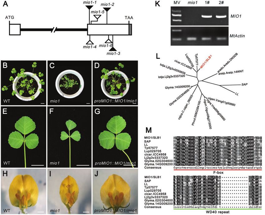
Page 4 of 17 | Zhou et al.

and MtKIX were inserted into the prey vector pGADT7. For the bi-              Protein degradation assay
molecular fluorescence complementation (BiFC) assay, the MIO1/                The His-BS1 fusion protein was expressed in E. coli, then extracted and
SLB1 coding sequence was inserted into the pFGC-YN173 vector                  purified with a His-tag protein purification kit (Beyotime, China) ac-
while MtASK and BS1 were inserted into the pFGC-YC155 vector.                 cording to the manufacturer’s instructions. Total protein from the WT,
To obtain a plasmid that expresses the GST-MIO1 WD40 fusion, the              mio1 mutant, and MIO1 overexpression lines (WT 35S::MIO1) was ex-
truncated coding sequence of Medtr5g097060 corresponding to the               tracted from an equal amount of leaf tissue. Then the purified His-BS1
WD40 repeat domain was cloned into pGEX4T-1 that harbored the                 protein was mixed with the total protein solution, in a 3:1 volume ratio.
GST coding sequence. Similarly, the coding sequence of BS1 was                For the control, the total protein was first pre-treated with MG132 before
inserted into pET28a that contained the His tag, to generate the              mixing it with His-BS1. All the mixtures were incubated at 4 °C with
His-BS1 fusion protein. All the primers used for the above are listed in      gentle shaking applied. Samples were removed at different time points

                                                                                                                                                           Downloaded from https://academic.oup.com/jxb/advance-article/doi/10.1093/jxb/erab033/6121689 by guest on 26 March 2021
the Supplementary Table S1.                                                   (0 min and 30 min), the catalytic reaction was stopped by boiling for
                                                                              5 min at 95 °C with SDS loading buffer. The denatured protein was
                                                                              separated on 10% SDS-PAGE and detected by an immunoblot analysis
Sub-cellular localization
                                                                              using anti-His antibodies (Transgene, China). Equal amounts of the total
The 35S::GFP-MIO1 and 35S::MIO1-GFP constructs were trans-                    proteins were used as loading controls.
formed into the Agrobacterium tumefaciens EHA105 strain, and then in-
filtrated into tobacco (Nicotiana benthamiana) leaves. The MIO1-GFP
and GFP-MIO1 signals were examined under a confocal laser scanning
microscope (Olympus, FV1000).                                                 Results
                                                                              Isolation and characterization of the M. truncatula
RNA isolation, RT–PCR, and quantitative RT–PCR                                mio1 mutant
Total RNA was isolated from plant tissues with a plant total RNA iso-
lation kit (Generay, China). The quantity and quality of extracted RNA        To identify the key regulators that control the organ size of
were assessed with Nanodrop 2000 (Thermo Scientific, USA). Next,              legumes, we screened the Tnt1 retrotransposon insertion mu-
the cDNA synthesis was performed with PrimeScriptTM RT reagent kit            tant population of the model legume M. truncatula (R108) for
(Takara, Japan), following the manufacturer’s instructions. For RT–PCR,
the cDNA was amplified in 30 cycles and the products were separated           any organ size mutants. Three independent mutant lines—mini
on a 1.5% sepharose gel. The MtActin gene was used as an internal con-        organ1 (mio1)-1, mio1-2, and mio1-3—exhibiting similarly de-
trol. Quantitative RT–PCR analysis was conducted by LightCycler 480           creased organ size, were isolated. A genetic analysis confirmed
engine (Roche, Switzerland), for which both MtActin and MtGAPDH               that they were allelic mutants. Then the mio1-1 mutant was
served as internal controls. The primers for this can be found in             backcrossed with the WT plants. In the BC1F2 population, the
Supplementary Table S1.
                                                                              plants displaying the WT-like and mutant phenotype showed
                                                                              a 3:1 (149:51) segregation ratio; this indicated that the mutant
GUS staining assay                                                            phenotype was caused by the mutation of a single recessive
Leaf samples of the proMIO1::GUS transgenic plants were collected and         gene. The mio1-1 was backcrossed with WT for three gener-
subjected to vacuum infiltration in a GUS staining solution, and then incu-   ations, and then the BC3F2 plants were used for phenotypic
bated overnight at 37 °C overnight.The WT served as the negative control.     analysis. Compared with the WT, the mio1 mutant was evi-
                                                                              dently reduced in its plant and organ size in both its vegetative
Yeast two-hybrid (Y2H) assay                                                  (Fig. 1A, B) and reproductive phases (Fig.1C, D). The sizes of
The constructed bait and prey plasmids were co-transformed into the           the representative lateral organs, namely the leaves and flowers,
Y2H Gold Yeast Strain (Clontech) and cultured on SD/-Leu-Trp media,           were markedly smaller in the mio1 mutant than the WT (Fig.
for three days, at 30 °C. The ensuing interactions were detected on se-       1E-G).The terminal leaflet length (TLL), terminal leaflet width
lective media -4 (SD/-Ade/-His/-Leu/-Trp).                                    (TLW), terminal leaflet area (TLA), and terminal leaflet perim-
                                                                              eter (TLP) of the mio1 mutant exhibited significant (P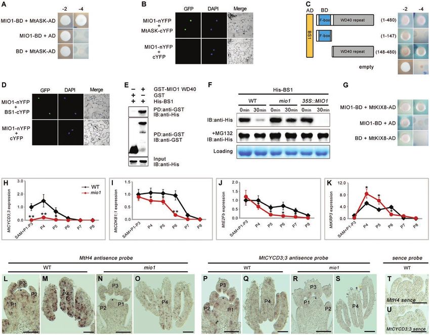
MIO1/SLB1 modulates organ size and pulvinus development | Page 5 of 17

                                                                                                                                                         Downloaded from https://academic.oup.com/jxb/advance-article/doi/10.1093/jxb/erab033/6121689 by guest on 26 March 2021
Fig. 1. Phenotype comparisons between the wild type (WT) and mio1 mutant. (A, B) Four-week-old seedlings of the WT (A) and mio1-1 mutant (B).
Scale bar=2 cm. (C, D) Branch of a 12-week-old plant of the WT (C) and mio1-1 mutant (D). Scale bar=2 cm. (E, F) The fifth compound leaf of five-week-
old seedlings of the WT (E) and mio1-1 mutant (F). Insets provide a close-up view of the stipules. Scale bar=1 cm for leaves, 0.5 cm for stipules. (G)
Flower (open petal) of the WT (upper) and mio1-1 mutant (bottom) plants. Scale bar=2 mm. (H) Comparison of the terminal leaflet length (TLL), terminal
leaflet width (TLW), terminal leaflet area (TLA), and terminal leaflet perimeter (TLP) of the WT and mio1-1 mutant plants. Values indicate the mean±SD
(n=3 biological replicates, with 20 plants per replicate); asterisks indicate significant differences from the WT (**P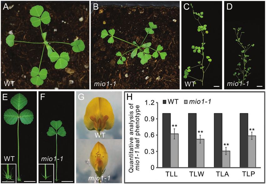
Page 6 of 17 | Zhou et al.

                                                                                                                                                        Downloaded from https://academic.oup.com/jxb/advance-article/doi/10.1093/jxb/erab033/6121689 by guest on 26 March 2021
Fig. 2. Molecular cloning and characterization of MIO1/SLB1. (A) The MIO1/SLB1 gene structure and Tnt1 retrotransposon insertion sites in the
mio1 mutant alleles. White and grey boxes represent the exon and 3’ UTR, respectively, and the bold line represents the intron. Triangles indicate
the Tnt1 insertion sites of mio1 mutant alleles, while the black and white ones indicate mutants obtained from forward and reverse screening,
respectively. (B-J) Genetic complementation of the mio1 mutant. Five-week-old seedlings of the WT (B), mio1 mutant (C), and complemented mio1 line
proMIO1::MIO1/mio1 (D). The fifth trifoliate leaf of five-week-old seedling of WT (E), mio1 mutant (F), and complemented mio1 line proMIO1::MIO1/mio1
(G). Flower (open petal) of the WT (H), mio1 mutant (I), and complemented mio1 line proMIO1::MIO1/mio1 (J). Scale bars=2 cm (B-D), 1 cm (E-G),
2 mm (H-J). (K) RT–PCR analysis of MIO1/SLB1 gene expression in the complemented mio1 lines. The expression of MIO1/SLB1 was restored in the
independent complemented line 1 (1#) and line 2 (2#). Total RNA was extracted from the shoot apices of five-week-old seedlings. MW, molecular weight
markers; MtActin was used as an internal control. (L) Phylogenetic analysis of MIO1/SLB1 and its closely related homologs. SAP from Arabidopsis
thaliana, LL from Cucumis sativus, with other homologs from several Fabaceae species including Trifolium pratense, Lupinus angustifolius, Cicer
arietinum, Lotus japonicus, Glycine max, Cajanus cajan, Phaseolus vulgaris, Vigna unguiculata, Vigna radiata, Vigna angularis, Arachis ipaensis, and
Arachis duranensis. (M) Amino acid sequence alignment of MIO1/SLB1 and its closely related homologs. The red and green underlining indicates the
F-box and WD40 repeat domain, respectively. SAP from A. thaliana, LL from C. sativus, Tp57577 from T. pratense, Lup029705 from L. angustifolius,
cicar.ICC4958 from C. arietinum, Lj2g3v3337320 from L. japonicus, and Glyma.02G304600 and Glyma.14G009200 from G. max. Dotted lines indicate
the abridged conserved amino acids.

locus corresponds to MIO1 that is allelic to the SLB1 gene                  2L; Supplementary Fig. S2), the key organ size regulators in
and required for organ size determination in M. truncatula.                 Arabidopsis and cucumber (Cucumis sativus), respectively
  Phylogenetic analysis revealed MIO1/SLB1 as the ortholog                  (Wang et al., 2016; Yang et al., 2018). Multiple amino acid se-
of STERILE APETALA (SAP) and LITTLE LEAF (LL; Fig.                          quence alignments of MIO1/SLB1 with its orthologs indicated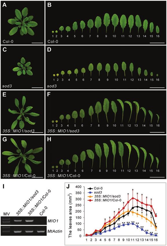
MIO1/SLB1 modulates organ size and pulvinus development | Page 7 of 17

that they shared the highly conserved N-terminal F-box and            (MIO1-GFP or GFP-MIO1) under the control of a 35S
C-terminal WD40 repeat domains (Fig. 2M; Supplementary                promoter in tobacco leaves. Both the MIO1-GFP and GFP-
Fig. S3). All of these features suggested that MIO1/SLB1 en-          MIO1 fusion proteins were localized to the nuclei of tobacco
codes an F-box protein. F-box proteins are known to form              leaf epidermal cells (Fig. 4M, N). Free GFP driven by the 35S
the SCF E3 ubiquitin ligase with ASK and Cullin through its           promoter was used as the negative control (Fig. 4O).
N-terminal F-box domain, and recognize specific substrates
for ubiquitination through its C-terminal domain, just like the       MIO1/SLB1 positively regulates primary cell division
WD40 repeat domain (Vierstra, 2003; Wang et al., 2016).
                                                                      Loss-of-function mutation of MIO1/SLB1 resulted in re-

                                                                                                                                          Downloaded from https://academic.oup.com/jxb/advance-article/doi/10.1093/jxb/erab033/6121689 by guest on 26 March 2021
                                                                      duced lateral organ size compared with the WT (Fig. 5B, C;
Ectopic expression of MIO1/SLB1 rescues the mutant
                                                                      Supplementary Fig. S5A, B).
phenotype of Arabidopsis sod3-3
                                                                         To gain insight into the mechanism by which MIO1/SLB1
The Arabidopsis MIO1/SLB1 ortholog, STERILE APETALA                   functions to regulate organ size, MIO1-overexpressing plants
(SAP)/SUPPRESSOR OF DA1 (SOD3), is known to posi-                     were generated by introducing the 35S::MIO1 construct into
tively regulate organ size during plant growth and development        WT. Ten independent transgenic lines exhibited enlarged or-
(Wang et al., 2016). Loss-of-function of SAP gave rise to de-         gans, especially for leaf (Fig. 5D; Supplementary Fig. S5C),
creased organ size in Arabidopsis (Wang et al., 2016).To investi-     floral organ (Fig. 5E), pod (Fig. 5F), and seed (Fig. 5G); these
gate whether MIO1/SLB1 performs a similar function as SAP             phenotypes were very similar to the reported M. truncatula bs1
for organ size control, the CDS of MIO1/SLB1 driven by the            mutant (Ge et al., 2016). MIO1/SLB1 expression increased by
cauliflower mosaic virus 35S (CaMV35S) promoter was intro-            more than twelve-fold when compared with the WT in the
duced into the Arabidopsis sod3-3 mutant (35S::MIO1/sod3-3)           MIO1-overexpressing transgenic lines (Fig. 5A; Supplementary
and the corresponding WT (35S::MIO1/Col-0). The smaller               Fig. S6).
organ size of sod3-3 was rescued in 35S::MIO1/sod3-3 trans-              Final organ size in plants is determined by a complex co-
genic plants (Fig. 3A-F, J). Furthermore, the 35S::MIO1/Col-0         ordination of cell division and expansion (Gonzalez et al.,
transgenic lines exhibited increased organ size compared with         2012). Microscopic examination of mature leaf epidermal
Col-0 (Fig. 3G, H, J). Interestingly, most of the transgenic plants   cells revealed that cell size was opposite to MIO1/SLB1 ac-
had twisted and dome-shaped leaves (Fig. 3F, H). This pheno-          tivity: epidermal cell size increased in the mio1 mutant while
type was similar to that reported for the Arabidopsis ppd mutant      it decreased in the MIO1-overexpressing plants in comparison
(White, 2006). The expression of MIO1/SLB1 was detected               with the WT (Fig. 5H-J; Supplementary Fig. S5D-F). Using
in both 35S::MIO1/sod3-3 and 35S::MIO1/Col-0 transgenic               the measurements of epidermal cell area (Fig. 5K) and leaflet
plants (Fig. 3I). These results suggested that MIO1/SLB1 and          area (Fig. 5L), we calculated the total number of epidermal cells
SAP play a conserved role in organ size regulation.                   per given leaflet (Fig. 5M). On average, this value for MIO1-
                                                                      overexpressing plants increased by more than two-fold com-
Expression pattern of MIO1/SLB1 and sub-cellular                      pared with WT on both adaxial and abaxial sides (Fig. 5M).
localization of MIO1/SLB1                                             However, the number of cells in the mio1 mutant was only
                                                                      about half that of the WT (Fig. 5M). These results suggested
Quantitative RT-PCR revealed that MIO1/SLB1 was highly                that MIO1/SLB1 augments leaf size mainly by increasing the
expressed in leaves, floral organs, and immature seeds (Fig. 4A;      abundance of cells (and not their individual area).
Supplementary Fig. S4). Further analysis with RNA in-situ hy-            The follow-up investigation of abaxial epidermal cell
bridization showed that the MIO1/SLB1 transcripts mainly              area (Fig. 5N), leaflet area (Fig. 5O), and the calculated total
accumulated in the early leaf primordia (P0-P2; Fig. 4B, C),          number of cells per leaflet (Fig. 5P) indicated that differences
pulvinus primordia (Fig. 4D), floral meristem, axillary bud           in leaf epidermal cell size among the MIO1-overexpressing,
(Fig. 4E), and petal meristem, developing carpel, and ovule           mio1 mutant, and WT plants had its origin around the P6
(Fig. 4F, G), with no signals detected in the control which hy-       leaf development stage, and this became magnified in later
bridized with a sense probe (Fig. 4H). To further confirm this,       stages. Differences in cell abundance began to emerge at P5,
but using a different approach, we introduced the GUS re-             and reach a plateau at P6 (Fig. 5N, P). All of these findings
porter gene driven by the MIO1/SLB1 promoter into WT                  implicated MIO1/SLB1 in influencing primary cell division
(proMIO1::GUS). GUS signals were detected in both single              during leaf growth and development. Although the cell size
and trifoliate leaves, and the signals were stronger in vascular      and number differed, the leaf epidermal cell pattern (Fig. 5H-J;
tissue and pulvinus (Fig. 4I-L). Taken together, the above ex-        Supplementary Figs S5D-F; S7) and stomatal index (Fig. 5Q)
pression pattern suggested a crucial role for MIO1/SLB1               were similar among the MIO1-overexpressing, mio1 mutant,
during lateral organ morphogenesis in M. truncatula.                  and WT plants, further pointing to the promotion of primary
   To determine its sub-cellular localization, we transiently ex-     cell proliferation by MIO1/SLB1 activity. The time point at
pressed MIO1/SLB1 fused with a green fluorescent protein              which leaflet length, width, and area peaked in size was likely
Page 8 of 17 | Zhou et al.

                                                                                                                                                            Downloaded from https://academic.oup.com/jxb/advance-article/doi/10.1093/jxb/erab033/6121689 by guest on 26 March 2021

Fig. 3 Ectopic expression of MIO1/SLB1 in Arabidopsis sod3-3 (35S::MIO1/sod3) mutant and wild type (35S::MIO1/ Col-0) plants. (A-H) Five-week-old
seedlings of Col-0 (A), sod3-3 mutant (C), 35S::MIO1/sod3 (E), and 35S::MIO1/Col-0 (G). Scale bar=2cm. The first to sixteenth leaves of five-week-
old seedlings of Col-0 (B), sod3-3 mutant (D), 35S::MIO1/sod3 (F), and 35S::MIO1/Col-0 (H). Scale bar=2cm. (I) RT–PCR analysis of MIO1/SLB1
expression in the complemented sod3 line and Col-0 plants. (J) The first to sixteenth leaves area of five-week-old seedlings of the Col-0, sod3-3 mutant,
35S::MIO1/sod3, and 35S::MIO1/Col-0. The leaves 1 and 2 are cotyledons. Values indicate the mean±SD (n=3 biological replicates; 10 plants per
replicate). Asterisks indicate significant differences from the WT (**P
MIO1/SLB1 modulates organ size and pulvinus development | Page 9 of 17
Downloaded from https://academic.oup.com/jxb/advance-article/doi/10.1093/jxb/erab033/6121689 by guest on 26 March 2021
Fig. 4. Expression pattern of MIO1/SLB1 and sub-cellular localization of MIO1/SLB1. (A) Expression of MIO1/SLB1 in different plant tissues analysed
by qRT–PCR, with MtActin used as an internal control. Values indicate the mean±SD (n=3). (B-H) RNA in situ hybridization of MIO1. Cross (B) and
longitudinal (C and D) sections of shoot apices from three-week-old wild type (WT) seedlings (vegetative stage) that were hybridized with the MIO1/SLB1
anti-sense probe. Longitudinal sections of shoot apices from 10-week-old WT plants (E), floral meristem (F), and 1 mm flower bud (G) that were
hybridized with the MIO1/SLB1 anti-sense probe. P+number, plastochron; FM, floral meristem; AB, axillary buds; P, petal; C, carpel; S, stamen; O, ovule;
asterisks denote the shoot apical meristem. The black arrow points to the pulvinus primordium. The MIO1/SLB1 sense probe was used as the control
(H). Scale bar=50 μm. (I-L) GUS staining of single leaf (I and J) and trifoliate leaf (K and L) of four-week-old seedlings of proMIO1::GUS transgenic plants.
J and L are the close-up views of the framed area in I and K, respectively. Scale bars=1 cm (I and K), 2 mm (J and L). (M-O) Sub-cellular localization of
the MIO1-GFP (M) and GFP-MIO1 (N) fusion proteins. Free GFP driven by the CaMV35S promoter was used as the control (O). Scale bar=20 μm.
MIO1/SLB1 positively regulates organ size mainly by pro- To uncover the mechanism underlying the control of organ
moting the process of primary cell proliferation during plant size by MIO1/SLB1, we performed protein-protein inter-
development. action assays to analyse the interaction between MIO1/SLB1
and other potential components of the ubiquitin-proteasome
F-box protein MIO1/SLB1 regulates the stability of pathway. MtASK, the ortholog of ASK1/2 in M. truncatula,
BS1 in vitro was identified by phylogenetic analysis and its interaction
with MIO1/SLB1 was tested using Y2H and BiFC assays.
Previous reports demonstrated that SAP interacts with ASK1/2 According to these results, MIO1/SLB1 physically interacted
and CUL1 (Cullin1), to form a conserved SCF E3 ubiquitin with MtASK in both yeast and tobacco leaf cells (Fig. 6A, B).
ligase, which targets the repressor PPD for degradation to Similarly, the interaction between MIO1/SLB1 and BS1 was
thereby enlarge the size of organs (Wang et al., 2016).The loss- also verified by the Y2H and BiFC assays (Fig. 6C, D). Further
of-function of BS1, the ortholog of PPD in M. truncatula, also analysis of the Y2H results revealed that MIO1/SLB1 is capable
led to enlarged lateral organs similar to the ppd mutant (Ge of interacting with BS1 through its C-terminal WD40 repeat
et al., 2016). These clues suggest that the role of MIO1/SLB1 domain (Fig. 6C), which was confirmed by the results from the
in regulating organ size may involve the conserved ubiquitin- in vitro pull-down experiment (Fig. 6E). Hence, MIO1/SLB1
proteasome at its core. could interact not only with MtASK, which is predicted toPage 10 of 17 | Zhou et al.
Downloaded from https://academic.oup.com/jxb/advance-article/doi/10.1093/jxb/erab033/6121689 by guest on 26 March 2021
Fig. 5. MIO1/SLB1 positively regulates primary cell division during leaf development. (A) MIO1/SLB1 transcript abundance in the wild type (WT) and
MIO1-overexpressing line, with MtActin used as an internal control. Values indicate the mean±SD (n=3); asterisks indicate significant differences
with respect to the WT (**PMIO1/SLB1 modulates organ size and pulvinus development | Page 11 of 17
form an SCF E3 ubiquitin ligase, but also with BS1 in a way juvenile leaves. By contrast, the expression of the predicted
possibly related to protein degradation. cell division repressor MtKRP3 (Fig. 6K; Supplementary Fig.
Given the similar phenotypes of MIO1-overexpressing and S10D) was increased in mio1 mutant at the P4 and P5 stages,
bs1 mutant plants (Ge et al, 2016), we suspected that MIO1/ where cell division activity is supposed to be high. We then
SLB1 might regulate the stability of BS1 protein to control carried out an in-situ hybridization assay to analyse the ex-
organ size in M. truncatula. To test this, total proteins were ex- pression of MtH4 (Medicago HISTONE4) (Fig. 6L-O) and
tracted from WT, mio1 mutant, and MIO1-overexpressing MtCYCD3;3 (Fig. 6P-S) at the early leaf development stages
plants, and mixed with equal amounts of E. coli-expressed of WT and mio1 mutant plants. These results showed that both
His-BS1 fusions, followed by incubation at 4 °C with gentle MtH4 and MtCYCD3;3 were expressed in the SAM and P1 to
Downloaded from https://academic.oup.com/jxb/advance-article/doi/10.1093/jxb/erab033/6121689 by guest on 26 March 2021
shaking. Samples were then removed at different time points P4 of both the WT and mio1 mutant, but the mio1 mutant had
for a gel quantification analysis. Evidently, the amount of much weaker signals. These findings suggested that MIO1/
His-BS1 decreased after 30 min of incubation compared with SLB1 could influence the expression of the core cell-cycle
what it was at the start (time 0: time point=0 min) in both the genes; hence, it was suspected to regulate cell proliferation and
WT and MIO1-overexpressing plants (Fig. 6F). However, the lateral organ size.
His-BS1 protein degraded more slowly when incubated with
the proteasome inhibitor MG132 (Fig. 6F), indicating that the MIO1/SLB1 influences pulvinus development and leaf
stability of BS1 is affected by the proteasome. movement
Additionally, the degradation of His-BS1 fusion protein ap-
peared to be much slower in samples incubated with total pro- Leaf movement is driven by a motor organ- the pulvinus - that
teins from the mio1 mutant but faster in samples incubated is commonly observed in legume species (Darwin, 1880; Satter
with proteins from the MIO1-overexpressing lines, when com- et al., 1990; Koller, 2011; Mancuso and Shabala, 2015). Besides
pared with the WT (Fig. 6F), indicating that the stability of BS1 its decreased organ size, the mio1 mutant also showed defective
protein is negatively regulated by MIO1/SLB1 in vitro. Taken leaf movement when compared with the WT (Fig. 7A, B).
together, these results suggested that MIO1/SLB1 might form Generally, all the leaflets of WT engaged in reversible
an SCF E3 ubiquitin ligase complex with MtASK so as to movement, that is, remaining in a horizontal (open) position
modulate BS1 stability and control the lateral organ size in during the day and vertical (closed) at night (Supplementary
M. truncatula. MtKIX8 was identified during the Y2H library Fig. S11). In contrast to the WT, leaves of the mio1 mu-
screening for potential interacting proteins of MIO1/SLB1, tant featured a suite of defects in terms of leaf closure
and was verified to physically interact with MIO1/SLB1 (Fig. (Supplementary Fig. S11C, D). Generally, leaf movement
6G). Since MIO1/SLB1 remarkably influences cell prolifer- was absent in the first and second trifoliate leaves (L1, L2),
ation and the number of cells, the expression of several rep- after which the degree of leaflet rotation gradually increased
resentative cell-cycle genes were analysed in the mio1 mutant in the third to sixth trifoliate leaves (L3–L6), followed by
and WT plants. The expression of MtCYCD3;2, MtCDKB1;1, it progressively decreasing in the seventh to tenth trifoliate
MtE2Fb, and MtKRP3 were analysed by qRT–PCR in leaves until it was completely lost (L7–L10; Supplementary
SAM (shoot apical meristem) and P1 (plastochron 1)-P8 of Fig. S11A, C, D). These defects in leaf movement were res-
six-week-old mio1 mutant and WT plants (Supplementary cued when the coding sequence of MIO1/SLB1, which was
Fig. S9). The expression of the predicted cell division acti- driven by its native promoter, was expressed in the mio1 mu-
vators MtCYCD3;2 (Fig. 6H; Supplementary Fig. S10A), tant (Fig. 7C; Supplementary Fig. S12).
MtCDKB1;1 (Fig. 6I; Supplementary Fig. S10B), and MtE2Fb To investigate the causes behind the defective leaf move-
(Fig. 6J; Supplementary Fig. S10C) were all decreased in the ment in the mio1 mutant, we analysed the structure of its
mio1 mutant when compared with the WT, especially in the pulvinus. The pulvini of mio1 mutants were shortened or
(H), mio1 mutant (I), and MIO1-overexpressing line (J). These pictures were outlined with Photoshop software based on photos from the microscope
observations. Green and yellow-colored cells represent epidermal and guard cells, respectively. (K) The leaf epidermal cell area of the WT, mio1 mutant,
and MIO1-overexpressing line. Values indicate the means±SD (n=3 biological replicates, with 10 plants per replicate); asterisks indicate significant
differences from the WT (*PPage 12 of 17 | Zhou et al.
Downloaded from https://academic.oup.com/jxb/advance-article/doi/10.1093/jxb/erab033/6121689 by guest on 26 March 2021
Fig. 6. F-box protein MIO1/SLB1 physically interacts with, and regulates BS1 stability. (A) Yeast two-hybrid (Y2H) assay showing the interaction between
MIO1/SLB1 and MtASK. -2, SD/-Leu/-Trp; -4, SD/-Ade/-His/-Leu/-Trp. (B) Bimolecular fluorescence complementation (BiFC) assay showing the
interaction between MIO1/SLB1 and MtASK in the nuclei of tobacco (Nicotiana benthamiana) leaf epidermal cells. MIO1-nYFP and MtASK-cYFP were
coexpressed in the leaves of tobacco; DAPI signals indicate the nuclei. (C) Y2H assay showing the interaction between MIO1/SLB1 and BS1. MIO1/
SLB1 interacts with BS1 through its WD40 repeat domain. AD+BD was used as the control. -2, SD/-Leu/-Trp; -4, SD/-Ade/-His/-Leu/-Trp. (D) BiFC
assay showing the interaction between MIO1/SLB1 and BS1 in the nuclei of tobacco leaf epidermal cells. MIO1-nYFP and BS1-cYFP are coexpressed in
leaves of tobacco, DAPI signals indicate the nuclei. (E) N-terminal WD40 repeat domain of MIO1/SLB1 (MIO1 WD40) interacts with BS1 in vitro. His-BS1
was pulled down (PD) by GST-MIO1 WD40 immobilized on glutathione sepharose, and analysed by immunoblotting (IB) using an anti-His antibody.
GST was used as a negative control. (F) MIO1/SLB1 regulates BS1 stability in vitro. The His-BS1 fusion protein was detected with the His antibody.
MG132 was used to inhibit the proteasome activity. The total proteins extracted from plants were used as a loading control. (G) Yeast two-hybrid (Y2H)
assay showing the interaction between MIO1/SLB1 and MtKIX. -2, SD/-Leu/-Trp; -4, SD/-Ade/-His/-Leu/-Trp. (H-K) Expression of MtCYCD3;3 (H),
MtCDKB1;1 (I), MtE2Fb (J), and MtKRP3 (K) in the WT and mio1 mutant, for which MtActin served as the internal control. SAM, shoot apical meristem; P,
plastochron. Values indicate the mean±SD (n=3); asterisks indicate significant differences from the WT (*PMIO1/SLB1 modulates organ size and pulvinus development | Page 13 of 17
found that the knitted wool-like structure of the pulvinus The loss-of-function of MIO1/SLB1 in M. truncatula and LL
was partially or completely absent from the boundary re- in C. sativus led to an increased and decreased cell size, respect-
gion between the leaflet and petiole in the mio1 mutant in ively, when compared with the WT (Yang et al., 2018). A prob-
comparison with the WT (Fig. 7D-G; Supplementary Fig. able explanation for this functional divergence in cell size is
S14). This abnormal pulvinus structure was restored to a the different mutational patterns and compensatory effects
WT-like phenotype in the complemented transgenic lines of mio1 and ll mutants. The mio1 mutant arose from the Tnt1
(Fig. 7H, I). retrotransposon insertion, which completely eliminated the
Moreover, the anatomical analysis uncovered a substantially MIO1/SLB1 transcript, so its cells may expand in an attempt
decreased number of motor cells in the typically defective to compensate for a drastic loss in cell division (Horiguchi and
Downloaded from https://academic.oup.com/jxb/advance-article/doi/10.1093/jxb/erab033/6121689 by guest on 26 March 2021
pulvinus of mio1 mutant when compared with those of the Tsukaya, 2011). Since the ll mutant arose from a single nu-
WT (Fig. 7J, K), but this was also fully rescued in the com- cleotide substitution (Yang et al., 2018), LL might retain some
plemented plants (Fig. 7L). These results indicated that the partial functioning: it might remain below the threshold to
defective pulvinus of mio1 mutant likely arose from reduced confer full function, yet still above the threshold needed to
motor cell division and final cell counts. Auxin was able to spe- resist compensation. Irrespective of compensation and species
cifically accumulate in the pulvinus of WT (Zhou et al., 2012; differences, both cell proliferation and cell expansion clearly
Supplementary Fig. S15A, B), but this signal disappeared in the contribute to determining organ size, and they account for the
mio1 mutant (Supplementary Fig. S15C, D). In conclusion, we conserved function of MIO1/SLB1, SAP, and LL in their na-
have shown that MIO1/SLB1 is also necessary for robust pul- tive plant species.
vinus development and leaf movement in M. truncatula.
MIO1/SLB1 controls final organ size by regulating BS1
stability and expression of cell-cycle genes
Discussion Based on previous reports (Ge et al., 2016; Wang et al., 2016),
MIO1/SLB1 positively regulates primary cell we proposed that the F-box protein MIO1/SLB1 could form
proliferation to control organ size part of an SCF E3 ubiquitin ligase complex, to regulate organ
size via targeting its potential substrate BS1 for ubiquitination
The final organ size of multicellular plants is determined and degradation in M. truncatula. Protein-protein interaction
by a complex coordination of cell division and expansion assays indicated that MIO1/SLB1 could directly interact with
(Gonzalez et al., 2012). Our results demonstrate that MIO1/ MtASK and BS1 (Fig. 6A-E). Our analysis also showed that the
SLB1 acts as a positive regulator of organ size by promoting protein stability of BS1 could be modulated by MIO1/SLB1
the number of cells in M. truncatula. The time-course analysis with sensitivity to the inhibitor MG132 (Fig. 6F). These ob-
of epidermal cell counts in developing leaflets indicated that servations are consistent with BS1 being recruited by MIO1/
MIO1/SLB1 promoted cell proliferation during the early de- SLB1 into the ubiquitin-proteasome pathway to control its
velopment stages (Fig. 5P); the stomatal index (a credible in- stability.
dicator of meristemoid cell division), epidermal cell pattern, Nevertheless, the His-BS1 fusion could still be degraded
and duration of leaf area expansion were not influenced by ei- by proteins extracted from the mio1 mutant, albeit at a slower
ther loss-of-function or overexpression of MIO1/SLB1 when rate (Fig. 6F), which suggests that additional factors may co-
compared with the WT (Fig. 5H–J, Q; Supplementary Figs S5; exist with MIO1/SLB1 to regulate the abundance of BS1. It is
S7; S8). These results suggest that MIO1/SLB1 functions as a thus conceivable that in M. truncatula, MIO1/SLB1 positively
facilitator of primary cell division, rather than meristemoid cell regulates organ size, in part, by forming the SCF E3 ubiquitin
proliferation, to modulate organ size during development. ligase to target the organ size repressor BS1 for degradation.
MIO1/SLB1 and its orthologs SAP (in Arabidopsis) and LL Identifying the additional factors that co-regulate BS1 and
(in cucumber) perform a conserved function - to positively elucidating their molecular mechanisms will enlighten our
regulate organ size during plant development, but interestingly, understanding of organ size regulation in legumes.
their effects on cell proliferation and cell size seem to follow Cyclin-dependent kinases (CDKs) are the primary regulators
different pathways. While MIO1/SLB1 positively controls pri- of eukaryotic cell cycle progression, whose catalytic activity de-
mary cell division, SAP mainly acts to promote meristemoid pends on the binding and activation of cyclins (CYCs; Joubès
cell proliferation during development (Wang et al., 2016). This et al., 2000). For example, the plant-specific CDKs are necessary
functional variation might be caused by the differences in for proper cell cycle progression (Andersen et al., 2008; Nowack
function of their downstream regulators between the Fabaceae et al., 2012), and CYCD3s are vital regulators that control cell div-
and Brassicaceae. Previous studies have confirmed that PPD ision and expansion by regulating the duration of mitotic phase
(the substrate of SAP) and its ortholog BS1 in M. truncatula and the mitosis-to-endocycle transition (Dewitte et al., 2007).
act as negative regulators of meristemoid proliferation and pri- Kip-related proteins (KRPs), another kind of essential
mary cell division, respectively (White, 2006; Ge et al., 2016). cell cycle regulator, which usually exhibit cyclin-dependentPage 14 of 17 | Zhou et al.
Downloaded from https://academic.oup.com/jxb/advance-article/doi/10.1093/jxb/erab033/6121689 by guest on 26 March 2021
Fig. 7. MIO1/SLB1 rescues the leaf movement and pulvinus of the mio1 mutant. (A-C) MIO1/SLB1 restored the defective leaf movement of the mio1
mutant. A representative trifoliate leaf of wild type (WT) (A), mio1 mutant (B), and complemented mio1 line proMIO1::MIO1/mio1 (C) at night. Scale
bar=1cm. (D-I) MIO1/SLB1 rescued the defective pulvinus structure of the mio1 mutant. Scanning electron microscope (SEM) images of WT (D and E),
mio1 mutant (F and G), and complemented mio1 line (H and I). The pulvini were highlighted by the red dotted line. Open boxes in D, F, and H, indicate
those areas shown in E, G, and I, respectively. Scale bar=100 μm (D, F, H),10 μm (E, G, I). (J-L) Pulvinus longitudinal-sections of the WT (J), mio1 mutant
(K), and complemented mio1 line (L). The pulvinus region in each longitudinal section is highlighted by the red dotted line. Scale bar=10 μm.
kinase binding specificity, are the negative regulators of cycle in M. truncatula, namely MtCYCD3;2, MtCDKB1;1,
CDKs (Verkest et al., 2005; De Almeida Engler et al., 2009). and MtE2Fb, were down-regulated in juvenile leaves of
Other work has revealed that the conserved transcription mio1 mutants (Fig. 6H-J; Supplementary Fig. S10A-C).
factors E2Fs play crucial roles in several pathways related to In contrast, the expression of MtKRP3, the predicted cell
plant cell division and differentiation; for instance, E2Fb is cycle repressor, was up-regulated in mio1 mutant (Fig. 6K;
an activator of cell cycle progression in Arabidopsis (Magyar Supplementary Fig. S10D). The in-situ hybridization assay
et al., 2005; Sozzani et al., 2006). The expression of three further confirmed the disrupted expression profiles of cell
genes encoding predicted positive regulators of the cell cycle genes in the mio1 mutant (Fig. 6L–U). These resultsMIO1/SLB1 modulates organ size and pulvinus development | Page 15 of 17
suggest that MIO1/SLB1 could regulate the expression of MIO1/SLB1 was transformed into the mio1 mutant (Fig. 7C,
cell cycle genes to manipulate cell proliferation and ex- H). These results provide evidence that MIO1/SLB1 func-
pansion in M. truncatula. The variation in expression pro- tion is necessary to ensure the pulvinus develops normally
files of cell cycle genes in the mio1 mutant is similar to in M. truncatula. Although the molecular mechanism under-
those resulting from the breakdown of LL, the ortholog of pinning how MIO1/SLB1 regulates pulvinus development is
MIO1/SLB1 in cucumber (Yang et al., 2018). BS1 and its still unclear, there are several possibilities based on the avail-
ortholog PPD also influence cell cycle gene expression in able clues. On the one hand, MIO1/SLB1 might directly or
M. truncatula and A. thaliana, respectively (Gonzalez et al., indirectly regulate certain pulvinus identity genes, such as
2015; Ge et al., 2016). So, collectively, these clues suggest ELP1 and GmILPA1 (Chen et al., 2012; Zhou et al., 2012;
Downloaded from https://academic.oup.com/jxb/advance-article/doi/10.1093/jxb/erab033/6121689 by guest on 26 March 2021
that the MIO1/SLB1-related pathway that is relevant to Gao et al., 2017). ELP1 is a plant-specific lateral organ bound-
organ size control through regulation of cell cycle gene ex- aries domain (LBD) transcription factor that determines the
pression during plant development, might be conserved. pulvinus identity in legumes through its conserved func-
Medicago truncatula KINASE-INDUCIBLE DOMAIN tion (Chen et al., 2012); GmILPA1 was found to ensure the
INTERACTING 8 (MtKIX8), the homolog of KIX8 in normal progression of the motor cell cycle during pulvinus
Arabidopsis, was identified during the Y2H library screening development in soybean (Gao et al., 2017). It would be worth
for potential interacting proteins of MIO1/SLB1, and then analysing the genetic interaction between MIO1/SLB1 and
verified to physically interact with MIO1/SLB1 via the Y2H ELP1, or the ortholog of GmILPA1, during pulvinus mor-
assay (Fig. 6G). In Arabidopsis, SAP interacts with PPD and phogenesis. On the other hand, our anatomical examinations
KIXs to target the PPD-KIX complex for degradation (Wang showed that the shortened pulvini of mio1 mutant mainly ori-
et al., 2016; Li et al., 2018). Accordingly, it is likely that the pu- ginate from a vastly decreased number of motor cells (Fig.
tative MIO1/SLB1-BS1-MtKIX module is playing a vital role 7K). It seems likely that MIO1/SLB1 regulates pulvinus de-
in plant organ size determination analogous to the SAP-PPD- velopment through modulating the cell division of the motor
KIX module. Yet interestingly, it is possible that the MIO1/ organ, similar to the regulation of organ size, by taking part
SLB1-BS1-MtKIX module has disappeared from the Poaceae, in the genetic pathway of cell cycle control. Loss-of-function
based on our inspection of available genome sequences of MIO1/SLB1 led to defective leaf movement in a devel-
(Supplementary Fig. S2). Further investigation of this complex opmental stage-dependent manner (Supplementary Fig. S11);
may advance our understanding of differences in molecular however, this differs from the completely lost pulvinus as
mechanisms of lateral organ expansion between eudicot and found in the elp1 mutant (Chen et al., 2012). This suggests
monocot plant species. that additional extant but unknown regulators ought to func-
The organ size of legume crops and forage is one of the tion synergistically with MIO1/SLB1 to regulate pulvinus
most important agronomic traits because it is closely related development in M. truncatula. Another attractive scenario is
to final yield and quality. Those previously reported genes in that auxin specifically accumulated in the WT pulvinus but
M. truncatula, such as SGL1 (SINGLE LEAFLET1), PALM1, disappeared in the mio1 mutant (Supplementary Fig. S15).
LLS1 (LATERAL LEAFLET SUPPRESSION1), and Auxin is known to have significant roles in the activation of
PINNA1 (PENTAFOLIATA1), all provide important clues as cell cycle processes (Perrot-Rechenmann, 2010). It would be
to how the total leaf area and biomass might be promoted very interesting to test how MIO1/SLB1 may regulate pul-
through manipulating the number and size of leaflets (Wang vinus development through auxin-related cell proliferation
et al., 2008; Chen et al., 2010; He et al., 2020; Zhao et al., processes.
2020). From the present study, the predicted MIO1/SLB1-BS1
module would be a useful candidate for the genetic manipu-
lation of crop yields, via increased seed size or total biomass. Supplementary data
The following supplementary data are available at JXB online.
MIO1/SLB1 influences pulvinus development in
Fig. S1. The expression of MIO1/SLB1 in WT and
M. truncatula
mio1 mutant.
The reversible leaf movement of legumes is driven by the pul- Fig. S2. MIO1/SLB1 is the ortholog of LL and SAP.
vinus, a specialized motor organ (Satter et al., 1990; Mancuso Fig. S3.The distribution of F-box and WD40 repeat domain
and Shabala 2015). An abnormal pulvinus initiation or devel- in MIO1/SLB1.
opment would directly influence leaf movement (Chen et al., Fig. S4. The expression pattern of MIO1/SLB1 in different
2012; Zhou et al., 2012; Gao et al., 2017). Loss-of-function organs of WT.
of MIO1/SLB1 led to impaired leaf movement which re- Fig. S5. Size changes of whole plants and epidermal cells in
sulted from the extremely shortened pulvinus in M. truncatula different backgrounds.
(Fig. 7B, F; Supplementary Fig. S13). However, this de- Fig. S6. The expression of MIO1/SLB1 in 35S::MIO1
fect was fully rescued when the coding sequence (CDS) of transgenic plants.Page 16 of 17 | Zhou et al.
Fig. S7. Time-course analysis of the adaxial leaf epidermal Data availability
cell pattern of WT and mio1 mutant.
No new sequence data were published in the present paper. All se-
Fig. S8. Quantitative analysis of leaflet growth during quence data included in our manuscript can be obtained from the
different stages. publicly available genome of M. truncatula (Mt4.0v1) (http://bioinfo3.
Fig. S9. Different stages of leaves were used to monitor the noble.org/doblast/) under the following accession numbers: MIO1
expression of cell-cycle genes. (Medtr5g097060), BS1 (Medtr1g102900), MtASK (Medtr5g022710),
Fig. S10. The expression of cell cycle genes in WT and MtKIX8 (Medtr4g114900).
mio1 mutant.
Fig. S11. Loss-of-function of MIO1/SLB1 results in the de-
Downloaded from https://academic.oup.com/jxb/advance-article/doi/10.1093/jxb/erab033/6121689 by guest on 26 March 2021
fect of leaf movement at different stages. References
Fig. S12. Complementation of leaf movement phenotype in Andersen SU, Buechel S, Zhao Z, Ljung K, Novák O, Busch W,
mio1 mutant. Schuster C, Lohmann JU. 2008. Requirement of B2-type cyclin-
Fig. S13. Loss-of-function of MIO1/SLB1 results in a series dependent kinases for meristem integrity in Arabidopsis thaliana. The Plant
Cell 20, 88–100.
of defective pulvini.
Andriankaja M, Dhondt S, De Bodt S, et al. 2012. Exit from proliferation
Fig. S14. Morphological changes of epidermal cells of pul- during leaf development in Arabidopsis thaliana: a not-so-gradual process.
vini in mio1 mutant. Developmental Cell 22, 64–78.
Fig. S15. The auxin reporter DR5rev::Green Fluorescent Baekelandt A, Pauwels L, Wang Z, et al. 2018. Arabidopsis leaf flatness
Protein (GFP) shows the distribution of auxin in the pulvini of is regulated by PPD2 and NINJA through repression of CYCLIN D3 genes.
Plant Physiology 178, 217–232.
WT and mio1 mutant.
Baute J, Polyn S, De Block J, Blomme J, Van Lijsebettens M, Inzé D.
Table S1. Sequence information of primers used in this study. 2017. F-Box protein FBX92 affects leaf size in Arabidopsis thaliana. Plant &
Table S2. Detailed information of different mio1 mutant Cell Physiology 58, 962–975.
alleles. Byzova MV, Franken J, Aarts MG, de Almeida-Engler J, Engler G,
Mariani C, Van Lookeren Campagne MM, Angenent GC. 1999.
Arabidopsis STERILE APETALA, a multifunctional gene regulating inflores-
cence, flower, and ovule development. Genes & Development 13, 1002–1014.
Acknowledgements Chen J, Moreau C, Liu Y, Kawaguchi M, Hofer J, Ellis N, Chen R.
2012. Conserved genetic determinant of motor organ identity in Medicago
We thank Noble Research Institute, USA for providing mio1 mutant truncatula and related legumes. Proceedings of the National Academy of
and Prof. Yunhai Li for providing Arabidopsis sod3 mutant. The au- Sciences, USA 109, 11723–11728.
thors thank Prof. Noel Ellis (John Innes Centre, UK) and Dr. Dimiru Chen J, Yu J, Ge L, et al. 2010. Control of dissected leaf morphology by a Cys(2)
Tadesse (Oklahoma State University, USA) for their critical reading and His(2) zinc finger transcription factor in the model legume Medicago truncatula.
improving this manuscript. We thank Zhijia Gu (Kunming Institute Proceedings of the National Academy of Sciences, USA 107, 10754–10759.
of Botany, Chinese Academy of Sciences) for providing SEM analysis. Cookson SJ, Radziejwoski A, Granier C. 2006. Cell and leaf size plas-
This research was supported by National Natural Science Foundation ticity in Arabidopsis: what is the role of endoreduplication? Plant, Cell &
Environment 29, 1273–1283.
of China grants 31471171, U1702234, 31700244, 32070204; West Light
Foundation of The Chinese Academy of Sciences, Natural Science Cosgrove DJ. 2005. Growth of the plant cell wall. Nature Reviews.
Molecular Cell Biology 6, 850–861.
Foundation of Yunnan Province of China (2018FB033), XDB27030106
Coté GG. 1995. Signal transduction in leaf movement. Plant Physiology
(to JC), the Strategic Priority Research Program XDA27030301 (to LH)
109, 729–734.
and Youth Innovation Promotion Association CAS (to BZ); JC is also
Czesnick H, Lenhard M. 2015. Size control in plants–lessons from leaves
supported by the ‘One Hundred Talent Project of the Chinese Academy and flowers. Cold Spring Harbor Perspectives in Biology 7, a019190.
of Sciences’, ‘High-end Scientific and Technological Talents in Yunnan
Darwin C. 1880. The power of movement in plants. London, UK: J. Murray.
Province’ and Core Botanical Gardens, Chinese Academy of Sciences
de Almeida Engler J, De Veylder L, De Groodt R, et al. 2009. Systematic
(CAS-CBG). analysis of cell-cycle gene expression during Arabidopsis development. The
Plant Journal 59, 645–660.
Dewitte W, Scofield S, Alcasabas AA, et al. 2007. Arabidopsis CYCD3
Author contributions D-type cyclins link cell proliferation and endocycles and are rate-limiting for
cytokinin responses. Proceedings of the National Academy of Sciences,
ZSL,YTQ and CJH conceived and designed the experiments; ZSL,YTQ, USA 104, 14537–14542.
MYW, LY, WRR, and FYGW performed the experiments; ZSL, YTQ, Disch S, Anastasiou E, Sharma VK, Laux T, Fletcher JC, Lenhard M.
ZBL, HLL, LYH, BQZ, WDF, WQ,YYF, and CJH analysed the data and 2006. The E3 ubiquitin ligase BIG BROTHER controls Arabidopsis organ
size in a dosage-dependent manner. Current Biology 16, 272–279.
wrote the paper. All authors reviewed and approved the final manuscript.
Dong H, Dumenil J, Lu FH, et al. 2017. Ubiquitylation activates a pep-
tidase that promotes cleavage and destabilization of its activating E3 lig-
ases and diverse growth regulatory proteins to limit cell proliferation in
Conflict of interest Arabidopsis. Genes & Development 31, 197–208.
Donnelly PM, Bonetta D, Tsukaya H, Dengler RE, Dengler NG. 1999.
The authors declare that they have no competing interests in relation to Cell cycling and cell enlargement in developing leaves of Arabidopsis.
this study. Developmental Biology 215, 407–419.You can also read