O rigin and use of embryonic and adult stem cells in differentiation
←
→
Page content transcription
If your browser does not render page correctly, please read the page content below
Cardiovascular Research 58 (2003) 324–335
www.elsevier.com / locate / cardiores
Review
Origin and use of embryonic and adult stem cells in differentiation
and tissue repair
Robert Passier, Christine Mummery*
Hubrecht Laboratory, Netherlands Institute for Developmental Biology, Uppsalalaan 8, 3584 CT Utrecht, The Netherlands
Received 6 September 2002; accepted 7 November 2002
Abstract
Stem cells are self-renewing, unspecialised cells that can give rise to multiple cell types of all tissues of the body. They can be derived
from the embryo, foetus and adult. The ability of stem cells to divide but also to differentiate to specialised cell types like nerve and
muscle, have made them candidates on which to base therapies for diseases and disorders for which no, or only partially effective,
therapies are available. Replacement of defective or absent cells in defective tissues and organs could represent a cure. Here, we introduce
the background to stem cell research and review the present state-of-the-art in stem cell biology, directed differentiation and tissue repair.
In particular, we distinguish embryonic versus adult sources of stem cells and data derived from animal versus human experiments in
order to place current research and perspectives for clinical application in their correct context.
2003 European Society of Cardiology. Published by Elsevier Science B.V. All rights reserved.
Keywords: Developmental biology; Stem cells
1. Introduction tive medicine; diseases such as diabetes, Parkinson,
rheumatoid arthritis and myocardial infarction caused by
Stem cells are primitive cells present in all organisms the loss or loss-of-function of specific cell types, could be
that can divide and give rise to more stem cells, or switch cured if healthy cells replaced defective cells. However,
to become more specialised cells, such as those of the ethical considerations question the instrumental use of
brain, heart, muscle and kidney. Stem cells in early embryos for the isolation of stem cells, even if those
embryos give rise to cells of all tissues of the adult body embryos are surplus to requirements for assisted reproduc-
and are therefore termed ‘pluripotent’. Stem cells in some tion and destined for destruction. The debate has been
adult tissues, involved in tissue replacement and repair, further fuelled by recent evidence that adult stem cells
usually give rise only to cell types already present in the have a greater capacity for differentiation than had previ-
surrounding tissue from which they are derived. Stem cells ously been thought; they have entered the discussion as
of the bone marrow, for example, give rise to hemato- alternatives. Although with obvious ethical advantages, the
poietic cells. These adult stem cells are generally regarded scientific question concerns whether embryonic and adult
as ‘multipotent’. stem cells are equivalent in their capacity to produce large
Since the first report of the isolation human stem cells numbers of specific cell types for transplantation, which
from surplus IVF embryos in 1998, their derivation and retain their function over long periods. Here, we first
use has been hotly debated by governments, press and introduce embryonic stem cell biology in its historical
society in many parts of the world. Driving the debate has context then consider current problems in controlling their
been the shortage of donor organs and tissues for regenera- growth and differentiation. Their potential in regenerative
medicine is considered in the light of state-of-the-art
advances in adult stem cell biology.
*Corresponding author. Tel.: 131-30-212-1800; fax: 131-30-251-
6464.
E-mail address: christin@niob.knaw.nl (C. Mummery). Time for primary review 31 days.
0008-6363 / 03 / $ – see front matter 2003 European Society of Cardiology. Published by Elsevier Science B.V. All rights reserved.
doi:10.1016 / S0008-6363(02)00770-8R. Passier, C. Mummery / Cardiovascular Research 58 (2003) 324–335 325
2. Embryonic stem cells and teratocarcinomas human IVF embryos has lead to exceptionally high
efficiencies for hES cell line isolation, with 50% of
Exactly 20 years ago, the first embryonic stem cells blastocysts now yielding cell lines. Since then, a handful of
were isolated from mouse preimplantation, blastocyst-stage publications have described their differentiation to various
embryos. The background to the discovery lay in the study cell types in culture in response to cytokines, hormones
of teratocarcinoma, a spontaneous tumour of the testis in and growth conditions. These include neural cells (neurons,
mice and humans, consisting of tissues as diverse as hair, glia, and oligodendrocytes) and, most recently, insulin-
muscle, bone and even complete teeth. They resemble a producing pancreas cells, cartilage and bone, car-
disorganised foetus and have fascinated pathologists for a diomyocytes, hematopoietic cells, endothelial cells and
century or more (reviewed in Refs. [1,2]). In the mid- hepatocytes. Some of these differentiated hES cells have
1970s, developmental biologists discovered that teratocar- been transplanted into mice but the majority of the studies
cinomas could be induced in mice by transferring embryos so far only differentiated phenotypes in vitro. The hype
to extra-uterine sites and that they contained undifferen- around embryonic stem cells derives largely from studies
tiated stem cells. These embryonal carcinoma (or EC) stem using mouse ES cells to derive differentiated derivatives
cells could be isolated and grown in culture without losing which when transplanted to ailing mice have cured or
the capacity to differentiate. This was most strikingly relieved their disease. These results have often been
demonstrated by introducing them into embryos; if derived extrapolated directly to the human equivalent, which
from a brown mouse and placed in a blastocyst from an cannot be justified without further validation using hES
albino, the pups delivered by the foster mother were brown cells. In the following sections, we will provide an
and white. The stem cells had formed ‘chimeras’ and had overview distinguishing results derived from human versus
contributed to all somatic tissues, most obviously in the mouse ES cells both in terms of their ability to grow and
melanocytes of the skin. After differentiation, EC cells are differentiate in culture as well as in their ability to
no longer malignant; they therefore became not only a contribute to tissue repair. We provide a similar overview
useful model for the study of development, but were also of the results in adult stem cells, largely concentrating on
of interest to oncologists testing differentiation-induction those derived from bone marrow as hematopoietic and
as therapy for teratocarcinoma. Although this ultimately mesenchymal stem cells are probably among the most
failed, pathologists gathered several diagnostic markers for accessible sources in humans and research on their po-
the undifferentiated cells, useful in determining therapy tential usefulness is more advanced than that using stem
and prognosis. Meanwhile, developmental biologists ad- cells from other adult tissues.
dressed the question of whether it would be possible to
isolate stem cells directly from mouse embryos, without an
intermediate teratocarcinoma stage. In 1981, two groups 3. Growth and differentiation of embryonic stem cells
succeeded in establishing mouse embryonic stem (or ES) in culture
cell lines.
In view of the similarities between mice and human 3.1. Growth
teratocarcinomas, it was predicted that ES cells could be
isolated from humans. The motivation initially was for The first mouse and human embryonic stem cell lines
studying early human development but later, the perspec- derived from blastocyst stage embryos were dependent on
tives for cell transplantation therapies became evident. mouse foetal fibroblasts for maintenance of growth in an
First attempts were made in the mid-1980s, when embryos undifferentiated state [3–6]. In 1988, three groups iden-
could not be cryopreserved and excess was discarded after tified the activity secreted by the fibroblast ‘feeder’ cells
IVF. The attempts were unsuccessful and were mostly (MEFs) as leukemia inhibitory factor (LIF) and showed
discontinued when freezing of embryos became common that this was sufficient to replace the feeder requirement
practice. Exceptionally, Thomson [3] in the US continued, for continuous undifferentiated growth [7–9] (see Smith
first in primates then in humans, and in 1998, his group [10] for review of signal transduction pathways involved in
published their breakthrough. In humans, verification of an stem cell self-renewal). Unfortunately, this is not the case
embryonic stem cell phenotype by generation of a chimeric for hES cells and LIF cannot supplant MEFs [3,4],
individual is of course not possible. The markers de- although semi-defined, feeder free conditions have recently
veloped by pathologists to diagnose EC stem cells in been described by Xu et al. [11] where 100% MEF-
tumours, thus provided the first evidence of their un- conditioned medium is used in combination with basic
differentiated phenotype; their capacity to form teratocar- fibroblast growth factor (bFGF) and a Matrigel substrate.
cinomas containing many tissue types in immunodeficient A possible clue to this LIF insensitivity was provided by a
mice confirmed their pluripotency. study in human EC cells, which express both the LIF
An Australian–Singaporean group lead by Bongso and binding receptor and it, co-receptor, gp130 [12]. Human
Trounson [4] later described the isolation of two human ES EC cells express elevated levels of the negative feedback
cell lines independently. As experts in human reproductive protein suppressor of cytokine signalling 1 (SOCS-1)
biology, their ability to assess the quality and stage of compared with mouse stem cells; this constitutive expres-326 R. Passier, C. Mummery / Cardiovascular Research 58 (2003) 324–335
sion inhibits LIF signalling via STAT3 and may be maturation of differentiated phenotypes is still limited and
applicable to hES given their similarities in other respects. essentially based on techniques developed with mouse EC
Amit et al. [13] also described a refinement to culture cells [24,25]. Cell aggregation in suspension culture trig-
conditions, which promoted clonal growth. Noting that gers differentiation in multilayered structures called em-
foetal calf was often inhibitory to colony formation, they bryoid bodies. Despite the absence of a body axis, dif-
used a commercial serum replacement in combination with ferentiation proceeds in a manner reminiscent of the early
bFGF to show a 3.5-fold increase in colony forming mouse embryo and results in a range of differentiated cell
efficiency above that under serum conditions. Apart from types, including yolk sac endoderm, cardiomyocytes, em-
being important in demonstrating the pluripotentiality of bryonic and definitive hematopoietic cells, endothelial
single cells and possibly yielding more phenotypically cells, skeletal myocytes, neurons and glia [26]. It is
stable cells lines [14], the ability to support clonal growth possible to bias the differentiation of mES cells using
is essential for selecting transgenic hES cell lines on the growth factors and / or retinoic acid [27–31] or blocking
basis of antibiotic resistance. Efficient stable transfection certain inhibitory pathways [32,33] or growing cells in
methods may be needed for creating or selecting pure co-culture with various differentiated cell types [34–37]
populations of cells [15–17], directed differentiation [18], but the final cultures are always a heterogeneous mixture
marking cells so that they can be recognised histologically of various cell types. Induction and selection
and examining the role of specific genes in early human [15,16,35,38,39] is probably the only way to obtain pure
development. populations of cells suitable for transplantation. In mono-
Despite these advances, the culture and frozen storage of layer culture in the absence of LIF or MEFs, mES cells
hES cells remains difficult, slow and labour intensive; also differentiate to cell types with various morphologies
some cell lines require manual dissection and transfer for expressing a mixture of endoderm and mesodermal
passage [4,19] while bulk culture often results in non- markers although no obvious mature phenotype [40].
specific differentiation. The presence of differentiated cells However, clonal progenitors of the endothelial and hema-
in mES cell cultures can in itself promote differentiation of topoietic lineages have been isolated more recently by
any remaining stem cells; their systematic removal as new FACS from these mixed monolayer cultures [41] and
lines are established in culture increases the efficiency of under appropriate conditions, these will go on to form an
their isolation [20,21]. Similar approaches may substantial- organised vasculature [42]. This demonstrates that somatic
ly improve hES culture methods. Likewise, identification differentiation can take place in the absence of complex
of the MEF-derived hES differentiation inhibiting factor(s) multicellular interactions and has practical implications
will represent an important breakthrough once available as since monolayer cultures are much more amenable to
a culture medium supplement. Culture quality remains experimental manipulation and analysis than aggregation.
inexplicably variable for many researchers and may be the An alternative approach would be forced expression of
reason that the distribution of the cell lines on the National transcription factors known to be essential for specific
Institutes of Health’s registry of lines accepted by Presi- differentiation events. A recent example is the ectopic
dent Bush as eligible for NIH funding has been slow [22]. expression of the transcription factors GATA-4 or GATA-
An important step forward has recently been described 6 in mES cells, which resulted in differentiation towards
where MEFs have been replaced by human foetal fi- visceral endoderm [18].
broblasts [23]. Not only has this eliminated the ‘roller- Whilst mES cells and many mEC cell lines undergo
coaster’ variability in cultures of existing hES cell lines, substantial differentiation in embryoid bodies, most hEC
the researchers have been able to derive a new cell line cell lines require aggregation and / or high concentrations
under these xenofree conditions. Although raising its own of retinoic acid [43]. They will then form extraembryonic
ethical questions, this new method represents a possible endoderm or neural cells but few, if any, other somatic
solution to the risks of cross-transfer of animal pathogens derivatives. By contrast, hES cells will undergo extensive
in xenosupport systems, which would have compromised (somatic) differentiation (Fig. 1) not only in vivo in
future clinical applications. The cell line derived has a teratomas in SCID mice [3,4,44] but also in culture where
normal karyotype, tested positive for alkaline phosphatase the formation of neurons, pancreas cells, cardiomyocytes,
activity, oct-4 expression and cell surface antigens SSEA- hematopoietic cells and endothelial cells has been de-
3, SSEA-4, Tra-1-60 and GCTM-2, the same panel of scribed [4,13,45–53]. It is clear that high local cell
markers used to validate hES cells in the first publications densities are required for differentiation to take place and,
of Thomson et al. [3] and Reubinoff et al. [4] and those on just as for mES cells, the additional presence of growth
the NIH list. In addition the new cell line formed teratomas factors can bias the differentiation program, but whether
in SCID mice containing multiple differentiated tissues. classical embryoid bodies actually form has been a point of
discussion between different research groups (see Ref.
3.2. Differentiation [54]). The groups working with the H9 hES cell line
derived by Thomson et al. [3] and its subclones, grow the
The ability to direct pluripotent stem cells into specific cells as aggregates in suspension to trigger differentiation
differentiation pathways and support the viability and (e.g. Ref. [49]) but it is not clear whether an outerR. Passier, C. Mummery / Cardiovascular Research 58 (2003) 324–335 327
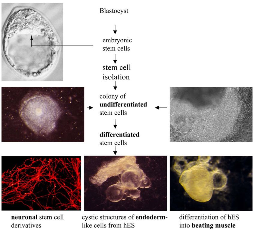
Fig. 1. From blastocyst, to embryonic stem cell line, to somatic cells. hES: human embryonic stem cell. Photographs D. Ward and L. Tertoolen, Hubrecht
Laboratory.
endoderm layer forms as in mES-derived embryoid bodies; their own weight [29,55] while more directed differentia-
the group working with the cells of Reubinoff et al. [4], tion of mES cells into motor neurons showed that they
however, grow the cells to high densities attached to a were able to repopulate the spinal cord, extend axons and
substrate. Overall, however, the extent to which the hES form synapses with target muscles [35]. Transplantation of
cell lines differentiate in vitro does not match their more ES-derived neural cells resulted in functional improvement
extensive differentiation capability in teratomas [44]. in mice with Parkinson-like lesions [28,56,57] and ES-
derived pancreas-like cells secreted insulin after trans-
plantation, although in insufficient amounts to cure their
4. Embryonic stem cells and tissue repair diabetes [31]. Myelin became detectable around axons in
mutant rats lacking myelin, after transfer of mouse ES-
The ability of human ES cells to differentiate to cells derived glial cells [58] suggesting a potential treatment for
from all three germ layers (ectoderm, endoderm and multiple sclerosis. Together, these results are highly en-
mesoderm) in culture is still a long way from contributing couraging of the feasibility of cell transplantation studies
to regenerative medicine. Speculation here is being driven but are still far from clinical application. The major
primarily by experiments in rodents with a variety of question is the extent to which these studies in rodents can
experimentally induced or genetic lesions. Mouse ES- be extended to humans. The differences in culture require-
derived cardiomyocytes have been transplanted to the heart ments between mES and hES have been described above
and survived [15], rats with spinal cord lesions receiving but obstacles related to directed differentiation of hES and
mES cell-derived embryoid bodies were again able to bear appropriate scale-up of transplantable cells to account for328 R. Passier, C. Mummery / Cardiovascular Research 58 (2003) 324–335 the differences in size of humans and rodents only multiply techniques to derive genetically matched stem cells for the problems to be overcome before clinical application is individual patients (therapeutic cloning). Both of these a reality. So far there are three reports of transfer of have their own particular disadvantages although a recent differentiated hES derivatives to rodent models: two experiment carried out in mice demonstrated a particularly describe the derivation of neural progenitors from hES elegant combination of nuclear transfer, stem cell isolation using a combination of high cell density and FGF and their and tissue transplantation [62]. Here, immune-deficient transfer to the brains of neonatal mice [46,50] while a third Rag22 / 2 mice were used as nuclear donors for transfer describes the seeding of human embryonic endothelial into enucleated oocytes, the resulting blastocysts were cells, selected from hES cell aggregates using antibodies cultured to isolate an isogenic embryonic stem cell line and against PE-CAM, on biodegradable polymer scaffolds one of the mutant alleles in the Rag22 / 2 ES cells was [51]. Upon subcutaneous insertion of these scaffolds in repaired by homologous recombination. Mutant mice were SCID mice, the (human) endothelial cells organise into then treated with repaired ES cells induced to differentiate vessel-like structures with mouse blood in their lumen, in vitro to hematopoietic precursors; 3–4 weeks after suggesting that microvessels had formed and anastomosed engraftment, mature myeloid and lymphoid cells as well as with the mouse vasculature, becoming functional blood- immunoglobulins became detectable, establishing a carrying microvessels. FGF and vascular endothelial paradigm for treatment of a genetic disorder by combining growth factor (VEGF) known to promote vasculogenesis therapeutic cloning with gene therapy. For the present in mES embryoid bodies had apparently no effect on the though, tissue matching is the major advantage that stem hES aggregates, suggesting different mechanisms might be cells from adult human tissues represent above hES cells, involved. In no case was there evidence of teratoma as will be described in the following sections. formation. There is one report of colonies of hES being grafted to somites in chick embryos where they were shown to divide and differentiate in the host tissue [59]. 5. Transdifferentiation of adult stem cells The host embryonic environment appeared to modulate their differentiation, suggesting that the chick may be a Success stories reported for embryonic stem cells have useful model system in which to study the integration of recently extended to adult stem cells. Adult stem cell hES cells and their derivative into tissues in vivo. function was assumed previously to be restricted to cell There has been no transfer of hES derivatives to humans lineages present in the organ from which they were derived to date although there has been a report of transfer of and to be involved solely in their repair. However, in neural derivatives of hEC cells to the brains of 11 stroke recent years several lines of evidence have suggested that victims in a phase I (safety) trial [60]. The efficiency with these adult stem cells are multipotent and can transdif- which EC cells form teratocarcinomas is much higher than ferentiate into different cell lineages (Table 1). Adult bone that of ES cells so this was as such a high-risk trial. In marrow, brain, skeletal muscle, liver, pancreas, fat, skin addition, and given the anxieties expressed over potential and skeletal muscle, have all been shown to possess stem risks of the present xenosupport systems used for hES or progenitor cells with the capacity to differentiate or cells, the hEC cell lines used had been derived through transdifferentiate into cell types other than their tissue of cloning via transfer to mice. Fortunately after two years origin. Among all presently known adult stem or there was no evidence of tumour formation or other progenitor cells, cell populations from bone marrow have pathologies and the transplanted cells were still detectable; shown the highest potential with respect to multilineage for two patients, a slight improvement in their symptoms differentiation and functional engraftment into host ani- was reported and a phase II trial has been initiated. mals. For human clinical use, graft rejection is likely to occur after transfer of differentiated hES derivatives to most 5.1. Bone marrow stem cells organs except the (immune privileged) central nervous system. Evidence from human embryos had raised the Studies with bone marrow stromal or mesenchymal stem possibility that hES cell themselves might be immune cells, a subset of cells that can be separated by plastic privileged since they did not appear to express MHC adherence, have shown differentiation into various cell proteins. However, a recent study has shown that MHC types, including bone [63,64], tendon, cartilage [65] and class I molecules are expressed on the undifferentiated hES fat [66]. A cardiomyogenic cell line was established by and that these increase after differentiation [61]. Among culturing mesenchymal stem cells from mice in the possible solutions to graft rejection would be building up a presence of the DNA demethylation agent 5-azacytidine. hES cell bank with a range of MHC profiles for matching Upon induction, the mesenchymal stem cells showed a to individual patients in combination with immunosuppres- cardiomyocyte phenotype, expressed cardiac differentia- sive drugs; less feasible alternatives would be genetic tion markers and began spontaneous beating [67]. Further- alteration of hES cells to develop a ‘universal donor’ that more, these cells displayed functional adrenergic and would not express MHC proteins or using nuclear transfer muscarinic receptors [68]. The cardiogenic potential of
R. Passier, C. Mummery / Cardiovascular Research 58 (2003) 324–335                                 329

Table 1
Transdifferentiation of adult stem cells
Tissue of origin                                   Newly formed tissue                                                                References
Bone marrow
Unfractionated                                     Brain                                                                              [72,73]
                                                   Kidney                                                                             [79,80]
                                                   Skeletal muscle                                                                    [113]
Mesenchymal /                                      Brain                                                                              [71]
stromal                                            Bone                                                                               [63,64]
                                                   Fat                                                                                [66]
                                                   Heart                                                                              [67–69]
HSC                                                Liver, lung, skin, gastro-intestinal tract                                         [77]
MAPC                                               Brain, retina, lung, heart, skeletal muscle,
                                                   liver, intestine, kidney, spleen, bone marrow, blood and skin                      [84]
Brain                                              Heart, skeletal muscle, kidney, stomach, intestine, liver                          [87]
                                                   Blood                                                                              [85,86]
                                                   Blood                                                                              [94,95]
Skeletal muscle                                    Heart                                                                              [99]
Liver                                              Bile duct                                                                          [97]
Fat                                                Pancreas                                                                           [98]
Skin                                               Bone, skeletal muscle                                                              [101,102]
Pancreas                                           Fat, brain, muscle                                                                 [103]
                                                   Liver                                                                              [100]
  HSC, hematopoietic stem cells; MAPC, multipotent adult progenitor cell.

mesenchymal stem cells was further demonstrated in vivo                           marrow at very low frequency and are able to repopulate
by injecting early-passage human mesenchymal stem cells,                          the hematopoietic system. Recent studies showed that
expressing b-galactosidase (b-gal), into the left ventricle                       transplanted bone marrow cells, enriched for HSC are able
of SCID mice. Although only a small percentage b-gal 1                            to differentiate into hepatocytes in liver of rodents [75,76].
donor cells could be detected in the heart (at the most                           Krause et al. [77] demonstrated that a single HSC was not
0.44% of injected cells) 60 days after injection, engrafted                       only able to repopulate the hematopoietic system in
b-gal 1 cells did express several cardiac markers, such as                        irradiated mice, but also differentiate into epithelium of
cardiac troponin T, alpha-actinin and phospholamban [69].                         lung, skin, liver and the gastro-intestinal tract. These HSCs
   Injection of human marrow stromal cells into rat brains                        showed increased expression of CD34 and SCA-1 during
showed that cells migrated from the injection site to                             homing to the bone marrow. Although HSC may have the
successive layers of the brain [70]. These findings were                          capacity to transdifferentiate, it was recently demonstrated
confirmed and extended by Kopen et al. [71]. Injection of                         that this is an extremely rare event. Injection of single
murine marrow stromal cells into the lateral ventricle of                         mouse green fluorescent protein (GFP) positive c-kit 1 Th-
neonatal mice, resulted in their migration throughout the                         y1.1 lo Lin 2 Sca-1 1 HSC into lethally irradiated mice
forebrain and cerebellum and their differentiation into                           showed reconstitution of peripheral blood leukocytes, but
astrocytes and presumably neurones. Transplantation of                            no (kidney, gut, muscle and lung) or only a few (liver and
unfractionated mouse bone marrow cells into mutant mice,                          brain) GFP 1 non-hematopoietic cells were detected. These
lacking the ability to develop cells of the myeloid and                           data were confirmed in GFP 1 :GFP 2 parabiotic (surgically
lymphoid lineages [72], or in lethally irradiated mice [73],                      joined) mice, which share a common anastomosed circulat-
resulted in their migration into the brain and, most                              ory system [78].
importantly, the donor-derived bone marrow cells differen-                           Transplantation of gender mismatched bone marrow
tiated into cells that expressed neuronal markers, such as                        cells into mouse and human resulted in engraftment of
NeuN. However, in a recent study, Castro et al. [74] did                          donor-derived cells into kidney. Using epithelial markers it
not observe transdifferentiation in a transplantation experi-                     was demonstrated that donor-derived CD45 2 engrafted
ment where they used a similar unfractionated or sub-                             cells had differentiated into renal parenchymal and tubular
population of mouse bone marrow cells. They came to the                           epithelial cells in mouse and humans, respectively [79]. In
same negative conclusion following bone marrow trans-                             accordance, transplantation of mouse bone marrow cells
plantation in a model of neural injury, raising the question                      expressing GFP into irradiated mice demonstrated the
whether the transformation from bone to brain is a general                        presence of donor-derived mesangial cells in the glomeruli
phenomenon or dependent on the experimental system.                               [80].
   Hematopoietic stem cells (HSC) are present in the bone                            The group of Verfaillie [81,82] has described another
330 R. Passier, C. Mummery / Cardiovascular Research 58 (2003) 324–335
sub-population of bone marrow cells. These cells, that into SCID mice [86]. Plasticity of neural stem cells was
copurify with mesenchymal cells, are found in human, also shown by injecting neurospheres derived from single-
mouse and rat and have been named multipotent adult cell cultures, into the amniotic cavity of chick embryos or
progenitor cells or MAPCs. Human and rodent MAPCs are aggregating them with mouse morulae. These chimeras
CD44 2 , CD45 2 , HLA class I 2 and II 2 and cKit 2 . Single were allowed to develop until different stages of develop-
MAPCs from human and rodents have been shown to ment [87]. Although neural stem cell progeny were found,
differentiate in vitro, not only into mesodermal and not only in ectodermal tissues, but also in mesodermal and
neuroectodermal cells, but also into endodermal cell types endodermal tissues, such as heart, kidney, skeletal muscle,
with hepatocyte phenotype and function. Optimal differen- lung stomach, intestine and liver, no neural stem cell
tiation was obtained by culturing MAPCs in the presence progeny were detected in the hematopoietic system, in
of FGF-4 and HGF on fibronectin, or a mixture of contrast with the studies described above. The contribution
extracellular matrix components (Matrigel) [83]. In addi- of neural stem cells to repopulation of the hematopoietic
tion to in vitro differentiation into endoderm, mesoderm system was further questioned by Morshead et al. [88],
and ectoderm derivatives, injection of a single mouse who used short-term and long-term passaged neurosphere
MAPC into mice blastocysts and transfer to foster mothers, cells for transplantation in irradiated mice. Although they
resulted in chimeric mice. When these chimeric mice were found neural stem cells continuing to generate neural
sacrificed MAPC-derived b-gal 1 cells were found in many progeny, no neural stem cell progeny could be detected in
tissues, including brain, retina, lung, myocardium, skeletal the hematopoietic system; they concluded that the findings
muscle, liver, intestine, kidney, spleen, bone marrow, of Bjornson et al. [85] represented a rare event, and
blood and skin. The same study also demonstrated that possibly caused by an accompanying genetic or epigenetic
mouse MAPCs engrafted and differentiated into tissue- change.
specific cells following injection into irradiated SCID
mice. Between 1 and 10% b-gal 1 cells were detected in 5.3. Skeletal muscle stem cells
blood, bone marrow, spleen and epithelium of lung liver
and intestine, whereas no contribution to skeletal or Adult skeletal muscle contains a population of myogenic
cardiac muscle was observed in mice analysed 4–24 weeks precursors, the so-called satellite cells, which are capable
after transplantation [84]. The single cell experiments have of self-renewal and myogenic differentiation in response to
provided the most rigorous proof to date of the capacity of physiological and pathophysiological stimuli. Furthermore,
MAPCs to transdifferentiate into several lineages. Without satellite cells or myoblasts have been shown to differen-
these experiments it remained possible that the bone tiate into slow-twitch muscle fibers when injected into the
marrow cell populations used in fact contained a mixture heart and improve cardiac function [89–91] (for reviews,
of precursor by various lineages. It is unclear why several see Refs. [92,93]). To investigate their possible plasticity,
months are necessary before MAPC colonies start to grow cells were isolated from skeletal muscle from adult mice,
in bone marrow cultures and it has been suggested that the cultured and injected retroorbitally in irradiated mice [94].
cells may represent a tissue culture specific cell with no in Six weeks after transplantation high percentages (.50%)
vivo counterpart. Nevertheless, if a true source of trans- of donor-derived B-, T-cells, granulocytes and macro-
plantable autologous cells then their exact identity may not phages were found by fluorescence activated cell sorting
be important. (FACS). The adult skeletal muscle was demonstrated to
contain a population of cells (approximately 1% of total)
5.2. Brain stem cells with a high efflux of the fluorescent dye Hoechst 33342
and expressing Sca-1 and c-Kit, characteristics of HSCs,
In addition to the observed plasticity of cell populations but lacking the hematopoietic marker CD45. The authors
in bone marrow, it has also been shown that adult neural suggested that these putative stem cells may be identical to
stem cells have a broader differentiation potential than satellite cells. In another study, a sub-population of skeletal
previously thought. Embryonic and adult stem cells, muscle cells, Sca-1 1 , lin 2 , c-Kit 2 and CD45 2 were
clonally derived from the forebrain of mice and expressing injected into the tail veins of lethally irradiated. Four to
b-gal ubiquitously, were systemically injected into subleth- eight weeks after transplantation regeneration of muscle
ally irradiated mice. Although normally giving rise to and blood cells, derived from donor cells, was detected
neurons, astrocytes and oligodendrocytes, neural stem cells [95]. However, recently, the group lead by Goodell [96]
were able to engraft into the hematopoietic system of came to different conclusions. They used freshly isolated
irradiated hosts to produce a range of blood cell types. adult skeletal muscle cells from mice and separated these
First b-gal 1 blood cells appeared 20–22 weeks after cells by FACS, based on the expression of CD45 and
injection, which was approximately 3 weeks later than Sca-1. CD45 1 and CD45 2 cell populations were injected
following bone marrow transplantation [85]. The contribu- into irradiated mice and their contribution to regeneration
tion of neural stem cells to the hematopoietic system was of muscle cells and cells of the hematopoietic system was
confirmed by transplantation of human brain derived cells studied. Only the CD45 1 fraction generated hematopoieticR. Passier, C. Mummery / Cardiovascular Research 58 (2003) 324–335 331
colonies in vitro and displayed a limited myogenic po- specific markers. Furthermore, cultured skin cells from
tential, whereas the CD45 2 fraction showed high human scalp were also able to produce neural proteins,
myogenic potential, but lacked hematopoietic reconstitu- when induced to differentiate. These skin stem cells were
tion. These findings made the authors conclude that different from mesenchymal or neural stem cells, as
muscle-derived HSCs are derived from the hematopoietic determined by their phenotype and the production of nestin
system rather than the adult muscle progenitor cell popula- and vimentin [103].
tion as originally proposed [94,96].
5.4. Liver stem cells
6. Adult stem cells and tissue repair
A population of small oval shaped cells in the liver
termed oval cells, is capable of proliferation and is thought A sub-population of Lin 2 and c-Kit 1 bone marrow cells
to be the stem-cell compartment in the liver. In addition to was obtained from transgenic mice, expressing GFP
the regeneration of hepatocytes, hepatic stem cells from rat ubiquitously. Following intra-ventricular injection of these
have also been shown to give rise to bile duct cells [97] cells in mice that had undergone experimental myocardial
and pancreatic cells, producing insulin and glucagon [98]. infarction, regeneration of cardiomyocytes was found in
Furthermore, cells from a clonal stem cell line (WB-F344), 40% of the animals [104]. Besides regeneration of car-
derived from an adult rat liver, were injected into the left diomyocytes, newly formed endothelial and smooth mus-
ventricle of mice. Before injection cells were infected with cle cells, organised in coronary vessels, were also demon-
a retrovirus expressing b-gal. b-gal 1 cells were detected in strated. Nine days after transplantation, the percentage of
the mouse heart and a cardiomyocyte phenotype was newly formed tissue, derived from the injected bone
observed as determined by the striated patterns and the marrow cells, was 68% of the infarcted region of the
co-localisation of cardiac troponin T and b-gal. Further- ventricle. Most importantly, cardiac function improved
more, electron microscopy showed the presence of well- following transplantation with the Lin 2 , c-Kit 1 sub-popu-
organised sarcomeres, intercalated discs and gap junctions, lation, as opposed to the effects of transplantation of the
co-localised with b-gal, suggesting but not proving their Lin 2 , c-Kit 2 sub-populations. In a subsequent study, the
multipotency [99]. same group used a non-invasive method to increase the
number of circulating stem cells in mice by treatment with
5.5. Pancreatic stem cells stem cell factor (SCF) and granulocyte-colony-stimulating
factor (G-CSF). Cytokine-treated mice displayed newly
Besides the ability of hepatic stem cells to differentiate formed cardiac tissue in the infarcted region, and as a
into pancreatic cells, cell fractions from the rat pancreas result decreased mortality and increased ejection fraction,
were shown to differentiate into hepatocytes after trans- parameters reflecting cardiac function. This experimental
plantation to the liver. Following transplantation donor- approach circumvented the high mortality of their previous
derived cells expressed liver-specific proteins and showed study [105] and demonstrated the successful homing of
phenotypical resemblance with hepatocytes [100] donor-derived bone marrow cells into the infarcted region
of recipient mice. However, it is not clear which sub-
5.6. Fat stem cells population of bone marrow cells is responsible for regene-
ration of cardiac tissue.
Human adipose tissue obtained from liposuction pro- Another sub-population (CD34 2 / low, c-Kit 1 , Sca-1 1 )
cedures has also been used to isolate a fibroblast-like cell of mouse bone marrow cells, also called side-population or
population, called processed lipoaspirate (LPA) cells. In SP, expressing b-gal were selected by FACS and injected
vitro studies with LPA cells demonstrated differentiation into irradiated mice. SP cells also expressed the adhesion
into adipogenic, chrondogenic, myogenic and osteogenic molecule PECAM-1 (CD31) and mRNA of Tie-2, Tal-2,
cells [101,102]. However, contamination with stem cells VEGF-A and angiopoietin, all features of endothelial
derived from peripheral blood can not be excluded in these precursors. After 10–12 weeks animals with high contribu-
studies. tions of engrafted cells were subjected to an experimental
ischemia–reperfusion regime to mimic cardiac infarct.
5.7. Skin stem cells Homing and engraftment of donor-derived SP cells were
found in the infarcted region of mice. These engrafted cells
Cells isolated from the skin of juvenile or adult mice had differentiated into both cardiac muscle and had formed
were cultured and studied for their ability to differentiate vessel-like structures [106]. Cardiac cells positive for b-gal
into cell-types of different lineages. In vitro experiments were predominantly located at the border zone of the
demonstrated differentiation of skin-derived cells and infarct region. A higher percentage was seen for endotheli-
clones of individual cells into neurones adipocytes, glial al engraftment (3.3%) than for cardiomyocyte engraftment
and smooth muscle cells, based on phenotype and cell- (0.02%). In all cases however the contribution of donor332 R. Passier, C. Mummery / Cardiovascular Research 58 (2003) 324–335 cells was low so that restoration of tissue function would embryonic stem-like (BMESL) cells. In vitro differentia- remain questionable tion of these BMESL cells into endoderm, ectoderm and Endothelial progenitor cells (EPC) have been identified mesoderm was observed. However, unexpectedly BMESL in adult peripheral blood, bone marrow and human umbili- cells showed tetraploid and hexaploid DNA content. The cal cord blood [107,108]. HSCs, which can also be isolated karyotype of two tetraploid cells was XXXY and these from human umbilical cord blood and EPCs have common expressed genetic markers for both bone marrow cells and cell markers, such as Flk-1, Tie-2 and CD34. Intravenous embryonic stem cells, indicating that fusion had occurred. injection of G-CSF-mobilised human CD34 1 bone marrow Blastocyst injection of BMESL failed to generate chimeric cells into rats, which were subjected to myocardial infarc- mice [116]. Similar findings were obtained with the co- tion, resulted in homing of these cells in the infarct zone culture of neural stem cells and embryonic stem cells. within 48 h after the ligation of the left descending Pluripotency of hybrid cells, expressing b-gal, was shown coronary artery. No infiltration was observed in non-infar- in vitro and by generating chimeric mice contributions to cted myocardium or in control hearts. The increased multilineage tissue in vivo was observed [115]. infiltration of CD34 1 cells in the infarct zone, lead to formation of capillaries of human origin, determined by DiI fluorescence labelling, within the central infarct zone. 7. Conclusions In addition, injection with CD34 1 cells also improved cardiac function in infarcted rats as opposed to injection of The results from the studies described in this overview CD34 2 cells or saphenous vein endothelial cells [109]. In suggest that undifferentiated stem cells derived from adult accordance, rat EPCs have also been shown to augment tissues are not determined progenitor cells with limited angiogenesis in ischemic tissues [110,111]. Furthermore, differentiation potential. Rather, these cells seem to pos- Condorelli et al. [112] showed that differentiated endo- sess a much broader capacity for cellular differentiation thelial cells from the human umbilical vein can trans- that is dependent on and responsive to specific cues present differentiate into beating cardiomyocytes when cocultured in the environment of the engrafted site. However, with the with neonatal rat cardiomyocytes or when injected near the exception of three studies with bone marrow cells border zone in infarcted rat hearts. [77,78,84], this has not been rigorously demonstrated for Unfractionated bone marrow cells obtained from trans- any other stem cell of adult origin. Questions of scale-up genic mice (under control of the muscle specific MLC3F and directed differentiation for transplantation and tissue promoter) were injected into injured muscle of SCID mice. repair remain to be answered, just as for embryonic stem In four of six b-gal 1 positive cells were found in injured cells, but their potentially autologous origin give them muscle, 2 weeks after injection. Furthermore, bone marrow extremely promising potential in the development of cell transplantation followed by induction of muscle injury was replacement therapies. studied. After 5 weeks bone marrow-derived myogenic Although considerable progress has been made towards progenitors had migrated into regenerating muscle and understanding the control of differentiation of adult and participated in the regeneration process, giving rise to fully embryonic stem cells, both have potential but a long way differentiated muscle fibers [113]. to go before clinical application. It is incorrect to say that Bone marrow transplantation experiments in a model for adult stem cells are equivalent to embryonic stem cells for hepatic injury, showed that new liver cells, partly derived treating diabetes, Parkinson and probably other diseases. from donor-bone marrow cells, were generated [75]. Bone Adult stem cells have tissue compatibility advantages in marrow transplantation in a mouse model (FAH mutants) transplantation but in certain genetic or autoimmune of a lethal hereditary liver disease resulted in improved diseases, (matched) tissue from ES cells and immuno- survival of mice. Seven months after transplantation, mice suppressive drugs may be preferable. Existing human ES were sacrificed and in all four mice b-gal 1 staining was cell lines are probably sufficient for current research if observed in 30–50% of the liver cells. They also observed made widely available; only when transplantation becomes increased biochemical liver function (assessed by measur- an issue may isolation of new lines for histocompatibility ing levels of serum transaminases, bilirubin and amino matching become necessary. acids) [114]. Recently, two research groups performed co-culture of mouse adult neural stem cells [115] or bone marrow cells Acknowledgements [116] with mouse embryonic stem cells. Both groups came to the same surprising conclusion: adult stem cells can fuse We thank colleagues at the Hubrecht Laboratory, Uni- with embryonic stem cells in culture. Co-culture of bone versity Hospital Medical School Utrecht, Interuniversity marrow cells with embryonic stem cells using selection Cardiology Institute of the Netherlands, Monash Universi- markers that would eliminate the growth of embryonic ty, Australia and Embryonic Stem Cell International for stem cells, lead to colonies with an embryonic stem cell their input in our stem cell projects. Leon Tertoolen and morphology, which they called bone marrow-derived Dorien Ward are thanked for contributions to Fig. 1.
R. Passier, C. Mummery / Cardiovascular Research 58 (2003) 324–335                                           333

References                                                                  [22] Holden C, Vogel G. Stem cell lines. ‘Show us the cells,’ US
                                                                                 researchers say. Science 2002;297:923–925.
                                                                            [23] Richards M, Fong CY, Chan WK, Wong PC, Bongso A. Human
 [1] Mummery CL, van den Eijnden-van Raaij AJ. Developmental                     feeders support prolonged undifferentiated growth of human inner
     tumours, early differentiation and the transforming growth factor           cell masses and embryonic stem cells. Nature Biotechnol
     beta superfamily. Int J Dev Biol 1999;43:693–709.                           2002;20:933–936.
 [2] Andrews PW, Przyborski SA, Thomson JA. Embryonal carcinoma             [24] Martin GR. The formation of embryoid bodies in vitro by homoge-
     cells as embryonic stem cells. In: Marshak HA, Gardner RL,                  neous embryonal carcinoma cell cultures derived from isolated
     Gottlieb D, editors, Stem cell biology, Cold Spring Harbor, NY:             single cells. Dev Biol 1975;59:169–187.
     Cold Spring Harbor Laboratory Press, 2001, pp. 231–265.                [25] Martin GR, Wiley LM, Damjanov I. The development of cystic
 [3] Thomson JA, Itskovitz-Eldor J, Shapiro SS et al. Embryonic stem             embryoid bodies in vitro from clonal teratocarcinoma stem cells.
     cell lines derived from human blastocysts. Science 1998;282:1145–           Dev Biol 1977;61:230–244.
     1147.                                                                  [26] Weiss MJ, Orkin SH. In vitro differentiation of murine embryonic
 [4] Reubinoff BE, Pera MF, Fong CY, Trounson A, Bongso A.                       stem cells. New approaches to old problems. J Clin Invest
     Embryonic stem cell lines from human blastocysts: somatic differen-         1996;97:591–595.
     tiation in vitro. Nature Biotechnol 2000;18:399–404.                   [27] Rohwedel J, Guan K, Wobus AM. Induction of cellular differentia-
 [5] Evans MJ, Kaufman MH. Establishment in culture of pluripotential            tion by retinoic acid in vitro. Cells Tissues Organs 1999;165:190–
     cells from mouse embryos. Nature 1981;292:154–156.                          202.
 [6] Martin GR. Isolation of a pluripotent cell line from early mouse       [28] Lee SH, Lumelsky N, Studer L, Auerbach JM, McKay RD. Efficient
     embryos cultured in medium conditioned by teratocarcinoma stem              generation of midbrain and hindbrain neurons from mouse em-
     cells. Proc Natl Acad Sci USA 1981;78:7634–7638.                            bryonic stem cells. Nature Biotechnol 2000;18:675–679.
 [7] Smith AG, Heath JK, Donaldson DD et al. Inhibition of pluripoten-      [29] Liu S, Qu Y, Stewart TJ et al. Embryonic stem cells differentiate
     tial embryonic stem cell differentiation by purified polypeptides.          into oligodendrocytes and myelinate in culture and after spinal cord
     Nature 1988;336:688–690.                                                    transplantation. Proc Natl Acad Sci USA 2000;97:6126–6131.
 [8] Moreau JF, Donaldson DD, Bennett F et al. Leukaemia inhibitory         [30] Buttery LD, Bourne S, Xynos JD et al. Differentiation of osteoblasts
     factor is identical to the myeloid growth factor human interleukin          and in vitro bone formation from murine embryonic stem cells.
     for DA cells. Nature 1988;336:690–692.                                      Tissue Eng 2001;7:89–99.
 [9] Williams RL, Hilton DJ, Pease S et al. Myeloid leukaemia inhibitory    [31] Lumelsky N, Blondel O, Laeng P et al. Differentiation of embryonic
     factor maintains the developmental potential of embryonic stem              stem cells to insulin-secreting structures similar to pancreatic islets.
     cells. Nature 1988;336:684–687.                                             Science 2001;292:1389–1394.
[10] Smith AG. Embryonic stem cells. In: Marshak HA, Gardner RL,            [32] Gratsch TE, O’Shea KS. Noggin and chordin have distinct activities
     Gottlieb D, editors, Stem cell biology, Cold Spring Harbor, NY:             in promoting lineage commitment of mouse embryonic stem (ES)
     Cold Spring Harbor Laboratory Press, 2001, pp. 205–230.                     cells. Dev Biol 2002;245:83–94.
[11] Xu C, Inokuma MS, Denham J et al. Feeder-free growth of                [33] Tropepe V, Hitoshi S, Sirard C et al. Direct neural fate specification
     undifferentiated human embryonic stem cells. Nature Biotechnol              from embryonic stem cells: a primitive mammalian neural stem cell
     2001;19:971–974.                                                            stage acquired through a default mechanism. Neuron 2001;30:65–
[12] Schuringa JJ, van der Schaaf S, Vellenga E, Eggen BJ, Kruijer W.            78.
     LIF-induced STAT3 signaling in murine versus human embryonal           [34] Kawasaki H, Mizuseki K, Nishikawa S et al. Induction of midbrain
     carcinoma (EC) cells. Exp Cell Res 2002;274:119–129.                        dopaminergic neurons from ES cells by stromal cell-derived induc-
[13] Amit M, Carpenter MK, Inokuma MS et al. Clonally derived human              ing activity. Neuron 2000;28:31–40.
     embryonic stem cell lines maintain pluripotency and proliferative      [35] Wichterle H, Lieberam I, Porter JA, Jessell TM. Directed differen-
     potential for prolonged periods of culture. Dev Biol 2000;227:271–          tiation of embryonic stem cells into motor neurons. Cell
     278.                                                                        2002;110:385–397.
[14] Amit M, Itskovitz-Eldor J. Derivation and spontaneous differentia-     [36] Mummery C, Ward D, van den Brink CE et al. Cardiomyocyte
     tion of human embryonic stem cells. J Anat 2002;200:225–232.                differentiation of mouse and human embryonic stem cells. J Anat
[15] Klug MG, Soonpaa MH, Koh GY, Field LJ. Genetically selected                 2002;200:233–242.
     cardiomyocytes from differentiating embryonic stem cells form          [37] Rathjen J, Lake JA, Bettess MD et al. Formation of a primitive
     stable intracardiac grafts. J Clin Invest 1996;98:216–224.                  ectoderm like cell population, EPL cells, from ES cells in response
[16] Li M, Pevny L, Lovell-Badge R, Smith A. Generation of purified              to biologically derived factors. J Cell Sci 1999;112:601–612.
     neural precursors from embryonic stem cells by lineage selection.      [38] Jones EA, Tosh D, Wilson DI, Lindsay S, Forrester LM. Hepatic
     Curr Biol 1998;8:971–974.                                                   differentiation of murine embryonic stem cells. Exp Cell Res
[17] Mountford P, Nichols J, Zevnik B, O’Brien C, Smith A. Mainte-               2002;272:15–22.
     nance of pluripotential embryonic stem cells by stem cell selection.   [39] Wiles MV, Keller G. Multiple hematopoietic lineages develop from
     Reprod Fertil Dev 1998;10:527–533.                                          embryonic stem (ES) cells in culture. Development 1991;111:259–
[18] Fujikura J, Yamato E, Yonemura S et al. Differentiation of em-              267.
     bryonic stem cells is induced by GATA factors. Genes Dev               [40] Slager HG, Freund E, Buiting AM, Feijen A, Mummery CL.
     2002;16:784–789.                                                            Secretion of transforming growth factor-beta isoforms by embryonic
[19] Reubinoff BE, Pera MF, Vajta G, Trounson AO. Effective cryop-               stem cells: isoform and latency are dependent on direction of
     reservation of human embryonic stem cells by the open pulled straw          differentiation. J Cell Physiol 1993;156:247–256.
     vitrification method. Hum Reprod 2001;16:2187–2194.                    [41] Nishikawa SI, Nishikawa S, Hirashima M, Matsuyoshi N, Kodama
[20] McWhir J, Schnieke AE, Ansell R et al. Selective ablation of                H. Progressive lineage analysis by cell sorting and culture identifies
     differentiated cells permits isolation of embryonic stem cell lines         FLK11VE-cadherin1 cells at a diverging point of endothelial and
     from murine embryos with a non-permissive genetic background.               hemopoietic lineages. Development 1998;125:1747–1757.
     Nature Genet 1996;14:223–226.                                          [42] Yamashita J, Itoh H, Hirashima M et al. Flk1-positive cells derived
[21] Brook FA, Gardner RL. The origin and efficient derivation of                from embryonic stem cells serve as vascular progenitors. Nature
     embryonic stem cells in the mouse. Proc Natl Acad Sci USA                   2000;408:92–96.
     1997;94:5709–5712.                                                     [43] Weima SM, van Rooijen MA, Feijen A et al. Transforming growth
334 R. Passier, C. Mummery / Cardiovascular Research 58 (2003) 324–335
factor-beta and its receptor are differentially regulated in human [66] Umezawa A, Maruyama T, Segawa K et al. Multipotent marrow
embryonal carcinoma cells. Differentiation 1989;41:245–253. stromal cell line is able to induce hematopoiesis in vivo. J Cell
[44] Odorico JS, Kaufman DS, Thomson JA. Multilineage differentiation Physiol 1992;151:197–205.
from human embryonic stem cell lines. Stem Cells 2001;19:193– [67] Makino S, Fukuda K, Miyoshi S et al. Cardiomyocytes can be
204. generated from marrow stromal cells in vitro. J Clin Invest
[45] Itskovitz-Eldor J, Schuldiner M, Karsenti D et al. Differentiation of 1999;103:697–705.
human embryonic stem cells into embryoid bodies compromising [68] Hakuno D, Fukuda K, Makino S et al. Bone marrow-derived
the three embryonic germ layers. Mol Med 2000;6:88–95. regenerated cardiomyocytes (CMG Cells) express functional adren-
[46] Reubinoff BE, Itsykson P, Turetsky T et al. Neural progenitors from ergic and muscarinic receptors. Circulation 2002;105:380–386.
human embryonic stem cells. Nature Biotechnol 2001;19:1134– [69] Toma C, Pittenger MF, Cahill KS, Byrne BJ, Kessler PD. Human
1140. mesenchymal stem cells differentiate to a cardiomyocyte phenotype
[47] Schuldiner M, Eiges R, Eden A et al. Induced neuronal differentia- in the adult murine heart. Circulation 2002;105:93–98.
tion of human embryonic stem cells. Brain Res 2001;913:201–205. [70] Azizi SA, Stokes D, Augelli BJ, DiGirolamo C, Prockop DJ.
[48] Assady S, Maor G, Amit M et al. Insulin production by human
Engraftment and migration of human bone marrow stromal cells
embryonic stem cells. Diabetes 2001;50:1691–1697.
implanted in the brains of albino rats—similarities to astrocyte
[49] Kehat I, Kenyagin-Karsenti D, Snir M et al. Human embryonic stem
grafts. Proc Natl Acad Sci USA 1998;95:3908–3913.
cells can differentiate into myocytes with structural and functional
[71] Kopen GC, Prockop DJ, Phinney DG. Marrow stromal cells migrate
properties of cardiomyocytes. J Clin Invest 2001;108:407–414.
throughout forebrain and cerebellum, and they differentiate into
[50] Zhang SC, Wernig M, Duncan ID, Brustle O, Thomson JA. In vitro
astrocytes after injection into neonatal mouse brains. Proc Natl Acad
differentiation of transplantable neural precursors from human
embryonic stem cells. Nature Biotechnol 2001;19:1129–1133. Sci USA 1999;96:10711–10716.
[51] Levenberg S, Golub JS, Amit M, Itskovitz-Eldor J, Langer R. [72] Mezey E, Chandross KJ, Harta G, Maki RA, McKercher SR.
Endothelial cells derived from human embryonic stem cells. Proc Turning blood into brain: cells bearing neuronal antigens generated
Natl Acad Sci USA 2002;99:4391–4396. in vivo from bone marrow. Science 2000;290:1779–1782.
[52] Kehat I, Gepstein A, Spira A, Itskovitz-Eldor J, Gepstein L. High- [73] Brazelton TR, Rossi FM, Keshet GI, Blau HM. From marrow to
resolution electrophysiological assessment of human embryonic brain: expression of neuronal phenotypes in adult mice. Science
stem cell-derived cardiomyocytes: a novel in vitro model for the 2000;290:1775–1779.
study of conduction. Circ Res 2002;91:659–661. [74] Castro RF, Jackson KA, Goodell MA et al. Failure of bone marrow
[53] Xu C, Police S, Rao N, Carpenter MK. Characterization and cells to transdifferentiate into neural cells in vivo. Science
enrichment of cardiomyocytes derived from human embryonic stem 2002;297:1299.
cells. Circ Res 2002;91:501–508. [75] Petersen BE, Bowen WC, Patrene KD et al. Bone marrow as a
[54] Pera MF. Human pluripotent stem cells: a progress report. Curr potential source of hepatic oval cells. Science 1999;284:1168–1170.
Opin Genet Dev 2001;11:595–599. [76] Theise ND, Badve S, Saxena R et al. Derivation of hepatocytes from
[55] McDonald JW, Liu XZ, Qu Y et al. Transplanted embryonic stem bone marrow cells in mice after radiation-induced myeloablation.
cells survive, differentiate and promote recovery in injured rat spinal Hepatology 2000;31:235–240.
cord. Nature Med 1999;5:1410–1412. [77] Krause DS, Theise ND, Collector MI et al. Multi-organ, multi-
[56] Bjorklund LM, Sanchez-Pernaute R, Chung S et al. Embryonic stem lineage engraftment by a single bone marrow-derived stem cell. Cell
cells develop into functional dopaminergic neurons after transplanta- 2001;105:369–377.
tion in a Parkinson rat model. Proc Natl Acad Sci USA [78] Wagers AJ, Sherwood RI, Christensen JL, Weissman IL. Little
2002;99:2344–2349. evidence for developmental plasticity of adult hematopoietic stem
[57] Kim JH, Auerbach JM, Rodriguez-Gomez JA et al. Dopamine cells. Science 2002;297:2256–2259.
neurons derived from embryonic stem cells function in an animal [79] Poulsom R, Forbes SJ, Hodivala-Dilke K et al. Bone marrow
model of Parkinson’s disease. Nature 2002;418:50–56. contributes to renal parenchymal turnover and regeneration. J Pathol
[58] Brustle O, Jones KN, Learish RD et al. Embryonic stem cell-derived 2001;195:229–235.
glial precursors: a source of myelinating transplants. Science [80] Imasawa T, Utsunomiya Y, Kawamura T et al. The potential of bone
1999;285:754–756. marrow-derived cells to differentiate to glomerular mesangial cells. J
[59] Goldstein RS, Drukker M, Reubinoff BE, Benvenisty N. Integration Am Soc Nephrol 2001;12:1401–1409.
and differentiation of human embryonic stem cells transplanted to [81] Reyes M, Lund T, Lenvik T et al. Purification and ex vivo
the chick embryo. Dev Dyn 2002;225:80–86. expansion of postnatal human marrow mesodermal progenitor cells.
[60] Kondziolka D, Wechsler L, Goldstein S et al. Transplantation of Blood 2001;98:2615–2625.
cultured human neuronal cells for patients with stroke. Neurology [82] Reyes M, Verfaillie CM. Characterization of multipotent adult
2000;55:565–569. progenitor cells, a subpopulation of mesenchymal stem cells. Ann
[61] Drukker M, Katz G, Urbach A et al. Characterization of the NY Acad Sci 2001;938:231–233.
expression of MHC proteins in human embryonic stem cells. Proc [83] Schwartz RE, Reyes M, Koodie L et al. Multipotent adult progenitor
Natl Acad Sci USA 2002;99:9864–9869. cells from bone marrow differentiate into functional hepatocyte-like
[62] Rideout WM, Hochedlinger K, Kyba M, Daley GQ, Jaenisch R. cells. J Clin Invest 2002;109:1291–1302.
Correction of a genetic defect by nuclear transplantation and [84] Jiang Y, Jahagirdar BN, Reinhardt RL et al. Pluripotency of
combined cell and gene therapy. Cell 2002;109:17–27. mesenchymal stem cells derived from adult marrow. Nature
[63] Rickard DJ, Sullivan TA, Shenker BJ, Leboy PS, Kazhdan I. 2002;418:41–49.
Induction of rapid osteoblast differentiation in rat bone marrow [85] Bjornson CR, Rietze RL, Reynolds BA, Magli MC, Vescovi AL.
stromal cell cultures by dexamethasone and BMP-2. Dev Biol Turning brain into blood: a hematopoietic fate adopted by adult
1994;161:218–228. neural stem cells in vivo. Science 1999;283:534–537.
[64] Friedenstein AJ, Chailakhyan RK, Gerasimov UV. Bone marrow [86] Shih CC, Weng Y, Mamelak A et al. Identification of a candidate
osteogenic stem cells: in vitro cultivation and transplantation in human neurohematopoietic stem-cell population. Blood
diffusion chambers. Cell Tissue Kinet 1987;20:263–272. 2001;98:2412–2422.
[65] Ashton BA, Allen TD, Howlett CR et al. Formation of bone and [87] Clarke DL, Johansson CB, Wilbertz J et al. Generalized potential of
cartilage by marrow stromal cells in diffusion chambers in vivo. adult neural stem cells. Science 2000;288:1660–1663.
Clin Orthop 1980;1:294–307. [88] Morshead CM, Benveniste P, Iscove NN, van der Kooy D. Hemato-You can also read