Findings from recent studies by the Japan Aerospace Exploration Agency examining musculoskeletal atrophy in space and on Earth - Nature
←
→
Page content transcription
If your browser does not render page correctly, please read the page content below
www.nature.com/npjmgrav

                  REVIEW ARTICLE                           OPEN

                  Findings from recent studies by the Japan Aerospace
                  Exploration Agency examining musculoskeletal atrophy in
                  space and on Earth
                  Satoshi Furukawa 1 ✉, Masahiro Chatani 2,3 ✉, Atsushi Higashitani 4 ✉, Akira Higashibata1, Fuminori Kawano5, Takeshi Nikawa6,
                  Takuro Numaga-Tomita7, Toshihiko Ogura8, Fuminori Sato9, Atsuko Sehara-Fujisawa9, Masahiro Shinohara10, Toru Shimazu11,
                  Satoru Takahashi 12 and Haruko Watanabe-Takano13

                      The musculoskeletal system provides the body with correct posture, support, stability, and mobility. It is composed of the bones,
                      muscles, cartilage, tendons, ligaments, joints, and other connective tissues. Without effective countermeasures, prolonged
                      spaceflight under microgravity results in marked muscle and bone atrophy. The molecular and physiological mechanisms of this
                      atrophy under unloaded conditions are gradually being revealed through spaceflight experiments conducted by the Japan
                      Aerospace Exploration Agency using a variety of model organisms, including both aquatic and terrestrial animals, and terrestrial
                      experiments conducted under the Living in Space project of the Japan Ministry of Education, Culture, Sports, Science, and
                      Technology. Increasing our knowledge in this field will lead not only to an understanding of how to prevent muscle and bone
1234567890():,;

                      atrophy in humans undergoing long-term space voyages but also to an understanding of countermeasures against age-related
                      locomotive syndrome in the elderly.
                      npj Microgravity (2021)7:18 ; https://doi.org/10.1038/s41526-021-00145-9

                  INTRODUCTION                                                                                muscle mass is the result of a balance between cellular protein
                  Almost 60 years have passed since Yuri Gagarin became the first                              synthesis and catabolism9. Bone and skeletal muscle mass can
                  human to fly in outer space. Since then, the duration of space                               be altered in response to increased loading with exercise or to
                  missions has increased, and nowadays it is common for astronauts                            decreased loading with spaceflight or disuse, which is summar-
                  to spend more than 6 months in outer space on the International                             ized as the “use it or lose it” concept of bone adaptation known
                  Space Station (ISS). In addition, plans are underway to build a                             as Wolff’s Law10. Various experiments examining the effects of
                  gateway to the moon and even to go to Mars, which will both                                 long-term spaceflight on the musculoskeletal system have been
                  necessitate long-term spaceflight missions1. However, the micro-                             conducted by the United States and Russia using space shuttles,
                                                                                                              unmanned satellites, and the ISS. The findings from these
                  gravity of space is a challenge because it induces bone loss and
                                                                                                              experiments have provided insights into how the muscles are
                  muscle atrophy in astronauts2. This atrophy is problematic
                                                                                                              changed during deloading. For example, analysis of the skeletal
                  because it not only reduces astronauts’ motor function, impairing
                                                                                                              muscle of mice exposed to microgravity has revealed that
                  their ability to perform daily activities once they return to Earth,                        muscle atrophy occurs mainly in the anti-gravity muscles such as
                  but it is also associated with negative changes in the cardiovas-                           the soleus muscle. Microgravity-induced atrophy is accompa-
                  cular system3. Interestingly, relationships between spaceflight-                             nied by an increase in the ratio of fast-twitch to slow-twitch
                  induced and aging-induced deconditioning have been sug-                                     muscle fibers11.
                  gested4–6. For example, there is a common problem of orthostatic                               The Japan Aerospace Exploration Agency (JAXA) has con-
                  intolerance in astronauts after spaceflight and in the elderly,                              ducted several spaceflight experiments to study the effects of
                  because muscle atrophy weakens venous return to the heart4,5,7.                             space environments on muscles and bones using the Space
                  Thus, countermeasures developed for use during spaceflight by                                Shuttle, Soyuz, SpaceX, and the ISS. As an example of a
                  astronauts may also be useful for the maintenance of elderly                                pioneering JAXA experiment conducted during mission STS-90
                  health on Earth.                                                                            of the Space Shuttle Columbia in 1998, Ikemoto et al. demon-
                     Bone and skeletal muscle are dynamic tissues that undergo                                strated increased degradation of fast-type myosin heavy chain
                  continuous synthesis and degradation. Bone remodeling is                                    protein and increased protease expression in atrophied gastro-
                  performed by bone-forming cells called osteoblasts and bone-                                cnemius muscles of neonatal rats exposed to 16 days of
                  resorbing cells called osteoclasts8, and maintenance of skeletal                            spaceflight; spaceflight was also found to stimulate the

                  1
                   Human Spaceflight Technology Directorate, Japan Aerospace Exploration Agency, Tsukuba, Ibaraki, Japan. 2Department of Pharmacology, Showa University School of Dentistry,
                  Tokyo, Japan. 3Pharmacological Research Center, Showa University, Tokyo, Japan. 4Graduate School of Life Sciences, Tohoku University, Sendai, Japan. 5Graduate School of Health
                  Sciences, Matsumoto University, Matsumoto, Nagano, Japan. 6Department of Nutritional Physiology, Institute of Medical Nutrition, Tokushima University Graduate School,
                  Tokushima, Japan. 7Department of Molecular Pharmacology, School of Medicine, Shinshu University, Matsumoto, Nagano, Japan. 8Department of Developmental Neurobiology,
                  Institute of Development, Aging and Cancer, Tohoku University, Sendai, Japan. 9Department of Growth Regulation, Institute for Frontier Life and Medical Sciences, Kyoto
                  University, Kyoto, Japan. 10Department of Rehabilitation for the Movement Functions, Research Institute, National Rehabilitation Center for Persons with Disabilities, Tokorozawa,
                  Saitama, Japan. 11Japan Space Forum, Tokyo, Japan. 12Department of Anatomy and Embryology, Faculty of Medicine, University of Tsukuba, Tsukuba, Ibaraki, Japan.
                  13
                    Department of Cell Biology, National Cerebral and Cardiovascular Center, Suita, Osaka, Japan. ✉email: furukawa.satoshi@jaxa.jp ; chatani@dent.showa-u.ac.jp ; atsushi.
                  higashitani.e7@tohoku.ac.jp

                  Published in cooperation with the Biodesign Institute at Arizona State University, with the support of NASA
S. Furukawa et al.
                  2
                       Table 1.   JAXA space missions related to musculoskeletal research.

                      Year          Transportation                        Subject                     Research target        Publications

                      1998          Space shuttle Columbia (STS-90)       Rats                        Muscle                 Ikemoto M. et al.12, Nikawa T. et al.24
                      2010          Space shuttle Discovery (STS-         Rat cells (myotubes) Muscle                        Uchida T. et al.22
                                    131), ISS
                      2004          Soyuz TMA-4, ISS                      C. elegans                  Muscle                 Higashibata A. et al.33,34, Selch F. et al.35
                      2009          Space shuttle Atlantis (STS-129), ISS C. elegans                  Muscle                 Etheridge T. et al.36, Higashibata A. et al.37, Harada
                                                                                                                             et al.38
                      1994          Space shuttle Columbia (STS-65)       Medaka                      Ontogeny               Ijiri K. et al.41
                      2012          Soyuz TMA-06M, ISS                    Medaka                      Bone                   Chatani M. et al.42
                      2014          Soyuz-U, Progress M-22M (54P), ISS Medaka                         Bone                   Chatani M. et al.43
                      2010          Space shuttle Atlantis (STS-132), ISS Goldfish scales              Scales                Ikegame M. et al.47
                      2014          Soyuz TMA-14M, ISS                    Zebrafish                    Muscle                Sato F. et al., in preparation
                      2018          SpaceX Falcon 9, ISS                  Zebrafish                    Muscle                 Sato F. et al., in preparation
                      2016          SpaceX Falcon 9, Dragon, ISS          Mice                        Bone, muscle           Shiba D. et al.18
                      2017          SpaceX Falcon 9, Dragon, ISS          Mice                        Bone                   Tominari T. et al.52
                      –*            ISS                                   Humans                      Bone, bisphosphonate Leblanc A. et al.100
                                                                                                                           Sibonga J. et al.101
                       *Ethical considerations.
1234567890():,;

                      ubiquitination of proteins such as myosin heavy chain, and the                      experiments conducted as part of the Living in Space project of
                      accumulation of myosin heavy chain degradation fragments in                         the Japanese Ministry of Education, Culture, Sports, Science and
                      muscle12. From 2008 to 2009, the Japanese Experiment Module                         Technology, and of spaceflight experiments conducted in the
                      (JEM), which is a part of the ISS, was assembled to create a                        “Kibo” Japanese Experiment Module of the ISS.
                      laboratory called “Kibo” on the ISS13. After that, Kibo was
                      equipped with cell biology experiment facilities (CBEF and
                      CBEF-L)14 and a fluorescent microscope, Confocal Space Micro-                        RECENT JAXA SPACE MISSIONS RELATED TO MUSCLE AND
                      scopy (COSMIC)15. JAXA also developed an aquatic habitat                            BONE
                      (AQH)16 for aquatic animals, a system for housing mice (Mouse                       Since 1998, JAXA has conducted 14 space missions onboard the
                      Habitat Unit: MHU)17 for use in space experiments that includes                     ISS to understand the causes of muscle atrophy and osteopenia
                      live imaging and an artificial gravity centrifuge and a multiple                     under microgravity environments (Table 1). So far, cultured cells,
                      artificial-gravity research system (MARS) for rodents18. In parti-                   Caenorhabditis elegans, medaka, goldfish scales, zebrafish, mice,
                      cular, MHU and MARS can create artificial gravity to properly                        and humans have been the subjects of these experiments. It is
                      study the effects of differences in gravity. These unique systems                   expected that the genetic, molecular, tissue and behavioral data
                      will allow scientists to investigate the effects of gravity                         accumulated under different gravitational conditions will allow
                      differences on rodents thoroughly.                                                  elucidation of the mechanisms underlying homeostasis of the
                         Although these spaceflight experiments have afforded a wealth                     musculoskeletal system. Here, we summarize the findings of the
                      of insights and novel findings, further studies are needed to fully                  most recent animal experiments conducted by JAXA in the
                      understand the molecular mechanisms of bone loss and muscle                         Japanese Experiment Module of the ISS.
                      atrophy during spaceflight and to determine the preventative
                      effects of exercise and other potential countermeasures. On the
                                                                                                          Cell culture experiments
                      ISS, crewmembers are scheduled 2.5 h of exercise a day, 6 days a
                      week, to prevent bone loss and muscle atrophy19. However, the                       In 2010, as part of space mission STS-131, JAXA conducted an
                      astronauts still lose an average of 3% of their bone mineral density                experiment to examine muscle atrophy-associated ubiquitin ligase
                      at the lumbar spine and 6% at the hip and pelvis after the typical                  casitas B-lineage lymphoma-b (Cbl-b) in rat myoblasts22. In this
                      6-month missions20. Furthermore, a marked variation has been                        experiment, L6 rat myoblasts at 100% confluence were allowed to
                      observed among individuals, with some astronauts losing up to                       differentiate for 5 days in a differentiation medium and were then
                      10–15% of their bone mineral density, which is equivalent to a loss                 sent to the ISS (Fig. 1a). Seven days after launch, the culture
                      of 1.7–2.5% per month during a 6-month mission20. This large                        medium was replaced with fresh DMEM with or without 40 ng/mL
                      variation in the effects of exercise is likely the result of a variety of           insulin-like growth factor-1, which induces skeletal myocyte
                      factors, including genetic background, exercise compliance,                         hypertrophy, and the cells were cultured for 10 days under
                      exercise intensity, and exercise dynamics.                                          microgravity or 1-g conditions, the latter achieved by an artificial
                         The next challenges for humankind include new missions to the                    gravity centrifuge in KIBO. In myoblasts and myotubes, gravitational
                      Moon, followed by human exploration to Mars. For the missions to                    unloading (with 3D clinorotation) resulted in a rapid increase in the
                      Mars, the long travel distance will make the total mission duration                 production of reactive oxygen species (ROS), which are critical
                      800–1100 days, of which approximately 500 days is expected to                       upstream mediators that link downstream atrophic signaling with
                      be spent on the planet’s surface21. As a result, astronauts will                    metabolic changes23. Flow cytometry with nitric oxide (NO)-specific
                      spend much more time under micro-gravity or low-gravity                             fluorescence indicator failed to detect NO after clinorotation in
                      compared with a mission on the ISS. Thus, one of the major                          myoblast, indicating that unloading stress did not increase NO
                      concerns for such long space missions is astronaut health with                      radicals. Administration of N-acetylcysteine or TEMPOL (1-oxyl-
                      respect to bone loss and muscle atrophy. In this review, we                         2,2,6,6-tetramethyl-4-hydroxypiperidine) to L6 myotubes signifi-
                      summarize the musculoskeletal findings of recent terrestrial                         cantly suppressed clinorotation-mediated activation of extracellular

                      npj Microgravity (2021) 18                    Published in cooperation with the Biodesign Institute at Arizona State University, with the support of NASA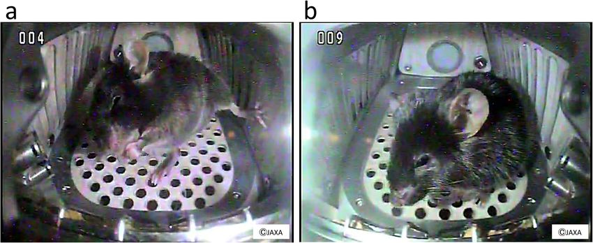
S. Furukawa et al.
3
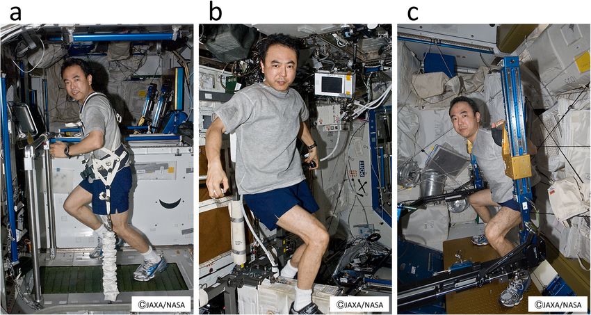
Fig. 1 Recent JAXA space missions in Kibo of ISS. a Cell culture system used in the Myo Lab project22. L6 rat myoblasts at 100% confluence in
a disposable cultivation chamber (DCC) were fused by changing the medium to a differentiation medium (i.e., Dulbecco’s modified Eagle’s
medium containing 0.5% fetal bovine serum). (Photo by Nikawa, T) b C. elegans culture system used in the CERISE project36–38. Upon reaching
microgravity at KIBO, flight crews activated the experiments by removing the U-pin. Holder was set into Meas Exp Unit A and transferred into
the Cell Biology Experiment Facility (CBEF) with or without 1-g rotation for either 4 or 8 days. (Photo by Higashitani, A). c Appearance of
medaka reared during spaceflight. Medaka was filmed for abnormal behavior to examine physiological changes under microgravity. The
recordings showed that the fish became accustomed to life under microgravity but displayed unique behavior such as swimming upside-
down (red arrow). Scale bar, 10 mm. d Medaka chamber for live imaging used on the ISS. Twelve larvae at stage 39 can be kept in each
“Medaka Chamber”, in which the larvae are placed in Mebiol Gel (Mebiol Inc., Kanagawa, Japan) and then covered with a gas-permeable
membrane. The chamber shown was carried aboard Soyuz flight progress M-22M (54 P) (Roscosmos, Russia) in 2014. After arrival at the
Japanese Experiment Module of the ISS, the chambers were set under a fluorescence microscope for live imaging for 8 days. The red dotted
area indicates an enlarged view (inset) showing three larvae. Scale bar, 10 mm.
signal-regulated kinase (ERK) 1/2; however, the suppressive effect of Furthermore, at the genetic level, spaceflight has been shown
TEMPOL on clinorotation-mediated ERK1/2 activation was stronger to significantly increase the mRNA levels of several muscle
than that of N-acetylcysteine. In addition, because TEMPOL mimics atrophy-associated ubiquitin ligases (E3), including muscle RING-
superoxide dismutase that catalyzes the dismutation of the finger protein-1 (MuRF-1), muscle atrophy F-box-1 (MAFbx-1)/
superoxide radicals23, microgravity and clinorotation resulted in atrogin-1, and Cbl-b24,26. In 2014, the first-ever spaceflight
the production of superoxide anions. experiment using gene-knockout mice was conducted using
The generation of ROS was found to activate two signaling MuRF1-deficient mice27. Under microgravity, these mice showed
pathways in the myotubes: (1) the ERK 1/2-early growth response muscle atrophy comparable with that of wild-type mice, suggest-
protein (Egr) 1/2-Cbl-b signaling pathway, which is involved in ing that a number of redundant systems are involved in
muscle volume loss, and (2) the mitochondrial aconitase- microgravity-induced muscular degradation. Other proteolytic
associated dynamin-related protein 1 signaling pathway, which pathways are also implicated in atrophy under spaceflight,
is a regulator of mitochondrial organization and function. The first including calpain-mediated pathways, apoptosis, and matrix
pathway was found to upregulate Cbl-b via an early growth metalloproteinase-mediated pathways28,29. In addition, down-
response protein consensus element at base pairs −110 to −60 of regulation of insulin-like growth factor-1-signaling mediated
the Cbl-b promoter, resulting in a potent oxidative stress response. MAFbx-1/atrogin-1 expression through dephosphorylation (activa-
This suggests that under microgravity, elevated levels of reactive tion) of FOXO3 has been shown to reduce the skeletal muscle
oxygen species in myotubes may be crucial mechanotransducers mitogen response30,31. This is consistent with previous observa-
that regulate muscle mass via expression of the ubiquitin ligase tions of suppression of the Akt-mTOR anabolic pathways under
Cbl-b. Previous experiments in Cbl-b-deficient mice have shown microgravity, suggesting that microgravity affects a complex
that Cbl-b plays an important role in not only microgravity- pathway that involves both anabolic and proteolytic systems30–32.
induced skeletal muscle atrophy but also in atrophy induced by
bed-rest24,25. The unique mechanism of this Cbl-b-mediated Caenorhabditis elegans experiments
muscle atrophy does not appear to involve the degradation of Model organisms, such as the nematode Caenorhabditis elegans,
structural muscle components; rather, it seems that Cbl-b impairs are useful to understand the effects of microgravity. Caenorhabdi-
muscular trophic signals in response to unloading conditions. tis elegans is particularly suitable for spaceflight experiments
Indeed, ubiquitinated Cbl-b has been shown to induce specific because they are easy to culture and require little space, there is a
degradation of insulin receptor substrate 1, a key signaling rich suite of genetic and molecular tools with which they can be
molecule in skeletal muscle growth, resulting in downregulation examined, and they have a common molecular basis for human
of insulin-like growth factor-1 signaling22,24,25. and rodent modeling. In 2004, the “International C. elegans
Published in cooperation with the Biodesign Institute at Arizona State University, with the support of NASA npj Microgravity (2021) 18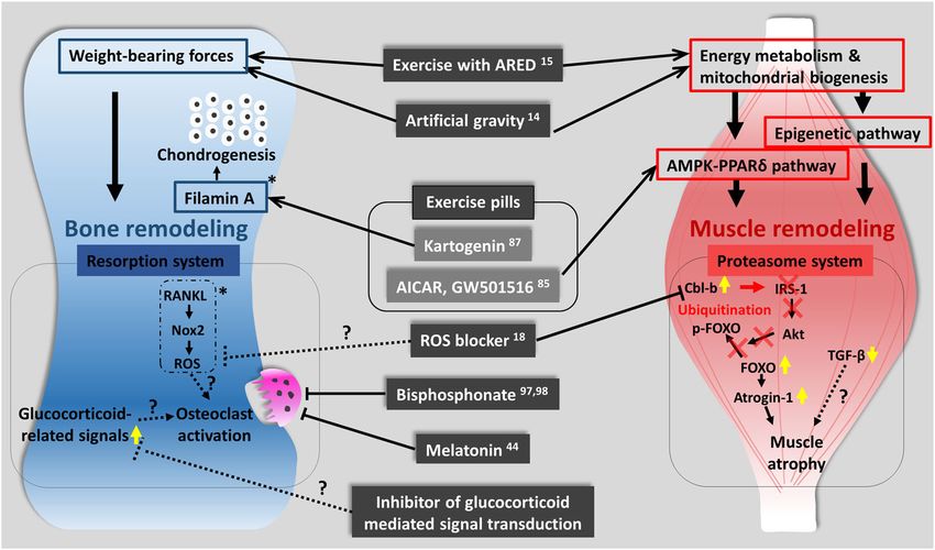
S. Furukawa et al.
4
    Experiment—First in Space” experiment (ICE-FIRST) was carried             these findings confirmed that medaka is an acceptable animal
    out by an international collaboration of laboratories. Transcrip-         model for the analysis of biological responses to altered gravity.
    tome analysis of C. elegans after 10 days of spaceflight revealed             To examine the initial action of microgravity, another space
    alteration of the expression of muscle-related genes, as well as          experiment was conducted in 2014. The osteoblast and osteoclast-
    genes that are regulated by insulin and transforming growth               specific transgenic medaka larvae housed in special chambers and
    factor-β signaling33–35.                                                  sent to the ISS (Fig. 1d). In live imaging for osteoblasts, the
       In 2009, the C. elegans RNA Interference Space Experiment              intensity of osterix-DsRed or osteocalcin-DsRed fluorescence in
    (CERISE) was conducted in which nematodes in a liquid medium              pharyngeal bones was significantly enhanced 1 day after launch.
    with food comprising bacteria expressing target double-stranded           In osteoclasts, the signals of TRAP-GFP and MMP9-DsRed were
    RNA were cultured for 4 days under microgravity or 1-g conditions         highly increased at days 4 and 6 after launch in flight. HiSeq
    (Fig. 1b). Upon return to Earth, RNA interference activity, and           analysis from pharyngeal bones of juvenile fish at day 2 after
    global gene and protein expression, were analyzed using DNA               launch showed significant changes in genes related to glucocorti-
    microarray and mass spectrometry techniques36,37. Gene silencing          coid signaling43. This experiment revealed that exposure to
    for ectopically expressed green fluorescent protein and endogen-           microgravity immediately induced a change in gene expression
    ously expressed genes encoding the RING finger protein RBX-1 for           levels in osteoblasts and osteoclasts.
    some complexes of E3 ubiquitin ligases and aspartic proteinases              In a different experiment examining the participation of
    ASP-4/6 occurred normally under microgravity38. Expression levels         osteoblasts and osteoclasts in bone healing, osterix-DsRed/TRAP-
    of gene and protein in muscular thick filaments were also                  EGFP double-transgenic medaka was treated with synthetic
    significantly reduced in C. elegans grown from the L1 larval stage         glucocorticoid, prednisolone. At 18 days after initiation of
    to adulthood under microgravity compared with those grown                 continuous prednisolone administration, a part of the bony fin
                                                                              ray was fractured. Bone fracture healing was significantly delayed
    under 1-g conditions37. In addition, the expression of cytoskeletal
                                                                              by up to 32 days and was accompanied by decreased osteoblast
    elements and mitochondrial metabolic enzymes were decreased
                                                                              and osteoclast signaling as compared with control fish. To confirm
    by microgravity and decreased locomotor activity (thrashing rate),
                                                                              the function of glucocorticoid receptors in bone healing, a
    body length, and fat accumulation were observed in the
                                                                              glucocorticoid receptor 2-deficient (gr2−/−) medaka line was
    nematodes cultured under microgravity compared with those                 constructed. The results showed that osterix-DsRed and TRAP-
    cultured under 1-g conditions37. Also, similar reductions in the          EGFP fluorescent signals were increased at the site of the bone
    expression of genes such as a myosin heavy chain 3 and a                  fracture in those fish. These results demonstrated negative
    transforming growth factor-β gene, dbl-1, were observed in                regulation of osteoclast recruitment by glucocorticoid receptors
    nematodes exposed to microgravity and those cultured under                and accumulation of osteoblasts during bone fracture healing44. It
    decreased fluid dynamic conditions (i.e., reduced viscosity/drag           is true that there is a change in a part of glucocorticoid signaling
    resistance or reduced depth of the liquid culture)38. dbl-1 is a          in space, but it is inferred that the phenomenon is slightly
    positive regulator of macromolecule biosynthetic processes such           different from that of glucocorticoid administration. It is expected
    as collagens, c-type lectin, mucin, catecholamine, and lysozymes,         to be clarified by detailed analysis in the future.
    and an ortholog of mammalian bone morphogenetic protein 10,                  Using a centrifuge designed for small-fish rearing, the effect of
    which is essential for maintaining cardiac growth and func-               hypergravity on medaka reared for 6 months under normal gravity
    tion39,40. These results suggest that there is a common mechanism         (1 g) or under gravity, approximately 5 times normal (5 g) was
    between C. elegans and humans that reduces muscle develop-                investigated45. Micro-computer tomography analysis revealed that
    ment and maintenance in response to microgravity. These                   although the fish were able to maintain body posture and
    findings support the use of C. elegans in future mechanistic               position, hypergravity gradually induced vertebral curvature
    studies to examine how microgravity impacts muscle mass,                  towards the dorsal side and asymmetric formation of otoliths in
    metabolism, and growth factor expression in humans. In addition,          which the cross-sectional area was increased. These findings
    future space experiments are awaited for the analysis of                  indicate that the process of adaptation to a hypergravity
    mechanisms and tissues related to gravitational effects, mechan-          environment results in spinal deformation and otolith abnormality
    ical stimuli, contact stimuli, and these responses using the              in medaka. Together, these experiments confirm the potential of
    nematode C. elegans.                                                      using medaka to elucidate the detailed mechanisms that underlie
                                                                              responses to altered gravity.
    Medaka, goldfish scale, and zebrafish experiments                              To examine the effects of melatonin, which has been reported
                                                                              to inhibit osteoclast function under microgravity conditions46,
    Medaka (Oryzias latipes) is a small freshwater teleost fish with           scales of goldfish were used as a model of coexisting osteoclasts
    properties suitable for studies in space, including a short               and osteoblasts in the experiment conducted on the ISS.
    generation time, compact size, and translucent body until the             Microgravity was found to stimulate osteoclast activity and
    larval stage. Medaka was first sent to space in 1994 and was               significantly increase the expression of genes involved in
    successfully mated under microgravity41. In 2012, medaka was              osteoclast differentiation and activation. Melatonin treatment
    raised for approximately 2 months in the Aquatic Habitat system           significantly increased expression of calcitonin (an osteoclast-
    onboard the ISS (Fig. 1c) and examinations revealed the reduced           inhibiting hormone) mRNA but decreased the mRNA expression of
    mineral density of the pharyngeal teeth and bone, as well as              receptor activator of nuclear factor kappa-B ligand (RANKL: a
    osteoclast activation42. In addition, electron microscopy revealed        promoter of osteoclastogenesis), and these changes coincided
    that the mitochondria of the osteoclasts were less circular. The          with suppression of the expression of genes associated with
    whole transcriptome analysis showed that fkbp5 and ddit4 genes,           osteoclastogenesis. The mRNA expression level of acetylserotonin
    known as the downstream of the glucocorticoid signaling, were             O-methyltransferase, an enzyme essential for melatonin synthesis,
    strongly upregulated in the flight group reared for 60 days at ISS42.      was significantly decreased under microgravity47. This was the
    Furthermore, the fish were filmed for abnormal behavior such as             original study to suggest an inhibitory effect of melatonin on
    swimming upside-down, and it was found that the medaka                    osteoclast activation by microgravity.
    tended to become motionless in the late stage of exposure.                   To examine the mechanisms of terrestrial age-related muscle
    Considering that all of the medaka remained healthy over the 2-           atrophy, an experimental system to investigate muscle atrophy in
    month period, it can be inferred that the fish changed their               zebrafish was developed. RNA-seq analyses revealed significant
    behavior in response to the microgravity environment. Together,           increases in the amounts of transcripts related to the proteasome

    npj Microgravity (2021) 18               Published in cooperation with the Biodesign Institute at Arizona State University, with the support of NASA
S. Furukawa et al.
5
system and to autophagy in skeletal muscle when the mobility of superfamily, the members of which are negative regulators of
zebrafish was decreased. To further elucidate the general vertebrate skeletal muscle mass. Myostatin inhibitors enhance
mechanisms of muscle atrophy, experiments on muscle atrophy muscle strength and functional performance against wasting
that occurs during spaceflight in 2014 were conducted. The aim of disorders57. In addition, increased bone mineral density in the
this project (named Zebrafish Muscle) was to examine whether femurs of myostatin-knockout mice has been reported58. It was
skeletal muscle atrophy occurs in adult zebrafish under micro- also found that the expression of TGF-β1 (a positive regulator of
gravity48. Zebrafish were bred on the ISS for 6 weeks and live myostatin59) decreased in response to microgravity. This is
monitoring revealed that the fish learned to swim under consistent with in vitro observations in human fetal osteoblastic
microgravity much quicker than expected49. To understand the cells grown in space60, in vivo observations in 11-day space-flown
mechanisms of skeletal muscle atrophy in space, transcriptome rat skeletal muscle61, and in vivo observations in colonic tissue
data were periodically obtained from the skeletal muscle of the and systemic lymph node levels of 91-day space-flown mice62.
fish during their stay in space and during their recovery after Together, these data suggest that microgravity does not induce
return to Earth; it is expected that analysis of this data will allow us muscular atrophy via direct upregulation of the negative regulator
to understand further the process of skeletal muscle atrophy in myostatin.
space and the role of gravity in the maintenance and homeostasis One of the cell types responsible for sensing physical loading is
of skeletal muscle on Earth. the skeletal stem cell (SSC) because loading-induced bone
formation requires activation of these cells in the periosteum to
Mouse experiments give rise to osteoblasts. Alteration of the gene expression pattern
A significant decrease in the mass of weight-bearing bone, but not and cell morphology within the periosteum after loading suggests
of non-weight-bearing bone, has been observed in space-flown that these skeletal stem cells sense loading stimuli and remodel-
mice50–52. Osteoclast activation may be one mechanism of this ing63–65. In addition, mitochondrial function is likely to be affected
bone loss;53 however, further analyses are needed to understand by microgravity24,37,66, and the role of mitochondrial energy
the mechanistic details. It has also been reported that the production mediated by oxidative phosphorylation in skeletal
signaling involved in this bone loss is compartmentalized to muscle stem cells (SMSCs) has been investigated. SMSCs are
discrete cells (e.g., osteoclasts) and signaling pathways54. For normally cell-cycle arrested but differentiate to generate myocytes
example, RANKL has been shown to activate NADPH oxidases upon muscle damage, and along with self-renewing SMSCs form
(e.g., Nox2), which produce ROS (primarily H2O2) that induce new myofibers. The end product of glycolysis, pyruvate was found
osteoclast proliferation and activation, as observed in osteoporo- to be a substance that stimulates their growth and differentia-
sis55. Thus, it is possible that similar signaling is involved in skeletal tion67. Pyruvate dephosphorylates and activates pyruvate dehy-
muscle atrophy under microgravity56. drogenase, which opens the gateway from glycolysis to the
In 2016, a 35-day space experiment in mice was conducted tricarboxylic acid cycle by producing acetyl coenzyme A from
using an artificial gravity generator developed by JAXA (Fig. 2). As pyruvate. Conditional deletion of pyruvate dehydrogenase in
in previous space experiments using mice, bone loss and muscle SMSCs reduces cell division associated with the generation of
atrophy were observed; however, the artificial 1-g condition myocytes and subsequent myotube formation, decreases skeletal
completely suppressed these changes18. It is expected that this muscle regeneration upon injury, and aggravates the pathogen-
system will allow more detailed analyses of the effects of the esis of Duchenne muscular dystrophy in dystrophin-deficient
space environment on mice to be performed in the future since mice. These results indicate that the flow from glycolysis to the
partial gravity can be used for quantitative analysis. Transcriptome tricarboxylic acid cycle mediated by pyruvate dehydrogenase
analysis of the soleus muscle of space-flown mice is currently plays a critical role in the differentiation of SMSCs67 and that
underway to study the effects of microgravity on skeletal muscle decreased oxidative phosphorylation affects SMSC differentiation
atrophy. Preliminary data show that the expression level of certain in space.
atrophy-related genes changes in the soleus of mice under
microgravity but the myostatin-encoding gene does not change
compared with in-ground controls or artificial 1-g controls COUNTERMEASURES TO PREVENT BONE AND MUSCLE
onboard the ISS. ATROPHY UNDER MICROGRAVITY
On the other hand, no upregulation of the myostatin gene is Current exercise program on the ISS
observed in the muscles of spaceflown mice. Myostatin is a highly The current exercise program on the ISS consists of two exercise
conserved member of the transforming growth factor (TGF)-β sessions per day (30–45 min of aerobic exercise and 45 min of
Fig. 2 Mice in space. a A still image captured from a video recording of a C57BL/6J male mouse in microgravity in the ISS. b A still image
captured from a video recording of a C57BL/6J male mouse under artificial 1 g conditions generated by a short-arm centrifuge in the ISS.
These mice were housed in the mouse habitat cage unit for 35 days and these images were taken on day 5.
Published in cooperation with the Biodesign Institute at Arizona State University, with the support of NASA npj Microgravity (2021) 18S. Furukawa et al.
6
Fig. 3 Japanese astronaut Satoshi Furukawa exercising on the T2 treadmill, the Cycle Ergometer with Vibration Isolation and
Stabilization System (CEVIS), and the Advanced Resistive Exercise Device (ARED) in the ISS. Astronauts have daily exercise sessions, which
are part of operational tasks onboard the ISS, to minimize muscle atrophy and bone loss risks. The first author and an astronaut S.F. agreed
and gave written consent to use these photographs. a T2 treadmill, (b) CEVIS, and (c) ARED.
resistance exercise) and exercise is scheduled 6 days a week19,68. issue that must be addressed before longer-term space missions
Many astronauts work out 7 days a week, although the seventh can be undertaken.
day is officially considered a rest day. The allotted exercise time It is reported that muscle mass gain after resistance exercise
includes time for changing clothes, exercise equipment setup and training is positively correlated with the expression level of a
stowing, and post-workout hygiene19. particular microRNA, miR-37871,72, and also positively correlated
In the United States’ on-orbit segment of the ISS, there are two with upregulation of mechanical-growth factor and myogenin
aerobic devices (a T2 treadmill and a Cycle Ergometer with gene expression73,74. DNA methylation in human skeletal muscles
Vibration Isolation and Stabilization system [CEVIS]) and one is also affected by resistance exercise training75,76. Seaborne et al.
resistance device (the Advanced Resistive Exercise Device [ARED]) have reported that the frequency of genome-wide hypomethyla-
(Fig. 3). For aerobic exercise, a Treadmill with a Vibration Isolation tion at CpG sites in human skeletal muscle is increased after
and Stabilization system was initially used but has now been chronic resistance exercise training, is maintained during a
replaced by a T2 or CEVIS system19. Aerobic sessions consist of detraining period, and is further enhanced in response to
steady-state and interval-type protocols, with target intensities of reloading75. Furthermore, Turner et al. reported that long-term
75–80% or 60–90% VO2 max68. Resistance exercise was initially resistance exercise training upregulated the expression of 592 of
performed with the interim Resistive Exercise Device but is now 5262 genes that were hypomethylated at their CpG sites76.
performed using the ARED19. Resistance protocols are multi-set, Together, these results suggest that epigenetic regulation results
multi-repetition for the lower and upper body, with initial loads in differences in responsiveness of skeletal muscle to exercise
calculated from a 10-repetition maximum load (plus 75% of body training.
weight to compensate for the absence of body weight) and In animal studies, Nakamura et al. have reported that disuse
adjusted thereafter based on actual performance68. In future atrophy is suppressed in the fast-twitch skeletal muscles of adult
space missions and analog studies, exercise programs could be rats with prior experience of running training using a treadmill77.
complemented with lower body negative pressure (LBNP) They also found that the expression of a subset of genes that are
application69 to assess whether this leads to a reduction of post- generally upregulated during disuse atrophy in sedentary rats was
spaceflight orthostatic intolerance and improvements in general less responsive to unloading in the fast-twitch fibers of the
astronaut health, including muscle and cardiovascular health. plantaris muscle of rats with training experience. Canonical
histone 3, a main component of nucleosomes, was replaced with
Exercise epigenetics in skeletal muscle the H3.3 variant at these loci after the training period. It has also
Exercise training during a long-term stay on the ISS has been been reported that these running training-associated exchanges
found to be effective, at least in part, in preventing disuse atrophy of histone components were induced relative to the amount of
in skeletal muscle. A previous study has reported that daily exercise78. Together, these results indicate that histone compo-
exercise (2.5 h per day) suppressed the decrease of slow-twitch- nent turnover is stimulated by running exercise and that this
fiber size in soleus muscle, with the astronauts retaining 67% of turnover alters gene responsiveness to later stimuli.
the pre-flight level after prolonged spaceflight (approx. Epigenetic regulation differs between fast-twitch and slow-
180 days)70. However, the exercise results differed depending on twitch skeletal muscle fibers. Transcriptionally active histone
the individual, with two of nine crew members exhibiting drastic modifications, such as acetylation and tri-methylation at lysine 4
loss of muscle fiber size (S. Furukawa et al.
7
in fast-twitch muscle fibers but, if muscle activity was enhanced by Kartogenin (KGN) is a small compound that promotes
surgery, the level of histone acetylation at the loci was decreased chondrocyte differentiation, chondroprotection, and cartilage
in rats79. Masuzawa et al. have reported that the transcriptional repair90. KGN binds filamin A, which disrupts its interaction with
activation of peroxisome proliferator-activated receptor-gamma the transcription factor CBFβ, and induces chondrogenesis by
coactivator 1-alpha gene in response to acute running exercise regulating CBFβ-RUNX1 transcriptional regulation90. KGN is a
using a treadmill in rats is greater in fast-twitch muscle fibers in potential lead for formulation into an exercise pill because it is
which acetylation of histones is more prevalent as compared to known that moderate exercise causes anabolic responses in
that in slow-twitch muscle fibers80. A unique regulation system of chondrocytes and cartilage exhibits increased proteoglycan
gene expression in slow-twitch skeletal muscle fibers is suggested content, decreased proteoglycan degradation, and increased
to be closely related to tonic neural activity, even in sedentary thickness91. In addition, it appears that traditional Chinese
posture under normal gravity81,82. medicine may play important role in alleviating muscle and bone
loss. Several traditional Chinese medicines are currently being
Exercise pill investigated in this regard30,92.
Comprehensive studies in this area should pave the way for a
Exercise is one way to prevent sarcopenia and osteoporosis, which
more scientific exercise assessment of individuals and the
are the most frequent symptoms observed in astronauts living
development of safe and effective exercise pills. This will help
under microgravity. Astronauts continue regular exercise in a small
humans live healthier and safer in microgravity and low-gravity
training facility built into their spacecraft, but the physical
environments for long periods of time without bone loss and
activities performed in this restricted space are insufficient, and
muscle atrophy.
they often show ataxy after returning to Earth. To address this
issue, novel medical aids must be developed. An exercise pill
(exercise mimetic) is a potential pharmaceutical approach that DISCUSSION
could address this issue in both astronauts and sedentary people New concepts in the context of muscle atrophy and
on Earth. osteopenia under microgravity
Skeletal muscle is highly adaptive to external stimuli, such as
mechanical, physiological, and nutritional demands. Exercise All living organisms are directly exposed to the effects of gravity.
remodels the energy metabolism of myofibers by converting In mammals, such as mice and humans, the body must work to
type-IIb glycolytic fibers to the more oxidative type-IIa fibers to overcome gravity and maintain posture. In nematodes and fish,
increase lipid metabolism and to stimulate mitochondrial activ- the different body construction compared with that of mammals
ity83. In the elderly, oxidative type-IIa muscle fibers are more means the effects of gravity are likely different. This same must
resistant to atrophy84 and show higher exercise endurance and also be true for cultured cells and fish scales. While the effects of
fat-burning metabolism; therefore, drugs that promote the gravity likely vary from species to species, it is interesting to note
formation of oxidative type-IIa muscle fibers are potentially that the JAXA space missions have shown in all the subjects
beneficial in combatting lifestyle-related diseases such as obesity examined that muscle and bone tend to become weaker under
and diabetes. Druggable targets for exercise pills could be the key microgravity. This indicates that it is important for cells, including
proteins of the regulatory system of this muscle remodeling, mammalian cells, to be under the direct effects of gravity. Indeed,
although the mechanism underlying how exercise boosts this it has been reported that the radius of people who have
adaptation is currently unknown85. experienced a microgravity environment has unexpectedly devel-
Recently, several key regulators of this adaptation have been oped post-flight fragility, especially in its cortical structure after
reported, and they in part involve ATP/AMP, NADH/NAD+ three months of landing93.
metabolism, and the metabolic sensors AMP-activated protein With respect to bone remodeling, it is considered that
kinase (AMPK) and sirtuin 186,87. In addition to these sensing osteocytes are a candidate mechano-sensing cell in mammals,
systems, several nuclear receptors, such as estrogen-related as they are localized in bone tissue and throughout the bone
receptors and peroxisome proliferator-activated receptors (PPARs), canal94. It is expected that specific markers for osteocytes (e.g.,
act as key transcriptional regulators in concert with the PPARγ co- sclerostin, a known marker of osteocytes, which are also expressed
activator α/β. Among the three PPARs (α, γ, and β/δ), PPARδ is in osteoblasts95) will be found in the near future to clarify the role
known to play a pivotal role in fatty acid metabolism of skeletal of osteocytes in response to mechanical stress95. With that said,
muscle. Its overexpression in muscle promotes the remodeling of medaka has no osteocytes but still shows altered bone
glycolytic myofibers into oxidative ones with higher fatty acid metabolism under microgravity, suggesting the existence of a
oxidation and mitochondrial biogenesis, resulting in super response mechanism to gravitational stress that does not involve
endurance performance (i.e., the so-called “marathon mice”). osteocytes.
In addition to genetic modification, pharmaceutical approaches
have also been reported88. Administration of a synthetic ligand of Activities in the world
PPARδ, GW501516, activates fatty acid oxidation and energy As shown below, interesting researches on muscles and bones are
expenditure in muscle, but when co-administrated with AICAR being conducted overseas. Italian Mice Drawer System and Bion-
(acadesine/AICA riboside), a potent AMPK activator, mitochondrial M1 missions have examined changes in the bone architecture of
biogenesis, and oxidative metabolism are synergistically boosted, spaceflight mice using high-resolution X-ray tomography96,97. The
suggesting that the AMPK-PPARδ pathway is a potential target for results of these studies clearly show that some (e.g., femur), but
an exercise pill to improve exercise endurance and fatty acid not all (e.g., parietal bone), weight-bearing bones (e.g., femurs and
oxidation. Extensive searches for druggable targets for an exercise VII lumbar ring) are significantly reduced during spaceflight.
pill have been made in the signaling cascades described above. Furthermore, the US National Aeronautics and Space Administra-
Natural compounds, such as urolithin A, are known to improve tion (NASA) has also sent 40 genetically modified mice to space to
exercise capacity in rodents and prevent the age-related decline evaluate whether blocking the actions of myostatin prevents
of muscle function89, although the primary protein target of this skeletal muscle atrophy in space98. Systemic inhibition of
compound is currently unknown. Similarly, some compounds are myostatin/activin A signaling using a soluble form of activin type
known to have anti-aging activities, particularly anti-sarcopenia IIB receptor, which can bind both these ligands, led to dramatic
effects, and the search for novel druggable targets should be increases in both muscle and bone mass in space-flown mice. Eli
expanded to finding targets for the so-called geroprotectors. Lilly and Company performed a similar mouse spaceflight
Published in cooperation with the Biodesign Institute at Arizona State University, with the support of NASA npj Microgravity (2021) 18S. Furukawa et al.
8
Fig. 4 Musculoskeletal atrophy and its countermeasures in space. Key pathways to prevent bone and muscle deterioration in the space
environment are represented. Yellow arrows indicate the molecules whose expression changes have been studied in space experiments.
Asterisks indicate ground-based experiments and candidates for future space experiments. Question marks indicate candidates for future
ground and space research.
experiment using a myostatin inhibitor that specific antibody- period of time. Recent research has shown that exercise together
mediated inhibition of myostatin restores skeletal muscle and with bisphosphonate treatment can partially ameliorate the bone
strength loss caused by microgravity, but not in the soleus loss that occurs in space100,101, however, the side effects
muscle99. Together, these findings suggest that inhibition of associated with bisphosphonate are a concern. Understanding
myostatin signaling may be an effective countermeasure against more about the mechanisms of gravitational stress at the
the skeletal muscle and bone loss experienced by astronauts molecular level will promote the development of small-molecule
under microgravity. drugs, such as those that selectively regulate epigenetics or
As a notable achievement, in the recent NASA Twins Study, in transcription factors90, as a means of protecting against muscle
which identical twin astronauts were examined before and after a atrophy and osteopenia.
1-year spaceflight, changes in immune responses, epigenetics, gut Here, we have provided a unique overview of the findings of
microbiota, body weight, and serum metabolites were observed JAXA missions (Fig. 4). The data presented here are important, as
after the astronauts’ return to Earth66. In addition, some changes, there is a need to maximize information from space data sources
including a small subset of changes in gene expression, telomere from different missions and from different agencies and across
dynamics, DNA disruption, carotid artery thickening, ocular tissues, research disciplines to ensure maintenance of astronaut health
and some cognitive functions, remained at 6 months after the during long-term missions to Mars or beyond102. In addition, some
astronauts’ return to Earth. The risk factors for the persistence of of the countermeasures used in spaceflight by astronauts could be
molecular changes (e.g., gene expression) after long-term space used to help in the maintenance of the health of older persons on
missions (>1 year) remain to be elucidated. The results from Earth. Thus, these findings could have an extensive application to
overseas and JAXA missions have points in common. Accumulat- improve elderly quality of life, prevent falls, and to ensure active
ing the common points between them will lead to the future and healthy aging103.
development of space biology researches.
Received: 21 February 2020; Accepted: 25 April 2021;
Limitations
Automatic analytical technologies such as single-cell RNA and
DNA sequencing, omics analysis, and imaging are advancing day
by day. They are also used for detailed analysis of biological
samples in space flight. By improving each of these devices so that REFERENCES
they can be used under microgravity and installing them at the ISS 1. Gateway | NASA. https://www.nasa.gov/gateway (2021).
laboratory, it is possible to immediately study biological phenom- 2. Vico, L. & Hargens, A. Skeletal changes during and after spaceflight. Nat. Rev.
ena that change in space. It is also expected to reduce the return Rheumatol. 14, 229–245 (2018).
3. Zizola, C. & Schulze, P. C. Metabolic and structural impairment of skeletal muscle
of in-flight samples for analysis on Earth and significantly reduce
in heart failure. Heart Fail Rev. 18, 623–630 (2013).
the time required for crew members to support experiments in 4. Goswami, N. Falls and fall-Prevention in older persons: geriatrics meets space-
space. Further innovations in these technologies and devices will flight! Front. Physiol. 8, 603 (2017).
help clarify the phenomena of life and will bring great progress in 5. Goswami, N., Blaber, A. P., Hinghofer-Szalkay, H. & Montani, J. P. Orthostatic
the field of space life science. intolerance in older persons: etiology and countermeasures. Front. Physiol. 8,
803 (2017).
6. Strollo, F., Gentile, S., Strollo, G., Mambro, A. & Vernikos, J. Recent progress in
Future directions
space physiology and aging. Front. Physiol. 9, 1551 (2018).
The ISS has already been developed as a space colony, and plans 7. Blaber, A. P., Goswami, N., Bondar, R. L. & Kassam, M. S. Impairment of cerebral
are now underway to build a gateway to the moon and even to go blood flow regulation in astronauts with orthostatic intolerance after flight.
to Mars, meaning that humans will have to live in space for a long Stroke 42, 1844–1850 (2011).
npj Microgravity (2021) 18 Published in cooperation with the Biodesign Institute at Arizona State University, with the support of NASAS. Furukawa et al.
9
8. Hadjidakis, D. J. Androulakis, II. Bone remodeling. Ann. N. Y Acad. Sci. 1092, 38. Harada, S. et al. Fluid dynamics alter Caenorhabditis elegans body length via
385–396 (2006). TGF-β/DBL-1 neuromuscular signaling. NPJ Microgravity 2, 16006 (2016).
9. Bajotto, G. & Shimomura, Y. Determinants of disuse-induced skeletal muscle 39. Chen, H. et al. BMP10 is essential for maintaining cardiac growth during murine
atrophy: exercise and nutrition countermeasures to prevent protein loss. J. Nutr. cardiogenesis. Development 131, 2219–2231 (2004).
Sci. Vitaminol. 52, 233–247 (2006). 40. Qu, X. et al. BMP10 preserves cardiac function through its dual activation of
10. Wolff, J. The Law of Bone Remodeling. (Press, Springer 1986). SMAD-mediated and STAT3-mediated pathways. J. Biol. Chem. 294,
11. Harrison, B. C. et al. Skeletal muscle adaptations to microgravity exposure in the 19877–19888 (2019).
mouse. J. Appl Physiol. 95, 2462–2470 (2003). 41. Ijiri, K. Fish mating experiment in space—what it aimed at and how it was
12. Ikemoto, M. et al. Space shuttle flight (STS-90) enhances degradation of rat prepared. Biol. Sci. Space 9, 3–16 (1995).
myosin heavy chain in association with activation of ubiquitin-proteasome 42. Chatani, M. et al. Microgravity promotes osteoclast activity in medaka fish
pathway. FASEB J. 15, 1279–1281 (2001). reared at the international space station. Sci. Rep. 5, 14172 (2015).
13. Japanese experiment module (Kibo). JAXA Homepage. Retrieved March 23, 43. Chatani, M. et al. Acute transcriptional up-regulation specific to osteoblasts/
2021, from https://iss.jaxa.jp/en/kibo/. osteoclasts in medaka fish immediately after exposure to microgravity. Sci. Rep.
14. Cell Biology Experiment Facility-Left (CBEF-L). JAXA Homepage. Retrieved March 6, 39545 (2016).
23, 2021, from https://iss.jaxa.jp/en/htv/mission/htv-8/payload/. 44. Azetsu, Y. et al. Treatment with synthetic glucocorticoid impairs bone meta-
15. A live imaging system “Confocal Space Microscopy” (“COSMIC”). JAXA Home- bolism, as revealed by in vivo imaging of osteoblasts and osteoclasts in medaka
page. Retrieved March 23, 2021, from https://iss.jaxa.jp/en/htv/mission/htv-9/ fish. Biomed. Pharmacother. 118, 109101 (2019).
payload/. 45. Chatani, M., Mitsuhashi, A., Dodo, Y., Sakai, N. & Takami, M. Hypergravity induces
16. AQH Outline. JAXA Homepage. Retrieved March 23, 2021, from https://iss.jaxa. vertebrae and otolith deformation in medaka fish. Biol. Sci. Space 33, 12–17
jp/en/kiboexp/pm/aqh/. (2019).
17. Mouse habitat unit (MHU). JAXA Homepage. Retrieved March 23, 2021, from 46. Suzuki, N. & Hattori, A. Melatonin suppresses osteoclastic and osteoblastic
https://iss.jaxa.jp/en/kiboexp/pm/mhu/. activities in the scales of goldfish. J. Pineal Res. 33, 253–358 (2002).
18. Shiba, D. et al. Development of new experimental platform ‘MARS’-Multiple 47. Ikegame, M. et al. Melatonin is a potential drug for the prevention of bone loss
Artificial-gravity Research System to elucidate the impacts of micro/partial during space flight. J. Pineal Res. 67, e12594 (2019).
gravity on mice. Sci. Rep. 7, 10837 (2017). 48. Niles, L. Zebrafish flex their muscles for research aboard the international
19. Loehr, J. A. et al. Physical training for long-duration spaceflight. Aerosp. Med. space station. https://www.nasa.gov/mission_pages/station/research/news/
Hum. Perform. 86, A14–A23 (2015). zebrafish_muscle (2015).
20. Sibonga, J. D., Spector, E. R., Johnston, S. L. & Tarver, W. J. Evaluating bone loss in 49. Sato, F. & Sehara-Fujiwara, A. Effects of the gravity on maintenance of muscle
ISS astronauts. Aerosp. Med. Hum. Perform. 86, A38–A44 (2015). mass in zebrafish (Zebrafish Muscle). Experiment report. http://iss.jaxa.jp/en/
21. Drake, B. G. Human exploration of Mars design reference architecture 5.0. kiboexp/theme/second/pmlatter/zebrafishmuscle/report/ (2014).
NASA Special Publication, NASA-SP-2009-566 https://www.nasa.gov/pdf/ 50. Macaulay, T. R., Siamwala, J. H., Hargens, A. R. & Macias, B. R. Thirty days of
373665main_NASA-SP-2009-566.pdf (2009). spaceflight does not alter murine calvariae structure despite increased Sost
22. Uchida, T. et al. Reactive oxygen species upregulate expression of muscle expression. Bone Rep. 7, 57–62 (2017).
atrophy-associated ubiquitin ligase Cbl-b in rat L6 skeletal muscle cells. Am. J. 51. Maupin, K. A. et al. Skeletal adaptations in young male mice after 4 weeks
Physiol. Cell Physiol. 314, C721–C731 (2018). aboard the International Space Station. NPJ Microgravity 5, 21 (2019).
23. Schnackenberg, C. G. & Wilcox, C. S. The SOD mimetic tempol restores vasodi- 52. Tominari, T. et al. Hypergravity and microgravity exhibited reversal effects on
lation in afferent arterioles of experimental diabetes. Kidney Int. 59, 1859–1864 the bone and muscle mass in mice. Sci. Rep. 9, 6614 (2019).
(2001). 53. Gerbaix, M. et al. One-month spaceflight compromises the bone microstructure,
24. Nikawa, T. et al. Skeletal muscle gene expression in space-flown rats. FASEB J. 18, tissue-level mechanical properties, osteocyte survival and lacunae volume in
522–524 (2004). mature mice skeletons. Sci. Rep. 7, 2659 (2017).
25. Nakao, R. et al. Ubiquitin ligase Cbl-b is a negative regulator for insulin-like 54. Győri, D. S. & Mócsai, A. Osteoclast signal transduction during bone metastasis
growth factor 1 signaling during muscle atrophy caused by unloading. Mol. Cell formation. Front. Cell Dev. Biol. 8, 507 (2020).
Biol. 29, 4798–4811 (2009). 55. Agidigbi, T. S. & Kim, C. Reactive oxygen species in osteoclast differentiation and
26. Sandonà, D. et al. Adaptation of mouse skeletal muscle to long-term micro- possible pharmaceutical targets of ROS-mediated osteoclast diseases. Int. J. Mol.
gravity in the MDS mission. PLoS ONE 7, e33232 (2012). Sci. 20, 3576 (2019).
27. Cadena, S. M. et al. Skeletal muscle in MuRF1 null mice is not spared in low- 56. Ferreira, L. F. & Laitano, O. Regulation of NADPH oxidases in skeletal muscle. Free
gravity conditions, indicating atrophy proceeds by unique mechanisms in Radic. Biol. Med. 98, 18–28 (2016).
space. Sci. Rep. 9, 9397 (2019). 57. Smith, R. C. & Lin, B. K. Myostatin inhibitors as therapies for muscle wasting
28. Riley, D. A. et al. Hypogravity-induced atrophy of rat soleus and extensor digi- associated with cancer and other disorders. Curr. Opin. Support Palliat. Care. 7,
torum longus muscles. Muscle Nerve. 10, 560–568 (1987). 352–360 (2013).
29. Allen, D. L. et al. Effects of spaceflight on murine skeletal muscle gene 58. Elkasrawy, M. N. & Hamrick, M. W. Myostatin (GDF-8) as a key factor linking
expression. J. Appl. Physiol. 106, 582–592 (2009). muscle mass and bone structure. J. Musculoskelet. Neuronal Interact. 10, 56–63
30. Gao, Y., Arfat, Y., Wang, H. & Goswami, N. Muscle atrophy induced by mechanical (2010).
unloading: mechanisms and potential countermeasures. Front. Physiol. 9, 235 59. Zhu, J. et al. Relationships between transforming growth factor-beta1, myos-
(2018). tatin, and decorin: implications for skeletal muscle fibrosis. J. Biol. Chem. 282,
31. Stitt, T. N. et al. The IGF-1/PI3K/Akt pathway prevents expression of muscle 25852–25863 (2007).
atrophy-induced ubiquitin ligases by inhibiting FOXO transcription factors. Mol. 60. Harris, S. A. et al. Effects of orbital spaceflight on human osteoblastic cell phy-
Cell. 14, 395–403 (2004). siology and gene expression. Bone 26, 325–331 (2000).
32. Rundfeldt, L. C., Gunga, H. C. & Steinach, M. Anabolic signaling and response in 61. Westerlind, K. C. & Turner, R. T. The skeletal effects of spaceflight in growing rats:
sarcopenia as a model for microgravity induced muscle deconditioning: a sys- tissue-specific alterations in mRNA levels for TGF-beta. J. Bone Min. Res. 10,
tematic review. REACH 13, 100025 (2019). 843–848 (1995).
33. Higashibata, A. et al. Decreased expression of myogenic transcription factors 62. McCarville, J. L. et al. Spaceflight influences both mucosal and peripheral
and myosin heavy chains in Caenorhabditis elegans muscles developed during cytokine production in PTN-Tg and wild type mice. PLoS ONE 8, e68961 (2013).
spaceflight. J. Exp. Biol. 209, 3209–3218 (2006). 63. Raab-Cullen, D. M., Thiede, M. A., Petersen, D. N., Kimmel, D. B. & Recker, R. R.
34. Higashibata, A. et al. Biochemical and Molecular Biological Analyses of space- Mechanical loading stimulates rapid changes in periosteal gene expression.
flown nematodes in Japan, the First International Caenorhabditis elegans Calcif. Tissue Int. 55, 473–478 (1994).
Experiment (ICE-First). Microgravity Sci. Technol. 19, 159–163 (2007). 64. Sakai, D. et al. Remodeling of actin cytoskeleton in mouse periosteal cells under
35. Selch, F. et al. Genomic response of the nematode Caenorhabditis elegans to mechanical loading induces periosteal cell proliferation during bone formation.
spaceflight. Adv. Space Res. 41, 807–815 (2008). PLoS ONE 6, e24847 (2011).
36. Etheridge, T. et al. The effectiveness of RNAi in Caenorhabditis elegans is 65. Duchamp de Lageneste, O. et al. Periosteum contains skeletal stem cells with
maintained during spaceflight. PLoS ONE 6, e20459 (2011). high bone regenerative potential controlled by Periostin. Nat. Commun. 9, 773
37. Higashibata, A. et al. Microgravity elicits reproducible alterations in cytoskeletal (2018).
and metabolic gene and protein expression in space-flown Caenorhabditis 66. Garrett-Bakelman, F. E. et al. The NASA Twins Study: a multidimensional analysis
elegans. NPJ Microgravity 2, 15022 (2016). of a year-long human spaceflight. Science 364, eaau8650 (2019).
Published in cooperation with the Biodesign Institute at Arizona State University, with the support of NASA npj Microgravity (2021) 18You can also read