Morphogenesis and Patterning of the Phallus and Cloaca in the American Alligator, Alligator mississippiensis
←
→
Page content transcription
If your browser does not render page correctly, please read the page content below
Sex Dev                                                                 Published online: June 28, 2014
                                                      DOI: 10.1159/000364817

Morphogenesis and Patterning of
the Phallus and Cloaca in the American
Alligator, Alligator mississippiensis
Marissa L. Gredler a Ashley W. Seifert a Martin J. Cohn a–c
Departments of a Biology and b Molecular Genetics and Microbiology, and c Howard Hughes Medical Institute,
UF Genetics Institute, University of Florida, Gainesville, Fla., USA

Key Words                                                                  dependent sex determination. Our analysis of alligator phal-
Alligator · Cloaca · Development · External genitalia · Gene               lus and cloaca development suggests that modifications of
expression · Genital tubercle · Phallus · Reptile                          an ancestral program of urogenital development could have
                                                                           generated the morphological diversity found in the external
                                                                           genitalia of modern amniotes.            © 2014 S. Karger AG, Basel
Abstract
In most animals, reproduction by internal fertilization is fa-
cilitated by an intromittent organ, such as the penis in amni-
ote vertebrates. Recent progress has begun to uncover the                      The emergence of external genitalia facilitated the
mechanisms of mammalian external genital development;                      transition from external fertilization (e.g. spawning) to
however, comparatively little is known about the develop-                  internal fertilization, a key innovation in the evolution of
ment of the reptilian penis and clitoris. Here, we describe the            terrestriality. In most amniotes, an intromittent organ
development of the phallus and cloaca in the American alli-                functions to transfer sperm from the male into the female
gator, Alligator mississippiensis. The embryonic precursor of              reproductive tract. Although many of the morphogenetic
the penis and clitoris is the genital tubercle, which forms by             and molecular mechanisms of phallus development are
the budding of genital mesenchyme beneath the ventral                      beginning to be understood in mammals, comparatively
body wall ectoderm, adjacent to the cloacal membrane. The                  little is known about development of the external genita-
cloacal lips develop from another pair of outgrowths, the lat-             lia in other amniotes.
eral swellings. Early development of the alligator phallus,                    Reptilian phalluses have diverse morphologies. A sin-
cloaca, and urogenital ducts generally resembles that of oth-              gle midline phallus, the male penis and female clitoris,
er reptiles, suggesting that differences in adult reptilian                occurs in most reptiles. Male squamates (lizards and
phallus and cloacal anatomy arise at later stages. The phallic             snakes) have 2 hemipenes that function as intromittent
sulcus is derived from the cloacal endoderm, indicating that               organs [Arnold, 1986; Card and Kluge, 1995; Böhme and
the crocodilian sulcus is functionally and developmentally                 Ziegler, 2009; Gredler et al., this issue; Leal and Cohn, this
homologous to the mammalian urethra. Initial external gen-                 issue]. Some birds have a single medial intromittent phal-
ital outgrowth and patterning occur prior to temperature-                  lus, but the majority of avian species lack an intromittent
                                                                                                                                                                132.187.19.80 - 6/30/2014 3:37:55 PM
                                                                                                                                                                Downloaded by: C. Steinlein - 60989
                                                                                                                                                                Universitätsbibliothek Würzburg

                           © 2014 S. Karger AG, Basel                      Martin J. Cohn
                           1661–5425/14/0000–0000$39.50/0                  Department of Molecular Genetics and Microbiology and Department of Biology
                                                                           University of Florida, PO Box 103610
E-Mail karger@karger.com
                                                                           Gainesville, FL 32610 (USA)
www.karger.com/sxd
                                                                           E-Mail mjcohn @ ufl.edu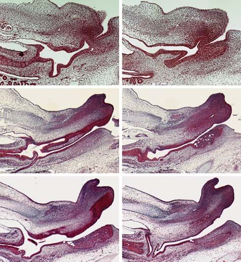
phallus altogether [King, 1979]. Archosauria is com-             and Cohn, this issue]. These swellings fuse to form the
prised of birds and crocodilians (alligators, crocodiles,        genital tubercle, the embryonic anlage of the penis in
and gharials); recent molecular phylogenetic analyses            males and the clitoris in females [Raynaud and Pieau,
support testudines (turtles) as the sister group to archo-       1985; Perriton et al., 2002; Herrera et al., 2013; Larkins
saurs [Chiari et al., 2012; Fong et al., 2012; Wang et al.,      and Cohn, this issue]. In non-mammalian amniotes, the
2013], although the turtle-archosaur clade remains con-          adult phallus resides inside the proctodeum, which is
troversial [reviewed in Hedges, 2012]. Male turtles and          formed by the cloacal lips and is the caudal-most of the 3
crocodilians have a single penis that is thought to develop      cloacal chambers [Gadow, 1887; King, 1979; Raynaud
by similar mechanisms [Gadow, 1887; Moens, 1912;                 and Pieau, 1985]. Proximal or anterior to the proctode-
King, 1979].                                                     um, the urodeum is the excretory cloacal chamber and
    During amniote copulation, the male intromittent or-         generally receives the urogenital ducts and the bladder
gan acquires and maintains rigidity in order to direct           [Gadow, 1887; Raynaud and Pieau, 1985]. The coprode-
sperm into the female reproductive tract [Gadow, 1887;           um connects to the rectum to function with the digestive
King, 1979; Powell, 2000; Kelly, 2002]. Erection and ever-       system, and is the most anterior cloacal chamber [Gadow,
sion by hydrostatic pressure and muscle contraction func-        1887; Raynaud and Pieau, 1985].
tion to achieve turgor and to create a functional channel            Morphogenesis of the phallus is physically, temporally,
for sperm transfer [Hart and Melese-D’Hospital, 1983;            and molecularly linked to development of the cloaca [Mo
Schmidt and Schmidt, 1993; Andersson and Wagner,                 et al., 2001; Dravis et al., 2004; Yucel et al., 2007; Seifert et
1995; Kelly, 2002, 2004, 2013; Hsu et al., 2005; Cabrera et      al., 2009b; Suzuki et al., 2012; Xu et al., 2012]. A portion
al., 2007]. Hydrostatic pressure is produced by blood vas-       of the cloacal epithelium extends into the genital tubercle
culature in penises of crocodilians, turtles and mammals         to form the penile sulcus (also known as the penile groove,
[Zug, 1966; King, 1979; Kelly, 2004; Moore et al., 2012], by     phallic sulcus or sulcus spermaticus) in birds and turtles
lymphatic vessels in bird penises, when present [King,           and the urethra in mammals [Seifert et al., 2008, 2009b;
1979], and by both blood and lymph in squamates [Dow-            Herrera et al., 2013; Larkins and Cohn, this issue]. Genet-
ling and Savage, 1960]. Internally, crocodilian, turtle, and     ic knockout experiments in mice have demonstrated that
mammalian penises have large fibroelastic, vascular, and         molecular perturbation of cloacal development affects
lacunar tissues that support vascular erection [Zug, 1966;       morphogenesis of the genital tubercle, and subsequently
King, 1979; Kelly, 2004]. Two regions of erectile tissue are     formation of the penis/clitoris [Perriton et al., 2002; Mi-
present; the corpus spongiosum is flexible, fibrous tissue       yagawa et al., 2009; Seifert et al., 2009a; Xu et al., 2012].
that surrounds the penile sulcus in non-mammalian am-                The developmental mechanisms that mediate forma-
niotes and the penile urethra in mammals, and the cor-           tion of the phallus have been studied most thoroughly in
pora cavernosa are large, paired regions of highly vascu-        the mouse. An evolutionarily conserved suite of gene net-
larized tissue that expand upon increased blood flow [Re-        works regulates organogenesis throughout the embryo,
ese, 1915; Zug, 1966; King, 1979; Powell, 2000; Kelly, 2004,     and a number of these pathways have been implicated in
2013; Cabrera et al., 2007]. The base of the alligator penis     external genital development. Members of the fibroblast
is formed from paired penile bodies called crura [Gadow,         growth factor (Fgf) family of signaling molecules mediate
1887; Reese, 1924; Kelly, 2013]. Proximally, each crus is        external genital development and formation of the ure-
connected to the pelvic girdle and adjacent cloacal mus-         thral tube in mice [Haraguchi et al., 2000; Petiot et al.,
cles; distally, the crura fuse to form the shaft of the corpus   2005]. Differential expression of Hox genes throughout
cavernosum [Powell, 2000; Kelly, 2013]. In crocodilians,         the embryo creates a combinatorial code that confers
eversion of the phallus is achieved by muscle contraction        identity on many organ systems, and the paralogous
[Reese, 1915; Powell, 2000; Kelly, 2013], and vascular dila-     Hoxd13 and Hoxa13 genes are required for normal devel-
tion inflates the distal portion of the penis (glans) and the    opment of distal appendages, including the digits and ex-
tissue adjacent to the penile sulcus [Gadow, 1887; Reese,        ternal genitalia [Sordino et al., 1996; Kondo et al., 1997;
1915; Cabrera et al., 2007; Ziegler and Olbort, 2007; Moore      Warot et al., 1997; Morgan, 2003; Scott et al., 2005]. Son-
et al., 2012].                                                   ic hedgehog (Shh) encodes a secreted signaling molecule
    In birds, turtles and mammals, development of the            that is expressed in the embryonic endoderm and its de-
phallus begins with the emergence of paired genital swell-       rivatives, and is required for cloacal septation and for
ings between the hindlimb buds [Raynaud and Pieau,               proper patterning and outgrowth of the murine genital
1985; Perriton et al., 2002; Herrera et al., 2013; Larkins       tubercle [Bitgood and McMahon, 1995; Perriton et al.,
                                                                                                                                     132.187.19.80 - 6/30/2014 3:37:55 PM
                                                                                                                                     Downloaded by: C. Steinlein - 60989
                                                                                                                                     Universitätsbibliothek Würzburg

2                    Sex Dev                                                           Gredler /Seifert /Cohn
                     DOI: 10.1159/000364817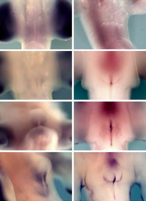
2002; Seifert et al., 2008, 2009a, 2010; Miyagawa et al.,             USA. Crocodilians undergo temperature-dependent sex determi-
2009; Runck et al., 2014]. Ligands and receptors of the               nation, and the thermosensitive period in A. mississippiensis oc-
                                                                      curs between Ferguson stages 21 [31–35 days post-oviposition
bone morphogenetic protein (Bmp) family of patterning                 (dpo)] and 24 (46–50 dpo) [Ferguson, 1985; Lang and Andrews,
genes frequently interact with Shh, are common media-                 1994]. A. mississippiensis embryos hatch at stage 28 (64–70 dpo).
tors of cell death during development, and have been                  Eggs from RWR were incubated in outdoor tanks at the wildlife
demonstrated to regulate external genital morphogenesis               refuge for 2 weeks (stage 12) before being transferred to the Uni-
in both mice and chickens [Bitgood and McMahon, 1995;                 versity of Florida for incubation. To investigate development of the
                                                                      phallus and cloaca prior to sex determination, we incubated eggs
Roberts et al., 1995; Sukegawa et al., 2000; Suzuki et al.,           at 33 ° C (which produces 100% male hatchlings, and maintains
2003; Sasaki et al., 2004; Bandyopadhyay et al., 2006; Sei-           consistency in developmental timing) until they reached stages
fert et al., 2008; Herrera et al., 2013; Lu et al., 2013]. Oth-       12.5–21, then harvested and dissected them in cold PBS [Ferguson,
er genes implicated in external genital development in-               1985; Lang and Andrews, 1994]. Eggs from SAAFZP were incu-
clude members of the Wnt/β-catenin pathway [Schwabe                   bated at 32 ° C for 1–2 months prior to transfer to the University of
                                                                      Florida where they were processed immediately for dissection. Af-
et al., 2004; Lin et al., 2008; Draaken et al., 2012; Guo et          ter removal from the egg, embryos at stage 23 and older were anes-
al., 2014], Dlx genes [Suzuki et al., 2008] and Eph/ephrins           thetized and euthanized by intracoelomic injection of tricaine
[Dravis et al., 2004].                                                methylsulfonate (MS222) (0.7% MS222, pH 7.0 at 250 mg/kg for
    In order to test the hypothesis that phallus develop-             anesthesia, 0.4 ml of 50% MS222 for euthanasia) based on pub-
ment in crocodilians is controlled by the same mecha-                 lished methods [Conroy et al., 2009].
                                                                          Tissue used for RNA extraction was stored in RNAlater at
nisms that pattern mammalian and avian external geni-                 – 20 ° C. Embryos used for histology were fixed overnight in 4%
talia [Kondo et al., 1997; Warot et al., 1997; Haraguchi et           paraformaldehyde at 4 ° C, rinsed in PBS and dehydrated in a grad-
al., 2000, 2001; Perriton et al., 2002; Morgan, 2003; Sasaki          ed ethanol series, then stored in 70% ethanol at 4 ° C. For whole
et al., 2004; Petiot et al., 2005; Scott et al., 2005; Seifert et     mount in situ hybridization, embryos were fixed overnight in 4%
al., 2008, 2009a, b; Miyagawa et al., 2009; Herrera et al.,           paraformaldehyde at 4 ° C, rinsed in PBS and dehydrated in a grad-
                                                                      ed methanol series, then stored in 100% methanol at –20 ° C. Tissue
2013], we performed a detailed analysis of American al-               used for scanning electron microscopy (SEM) was fixed and stored
ligator (Alligator mississippiensis) phallus development              in 1% glutaraldehyde at 4 ° C.
from the time that outgrowth is initiated through the on-
set of sexual differentiation. We show that initiation and                Cell Death Analysis
early development of the alligator genital tubercle is strik-             Stage 15 embryos were quickly dissected in PBS and incubated
                                                                      in LysoTracker red (Molecular Probes) staining solution (25 μl Ly-
ingly similar to that of turtles, birds, and mammals. More-           soTracker red in 15 ml PBS) at 37 ° C for 30 min, then rinsed in PBS
over, gene expression patterns in the developing alligator            and fixed overnight in 4% paraformaldehyde at 4 ° C. Epifluores-
phallus are similar to those that have been observed for              cent and brightfield images were acquired after fixation.
orthologous genes in turtles, birds, and mice, suggesting
that the molecular mechanisms of phallus formation are                   Scanning Electron Microscopy
                                                                         After glutaraldehyde fixation, embryos were washed with PBS,
conserved across amniotes. Morphogenesis of the cloaca                osmicated in 2% osmium tetroxide for 1 h, and dehydrated to ab-
occurs by interaction between the posterior limits of the             solute ethanol. The embryos were then critical-point dried and
digestive and excretory tracts, and a portion of the cloacal          mounted on stubs, sputter coated with gold/palladium, and im-
endoderm extends into the genital tubercle to form the                aged on a Hitachi S-4000 FE-SEM.
phallic sulcus. Our results show that the crocodilian phal-
                                                                          Histology
lus and cloaca develop by morphogenetic and molecular                     Paraffin histology was performed according to standard meth-
mechanisms that closely resemble those of other amni-                 ods. Lower bodies were dehydrated to absolute ethanol, permeabi-
otes. Viewed in a phylogenetic context, these similarities            lized in xylene, then infiltrated with and embedded in paraffin.
are consistent with the hypothesis that the genital tuber-            Sections were cut 5 μm thick and slides were stained with Masson’s
cle of crocodilians is homologous to that of birds, turtles,          trichrome (Richard Allen Scientific).
and mammals.                                                              In situ Hybridization
                                                                          RNA extracted (Qiagen RNeasy Plus Micro kit) from stage 15
                                                                      A. mississippiensis tail and forelimb tissue was used to synthesize
   Methods                                                            cDNA (BioRad iScript cDNA synthesis kit) that served as template
                                                                      for PCR cloning. Primers were designed from published sequenc-
    Egg Collection and Embryo Dissection                              es (Bmp4, EF527278; Shh, EF527277). PCR products were ligated
    A. mississippiensis eggs were provided by the Rockefeller Wild-   into the pGEM-T Easy Vector (Promega), PCR-amplified using
life Refuge (RWR) in Grand Chenier, La., and the St. Augustine        M13 primers, and used as templates for transcription of digoxigen-
Alligator Farm Zoological Park (SAAFZP) in St. Augustine, Fla.,       in-labeled antisense riboprobes for Bmp4 and Shh. The remaining
                                                                                                                                              132.187.19.80 - 6/30/2014 3:37:55 PM
                                                                                                                                              Downloaded by: C. Steinlein - 60989
                                                                                                                                              Universitätsbibliothek Würzburg

Alligator Phallus and Cloaca                                          Sex Dev                                                            3
Development                                                           DOI: 10.1159/000364817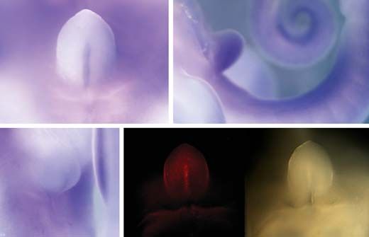
probes were generated by transcription from linearized plasmids ventral body wall ectoderm between the genital tubercle
for Hoxd13 (Trachemys scripta, kindly provided by C. Larkins) and and the tail (fig. 1H).
Fgfr2 and Fgf10 (kindly provided by T. Iguchi). Whole mount in
situ hybridization was performed according to published methods At stage 17 (22 dpo), the genital tubercle bends cau-
[Nieto et al., 1996] with the following modifications: BM purple dally, coincident with deepening of the depression be-
(Roche) was used as a color substrate in place of NBT/BCIP, Triton tween the genital tubercle and the ventral body wall
X-100 was replaced with Tween-20 in KTBT solution, and the con- (fig. 1I). Two crests of tissue are present at the lateral mar-
centration of Triton X-100 in NTMT solution was increased from gins of this fold, forming the anlagen of the cloacal lips
0.1 to 1%.
(fig. 1I, J). At stage 18, the cloacal lips are thinner medio-
laterally and are closer to the genital tubercle than at stage
17 (fig. 1K, L). The posterior-most cloacal chamber, the
Results proctodeum, appears as the cloacal lips emerge and the
genital tubercle bends caudally (fig. 1L). The cranial ra-
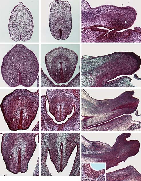
Early Development of the Alligator Phallus phe persists anterior to the genital tubercle (fig. 1L). At
SEM was used to characterize development of the ex- stage 19 (27 dpo), a crescent-shaped outgrowth, which we
ternal genitalia in alligator (A. mississippiensis) embryos have termed the phallic ridge, develops along the dorsal
between stage 12.5, prior to the appearance of the genital margin of the distal genital tubercle (fig. 1M). The phallic
tubercle, and stage 20, before the onset of sexual differ- sulcus is visible on the ventral side of the genital tubercle,
entiation [staging was performed according to Fergu- extending into the proctodeal chamber (fig. 1M, N). The
son, 1985; Lang and Andrews, 1994]. At stage 12.5 (14 cloacal lips extend farther laterally than at previous stages
dpo), genital mesenchyme and the overlying ectoderm (fig. 1N). At stage 20, the phallic sulcus extends distally
protrude from the ventral body wall, anterior and lateral into the glans, which appears to consist of 2 lobes and is
to the cloacal membrane (fig. 1A, B). A small crease in distinct from the shaft of the phallus (fig. 1O, P). Matura-
the midline of this early genital tissue delineates the po- tion of the distal genital tubercle has resulted in formation
sition of the cloacal membrane, a transient structure of an arch-shaped pocket between the phallic ridge and
formed where the cloacal endoderm meets the overlying the apex of the glans (fig. 1P).
surface ectoderm. An additional pair of swellings forms
lateral to the cloacal membrane and caudal to the genital Maturation of the Genital Tubercle
swellings, at the level of the posterior hindlimb bud We used histology to examine the internal anatomy of
(fig. 1A, B). By stage 13.5 (15 dpo), a single genital tu- the developing phallus. At stage 17, the phallic sulcus is a
bercle is apparent (fig. 1C, D). The apex of the genital bilaminar epithelial plate that extends from the ventral
tubercle is slightly bifid at this stage, which may repre- surface into the center of the genital tubercle (fig. 2A–C).
sent its formation by fusion of the 2 genital swellings A ring of dense connective tissue, the anlage of the cor-
(fig. 1D). A third pair of swellings emerges anterior to pora cavernosa (alternatively known as fibrovascular bod-
the genital tubercle (fig. 1C, D). Outgrowth of the genital ies or corpora fibrosa), is visible in the mesenchyme dorsal
tubercle continues and, by stage 15 (18 dpo), it has a to the phallic sulcus (fig. 2B, C). A second region of lacunar
rounded, cylindrical appearance (fig. 1E). By stage 15, connective tissue, the corpus spongiosum, is positioned
the genital tubercle has extended beyond the lateral ventral to the corpus cavernosum (fig. 2C). At stage 18, the
swellings (fig. 1E, F). The anterior swellings, which are distal phallic sulcus appears as a bilaminar plate dorsally
positioned immediately cranial to the base of the genital and the ventral margin is an open groove (fig. 2D). In the
tubercle, are elongated anteroposteriorly and com- proximal genital tubercle, the dorsal and lateral sides of the
pressed laterally (fig. 1F). A small remnant of tissue, the phallic sulcus are bordered by the dense mesenchyme of
cranial raphe, is evident between the 2 anterior swell- the corpora cavernosa, and the center of the phallic sulcus
ings, at the base of the dorsal side of the genital tubercle begins to delaminate to form an internal lumen (fig. 2E).
(fig. 1F). At stage 16 (21 dpo), a fold has developed at the Blood vessels can be observed in mesenchyme lateral to the
junction between the ventral side of the genital tubercle phallic sulcus (fig. 2E). The cranial raphe is present ante-
and the body wall (fig. 1G, H). The cranial raphe persists rior to the genital tubercle (fig. 2F). Ventral and posterior
anterior to the genital tubercle (fig. 1H). An indentation to the genital tubercle, an epithelial invagination indicates
of the surface epithelium along the ventral midline of the formation of the proctodeum (fig. 2F). At stage 19, the cor-
genital tubercle indicates the position of the phallic sul- pora cavernosa are present in both the proximal and distal
cus (fig. 1G, H). This furrow extends posteriorly into the genital tubercle and are continuous with the paired penile
132.187.19.80 - 6/30/2014 3:37:55 PM
Downloaded by: C. Steinlein - 60989
Universitätsbibliothek Würzburg
4 Sex Dev Gredler /Seifert /Cohn
DOI: 10.1159/000364817st12.5 st13.5 st15 st16
A C E G
as as
gt
hlb hlb
gt
ls ls
tail
B D F H
hlb as as as as
ls
gt
tail
ls ls ls ls ls
hlb
st17 st18 st19 st20
I K M O
pr
g
cl cl cl cl
J L N P phallic
pr
ridge
g
glans
cl shaft
cl
Fig. 1. Morphogenesis of the alligator phallus. Scanning electron cloacal lips (cl). The cranial raphe (white arrowheads in F, H and
micrographs of developing external genitalia in A. mississippiensis L) is located between the anterior swellings (as). The phallic sulcus
from stages 12.5 through 20. Panels in the first and third rows show (yellow arrows) is visible along the ventral midline of the genital
ventral views of the external genitalia with anterior to the top. Pan- tubercle, the proctodeum (white arrows) develops as a crease on
els in the second and fourth rows show alternate views of the em- the caudal side of the genital tubercle, and a caudal extension of
bryos in the first and third rows, respectively. Alternate views are the cloacal epithelium (yellow arrowhead) is visible between the
oriented laterally with anterior to the left, except for panels D, F genital tubercle and the tail. The phallic ridge (pr) develops at the
and P, which show top views with anterior to the top. Outgrowth dorsal side of the glans (g). At stage 20, the proximal glans is bifid
of the genital ectoderm and mesenchyme (white asterisks in A and (asterisks in O), and maturation of the phallic ridge has resulted in
B) forms the genital tubercle (gt), which is initially forked distally formation of a pocket in the distal glans (blue arrowheads). hlb =
(white arrowheads in D). The lateral swellings (ls) develop into the Hindlimb bud. Scale bars = 50 μm.
crura internally (fig. 2G–I). Each crus is positioned caudal genital tubercle than at stage 18 (fig. 2I). The surface epi-
and slightly dorsal to the ischium, and the sphincter cloa- thelium between the newly formed phallic ridge and glans
cae muscles are visible between the crus and the ventral appears thicker and more stratified than neighboring cell
body wall (fig. 2I). A pair of large blood vessels is visible populations (fig. 2H, I). At stage 20, the phallic sulcus re-
adjacent to the phallic sulcus on the ventral side of the mains a bilaminar epithelial plate, except at the dorsal tip,
genital tubercle (fig. 2G). where a small lumen has formed along the proximodistal
By stage 19, the proctodeal chamber has grown larger axis of the genital tubercle (fig. 2J, K). An epithelial in-
ventrolaterally, extending farther along the base of the vagination and thickening is apparent in the proctodeal
132.187.19.80 - 6/30/2014 3:37:55 PM
Downloaded by: C. Steinlein - 60989
Universitätsbibliothek Würzburg
Alligator Phallus and Cloaca Sex Dev 5
Development DOI: 10.1159/000364817Transverse Sagittal crus, and the corpus spongiosum is positioned immedi-
A B C ately interior to the phallic sulcus (fig. 2L). The depres-
sion between the phallic ridge and the glans is lined by
cc
cs thickened epithelium (fig. 2L, inset). The anteroposterior
st17
cc extent of the proctodeum has increased and further sepa-
rates the phallus from the proctodeal epithelium (fig. 2L).
D E F Gene Expression and Cell Death
Fgf signaling regulates development of a closed ure-
cc
st18
cc thral tube in mice; Fgf10 is expressed in the genital tuber-
cle mesenchyme adjacent to the urethral plate, and its re-
ceptor, Fgfr2, is expressed in the mouse urethral plate ep-
ithelium [Petiot et al., 2005]. Loss of function mutations
G H I pr in either of these genes result in the formation of an open
cc isc urethral sulcus instead of a closed urethral tube; in mam-
cc sc
g
st19
crus mals, this type of birth defect is defined as hypospadias
[Haraguchi et al., 2000; Petiot et al., 2005]. Since the croc-
odilian sulcus has the same function and embryonic origin
as the mammalian urethra, we investigated the expression
J K L pr
cc of Fgf10 and Fgfr2 in the developing alligator phallus. At
cc sc
cc g stage 12.5, whole mount in situ hybridization for Fgf10
crus
st20
showed staining in the mesenchyme on either side of the
cs
cloacal membrane, in the region of the genital tubercle
primordia (fig. 3A). Fgfr2 is expressed in a complemen-
Distal Proximal tary pattern along the cloacal epithelium, between the 2
Fgf10 domains (fig. 3B). By stage 13, Fgf10 is expressed in
Fig. 2. Internal development of the alligator genital tubercle. His- the mesenchyme of the genital tubercle on either side of
tological sections of the developing genital tubercle at stages 17– the cloacal/sulcus epithelium (fig. 3C). Fgfr2 expression
20. Transverse (cross) sections are perpendicular to the long (prox- occurs along the sulcus epithelium and cloacal membrane
imodistal) axis of the genital tubercle. A, D, G, and J are through
the distal tip of the tubercle and B, E, H, and K are proximal sec- of the genital tubercle, and extends caudally along the ven-
tions. Sections in C, F, I, and L are through the sagittal plane of the tral surface ectoderm (fig. 3D). Fgfr2 transcripts can also
main body axis and the genital tubercle. The phallic sulcus (yellow be detected in the ectoderm along the lateral margins of
arrows) is a bilaminar epithelial plate, which delaminates (yellow the genital tubercle (fig. 3D). An additional broad domain
arrowheads) at its dorsal margin at stage 20. The proximal end of of Fgfr2 expression is present anterior to the genital tu-
the corpus cavernosum (cc) abuts the crus, and the corpus spon-
giosum (cs) is visible immediately adjacent to the phallic sulcus. bercle, in the region where the anterior swellings will form
Blood vessels (blue arrowheads) can be seen in the genital tubercle (fig. 3D). At stage 14, the Fgf10 expression domain has
mesenchyme. The cloacal gland (black arrowhead in K) develops expanded laterally within the genital tubercle mesen-
inside of the proctodeal epithelium (white arrowheads). The chyme, but no expression is detectable in the lateral swell-
sphincter cloacae (sc) muscles develop ventral to the crus. Thick- ings (fig. 3E). Each of the Fgfr2 expression domains pres-
ened epithelium (red arrowheads, inset in L) develops between the
phallic ridge (pr) and glans (g). The black arrow denotes the cra- ent at stage 13 persists through stage 14, and the expres-
nial raphe. isc = Ischium. Scale bars = 50 μm. sion domain along the lateral ectoderm of the phallus
appears broader and stronger than at previous stages
(fig. 3F). At stage 15, transcription of Fgf10 is visible in the
genital tubercle on either side of the phallic sulcus, and
epithelium, adjacent to the proximal genital tubercle; this this expression extends posteriorly along the margin of
marks development of the cloacal musk glands (fig. 2K). the proctodeum (fig. 3G). Fgfr2 continues to be expressed
The sphincter cloacae muscles have matured and the cru- in the phallic sulcus/cloacal membrane, in the ectoderm
ra are larger, extend farther anteriorly towards to the is- of the lateral swellings, in a broad domain anterior to the
chium, and are better defined than at stage 19 (fig. 2L). genital tubercle, and on the surface ectoderm of the ven-
The corpus cavernosum abuts the distal margin of the trolateral sides of the genital tubercle (fig. 3H).
132.187.19.80 - 6/30/2014 3:37:55 PM
Downloaded by: C. Steinlein - 60989
Universitätsbibliothek Würzburg
6 Sex Dev Gredler /Seifert /Cohn
DOI: 10.1159/000364817Fgf10 Fgfr2 A Shh B Hoxd13
A B
gt
st12.5
tail
hlb
st16 st15
C Bmp4 D LysoTracker Brightfield
C D *
tail
st13
st15 st15
*
Fig. 4. Gene expression and cell death in the embryonic alligator
E F * phallus. A–C Whole mount in situ hybridization for Shh at stage 16
(A), Hoxd13 at stage 15 (B) and Bmp4 at stage 15 (C). A Ventral
view with anterior to the top. B Lateral view with anterior to the
st14
left. C Ventrolateral view with anterior to the top. Expression in the
phallic sulcus (white arrows) was detected for Shh at stage 16 and
* for Bmp4 at stage 15. At stage 15, Hoxd13 showed expression in 3
domains of the posterior embryo: the genital tubercle (gt), the
H
hindlimb bud (hlb) and the ventral side of the tail. A region of
G
* Bmp4 expression was detected in the tail at stage 15. D Whole
mount fluorescent (left) and light (right) micrographs of a stage 15
American alligator genital tubercle stained with LysoTracker red
st15
to reveal apoptotic cells. LysoTracker red staining was detected in
the distal region of the phallic sulcus (white arrow), which co-lo-
* calizes with the expression of Bmp4. Scale bars = 20 μm.
Fig. 3. Fgf10 and Fgfr2 expression in the developing alligator exter-
nal genitalia. Whole mount in situ hybridization for Fgf10 (A, C, E,
G) and Fgfr2 (B, D, F, H) on A. mississippiensis genital tubercles at out the genital tubercle, as well as in the tail (fig. 4B).
stages 12.5 (A, B), 13 (C, D), 14 (E, F), and 15 (G, H). Fgf10 mRNA Bmp4 is expressed weakly in the region of the phallic
was detected in the mesenchyme of the lateral swellings (white ar- sulcus, and a stronger domain exists in the distal tip of the
rowheads in A), paired mesenchymal domains in the genital tu-
bercle at stages 13–15 (white arrowheads in C, E, G), and in an ad- tail (fig. 4C). In addition to playing a role in the pattern-
ditional caudal pair of regions between the genital tubercle and the ing of multiple developmental systems, Bmp4 has been
tail at stage 15 (bottom white arrowheads in G). Fgfr2 mRNA was shown to promote apoptosis in the genital tubercle of
detected in the developing cloacal membrane at stage 12.5 (white mouse, chick, and duck embryos [Suzuki et al., 2003;
arrow in B) and in the phallic sulcus at stages 13–15 (white arrows Herrera et al., 2013]. To determine whether Bmp4 ex-
in D, F, H). At stages 13–15 (D, F, H), Fgfr2 was also detected in a
thin domain extending from the base of the phallic sulcus towards pression is associated with cell death in the phallic sulcus
the tail (black asterisks), in lateral regions of the surface epithelium of alligator embryos, we performed LysoTracker red
of the genital tubercle (black arrowheads), in a diffuse area ante- staining at stage 15. We detected a region of LysoTracker-
rior to the genital tubercle (white asterisks), and in the epithelium labeled cells in the phallic sulcus, which co-localizes with
of the lateral swellings (black arrows) including a caudally extend- the region of Bmp4 expression in the developing phallus
ed domain at stage 15 (bottom black arrow in H).
(fig. 4D).
Cloacal Development
We next assayed alligator genital tubercles at stage In mammals, development of the phallus occurs to-
15/16 for expression of 3 other genes, Shh, Hoxd13, and gether with morphogenesis of the cloaca [Seifert et al.,
Bmp4, which are known to be involved in development of 2008, 2009a]. Histological analysis was used to investigate
mouse and bird external genitalia. Shh is expressed in the alligator cloacal development and to determine its rela-
phallic sulcus (fig. 4A), and Hoxd13 is expressed through- tionship to morphogenesis of the phallus. At stage 17, the
132.187.19.80 - 6/30/2014 3:37:55 PM
Downloaded by: C. Steinlein - 60989
Universitätsbibliothek Würzburg
Alligator Phallus and Cloaca Sex Dev 7
Development DOI: 10.1159/000364817cloaca is a single chamber (fig. 5A). The allantois connects A B
to the ventral cloaca and has a thinner epithelium than
the heavily stratified cloacal (urodeal) epithelium. Ante- crus ps
st17
al al
rior to the cloaca, the urorectal septum separates the al- *
urs * UR cg
lantois and hindgut (fig. 5A). The caudal side of the uro- UR
hg
deum is separated from the surface ectoderm by a layer of *
* *mt *
wd
dense mesenchyme (fig. 5A). The ventral portion of the
posterior cloacal epithelium extends into the proximal C D
genital tubercle, forming the anlage of the phallic sulcus cc cc
isc sc isc sc
(fig. 5B). The cloacal gland (or cloacal musk gland) is vis- crus crus cs
st19
al ps al
ible posterior to the urodeum, adjacent to the developing LR
urs cg
proctodeum (fig. 5B). On the dorsal side of the cloaca, a UR
CO * hg CO UR
region of the urodeum evaginates to form a channel that PR
wd PR
will connect the cloacal sinus to the Wolffian (mesoneph-
ric) duct (fig. 5B). Ventral to the developing cloaca, a con- E F s
sc i m
densation of mesenchymal cells indicates formation of cc cc
isc sc ps isc
the crura (fig. 5A).
st20
crus cs
al al
By stage 19, the coprodeum has begun to form cranial LR
ps
and dorsal to the urodeum (fig. 5C, D). The uroprocto- cg cg
CO UR hg
PR
deal fold marks the junction of the urodeum and procto-
deum (fig. 5C). The Wolffian duct joins the proctodeum
at the base of the phallus, caudal to the uroproctodeal fold Fig. 5. Ontogeny of the alligator cloaca. Histology of the develop-
(fig. 5C). The allantois remains in contact with the uro- ing alligator cloaca and genital tubercle. All panels are sagittal sec-
deum, and caudal growth of the urorectal septum has re- tions stained with Masson’s trichrome. A, B At stage 17, the allan-
tois (al) and hindgut (hg) are separated by the urorectal septum
sulted in reduction of the anteroposterior length of the (urs). An evagination (blue asterisks) of the dorsal urodeum (UR)
urodeum (compare fig. 5A and C). The urethra connects lies in close proximity to the developing Wolffian duct (wd), into
the urodeum to the phallic sulcus, which extends to the which the mesonephric tubules (mt) empty. The phallic sulcus (ps)
distal tip of the glans (fig. 5C). The corpus cavernosum is extends from the urodeum into the genital tubercle, and a popula-
visible along the length of the phallus; proximally it con- tion of condensed cells is visible between the urodeum and the
surface ectoderm (white arrow in A). The cloacal gland (cg) devel-
nects to the crus, and distally it abuts the thickened ecto- ops on the dorsal wall of the proctodeum. C, D By stage 19, the
derm between the phallic ridge and glans (fig. 5C, D). The urodeum connects with the coprodeum (CO) on its anterior side
corpus spongiosum is visible as a population of dense and the proctodeum (PR) on its posterior side, and the uroprocto-
connective tissue along the shaft of the phallus, between deal fold (yellow asterisk in C) projects between the urodeum and
the phallic sulcus and the crus (proximally) or corpus ca- proctodeum. The phallic sulcus is adjacent to the corpus spongio-
sum (cs). The corpus cavernosum (cc) is visible in the mesenchyme
vernosum (distally) (fig. 5D). The sphincter cloacae mus- of the distal genital tubercle, the sphincter cloacae (sc) muscles are
cles are visible just ventral to the crura (fig. 5C, D). The positioned ventral to the crus, and the ligamentum ramus (LR)
genital tubercle projects from the anlage of the ventral connects the crus to the ischium (isc). E, F At stage 20, the sphinc-
proctodeal wall, and the cloacal gland extends internally ter cloacae muscles have begun to differentiate into the pars super-
on the dorsal side of the proctodeum (fig. 5D). ficialis (s), pars medialis (m) and pars intermedius (i). Black aster-
isks = peritoneum. A, B Scale bars = 20 μm. C–F Scale bars = 50 μm.
The proctodeal chamber has enlarged by stage 20 (fig.
2H, 5E). The coprodeal and rectal epithelia are heavily
stratified and convoluted (fig. 5E). The ligamentum ra-
mus connects the dorsal side of the caudal ischium to the
crus (fig. 5F). The sphincter cloacae muscles have subdi- Differentiation of the Alligator Penis
vided into the pars superficialis, pars medialis and pars To investigate early sexual differentiation of the penis,
intermedius (fig. 5F). The corpus cavernosum is visible in we used SEM to examine embryos at 2 stages: during
the distal shaft, and the corpus spongiosum is positioned (st23) and after (st25) the thermosensitive period [Smith
between the crus and the phallic sulcus (fig. 5E, F). By this and Joss, 1993]. Initial development of the phallus occurs
stage, the phallic sulcus has formed an open groove externally, but at the time of sexual differentiation, it is
(fig. 5E). partially enclosed within the proctodeum. At stages 19
132.187.19.80 - 6/30/2014 3:37:55 PM
Downloaded by: C. Steinlein - 60989
Universitätsbibliothek Würzburg
8 Sex Dev Gredler /Seifert /Cohn
DOI: 10.1159/000364817stage 23 stage 25 sulting in the appearance of a morphological tube instead
A B D of a groove, while the terminus of the sulcus is an open
pr groove (fig. 6D, inset).
g prc
g
pr
prc C Discussion
s Ontogeny of the Alligator Phallus
Morphogenesis and anatomy of crocodilian genitalia
are generally thought to resemble that of turtles; however,
Fig. 6. Sexual differentiation of the alligator penis. Scanning elec-
tron micrographs of American alligator genital tubercles incubat-
since the late 19th and early 20th centuries, few studies
ed at a male-producing temperature during (stage 23) and after have investigated penis, clitoris, and cloaca development
(stage 25) the thermosensitive period of sexual differentiation. in crocodiles and alligators [Clarke, 1891; Reese, 1908,
A Ventral view with anterior to the top. B Lateral view with an- 1915, 1924; Moens, 1912; Ferguson, 1985]. Our analysis
terior to the top left. C Top view with anterior to the top. D Lat- of external genital development in the American alligator
eral view with anterior to the left. A–C During the thermosensitive
period, the shaft of the genital tubercle is enclosed within the proc- provides new insight into morphogenesis of the embry-
todeum (prc), while the glans (g) and phallic ridge (pr, yellow ar- onic genital tubercle and ontogeny of the cloaca. Early
rowheads in A) remain outside of the cloaca. The phallic sulcus development occurs via the formation of 3 sets of out-
(yellow arrow) extends distally from the base of the phallus into growths: the genital tubercle, lateral swellings, and ante-
the glans, which is bifid distally (yellow asterisks in C). D After the rior swellings. Budding of the external genitalia from the
thermosensitive period, the penis is composed of the phallic ridge,
glans, and shaft (s), and is enclosed within the proctodeum. The pericloacal region of the ventral body wall begins at stage
penile sulcus (inset in D, yellow arrow) is visible along the ventral 12.5, and the genital tubercle, the anlage of the penis and
midline of the penis. In the distal glans, the lateral margins of the clitoris, is apparent by stage 13.5. The lateral swellings
surface epithelium are in contact at the midline over the penile develop at the level of the posterior hindlimb bud, caudal
sulcus, and a circular opening (inset in D, white arrowhead) is vis- and lateral to the genital swellings, and develop into the
ible at the terminus of the penile sulcus. Scale bars = 100 μm.
cloacal lips. Development of the cloacal lips from the lat-
eral swellings has been described in caimans, lending sup-
port to our hypothesis that the cloacal lips in A. mississip-
piensis also are derived from the lateral swellings [Reese,
and 20, the genital tubercle has begun to withdraw into 1924]. Maturation of the cloacal lips forms the procto-
the proctodeum; the ventral side of the shaft lies within deum, the cloacal chamber that encloses the adult phallus.
the proctodeum, but the dorsal side is visible externally The anterior swellings are evident at stage 13.5, cranial to
and is contiguous with the ventral body wall ectoderm the genital tubercle.
(fig. 1N, P). By stage 23, the proctodeal chamber encloses The unpaired, medial phallus in non-squamate amni-
the shaft on all sides, while the phallic ridge and glans are otes develops from the cloacal epithelium and the so-
still located outside of the proctodeum (fig. 6A–C). The matopleure (lateral plate mesoderm and surface ecto-
glans elongates and is now longer than the phallic ridge, derm) on the left and right sides of the cloaca [Raynaud
whereas the stage 20 glans and phallic ridge are similarly and Pieau, 1985; Perriton et al., 2002; Herrera et al., 2013
sized (compare fig. 6B to 1P). Bifurcation of the distal and this issue; Larkins and Cohn, this issue]. In birds,
glans occurs as the phallic sulcus extends distally into the turtles and mice, the left and right somatopleural portions
glans (fig. 6A, C). The lateral margins of the phallic ridge of the genital tubercle emerge as morphologically distinct
extend cranially and the medial portion remains tethered paired outgrowths, the genital swellings, that fuse to form
to the underlying glans (fig. 6A). By stage 25, the penis is the genital tubercle [Raynaud and Pieau, 1985; Perriton
enclosed within the proctodeum and is regionalized into et al., 2002; Herrera et al., 2013; Larkins and Cohn, this
the shaft, phallic ridge and glans (fig. 6D). The glans con- issue]. Although we found similar projection of external
stitutes approximately half of the length of the male phal- genital tissue adjacent to the cloacal membrane in the al-
lus (fig. 6D). The phallic sulcus remains a continuous ligator, we could not resolve whether the tubercle arises
structure along the ventral side of the shaft and glans from 2 morphologically distinct buds or a single region of
(fig. 6D). The lateral margins of the penile sulcus make outgrowth. Other reports of crocodilian phallus develop-
contact along the ventral midline of the distal glans, re- ment describe a single genital eminence [Reese, 1910,
132.187.19.80 - 6/30/2014 3:37:55 PM
Downloaded by: C. Steinlein - 60989
Universitätsbibliothek Würzburg
Alligator Phallus and Cloaca Sex Dev 9
Development DOI: 10.1159/0003648171924; Ferguson, 1985 and citations therein], although it is The position, morphology, and relative size of the
possible that embryos in those studies were examined af- phallic ridge appear to vary among crocodilian species. In
ter initiation stages, when paired buds are apparent in the Nile crocodile, Australian freshwater (Crocodylus po-
other taxa. On the homology of amniote intromittent or- rosus), and Australian saltwater (Crocodylus johnsoni)
gans, Gadow [1887] argues that the ‘original duplicity’ of crocodiles, the proximodistal length of the phallic ridge is
the unpaired (avian, turtle, crocodilian, and mammalian) more than half of the length of the penis, and the mor-
phallus is manifested in paired anatomical structures phological transition between the ridge and the neighbor-
such as the copulatory nerve supply and vasculature, cor- ing glans is gradual [Webb et al., 1984; Ziegler and Olbort,
pora cavernosa, and crura. We posit that in all amniotes 2007]. In contrast, the spectacled, smooth-fronted (Paleo-
with a single phallus, the genital tubercle develops by co- suchus trigonatus), and Cuvieri dwarf (Paleosuchus palpe-
ordinated outgrowth of homologous regions of lateral brosus) caimans have a shorter phallic ridge that is posi-
plate mesoderm and surface ectoderm adjacent to the clo- tioned at the distal tip of the penis, resulting in a com-
acal membrane. paratively thin phallus with a bulbous extremity [Ziegler
The anterior swellings also appear for only a short pe- and Olbort, 2007; Cabrera and García, 2010]. The phallic
riod of development, forming in association with the gen- ridge of the smooth-fronted caiman extends completely
ital tubercle and cranial raphe. The cranial raphe that we over the glans, whereas the glans is longer than the ridge
observed in association with the anterior swellings resem- in the Cuvieri dwarf caiman [Ziegler and Olbort, 2007].
bles an epithelial tag that has been described in develop- Finally, the phallic ridge of Chinese (Alligator sinensis)
ment of the chicken external genitalia [Bakst, 1986]. Our and American alligators is short relative to the length of
analysis suggests that the anterior swellings are either in- the penis, similar to the pattern in caimans, although it is
corporated into the cranial side of the growing genital positioned more proximally, approximately halfway
tubercle, regress, or contribute to the cloacal lips. While along the length of the phallus [Allsteadt and Lang, 1995;
it is possible that cells of the anterior swellings give rise to Ziegler and Olbort, 2007; Moore et al., 2012]. In order to
the phallic ridge later in development, the fate and func- facilitate future comparisons between the sexes and
tion of these swellings remains unclear. Fate-mapping ex- among species of crocodilians, we propose that the term
periments will be necessary to determine the definitive phallic ridge be used to describe this structure. Future
fate of the anterior swellings in A. mississippiensis. analysis of phallic ridge development among different
Our data demonstrate that, at stage 19, a crescent- species will help identify homologous structures within
shaped ridge forms on the dorsal side of the distal glans. It the crocodilian phallus.
has been suggested that formation of this tissue may reflect
homology with the mammalian prepuce [Reese, 1924]. A Morphogenesis of the Alligator Cloaca and Phallic
comparable feature has been described in turtles; Raynaud Sulcus
[1985] characterizes the ‘ridge on the phallic primordium’ Formation of the phallus is associated with cloacal de-
of Testudo graeca as a ‘horseshoe-shaped fold that envelops velopment. The vertebrate cloaca comprises 3 chambers;
the urodeal furrow (sulcus) and lobes (bifurcated distal tu- the coprodeum is the anterior-most cloacal chamber and
bercle)’, and a similar structure has been recently described communicates with the digestive system, the urodeum
in the developing red-eared slider turtle, T. scripta [Larkins performs urinary and excretory functions, and the proc-
and Cohn, this issue]. Based on its morphological resem- todeum is the caudal-most chamber and houses repro-
blance to the ridge on the turtle genital tubercle, we favor ductive structures [Gadow, 1887; King, 1979]. Our data
the use of the term ‘phallic ridge’ to describe this structure show that the early embryonic cloaca in A. mississippien-
in the crocodilian phallus. Among adult crocodilians, this sis is composed of the urodeum; its anterior wall is con-
structure has been referred to as the cuff in the American nected to the allantois on the ventral side and the hindgut
alligator [Moore et al., 2012], the head in the broad-snout- on the dorsal side. Cranial to the urodeum, the urorectal
ed caiman (Caiman latirostris, together with the adjacent septum separates the allantois and hindgut. In mammals,
portion of the glans [Nuñez Otaño et al., 2010]), the base caudal elongation of the urorectal septum (or, alterna-
in the American alligator (with all other phallic tissue ex- tively, medial migration of lateral folds) results in forma-
cept the distal-most projection of the glans [Allsteadt and tion of distinct urogenital and anorectal tracts [Qi et al.,
Lang, 1995]), and the glans penis in the spectacled caiman 2000; Seifert et al., 2009a; Kluth, 2010; Xu et al., 2012].
(Caiman crocodilus, with the central portion of the glans Our findings support previous reports that the urorectal
[Reese, 1924; Cabrera et al., 2007]). septum grows caudally during development of the croco-
132.187.19.80 - 6/30/2014 3:37:55 PM
Downloaded by: C. Steinlein - 60989
Universitätsbibliothek Würzburg
10 Sex Dev Gredler /Seifert /Cohn
DOI: 10.1159/000364817dilian cloaca but never reaches the proctodeal epithelium. from the embryonic cloaca develops inside the genital tu-
A consequence of the early arrest of urorectal septum de- bercle as a bilaminar epithelial plate which later opens to
scent in crocodilians is persistence of a shared urinary form the tubular urethra [Perriton et al., 2002; Seifert et
and anorectal chamber at the terminal end of the cloaca, al., 2008]. The data presented here and in our studies of
in contrast to the distinct urogenital and anorectal ori- external genital development in turtles and birds [Her-
fices that develop as a result of complete cloacal septation rera et al., 2013 and this issue; Larkins and Cohn, this is-
in mammals [Gadow, 1887; Reese, 1908, 1910, 1924; Sei- sue] are consistent with the hypothesis that the phallic
fert et al., 2009a]. sulcus of non-squamate reptiles is homologous to the
In squamates, turtles, and mammals, the allantois has urethral tube of mammals, with the primary difference
been reported to contribute to the bladder [Gadow, 1887; being the formation of an open groove in the former and
King, 1979; Raynaud and Pieau, 1985; Beuchat, 1986]. a closed tube in the latter.
Similar to birds, the crocodilian allantois regresses before
hatching; storage of urine occurs in a large chamber Molecular Genetic Mechanisms of Alligator External
formed by the post-hatch fusion of the coprodeum and Genital Development
urodeum [Gadow, 1887; Kuchel and Franklin, 2000]. Our A number of similarities exist between limb and exter-
data demonstrate that the allantois is still present at stage nal genital development, including shared patterns of
20, but its epithelium is less stratified than that of the ad- gene expression [reviewed in Cohn, 2011]. The expres-
jacent urodeal epithelium. This finding suggests that the sion patterns of Hox genes confer positional identity on
transition between cloacal and allantoic epithelia be- undifferentiated embryonic tissue, particularly with re-
comes increasingly pronounced due to concomitant thin- spect to axial position [Burke et al., 1995; Roberts et al.,
ning of the allantois epithelium and stratification of the 1995; Kondo et al., 1997; Warot et al., 1997; Mansfield and
urodeal epithelium, which is consistent with previous de- Abzhanov, 2010]. Hoxd13 is an AbdB-related Hox gene
scriptions of caiman and alligator cloacal development that is required for the development of distal appendages
[Reese, 1908, 1910, 1915, 1924]. in vertebrates, including the digits, fins, tail, terminal
Early communication between the cloaca and meso- hindgut, and genital tubercle [Sordino et al., 1996; Kondo
nephros is established in A. mississippiensis by mecha- et al., 1997; Warot et al., 1997; Morgan, 2003; Scott et al.,
nisms similar to those that have been described for other 2005]. In mice, expression of Hoxd13 in the digits and
reptiles. At stage 17, the craniolateral wall of the urodeum genital tubercle is controlled by a conserved cis regula-
evaginates dorsally and anteriorly to form a small vesti- tory sequence located within the global control region, an
bule, and the Wolffian ducts have not yet contacted the enhancer-containing region upstream of Hoxd13 [Gon-
developing cloaca. This structure is similar to the embry- zalez et al., 2007]. In A. mississippiensis, we found expres-
onic ‘urogenital pocket’ of the turtle cloaca and the ‘cloa- sion of Hoxd13 in the genital tubercle and distal limb
cal horn’ that forms in squamate embryos [Raynaud and buds (as well as the tail). These results are consistent with
Pieau, 1985]. We posit that this region contacts the devel- the hypothesis that Hoxd13 defines the terminus or distal
oping Wolffian duct in crocodilians, as in turtles [Ray- domain of appendages [Warot et al., 1997], and suggest
naud and Pieau, 1985]. By stage 19, the Wolffian duct that the gene regulatory controls identified in mice are
opens into the proctodeum adjacent to the uroproctodeal conserved in alligators.
fold. These findings are consistent with previous reports In mice, lineage-tracing experiments have demon-
that the genital ducts of crocodilians empty caudally into strated that the urogenital and anorectal epithelia are de-
the proctodeum and not the urodeum, a trait that is rived from Shh-expressing endodermal cells [Seifert et al.,
unique among reptiles [Gadow, 1887; Forbes, 1940; King, 2008]. Our finding that Shh is expressed in the developing
1979; Ferguson, 1985; Kuchel and Franklin, 2000; Olivei- phallic sulcus in A. mississippiensis, together with histo-
ra et al., 2004; Cabrera et al., 2007; Cabrera and García, logical data demonstrating that the sulcal epithelium is
2010]. contiguous with the cloacal epithelium, is consistent with
The phallic sulcus forms as an extension of the endo- the alligator sulcus having an endodermal origin. Shh is
dermally-derived cloaca into the developing phallus as a expressed in the embryonic phallic sulcus of birds and
bilaminar epithelial plate that first extends proximodis- turtles [Herrera et al., 2013 and this issue; Larkins and
tally and subsequently ventrodorsally through the genital Cohn, this issue], suggesting that mechanisms of genital
tubercle. A similar developmental progression occurs in tubercle patterning are conserved among amniotes. Al-
development of the mouse urethra; a population of cells though our study examined a small subset of genes im-
132.187.19.80 - 6/30/2014 3:37:55 PM
Downloaded by: C. Steinlein - 60989
Universitätsbibliothek Würzburg
Alligator Phallus and Cloaca Sex Dev 11
Development DOI: 10.1159/000364817plicated in mammalian genital development, the results 1910]. Thus, the caudal domain of Fgfr2 expression that
suggest that the Shh pathway is active in the crocodilian we observed in alligators may reflect formation of a post-
genital tubercle. Epithelial-mesenchymal interactions be- anal gut. Anterior to the genital tubercle, a broad region
tween the (endodermal) epithelially-expressed Shh and of Fgfr2 expression was observed in the region of the an-
mesenchymally-expressed Bmp4 are required for devel- terior swellings. However, Fgfr2 expression in this region
opment of the gut, urogenital system, multiple epithelial appears to be mesenchymal, in contrast to the epithelial
appendages, and external genitalia [Bitgood and McMa- domains of Fgfr2 activity in the phallic sulcus and genital
hon, 1995; Roberts et al., 1995; Sukegawa et al., 2000; Su- ectoderm. Since Fgf10 is expressed only in mesenchymal
zuki et al., 2003; Sasaki et al., 2004; Bandyopadhyay et al., tissue and Fgf signaling generally proceeds by epithelial-
2006; Seifert et al., 2008; Herrera et al., 2013; Lu et al., mesenchymal crosstalk, a different Fgf ligand would be
2013]. Therefore, our detection of Bmp4 expression in the required to activate Fgfr2 anterior to the genital tubercle
phallic sulcus of the alligator genital tubercle at stage 15 [Peters et al., 1992; De Moerlooze et al., 2000; reviewed in
was somewhat unexpected; however, Bmp4 has been Itoh, 2007]. Future work investigating the expression pat-
shown to promote apoptosis in the genital tubercle of terns and functions of different Fgf family members in
mouse, chick, and duck embryos [Suzuki et al., 2003; Her- external genital development may shed light on the roles
rera et al., 2013]. Cell death analysis in alligator genital of these unique Fgfr2 expression domains in alligators.
tubercles revealed apoptosis in the phallic sulcus at stage
15, in a region that overlaps with Bmp4 expression. Taken Environmental Influence on Development of the
together with the studies cited above, this finding raises Crocodilian Phallus
the possibility that Bmp4 expression could act to promote Alligators undergo temperature-dependent sex deter-
apoptosis during development of the open phallic sulcus. mination, and the development of sexually dimorphic
In mice, Fgf10 is expressed in the genital tubercle mes- structures, including the external genitalia, is sensitive
enchyme, and its primary receptor, Fgfr2, is expressed in to environmental cues [Ferguson, 1985; Hutton, 1987;
both the endodermally-derived urethral epithelium and Smith and Joss, 1993; Lang and Andrews, 1994; Allsteadt
along the genital (surface) ectoderm [Haraguchi et al., and Lang, 1995; Western et al., 1999, 2000]. Fgfr2 and
2000; Perriton et al., 2002; Petiot et al., 2005]. Genetic Fgf10 are required for normal development of the phallus
knockout studies have demonstrated that these regions of in mice [Haraguchi et al., 2000; Petiot et al., 2005], and
Fgf10 and Fgfr2 expression mediate maturation and pro- our findings suggest that these genes might have con-
liferation of the urethral epithelium, development of a served functions in crocodilian external genital develop-
closed urethral tube, and formation of the ventral aspect ment. We showed previously that Fgfr2 expression in
of the prepuce [Haraguchi et al., 2000; Petiot et al., 2005]. mouse external genitalia is responsive to treatment with
In the American alligator, Fgf10 is expressed in the mes- anti-androgens [Petiot et al., 2005], and other studies
enchyme neighboring the cloacal membrane during ini- have demonstrated its ability to mediate androgen signal-
tial outgrowth of the external genitalia. Fgfr2 is expressed ing in the prostate, which has led to it being called an ‘an-
in the adjacent cloacal (and later, sulcus) epithelium, and dromedin’ [Lu et al., 1999]. Nile crocodile embryos treat-
an additional region of Fgfr2 expression occurs in the ed with testosterone in ovo develop larger penises than
ventral and lateral surface ectoderm of the genital tuber- controls [Ramaswami and Jacob, 1963], and American
cle. The similarity of these expression patterns in alligator alligators collected from Florida lakes with high levels of
and mouse genital tubercles suggests that Fgf10-Fgfr2 sig- endocrine disrupting chemicals (EDC) have smaller pe-
naling may play conserved roles in maturation of the nises than animals living in uncontaminated environ-
(phallic) sulcal epithelium, outgrowth of the genital tu- ments [Guillette et al., 1996, 1999; Gunderson et al.,
bercle, and development of the ventral phallus in A. mis- 2004]. In light of earlier demonstrations that deletion of
sissippiensis. Fgfr2 or Fgf10 in mice causes feminization of male exter-
We identified 3 additional domains of Fgfr2 expression nal genitalia, and that Fgfr2 is a target of androgen signal-
in the alligator genital tubercle: a caudal extension of the ing, the results presented here raise the possibility that
cloacal membrane, the surface ectoderm of the lateral EDC-induced downregulation of Fgfr2 may be a mecha-
swellings, and in a broad domain anterior to the genital nism by which environmental contaminants induce fem-
tubercle. Over a century ago, it was reported that a portion inization of the phallus in crocodilian embryos.
of the cloacal endoderm extends into the tail of crocodil- External genital morphology is sexually dimorphic in
ian embryos to form the transient post-anal gut [Reese, hatchlings of some crocodilian species, such as the Aus-
132.187.19.80 - 6/30/2014 3:37:55 PM
Downloaded by: C. Steinlein - 60989
Universitätsbibliothek Würzburg
12 Sex Dev Gredler /Seifert /Cohn
DOI: 10.1159/000364817You can also read