Spitzenkörper assembly mechanisms reveal conserved features of fungal and metazoan polarity scaffolds - Nature
←
→
Page content transcription
If your browser does not render page correctly, please read the page content below
ARTICLE
                  https://doi.org/10.1038/s41467-020-16712-9                 OPEN

                  Spitzenkörper assembly mechanisms reveal
                  conserved features of fungal and metazoan
                  polarity scaffolds
                  Peng Zheng1, Tu Anh Nguyen 1, Jie Yun Wong1, Michelle Lee1, The-Anh Nguyen1, Jing-Song Fan2,
                  Daiwen Yang2 & Gregory Jedd1,2 ✉
1234567890():,;

                  The Spitzenkörper (SPK) constitutes a collection of secretory vesicles and polarity-related
                  proteins intimately associated with polarized growth of fungal hyphae. Many SPK-localized
                  proteins are known, but their assembly and dynamics remain poorly understood. Here, we
                  identify protein-protein interaction cascades leading to assembly of two SPK scaffolds and
                  recruitment of diverse effectors in Neurospora crassa. Both scaffolds are transported to the
                  SPK by the myosin V motor (MYO-5), with the coiled-coil protein SPZ-1 acting as cargo
                  adaptor. Neither scaffold appears to be required for accumulation of SPK secretory vesicles.
                  One scaffold consists of Leashin-2 (LAH-2), which is required for SPK localization of the
                  signalling kinase COT-1 and the glycolysis enzyme GPI-1. The other scaffold comprises a
                  complex of Janus-1 (JNS-1) and the polarisome protein SPA-2. Via its Spa homology domain
                  (SHD), SPA-2 recruits a calponin domain-containing F-actin effector (CCP-1). The SHD NMR
                  structure reveals a conserved surface groove required for effector binding. Similarities
                  between SPA-2/JNS-1 and the metazoan GIT/PIX complex identify foundational features of
                  the cell polarity apparatus that predate the fungal-metazoan divergence.

                  1 Temasek Life Sciences Laboratory, 1 Research Link, National University of Singapore, Singapore 117604, Singapore. 2 Department of Biological Sciences,

                  National University of Singapore, 16 Science Drive 4, Singapore 117558, Singapore. ✉email: gregory@tll.org.sg

                  NATURE COMMUNICATIONS | (2020)11:2830 | https://doi.org/10.1038/s41467-020-16712-9 | www.nature.com/naturecommunications                                   1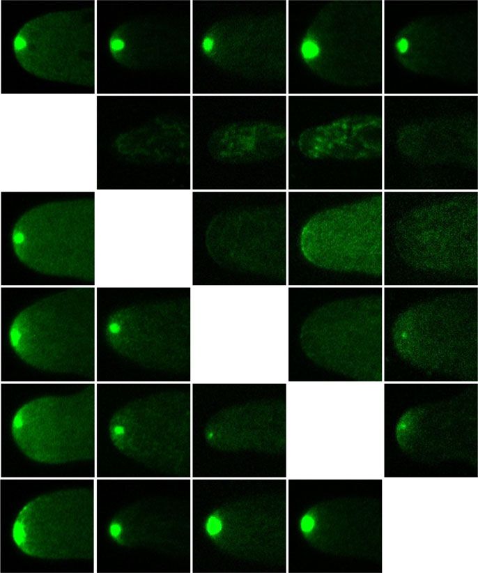
ARTICLE                                                                  NATURE COMMUNICATIONS | https://doi.org/10.1038/s41467-020-16712-9

E
      ukaryotic diversity is manifest in a stunning variety of cel-           for effector binding. Similar interactions and sequence features of
      lular form and function. From unicellular yeasts to multi-              SPA-2/JNS-1 and the mammalian G protein-coupled receptor
      cellular plants and animals, the ability to polarize signaling,         kinase interacting ArfGAP (GIT)/ p21-activated kinase-interact-
cytoskeleton and endomembrane trafficking underlies the fun-                   ing exchange factor (PIX) scaffold suggests an ancestral rela-
damental processes of morphogenesis and differentiation1–3. In a              tionship that predates the fungal/metazoan split. By contrast,
given cell type, polarization appears to involve the combined                 SPZ-1 and LAH-2 appear to have evolved at key junctures leading
action of ancient functional modules, such as Rho GTPases and                 to multicellularity in the Ascomycota.
cytoskeletal elements, operating under the control of lineage-specific
regulatory components4,5. Intensively studied in the yeasts, polar-
                                                                              Results
ization is a sequential process consisting of the selection of a defined
                                                                              Identification of SPZ-1 interacting proteins. To identify N.
cortical site, recruitment of polarity establishment proteins, F-actin
                                                                              crassa SPZ-1 interacting proteins, we employed immunoprecipi-
polymerization, cytoskeleton-dependent recruitment of secretory
                                                                              tation (IP) and mass spectrometry. SPZ-1 co-precipitating pro-
and endocytic machineries, and reinforcement of the polarity axis
                                                                              teins include MYO-5, SPA-2, and an uncharacterized protein,
through scaffold assembly and transport-mediated positive feedback
                                                                              NCU03458. Based on its role in SPA-2 transport (see below), we
(reviewed in refs. 2,3,5).
                                                                              name the latter JANUS-1 (JNS-1) after the Roman god of pas-
   In certain cell types, such as neurons and fungal hyphae, per-
                                                                              sages and transition. An epitope-tagged version of each pulls
sistent vesicle organizing centres assemble at sites of polarization.
                                                                              down the others (Fig. 1a and Supplementary Table 1), suggesting
In the fungi, the Spitzenkörper (SPK) is a phase-dark structure
                                                                              that they form a stable complex. SPZ-1 and LAH-236 also co-
seen by light microscopy at the growing hyphal tip in multi-
                                                                              precipitate (Fig. 1a and Supplementary Table 1). All of these
cellular Ascomycetes (Pezizomycotina) and Basidiomycetes
                                                                              proteins possess conserved predicted coiled-coil domains
(Agaricomycotina)6–8, (reviewed in refs. 9–12). Electron micro-
                                                                              (Fig. 1b), suggesting a basis for their interaction. In keeping with
scopy reveals an actin filament-containing core13, which is likely
                                                                              their co-precipitation, mGFP fusions produced from chromoso-
the phase-dark region, surrounded by ~70–100 nm secretory
                                                                              mal loci all localize to the SPK (Fig. 1c). Deletion strains reveal
vesicles14. In the Pezizomycotina, the core also contains ~40 nm
                                                                              diminished growth rates for Δspz-1, Δspa-2, Δjns-1, and Δlah-2,
micro-vesicles14. Interestingly, macro- and micro-vesicles appear
                                                                              indicating that each plays an important cellular role (Fig. 1d). To
to transport distinct secreted cargoes, suggesting that they com-
                                                                              assess their ability to nucleate complex assembly, we ectopically
prise different types of post-Golgi secretory vesicles15,16.
                                                                              targeted SPZ-1, SPA-2 and JNS-1 to the peroxisome surface (see
   Vesicles are delivered to the SPK through long-range micro-
                                                                              Methods). Each protein is sufficient to recruit GFP-tagged ver-
tubule-mediated transport, followed by short-range transport via
                                                                              sions of the others to the peroxisome membrane (Fig. 1e).
type V myosin motors and actin filaments17–22. The conserved
                                                                              Moreover, each promotes the aberrant accumulation of peroxi-
exocyst complex promotes vesicle fusion with the plasma mem-
                                                                              somes at the SPK, indicating that the complexes formed are
brane and also appears to be required for SPK stability23. Various
                                                                              competent for MYO-5 engagement and transport (Fig. 1f).
polarity-related signaling proteins accumulate at the SPK. These
include the nuclear dbf2-related (NDR) kinase COT-1 and
associated proteins24, and the polarisome protein SPA-225–27. In              Ordered dependencies leading to SPK-localization. We next
budding yeast, the polarisome comprises Spa2, Pea2, and the F-                employed sexual crossing to combine the tagged proteins and
actin polymerization factors Bni1 and Bud628, all of which                    deletion strains in all possible combinations. These data reveal
colocalize at sites of cell growth and are required to maintain               hierarchical relationships leading to SPK-residency (Fig. 2a).
proper cell shape. Recent work has identified an additional                    MYO-5 localizes to the SPK independently of all the other pro-
polarisome component, Aip5, which synergizes with Bni-1 to                    teins. SPZ-1 only depends on MYO-5, while all others depend on
promotes F-actin polymerization29. Spa2 binds to Bni1 and Aip5                SPZ-1. These findings suggest that SPZ-1 acts as a cargo adaptor
through a C-terminal domain29,30, while its conserved Spa                     allowing MYO-5 to transport SPA-2, JNS-1 and LAH-2. Rela-
homology domain (SHD) interacts with MAP kinase compo-                        tionships between SPA-2, JNS-1 and LAH-2 are more complex.
nents28,31 and Rab GTPase activating (GAP) proteins32, sug-                   SPA-2 depends on JNS-1, but JNS-1 retains weak SPK-residency
gesting that it plays a central scaffolding role. Pea2 is required for        in the absence of SPA-2. LAH-2 also retains weak SPK-
SPA-2 tip-localization33. However, the precise function of Pea2 is            localization in the absence of SPA-2 and JNS-1. None of the
unknown. The majority of characterized SPK proteins are con-                  other proteins depend on LAH-2, suggesting that it is a terminal
served in budding yeast34, which does not produce a persistent                component of the localization pathway. Together, these data place
vesicle supply centre. In Neurospora crassa (N. crassa) and                   MYO-5 upstream of SPZ-1, SPZ-1 upstream of SPA-2, JNS-1 and
Aspergillus nidulans, BUD-6/BudA do not colocalize with SPA-2/                LAH-2, and JNS-1 upstream of SPA-2.
SpaA in the SPK25,27. Moreover, Pea2 has not been identified                      Native polyacrylamide gel electrophoresis (native PAGE) was
outside close relatives of budding yeast27. Thus, the role of the             next used to investigate the formation of complexes by SPZ-1
polarisome, and determinants of the SPK’s unique features                     interacting proteins (Fig. 2b and Supplementary Fig. 1). MYO-5
remain unclear.                                                               and SPZ-1 both migrate at approximately 800 kDa and their
   Using the N. crassa model system, we previously identified                  banding patterns are unaffected in the absence of the other proteins
Spitzenkörper-1 (SPZ-1) as a novel coiled-coil SPK protein pre-               (Supplementary Fig. 1). By contrast, the banding patterns of JNS-1
sent in the SPK-containing multicellular Ascomycota, but absent               and SPA-2 are interdependent (Fig. 2b). Both migrate as three
in budding and fission yeasts35. Here, we show that SPZ-1 acts as              bands of approximately 600, 900 and 1100 kDa. When either JNS-1
a cargo adaptor allowing the MYO-5 motor to transport two                     or SPA-2 is absent, the larger species of the other collapse to the
distinct scaffold complexes to the SPK. One consists of Leashin-2             600 kDa band. This molecular weight is significantly higher than
(LAH-2), which is required for SPK-residency of the signalling                the ~100 kDa predicted molecular weights of SPA-2 and JNS-1,
kinase COT-1, and the glycolysis enzyme GPI-1. The other is                   suggesting that they may both form homo-oligomers. To examine
made up of a megadalton hetero-oligomer composed of SPA-2                     this possibility, we used heterokaryons to combine mGFP- and
and Janus-1 (JNS-1). SPA-2 employs its conserved SHD to recruit               HA-tagged versions in the deletion background of the other. For
a novel calponin domain-containing F-Actin effector, CCP-1. The               both proteins, when precipitation is carried out with anti-GFP
SHD NMR structure reveals a conserved surface groove required                 antibodies, HA-tagged versions are co-precipitated (Fig. 2c).

2                 NATURE COMMUNICATIONS | (2020)11:2830 | https://doi.org/10.1038/s41467-020-16712-9 | www.nature.com/naturecommunications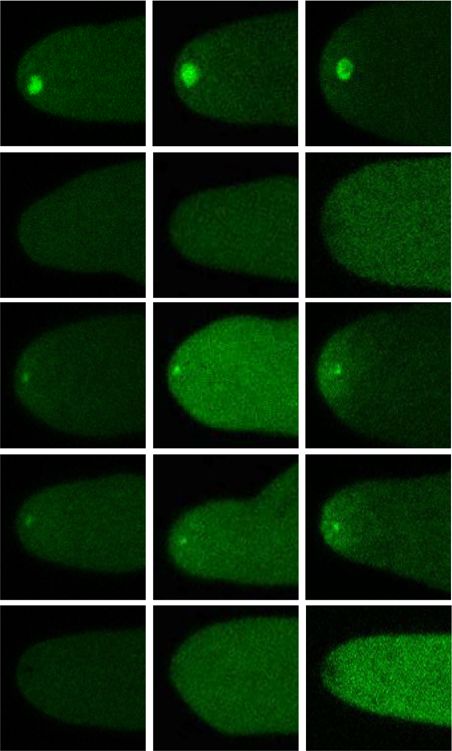
NATURE COMMUNICATIONS | https://doi.org/10.1038/s41467-020-16712-9 ARTICLE
a e SPZ-1-TAperoxisome
MYO-5 SPZ-1 LAH-2
mCh-PTS1 GFP Bright field
SPA-2 JNS-1 JNS-1
Conservation
Coiled coil probability
b SPA-2
1
SPZ-1
0 MYO-5
2 μm
1 1
Coiled coil dimer
JNS-1 JNS-1-TAperoxisome
probability
0 mCh-PTS1 GFP Bright field
0.5
SHD
1
SPZ-1
SPA-2
0
0
0 200 400 600 800 1000 1200 1400
SPA-2
1
LAH-2
(CTD)
MYO-5
0 2 μm
9200 9600 10,000 10,400
SPA-2-TAperoxisome
c d 100
mGFP-tagged proteins mCh-PTS1 GFP Bright field
Growth rate (% of WT)
MYO-5 SPZ-1 JNS-1 80
SPZ-1
60
40 JNS-1
SPA-2 LAH-2
20
MYO-5
10 μm 0 2 μm
e
z-1
s-1
-2
h-2
-5
typ
pa
yo
p
Δjn
Δla
ld
Δs
Δs
Δm
Wi
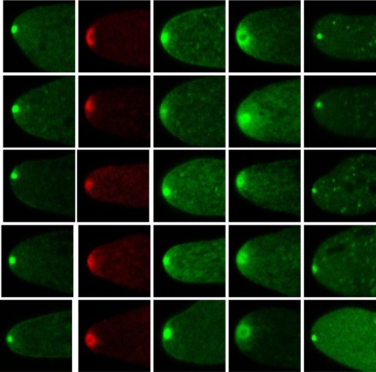
f no ectopic targeting SPZ-1-TAperoxisome JNS-1-TAperoxisome SPA-2-TAperoxisome
10 μm
Fig. 1 Identification of the SPZ-1 interacting proteins. a The indicated proteins were HA-epitope tagged at their endogenous loci and co-precipitating
proteins were identified by mass spectrometry. Double arrows identify mutual co-precipitators. Details are provided in Supplementary Table 1. b Sequence
conservation (grey) and coiled-coil probability (black) of the indicated proteins. The horizontal bar above each graph shows the coiled-coil dimer
probability according to the greyscale shown in the legend. c The localization of chromosomally-encoded mGFP fusion proteins is shown for the indicated
proteins. Dotted white lines show the hyphal outline. Scale bar = 10 μm. d For the indicated strains, the average growth rate is shown as mean values ± SD
(n = 3 independent measurements shown as opaque red dots). e The indicated HA-epitope tagged proteins were fused to a C-terminal peroxisome tail-
anchor (TAperoxisome) and combined through sexual crosses with the indicated mGFP-fusion proteins. Under this condition, SPZ-1, JNS-1 and SPA-2 can
each recruit the others to the surface of the peroxisome. mCherry-PTS1 provides a marker of the peroxisome matrix. Scale bar = 2 μm. f Aberrant
peroxisome accumulation at the SPK shows that ectopically assembled complexes are all competent for transport. The no ectopic panel shows the normal
distribution of peroxisomes. Dotted white lines show the hyphal outline. Scale bar = 10 μm. Source data are provided as a Source Data file.
Together, these data show that SPA-2 and JNS-1 form homo- Two functionally distinct domains in SPZ-1, SPA-2 and JNS-1.
oligomers, which further associate to form two stable hetero- Data presented thus far show that SPZ-1 acts as a cargo adaptor
oligomeric species (Fig. 2b). The intimate relationship between allowing MYO-5 to transport SPA-2/JNS-1 and LAH-2 to the
JNS-1 and SPA-2 is further demonstrated by their dependency on SPK. To investigate the basis for these interactions, we deleted
one-another for pull-down by SPZ-1 (Fig. 2d). For JNS-1, this does discrete regions of SPZ-1 selected based on a combination of
not appear to be consistent with its weak SPK-localization in the coiled-coil prediction and evolutionary conservation (Fig. 3a).
SPA-2 mutant, which is presumably dependent on SPZ-1. Steady- Deletions were constructed at endogenous loci by replacing
state levels of JNS-1 appear to be diminished in the SPA-2 mutant selected coding sequences with an in-frame mCherry-selectable
(Fig. 2b), suggesting that it requires SPA-2 for stability. Thus, in the marker fusion cassette37. The resulting variants were analysed for
absence of SPA-2, JNS-1’s SPK-localization may be the result of its loss-of-function, localization, interaction, and steady-state protein
aberrant interactions. Alternatively, weak binding to SPZ-1 may levels. This analysis identifies two key regions whose absence
not be captured by IP. leads to distinct SPZ-1 loss-of-function phenotypes (Fig. 3b, c and
NATURE COMMUNICATIONS | (2020)11:2830 | https://doi.org/10.1038/s41467-020-16712-9 | www.nature.com/naturecommunications 3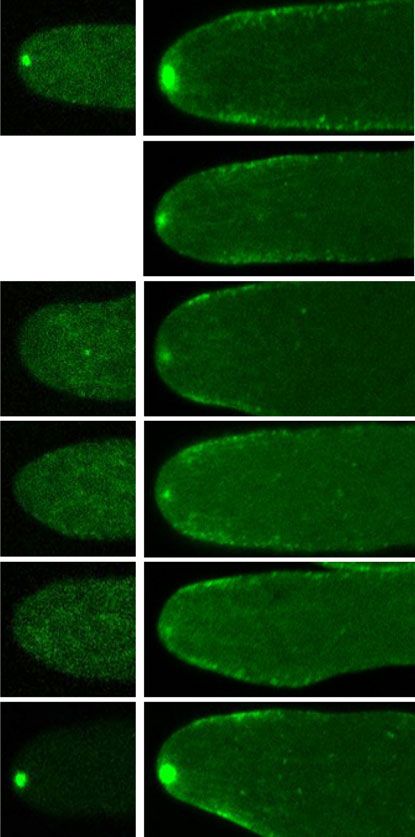
ARTICLE NATURE COMMUNICATIONS | https://doi.org/10.1038/s41467-020-16712-9
a b
-5
-5
mGFP-tagged proteins
Δm 2
Δs 1
Δj 1
-1
-
-
yo
yo
-
pa
pz
pz
ns
Δm
T
T
Δs
Δs
W
W
MYO-5 SPZ-1 JNS-1 SPA-2 LAH-2 kDa
1236
1048 HO-2
HO-1
WT
10 μm 720
480
Δmyo-5 JNS-1 SPA-2
c Δjns-1 Δspa-2
Δspz-1
1
FP
2-G ,
JN ,
S-
SP -2-HA
A
A
GF 1-HA
1-H
2-H
P-
A-
S-
A-
S-
A
SP
JN
SP
JN
kDa
150
Δjns-1 100 Input
150 Pulled
100 Down
α-HA
Δspa-2
d 1
co-IP relative to WT
5
A-2
Z-1
2
S-1
IP target
O-
H-
MY
SP
SP
Δlah-2
JN
LA
Strain
0.5
WT
Δjns-1
Δspa-2 0
Fig. 2 SPK-localization dependencies and complex formation. a The localization of the indicated mGFP-tagged proteins was determined by confocal
microscopy in the indicated strains. Dotted white lines show the hyphal outline. Scale bar = 10 μm. b Native PAGE identifies two stable hetero-oligomeric
complexes formed by JNS-1 and SPA-2 (HO-1 and HO-2). This figure is related to Supplementary Fig. 1. c SPA-2-mGFP precipitates SPA-2-HA in the
absence of JNS-1 (left panels), and JNS-1-mGFP precipitates JNS-1-HA in the absence of SPA-2 (right panels). d SPA-2 and JNS-1 depend on each other for
interaction with SPZ-1. The signal from mass spectrometry is compared to the wild type (WT) precipitation according the scale shown in the legend.
Source data are provided as a Source Data file.
Supplementary Fig. 2a). Deletion of the highly conserved region 3 (Fig. 4b). The conserved direct repeats encode α-2 and α-3 (repeat
coiled-coil domain abolishes the ability of SPZ-1 to precipitate 1), and α-4 and α-5 (repeat 2) (Fig. 4b, c). Conserved residues in
SPA-2/JNS-1 and LAH-2. However, MYO-5 interaction (Fig. 3c) these segments form a surface groove with a partially hydro-
and SPK-localization (Fig. 3b) are retained, albeit at somewhat phobic base and positively charged rims. Antiparallel arrange-
diminished levels, possibly due to diminished steady-state accu- ment of α-3 and α-5 form the groove base, while antiparallel α-2
mulation of this variant as compared to wild-type SPZ-1 (Sup- and α-4 form the rims (Fig. 4b–d). In the mammalian SHD, the
plementary Fig. 2a). By contrast, the coiled-coil region 6 deletion L288A mutation abolishes binding to Piccolo and FAK, but not to
variant retains cargo binding, but abolishes MYO-5 binding GIT39. Sequence alignment shows that L288 is conserved in the
(Fig. 3c) and SPK-localization (Fig. 3b), indicating that it is N. crassa SHD (L133) where it contributes hydrophobicity to the
responsible for motor engagement. These data show that SPZ-1 groove’s base (Fig. 4c, d). The L133A mutation in N. crassa SPA-2
binds to cargos and MYO-5 through distinct coiled-coil domains. leads to a full loss-of-function (Supplementary Fig. 4a), suggesting
JNS-1 and SPA-2 can also be dissected into two discrete that fungal and metazoan SHD domains recruit effectors through
functional regions (Fig. 3d, g and Supplementary Fig. 2b,c). N- a similar structural moiety.
and C-terminal coiled-coil domains of JNS-1 are essential for
SPK-residency (Fig. 3e). However, only the N-terminal domain is
required for hetero-oligomer formation with SPA-2 (Fig. 3f). Identification of a new SHD effector. Initial SPA-2 IP experi-
SPA-2 possesses an essential coiled-coil domain and neighbour- ments did not identify N. crassa SHD effectors. We reasoned that
ing sequences (regions 3 and 4), required for hetero-oligomer this might be due to interference of detergents with binding to the
formation with JNS-1 (Fig. 3h, j) and SPK-residency (Fig. 3i). By SHD groove. Indeed, when detergent is excluded from IP washes,
contrast, deletion of the N-terminal SHD containing region 1 an uncharacterized protein (NCU00277) co-precipitates with
results in loss-of-function, but does not impair SPK-residency or SPA-2 (Fig. 5a, b). NCU00277 contains an N-terminal calponin
hetero-oligomer formation with JNS-1, suggesting its exclusive homology domain and central coiled-coil domain (Fig. 5c). We
association with effector recruitment. IP of variants corroborates therefore named it calponin coiled-coil protein-1 (CCP-1). The
these conclusions: the SPA-2 region 3/4 deletions impair L133A mutation significantly diminishes the ability of SPA-2 to
precipitation with JNS-1, SPZ-1 and MYO-5, while the region 1 bind CCP-1, indicating that they interact through the SHD
SHD deletion retains this ability (Fig. 3j). (Fig. 5a, b). The calponin homology domain and coiled-coil
region are both essential for CCP-1 function and localization to
NMR structure of the SPA homology domain. The SHD was the SPK (Fig. 5c). Calponin homology domains occur in diverse
originally identified as a direct repeat conserved between the actin regulatory proteins40. Thus, we next examined the impact of
mammalian polarity scaffold protein GIT and yeast Spa238. ccp-1 deletion on F-actin distribution. Loss of CCP-1 and its
Because of its conserved and apparently central role in effector upstream regulators, lead to significantly diminished levels of SPK
recruitment, we purified the N. crassa SHD and determined its F-actin (Fig. 5d, e). Interestingly, CCP-1 loss-of-function also
NMR solution structure (Fig. 4 and Supplementary Fig. 3, PDB impairs SPK-incorporation of LAH-2, but does not affect SPK-
ID: 6LAG). The overall fold consists of six alpha-helical segments residency of SPZ-1, SPA-1 or JNS-1 (Fig. 5f). Together, these data
4 NATURE COMMUNICATIONS | (2020)11:2830 | https://doi.org/10.1038/s41467-020-16712-9 | www.nature.com/naturecommunications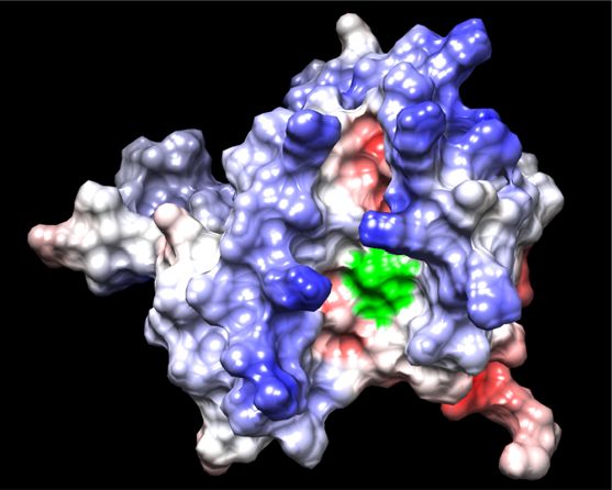
NATURE COMMUNICATIONS | https://doi.org/10.1038/s41467-020-16712-9 ARTICLE
a 0 0.5 1
SPZ-1 coiled-coil dimer probability 1 0.5 0
1 201 401 601 801 1001 1201
Conservation
Variant functionality
Coiled coil
probability
c
5
A-2
S-1
Z-1
2
O-
H-
MY
SP
SP
JN
LA
Strain
r1 r2 r3 r4 r5 r6 r7 WT
1.01 1.13 1.02 0.99 0.18 0.66 spz-1Δr3
–0.57
spz-1Δr6 1.37
b spz-1Δr7 1.63
wt Δr1 Δr2 Δr3 Δr4 Δr5 Δr6 Δr7
0 0.5 1
IP target
SPZ-1 co-IP relative to WT
10 μm
d g
r1 r2 r3 r1 r2 r3 r4 r5
JNS-1 0.04 0.94 0.77 –0.05 SPA-2 –0.01 0.96 0.03 0.59 0.96
1 201 401 601 801 1 201 401 601 801
1 1
0 0
SHD
e JNS-1 f h i SPA-2 j
JN Δr1
r2
r3
r5
SP Δr1
SP Δr2
r3
SP Δr4
5
A-2
Z-1
S-1
O-
1Δ
1Δ
2Δ
2Δ
wt Δr1 wt Δr1 Δr2
JN 1
1
SP -2
2
2
2
MY
S-
S-
S-
S-
A-
A-
A-
A-
A-
SP
SP
JN
A
Strain
JN
JN
SP
SP
WT
kDa
spa-2Δr1
1236
1048
spa-2Δr2
Δr2 Δr3 Δr3 Δr4 Δr5
spa-2Δr3
720
spa-2Δr4
480
10 μm SPA-2 JNS-1 10 μm spa-2Δr5
Fig. 3 Functional dissection of SPZ-1, JNS-1 and SPA-2. a Conservation, coiled-coil, and coiled-coil dimer probability is shown for SPZ-1. The indicated
regions of SPZ-1 (r1–r7) were replaced by an mCherry-selectable marker fusion as described in materials and methods. Functionality of the variants is
scored according to the color scale, where 1 and 0 represent wild type and deletion mutant growth rates, respectively. This figure is related to
Supplementary Fig. 2a. b Localization of the indicated SPZ-1 deletion variants. Dotted white lines show the hyphal outline. Scale bar = 10 μm. c The ability of
the indicated SPZ-1 variants to co-precipitate interacting proteins is compared to full-length SPZ-1 (WT). The signal from mass spectrometry is compared
to WT according the scale shown in the legend. If the signal from mass spectrometry exceeds that of the WT, this value is identified with white numbers.
d Conservation, coiled-coil and coiled-coil dimer prediction for JNS-1. The indicated regions of JNS-1 (r1-r3) were replaced by an mCherry-selectable
marker fusion as described in materials and methods. Variant functionality is indicated as in a. This figure is related to Supplementary Fig. 2b. e Localization
of the indicated JNS-1 deletion variants. Dotted white lines show the hyphal outline. Scale bar = 10 μm. f Native PAGE analysis of SPA-2 mobility in the
indicated JNS-1 deletion variants. g Conservation, coiled-coil and coiled-coil dimer probability for SPA-2. The indicated regions of SPA-2 (r1–r5) were
replaced by an mCherry-selectable marker fusion as described in materials and methods. Variant functionality is indicated as in (a). This figure is related to
Supplementary fig. 2c. h Native PAGE analysis of JNS-1 mobility in the indicated SPA-2 deletion variants. i Localization of the indicated SPA-2 deletion
variants. Dotted white lines show the hyphal outline. Scale bar = 10 μm. j The ability of the indicated SPA-2 variants to co-precipitate interacting proteins is
compared to full-length SPA-2 (WT). The mass spectrometry is quantified as in (c). Source data are provided as a Source Data file.
suggest that CCP-1 participates in transport-mediated positive (GPI-1) is localized to the N. crassa SPK. It also depends on LAH-
feedback to stabilize SPK F-actin. 2. All three proteins depend on SPZ-1, and like LAH-2 show
diminished SPK-residency in the absence of SPA-2 and JNS-1
(Fig. 6a).
Identification of LAH-2 effectors. Data presented thus far
identify an ordered cascade of protein-protein interactions lead-
ing to assembly and SPK-residency of two distinct SPK scaffolds. Scaffold mutants are not impaired in vesicle accumulation.
The SHD domain of the JNS-1/SPA-2 complex recruits the actin Scaffold clients identified here are associated with signaling (COT-
effector CCP-1 to the SPK (Fig. 5). By contrast, the role of LAH-2 1/MOB-2A), actin regulation (CCP-1), and metabolism (GPI-1).
remains unclear. We therefore screened proteins known to reside However, none appears to be directly associated with vesicle
at the SPK for LAH-2 dependency (Fig. 6a). This identified the trafficking. We concluded this study by examining the effect of
polarity-associated NDR kinase, COT-1 and its regulatory bind- scaffold loss-of-function on secretory markers. Remarkably, SPK-
ing partner MOB-2A24 as LAH-2 clients. In unrelated work we localization of markers associated with post-Golgi micro- (CHS-1)
found that the glycolysis enzyme glucose-6-phosphate isomerase and macro-vesicles (GS-1)15 as well as markers of early- (YPT-1)
NATURE COMMUNICATIONS | (2020)11:2830 | https://doi.org/10.1038/s41467-020-16712-9 | www.nature.com/naturecommunications 5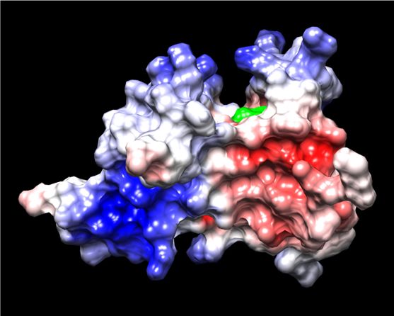
ARTICLE                                                                              NATURE COMMUNICATIONS | https://doi.org/10.1038/s41467-020-16712-9

                                                                                                                                L133
                              a                                                                 c                α2                    α3
                                                                                                           Nc                                         144
                             SPA-2                                                                         Hs                                         299

                                                                                                repeat 1
                                                                                                           Mm                                         299
                              GIT1
                                                                                                            Sc                                        69
                                     0          200        400          600         800                     Sp                                        59
                                                                                                            Ni                                        108
                                                     ARF-GAP                                                Ca                                        62
                                                     ankyrin repeats
                                                                                                           Nc                                         203
                                                     SHD tandem repeat                                                                                363
                                                                                                           Hs

                                                                                                repeat 2
                                                     FAT homology domain                                   Mm                                         363
                                                 0          0.5            1                                Sc                                        119
                                                                                                            Sp                                        104
                                                                                                            Ni                                        158
                                                coiled-coil dimer probability                               Ca                                        112
                                                                                                                 α4               α5
                             b
                                                                        α2
                                                                                                                      α5
                                                α4
                                                                        α5                                                                   α6
                                                                                          90°
                                                                                                                                            α2
                                                                               N′
                                                                                                                                            α1
                                                                             α6                                             α3
                                                              α1
                                           α3
                                                                   C’                                                      α4                    N′

                             d

                                         L133

                                                                                          90°

Fig. 4 Solution structure of the Neurospora crassa SPA-2 SHD (Spa homology domain). a The cartoon shows the position of domains in N. crassa SPA-2
and Human GIT1. Domains are identified according to the legend. b The SHD structure (G84-E211) is shown in a rainbow-colored ribbon diagram. Alpha-
helical segments and N- and C-terminal ends are labeled. The right panel shows the structure after the indicated rotation. The N- to C-terminal directionality
of selected helices is indicated with opaque white arrows. The surface groove whose rims are formed by antiparallel α-2 and α-4 is identified with an asterisk.
c The SHD tandem repeat sequences from representative metazoan and fungal SHDs are aligned. Identical residues are shaded black and conserved residues
grey. Alpha helices are labeled according to colors shown in c. The L133 residue associated with effector binding is identified with a grey arrow. Neurospora
crassa (Nc), Homo sapiens (Hs), Mus musculus (Mm), Saccharomyces cerevisiae (Sc), Schizosaccharomyces pombe (Sp), Neolecta irregularis (Ni) and Candida
albicans (Ca). d The SHD electrostatic surface projection reveals positive charge at the groove’s rims. The perspective of the two panels are the same as
shown in (b). The surface groove is identified with an asterisk. The L133 residue is shown in green. This figure is related to Supplementary Fig. 3.

and late-Golgi/post-Golgi vesicles (YPT-31 and SYN-1), do not                              of vesicles to the SPK (Fig. 7). This has important implications,
appear to be significantly altered in the mutants (Fig. 6b). More-                          suggesting that parallel transport pathways can allow for SPK-
over, the inner and out layers of the SPK also appear to form                              specific regulatory interactions between scaffold clients and vesi-
normally (Fig. 6c). Thus, while MYO-5 is known to be required                              cles (see below).
for delivery of post-Golgi secretory vesicle delivery to the N. crassa                        Scaffold effectors support distinct activities associated with cell
SPK21, the scaffolds identified here do not appear to be directly                           polarity. CCP-1 loss-of-function leads to significantly diminished
related to this process.                                                                   SPK F-actin, suggesting that it acts as part of an transport-
                                                                                           mediated positive feedback loop (Fig. 7a). Such a role for CCP-1
Discussion                                                                                 is consistent with findings in metazoan systems where non-
Cell polarity requires the coordinated regulation of signaling,                            muscle calponin proteins stabilize F-actin networks41,42. COT-1
cytoskeletal dynamics, and membrane trafficking. Protein scaf-                              is a member of the ancient NDR kinase family and plays an
folds act as points of convergence to organize these diverse                               essential role in cell polarity43. By contrast, LAH-2 is non-
activities. However, an overall understanding of these complex                             essential, suggesting that its scaffold function plays a regulatory
systems is lacking. Here, we characterize the assembly of Spit-                            role to promote COT-1 activity. GPI-1 catalyses the second step
zenkörper scaffold complexes and effectors associated with F-actin                         in glycolysis. Its association with the SPK suggests coordination
reinforcement (CCP-1), signalling (COT-1) and metabolism (GPI-                             between metabolism and tip-growth. Resolving the functional
1). SPZ-1 plays a key role as cargo adaptor allowing MYO-5 to                              consequence of this intriguing association will require more work.
promote SPK-residency of the ancient polarisome-related SPA-2/                                Several findings support a model in which overall SPK
JNS-1 complex and the Pezizomycotina-specific LAH-2 scaffolds.                              assembly occurs through the concerted action of independent
Neither appears to be associated with MYO-5 dependent transport                            functional modules. The SPK-residency of LAH-2 depends on

6                   NATURE COMMUNICATIONS | (2020)11:2830 | https://doi.org/10.1038/s41467-020-16712-9 | www.nature.com/naturecommunications
NATURE COMMUNICATIONS | https://doi.org/10.1038/s41467-020-16712-9 ARTICLE
a0 0.5 1 b d
CCP-1 IP
IP target CCP-1 F-actin
L1 -2
A-2
co-IP relative to WT
A
A
33
SP
SP
5
1
2
Z-1
1
O-
P-
WT
A-
S-
MY
CC
SP
SP
JN
Strain Input
WT α-SPA-2
spa-2 L133A Precipitated
Δccp-1
c CCP-1
CHD
Conservation
1
Coiled coil
probability
Δspz-1
0
0 200 400 600
Δ1 Δ2 Δ3
Δjns-1
1 Variant functionality 0
WT Δ1 Δ2 Δ3 Δspa-2
10 μm Δlah-2
10 μm
f MYO-5 SPZ-1 JNS-1 SPA-2 LAH-2
e
WT Δccp-1
9.73 × 10⁻⁶
WT
0 200 400 600 800 1000
Δccp-1 SPK F-actin signal intensity
10 μm
Fig. 5 Identification of the Neurospora crassa SHD binding effector CCP-1. a The ability of the SPA-2L133A mutant to co-precipitate interacting proteins is
compared to full-length SPA-2 (WT). The signal from mass spectrometry is compared to the WT precipitation according the scale shown in the legend.
b CCP-1-mGFP precipitates SPA-2, but interacts weakly with the SPA-2 L133A mutant. Input and precipitated fractions are analyzed by western blotting for
SPA-2. c Conservation, coiled-coil and coiled-coil dimer prediction for CCP-1. The calponin domain is boxed with a dashed line. The indicated regions of
CCP-1 (r1-r3) were deleted using an mCherry-selectable marker fusion as described in Materials and Methods. Variant functionality is scored according to
the color scale where 1 and 0 represent wild type and deletion mutant growth rates, respectively. Lower panels show localization of the indicated deletion
variants. Dotted white lines show the hyphal outline. Scale bar = 10 μm. d CCP-1 tip-localization depends on SPZ-1, JNS-1 and SPA-2, but not LAH-2. The
distribution of F-actin is shown for the indicated strains. Dotted white lines show the hyphal outline. Scale bar = 10 μm. e SPK F-actin signal intensity
(arbitrary units) is quantified for wild type and ccp-1 deletion strains. Data are shown as mean values ± SD (n = 5 independent measurements shown as
opaque red dots). The p-value calculated from 2-sided Student’s t-test is indicated. f LAH-2 depends on CCP-1, but other scaffold components do not.
Localization of the indicated GFP-tagged proteins is shown in wild type and ccp-1 deletion mutant. Dotted white lines show the hyphal outline. Scale bar =
10 μm. Source data are provided as a Source Data file.
SPA-2/JNS-1 (Fig. 2a). However, this relationship is not reci- A number of observations support an ancestral relationship
procal. Loss of the SPA-2 effector CCP-1 also leads to diminished between SPA-2/JNS-1 and the mammalian GIT/PIX polarity
SPK-localization of LAH-2, but does not affect SPA-2/JNS-1. scaffolds. The SHD was first identified as a sequence repeat
Together, these results support a model in which LAH-2’s shared by metazoan GIT and yeast Spa-2 proteins38,39,46. Pre-
dependence on SPA-2/JNS-1 is an indirect consequence of CCP- vious work showed that Spa-2 and GIT recruit a variety of
1’s absence at the SPK and a resulting diminishment in SPK F- effectors through this domain31,32,39. Here we show that as with
actin. This may also be true of BNI-1 which displays similar GIT and PIX39,47, N. crassa SPA-2 and JNS-1 form homo-
dependencies as LAH-2 (Supplementary Fig. 4b). The apparently oligomers (Fig. 2c) which further associate to produce a mega-
normal stratification of SPK secretory markers in scaffold deletion dalton hetero-oligomer (Figs. 2b and 3). The SHD structure
mutants (Fig. 6b, c) further attests to modular SPK assembly and shows how the direct repeat contributes to the formation of an
the independent accumulation of scaffolds and vesicles, as well as amphipathic surface groove (Fig. 4). A mutation previously
differential sensitivity of SPK constituents to levels of SPK F- shown to abolish GIT1 binding to its effectors Piccolo and FAK39,
actin. contributes hydrophobicity to the groove’s base, and the corre-
Scaffold deletion mutants retain residual levels of SPK F-actin sponding mutation in N. crassa SPA-2 disrupts interaction with
(Fig. 5d) and BNI-1 (Supplementary Fig. 4b). The ability of its effector, CCP-1 (Fig. 5a, b). Thus, fungal and metazoan SHDs
Formin proteins like BNI-1 to nucleate F-actin polymerization is employ a similar fold to associate with effectors. An ancestral
well-established44, and in N. crassa BNI-1 is known to be an relationship between SPA-2 and GIT is further supported by
effector of RHO-1 and its nucleotide exchange factor LRG, which significant sequence similarity between the C-terminal GIT focal
localize to the SPK and cell cortex, respectively45. Thus, normal adhesion targeting domain (FAT, also known as paxillin-binding
levels of SPK F-actin appear to be the product of the converging domain) and C-terminal domains of fungal SPA-2 proteins
activities of BNI-1 controlled polymerization, originating from (Supplementary Fig. 6). However, we note that deletion of the
the cell cortex, and CCP-1 mediated stabilization, which depends SPA-2 FAT domain does not produce significant loss-of-function
on MYO-5 transport from sub-apical regions of the hypha. (Fig. 3g), suggesting that it plays a minor role in N. crassa.
NATURE COMMUNICATIONS | (2020)11:2830 | https://doi.org/10.1038/s41467-020-16712-9 | www.nature.com/naturecommunications 7
ARTICLE                                                                     NATURE COMMUNICATIONS | https://doi.org/10.1038/s41467-020-16712-9

                           a                                            b                 Secretory pathway

                                     COT-1      MOB-2A      GPI-1        CHS-1       GS-1       SYN-1      YPT-31      YPT-1

                               WT

                           Δspz-1

                            Δjns-1

                           Δspa-2

                           Δlah-2
                                                           10 μm                                                       10 μm

                           c         CHS-1       GS-1      Merged

                               WT

                           Δspz-1
                                                           10 μm

Fig. 6 Identification of LAH-2 effectors, and the influence of scaffolds on secretory markers. a COT-1, MOB-2A and GPI-1 depend on LAH-2 for tip-
localization. The indicated mGFP fusion proteins were combined with the indicted deletion strains and imaged by confocal microscopy. Note that the partial
SPA-2 and JNS-1 dependence of the LAH-2 effectors is similar to that displayed by LAH-2 itself (see Fig. 2a). Dotted white lines show the hyphal outline.
Scale bar = 10 μm. b Secretory vesicles and organelles do not depend on any of the scaffolds for SPK-localization. The indicated mGFP or mCherry fusion
proteins were combined with the indicted deletion strains and imaged by confocal microscopy. Dotted white lines show the hyphal outline. Scale bar =
10 μm. c Co-expression of a marker of micro-vesicles (CHS-1) and macro-vesicles (GS-1) in the spz-1 deletion mutant reveals localization to inner and outer
layers of the SPK, respectively. Wild-type (WT) accumulation is shown for comparison. Dotted white lines show the hyphal outline. Scale bar = 10 μm.
Source data are provided as a Source Data file.

   JNS-1 possesses two essential coiled-coil domains. The N-                   the dimerization domain interacts with piccolo more strongly
terminal region 1 is required for complex formation with SPA-2,                than monomeric SHD39, suggesting that this may indeed be the
while both region 1 and the C-terminal region 3 are required for               case. Alternatively, the dimeric arrangement of SHDs could be
SPK-localization (Fig. 3d–f). These data suggest that JNS-1 has a              exploited to promote interaction between distinct SHD-bound
primary function in promoting SPA-2 accumulation at the SPK.                   clients. In the future, powerful haploid genetics of N. crassa can
Despite an absence of primary sequence similarity, several com-                be used to investigate how the oligomeric presentation of SHD
monalities suggest that budding yeast Pea2 performs an analo-                  influences its activity.
gous function to JNS-1. Both possess predicted coiled-coil                        In metazoans, GIT and PIX regulate diverse polarity-related
domains, and as with N. crassa SPA-2 and JNS-1, yeast Spa2                     processes that include focal adhesion dynamics and cell migra-
depends on Pea2 for localization to the bud tip33, Spa2 and Pea2               tion49–51, organ development52–54, and synapse formation55 and
co-sediment in a large complex (12S by velocity sedimentation)28,              dynamics56–58. From the perspective of domain organization,
and as with JNS-1, steady-state levels of Pea2 are diminished in               GIT/PIX are significantly more complex than their fungal coun-
the absence of Spa233. Furthermore, in yeast, Spa2 co-precipitates             terparts. In addition to the coiled-coil, SHD and FAT domain, GIT
with Myo2 and depends on it for tip-localization48. Whether the                proteins contain Arf GAP and Ankyrin repeat domains, while PIX
Myo2 interaction is direct or requires Pea2 or another inter-                  contains calponin homology, SH3, Rho GEF, and PH domains59.
mediary remains unclear. Nevertheless, combined with findings                   These domain gains are likely to reflect complexification of
presented here, these observations suggest that SPA-2 proteins                 polarity signaling during metazoan evolution. Despite this, a dual
generally require coiled-coil binding partners and Myosin V                    function in promotion of F-actin polymerization and regulation of
motors for polarized accumulation. Future work can address                     membrane trafficking appears to be a conserved feature of fungal
whether a similar relationship exists with the GIT/PIX complex.                and metazoan polarisome-related scaffolds. In metazoans, reg-
   Surface features and dimensions of the SHD groove suggest a                 ulation of F-actin dynamics is achieved through a complex
potential to bind an amphipathic alpha-helical segment through                 interplay between activation of Rac1 and Cdc42 through the PIX
hydrophobic contacts with the base and charged interactions with               Rho GEF domain and recruitment of the RAC/CDC42 effector
the rim (Fig. 4). GIT forms a parallel dimer through its coiled-coil           PAK via the PIX SH3 domain59,60. By contrast, in N. crassa, F-
domain47. This arrangement positions two SHDs to potentially                   actin regulation is achieved through the SPA-2 client CCP-1
bind clients cooperatively. Interestingly, the GIT SHD linked to               (Fig. 5), while in yeast Bni1, Bud6 and Aip5 are involved28,29.

8                  NATURE COMMUNICATIONS | (2020)11:2830 | https://doi.org/10.1038/s41467-020-16712-9 | www.nature.com/naturecommunications
NATURE COMMUNICATIONS | https://doi.org/10.1038/s41467-020-16712-9 ARTICLE
a
F-actin
macro-vesicle SPZ-1 MYO-5 JNS-1 SPA-2
micro-vesicle
SPZ-1/MYO-5 JNS-1/SPA-2
LAH-2
MYO-5
RABGTP
SPZ-1/MYO-5 SPZ-1/MYO-5
LAH-2 JNS-1/SPA-2
Signalling
[+] SPA-2
LAH-2
[+]
MYO-5 CCP-1
COT-1 RABGTP GYP
? GPI-1
BNI v
-1
RHO t
-1 cys
Exo
LRG
b Opisthokonta SPA-2/GIT ancestor
Fungi Metazoa
Ascomycota JNS-1
SPZ-1
LAH-2
CCP-1
T.melanosporum
D.melanogaster
S.sclerotiorum
A.macrogynus
C.neoformans
D.discoideum
F.oxysporum
S.complicata
T.deformans
S.cerevisiae
N.irregularis
M.musculus
A.fumigatus
Y.lipolytica
C.albicans
C.glabrata
A.gossypii
C.elegans
H.sapiens
A.thaliana
C.cinerea
U.maydis
M.oryzae
S.pombe
R.oryzae
N.crassa
S.roseus
L.bicolor
D.rerio
LAH-2
CCP-1(coiled-coil)
SPZ-1
JNS-1
* Pea2
PIX-1
SPA-2/GIT (SHD)
SPA-2 (FAT)
MYO-5
amino acid substitutions/site
0 1 2 3 4 5
Fig. 7 A blueprint for assembly of the of SPK proteinaceous scaffold. a The cartoon depicts scaffold assembly, MYO-5 dependent transport and effector
recruitment. Parallel transport of secretory vesicles, and scaffold complexes is indicated with dashed orange arrows. Complexes are boxed. Secretory
vesicles and F-actin are depicted according to the legend. The positive feedback from CCP-1 to F-actin is shown with a solid arrow from CCP-1 to an F-actin
filament. The dependency of LAH-2 on CCP-1 is likely to be an indirect consequence of diminished SPK F-actin. This relationship is depicted with a dashed
arrow from an F-actin filament to LAH-2. See discussion for additional information. b Phylogenetic distribution of proteins examined in this study. Filled and
empty squares indicate the presence and absence of the indicated protein, respectively. Arrows to branch nodes identify the likely origin of the indicated
proteins. Note that with a few exceptions, all the SHD-containing proteins co-occur with coiled-coil binding partners (asterisk). Squares with colored
borders identify complex multicellular fungi: the Pezizomycotina (orange), Neolecta (blue) and the Agaricomycotina (purple). Amino acid substitution per
site is shown according to the indicated greyscale, which represents the degree of divergence from the reference Pezizomycotina sequences (see
Methods). In the case of Pea2 and PIX the reference group is the Saccharomycotina and Metazoans, respectively.
Interestingly, mammalian PIX contains a calponin domain, while membrane trafficking events at the plasma membrane62,63. In
in N. crassa and presumably other filamentous fungi, the calponin yeast, the Spa2 SHD binds two related Rab GTPase activating
domain in CCP-1 is recruited through the SHD (Fig. 5). proteins (GAPs), Msb3 and Msb432, which display GAP activity
With respect to regulation of membrane trafficking, in the GIT/ towards Sec464. In budding yeast, Myo2 transports post-Golgi
PIX complex, the GIT ARF-GAP domain61 influences a variety of secretory vesicles through sequential association with activated
NATURE COMMUNICATIONS | (2020)11:2830 | https://doi.org/10.1038/s41467-020-16712-9 | www.nature.com/naturecommunications 9
ARTICLE                                                                    NATURE COMMUNICATIONS | https://doi.org/10.1038/s41467-020-16712-9

Rab GTPases, Ypt31/32 and Sec465,66. Neurospora encodes a                       agar block containing live hyphae was then excised from the growth front and
single Msb homolog, GYP-3, which was not captured in our pull-                  inoculated on a race tube containing VN medium. The growth rate was measured
                                                                                after incubation of race tubes for two days at 30 °C. Epitope and fluorescent protein
down experiments. However, recent work has shown that it                        tags and partial deletions were generated using Marker Fusion Tagging37. The
indeed depends on SPA-2 for SPK-residency67 (Supplementary                      strains employed in this study can be found in Supplementary Data 1.
Fig. 4d). Furthermore, GYP-3 SPK-residency is also abolished in                     For the functional dissection of proteins by MFT, deletion variants were selected
the SPA-2 L133A mutant (Supplementary Fig. 4e), indicating that                 based on a combination of protein sequence conservation35, coiled-coil probability
recruitment occurs through the SHD. N. crassa MYO-5 has been                    and dimer probability as predicted by MultiCoil75. Precise deletion breakpoints are
                                                                                identified in the strain genotypes found in Supplementary Data 1. Ectopic targeting
shown to deliver post-Golgi secretory vesicles to the SPK21.                    to the peroxisomal surface (Fig. 1e,f) was accomplished by appending the tail-
However, scaffolds identified here do not appear to play a role in               anchor from N. crassa PEX-26 to the C-terminus of the indicated proteins. All
vesicle transport (Fig. 6b). These observations lead to a model in              tagged and mutant strains were backcrossed with wild-type strains FGSC465 or
which MYO-5/SEC-4 tethered post-Golgi vesicles are likely to                    FGSC466 to obtain homokaryons. Additional crosses were then conducted to
                                                                                combine various tagged and mutant strains.
encounter high concentrations of SPA-2 clients such as GYP-3                        Images of hyphal tips were obtained using a Leica SP8 inverted confocal
only after delivery to the SPK. Such an arrangement could ensure                microscope with the HCX PL APO 100 /1.40 OIL objective. Primary peripheral tips
that the link between vesicle and motor is only terminated in close             were identified and watched to ensure that they were growing. Images of five tips
proximity to the site of exocytosis.                                            were taken for each strain and representative tips are shown. Images were exported
                                                                                using ImageJ (http://rsb.info.nih.gov/ij/) and converted into figures with Adobe
   In budding yeast, polarisome loss-of-function leads to abnor-                Illustrator (Adobe Illustrator CS6). Quantitation of SPK F-actin in wild type and
mally shaped and enlarged cells, suggesting a primary function in               the ccp-1 deletions strain (Fig. 5e), was conducted using the linescan function of
morphogenesis2. By contrast, in N. crassa polarisome loss-of-                   ImageJ.
function leads to diminished growth rate (Fig. 1d). An analysis of
hyphal shape reveals minor defects in morphology as compared
to loss-of-function in the endocytic component coronin (Sup-                    Immunoprecipitation (IP) and mass spectrometry. To prepare N. crassa
plementary Fig. 7), which affects both growth rate and mor-                     extracts, mycelium was grown in liquid VN and harvested, washed and ground to a
                                                                                fine powder in liquid nitrogen as previously described71. Extracts were prepared by
phogenesis68. Filamentous fungi such as N. crassa can display                   adding 10 ml of IP buffer (20 mM HEPES pH 7.4, 150 mM KOAc, 2 mM MgCl2,
remarkably high rates of tip-growth, which can approach 1 µm                    1 mM DTT, 0.5 mM PMSF and protease inhibitor cocktail (Roche, 04693159001)
per second69 and unlike yeast, morphogenesis and nuclear divi-                  to an equal volume of frozen powder. Extraction was conducted with end-over-end
sion are not coupled. Thus, the polarisome is likely to have                    inversion for 30 min at 4 °C. Insoluble cell debris was subsequently removed by
                                                                                filtration through a 40 µm cell strainer (SPL Life Sciences, 93040). This crude
diverged at the level of effectors to accommodate differing exi-                extract was centrifuged at 16,000 × g for 30 minutes to obtain a soluble supernatant
gencies in hyphal fungi and budding yeast. This idea is further                 fraction for IP. Soluble protein fractions from different strains were diluted to the
supported by work in Ashbya gossypii and N. crassa, where SPA-2                 same concentration prior to IP. DynaBeads were coupled with the anti-HA epitope
localization transitions from a yeast-like tip crescent to a SPK                3FA antibody (Roche, 11867423001) according to the manufacturer’s instructions
sub-apical dot as the rate of hyphal tip growth increases27,70.                 (Invitrogen,14311D). For each sample, 7.5 mg of the coupled beads were added to
                                                                                1 ml of the extract and placed on a roller at 4 °C for 1 h. The beads were then
   To better understand the evolutionary history of SPK com-                    washed once with IP buffer containing 0.1% (w/v) Tween-20, twice with IP buffer,
ponents, we searched for related sequences in representative                    once with the manufacturer’s LWB buffer supplemented with 0.02% (w/v) Tween-
fungal and metazoan proteomes (Fig. 7b). SPA-2 SHD and FAT                      20, and thrice with LWB. Bound proteins were eluted twice according to the
domains identify relatives throughout the fungi and metazoans.                  manufacturer’s instructions, after which, they are combined and dried overnight at
                                                                                room temperature using a vacuum concentrator. Dried proteins were suspended in
JNS-1, Pea2 and PIX distribution suggests that SPA-2/GIT pro-                   sodium dodecyl sulfate (SDS) polyacrylamide gel electrophoresis (PAGE) loading
teins generally require a coiled-coil binding partner. Together,                buffer. IP with GFP-Trap/RFP-Trap M beads (ChromTech, gtma-10/ rtma-10) was
these findings point to an ancestral hetero-oligomeric polarisome                done similarly, except detergents were excluded, and the washed beads and bound
scaffold that predates the divergence of fungi and metazoans. The               proteins were directly suspended in SDS-PAGE loading buffer. Samples were run
                                                                                into the resolving gel by approximately 1 cm after which gels were stained with
analysis further implicates the serial advent of new protein                    Coomassie blue, de-stained and extensively washed with de-ionized water. The
functions in SPK evolution. SPZ-1 maps to the origin of the                     entire Coomassie staining regions was excised and processed for mass spectro-
Ascomycota and is conserved in multicellular filamentous species,                metry. Reduction, alkylation, trypsin digestion and analysis by mass spectroscopy
but was apparently lost in budding and fission yeast as they                     were carried out by the Protein and Proteomics Centre, National University of
                                                                                Singapore. To graphically present the IP results, for each experiment, the Max-
independently underwent simplification35. Its role in SPK com-                   Quant signal intensity76 of each interactant is normalized against the intensity of
plexification is supported by a dual function in transporting                    the IP target to account for differences in IP efficiency. The normalized intensities
ancient (SPA-2/JNS-1), and Pezizomycotina-specific (LAH-2),                      of interactants in the wild-type IP are then used as reference (i.e, set to 1), to which
scaffold components. A number of innovations appear to have                     signal intensities in mutant IPs are compared. These values are represented in a
been fixed prior to radiation of the Pezizomycotina. CCP-1 plays                 linear grayscale with 0 equal to white and 1 equal to black. Values that exceed 1 are
                                                                                indicated numerically on the figure.
a fundamental role in maintaining F-actin in the SPK core and it
acquired a functionally important coiled-coil domain at this
juncture (Fig. 7b). LAH-2 is related to the tether linking Woronin              Native PAGE and Western blotting. To prepare extracts, frozen N. crassa powder
body septal pore-plugging organelles to the cell cortex71. Its role             was added to an equal volume of native sample buffer (Invitrogen, BN2008) and
as scaffold suggests co-option to assume additional functionality               thawed on ice for 30 minutes. This mixture was centrifuged at 16,000 × g, 4 °C for
in the SPK. The advent of several genes required to build Wor-                  30 min and the supernatant was loaded on 3–12% blue native PAGE gels (Invi-
onin bodies also maps to the Pezizomycotina common ances-                       trogen, BN1001BOX). Electrophoresis was carried out at 4 °C at 100 V for 40 min
                                                                                in dark blue cathode buffer, followed by 150 V for 100 minutes in light blue
tor35. Thus, the emergence of new proteins leading to integrated                cathode buffer (Invitrogen, BN2007).
novelties in organelle function and cell polarity appears to have                  For first dimension blotting, proteins separated by native PAGE were
preceded a transition to complex multicellular organization and                 transferred to PVDF membrane with native transfer buffer at 10 V for 10 h at 4 °C.
extensive evolutionary radiation.                                               The membrane was de-stained with 100% methanol before it was blocked in tris-
                                                                                buffered saline (10 mM tris pH 7.4, 150 mM NaCl), 0.1% Triton X-100 (TBS-T),
                                                                                5% nonfat milk (Bio-RAD, #170–6404) for 1 hour at room temperature. The
                                                                                membrane was probed overnight with anti-HA-HRP antibody (#, 1:2500 dilution)
Methods                                                                         in TBS-T, 1% non-fat milk, and imaged with a gel imaging system (ChemiDoc
N. crassa growth, genetic manipulation and microscopy. N. crassa strains were   Touch, Bio-RAD). For second dimension blotting, the whole lane of each sample
grown in synthetic Vogel’s N (VN)72. N. crassa deletion mutants were obtained   was cut from the BNP gel and soaked in SDS loading buffer for 15 minutes at room
from the Fungal Genetics Stock Centre73. Crosses were performed as previously   temperature. This gel strip is then loaded on the top of an SDS-PAGE gel and
described74. Growth rates were measured using the race tube method69. Here,     sealed with 1% agarose in 125 mM Tris pH 6.8. Standard electrophoresis and
conidia were germinated on solid VN medium and grown at 30 °C overnight. An     blotting procedures were carried out following this step.

10                  NATURE COMMUNICATIONS | (2020)11:2830 | https://doi.org/10.1038/s41467-020-16712-9 | www.nature.com/naturecommunications
NATURE COMMUNICATIONS | https://doi.org/10.1038/s41467-020-16712-9 ARTICLE
Solution nuclear magnetic resonance (NMR) analysis. The SHD from N. crassa Homology Domain (SHD) NMR structure is deposited with the Protein Data Bank under
SPA-2 (from glycine-84 to serine-217) was expressed as a HIS-tagged protein in accession number PBD 6LAG [https://doi.org/10.2210/pdb6LAG/pdb] and the assigned
Escherichia coli BL21 in M9 medium (12.8 g/L Na2HPO4.7H2O, 3 g/L KH2PO4, 0.5 chemical shifts are deposited with the Biological Magnetic Resonance Bank under
g/L NaCl, 2 mM MgSO4, 0.1 mM CaCl2, 0.2% 13C-labeled glucose (Cambridge accession number BMRB ID 36299 [https://doi.org/10.13018/BMR36299]. Other data
Isotope Laboratories, Inc, CLM-1396-5) and 0.1% 15N-labelled NH4Cl (Sigma- supporting the findings of this manuscript are available from the corresponding author
Aldrich, 299251)). The protein was purified under native conditions using Ni-NTA upon request. Source data are provided with this paper.
resin (QIAGEN, 30230), followed by gel filtration using Superdex75 column (GE
Healthcare). Fractions containing the SHD protein were concentrated to 2 mM in
10 mM phosphate buffer at pH 6.5 with 1 mM EDTA, 1 mM DTT, 0.05% NaN3 Received: 18 December 2019; Accepted: 14 May 2020;
and 5% D2O. All NMR experiments were performed on a Bruker Avance
800 spectrometer equipped with a cryo-probe at 25 °C. 2D HSQC, 3D HNCA77,
HNCOCA78, MQ-(H)CCH-TOCSY79 and 4D NOESY80 were recorded using
TOPSPIN software (www.bruker.de) without non-uniform sampling scheme. NMR
spectra were processed using NMRPipe v10.881 and analysed using NMRFAM-
Sparky v3.10882. Backbone and side-chain resonance assignments were achieved
using the 4D NOESY-based strategy83. Unambiguous NOEs were obtained from References
three sub-spectra: 13C,15N-edited, 13C,13C-edited, and 15N,15N-edited 4D NOESY. 1. Nelson, W. J. Adaptation of core mechanisms to generate cell polarity. Nature
Distance constraints were obtained from the NOEs assigned, while dihedral angle 422, 766–774 (2003).
restraints of φ and ψ were calculated with TALOS+84 using the assigned chemical 2. Bi, E. & Park, H. O. Cell polarization and cytokinesis in budding yeast.
shifts of Cα, Cβ, N, Hα, and HN. Genetics 191, 347–387 (2012).
The structure was determined using distance and dihedral angle constraints 3. Li, R. & Gundersen, G. G. Beyond polymer polarity: How the cytoskeleton
derived from NOEs and chemical shifts (Supplementary Table 2). Except 15 builds a polarized cell. Nat. Rev. Mol. Cell Biol. 9, 860–873 (2008).
proline residues, five N–H correlations (M1, R25, N34, K35 and G103) were not 4. Wedlich-Soldner, R. & Li, R. Yeast and fungal morphogenesis from an
observable in the 2D HSQC spectrum and thus could not be assigned. The initial evolutionary perspective. Semin. Cell Developmental Biol. 19, 224–233 (2008).
structure calculation was employed with Xplor-NIH85 using the conventional 5. Chiou, J., Balasubramanian, M. K. & Lew, D. J. Cell Polarity in Yeast.
simulated annealing protocol from an extended conformation of SHD. Then the Annu. Rev. Cell Dev. Biol. 33, 77–101 (2017).
best folded models with the lowest total energy were selected for EEFX force-field 6. Girbardt, M. Der Spitzenkörper von Polystictus versicolor (L.). Planta 50,
implicit refinement86 using Xplor-NIH. Both protocols employ the internal 47–59 (1957).
variable module and share the same basic scheme: (i) torsion angle dynamics at 7. Bracker, C. E., Murphy, D. J. & Lopez-Franco, R. in Functional Imaging and
high-temperature (3,500 K) for 15,000 timesteps; (ii) torsion angle dynamics with Optical Manipulation of Living Cells Vol. 2983 (eds Farkas, D. L. & Tromberg,
simulated annealing, where the temperature is reduced from the initial high B. J) 67–80 (SPIE, 1997).
temperature value to 25 K in steps of 12.5 K, for a time of 0.4 ps per temperature 8. Bartnicki-Garcia, S., Bartnicki, D. D., Gierz, G., López-Franco, R. & Bracker,
step (refinement protocol); (iii) 500 steps of Powell torsion angle minimization; and
C. E. Evidence That Spitzenkörper behavior determines the shape of a fungal
(iv) 500 steps of Powell Cartesian minimization.
hypha: a test of the hyphoid model. Exp. Mycol. 19, 153–159 (1995).
In the high temperature stage, experimental dihedral angle restraints and distance
9. Riquelme, M. & Sánchez-León, E. The Spitzenkörper: a choreographer of
restraints were applied with respective force constants of kCDIH = 10 kcal mol−1 rad−2
fungal growth and morphogenesis. Curr. Opin. Microbiol. 20, 27–33 (2014).
and kDIST = 2 kcal mol−1 rad−2. In the simulated annealing stage, kCDIH was set to
10. Riquelme, M. et al. Fungal Morphogenesis, from the Polarized Growth of
200 kcal mol−1 rad−2 and kDIST was increased geometrically from 2 to 30 kcal mol−1
rad−2. The torsionDB statistical torsion angle potential was included with a force Hyphae to Complex Reproduction and Infection Structures. Microbiol. Mol.
constant set to ktDB = 0.02 kcal mol−1 rad−2 in the high temperature stage and Biol. Rev. 82, e00068 (2018).
ramped geometrically from 0.02 to 2 kcal mol−1 rad−2 during simulated annealing. 11. Steinberg, G. Hyphal growth: A tale of motors, lipids, and the spitzenkörper.
A total of 100 EEFX force-field refined structures was calculated and 20 Eukaryot. Cell. 6, 351–360 (2007).
conformers with the lowest total energy were deposited with the PBD ID 6LAG and 12. Harris, S. D. et al. Polarisome meets Spitzenkörper: Microscopy, genetics, and
the assigned chemical shifts are deposited with the BMRB ID 36299. genomics converge. Eukaryot. Cell 4, 225–229 (2005).
13. Howard, R. J. Ultrastructural analysis of hyphal tip cell growth in fungi:
Spitzenkorper, cytoskeleton and endomembranes after freeze-substitution. J.
Bioinformatics. Sequence conservation shown in Figs. 1b, 4a, 5a,d, and 7c was Cell Sci. 48, 89–103 (1981).
determined from the multiple sequence alignments which were constructed using 14. Grove, S. N. & Bracker, C. E. Protoplasmic organization of hyphal tips among
MAFFTv6.24087. For each position in the alignment, the percentage of amino acids
fungi: vesicles and Spitzenkörper. J. Bacteriol. 104, 989–1009 (1970).
in each of the following groups was calculated: aromatic (phenylalanine, tyrosine,
15. Verdín, J., Bartnicki-Garcia, S. & Riquelme, M. Functional stratification of the
tryptophan), polar (serine, threonine, glutamine, asparagine), negatively charged
Spitzenkörper of Neurospora crassa. Mol. Microbiol 74, 1044–1053 (2009).
(aspartic acid, glutamic acid), positively charged (lysine, arginine, histidine),
16. Schultzhaus, Z., Yan, H. & Shaw, B. D. Aspergillus nidulans flippase DnfA is
hydrophobic (alanine, valine, leucine, isoleucine), others (glycine, cysteine,
cargo of the endocytic collar and plays complementary roles in growth and
methionine, proline). The highest percentage will be used as the conservation score
for that particular position. Potential metazoan homologs of fungal SPA-2 were phosphatidylserine asymmetry with another flippase, DnfB. Mol. Microbiol.
identified by searching the human reference proteome (UP000005640) using an 97, 18–32 (2015).
hmm profile constructed from the alignment of fungal SPA-2 sequences (Sup- 17. Brand, A. & Gow, N. A. Mechanisms of hypha orientation of fungi. Curr.
plementary Fig. 7). Phylogenetic distribution, and sequence divergence of proteins Opin. Microbiol. 12, 350–357 (2009).
indicated in Fig. 7b were determined from branch lengths of maximum likelihood 18. Zekert, N. & Fischer, R. The Aspergillus nidulans kinesin-3 UncA motor
trees inferred with RAxML v8.1.1588 using input alignment constructed with moves vesicles along a subpopulation of microtubules. Mol. Biol. Cell 20,
MAFFT v6.240 and trimmed with TrimAl v1.189 as previously described35. Indi- 673–684 (2009).
vidual protein domains shown in Fig. 7b correspond to the following regions of the 19. Taheri-Talesh, N., Xiong, Y. & Oakley, B. R. The Functions of Myosin II and
N. crassa sequences, SPA-2 SHD: amino acids 118–180, SPA-2 FAT: amino acids Myosin V Homologs in Tip Growth and Septation in Aspergillus nidulans.
753–873 (Unitprot ID: V5IQM7). CCP-1 Pezizomycotina-specific coiled-coil PLoS One 7, e31218 (2012).
containing region: amino acids 274-709. All proteomes used in these analyses are 20. Pantazopoulou, A., Pinar, M., Xiang, X. & Peñalva, M. A. Maturation of late
listed in Supplementary Table 3. Golgi cisternae into RabERAB11 exocytic post-Golgi carriers visualized
in vivo. Mol. Biol. Cell 25, 2428–2443 (2014).
21. Ramírez-del Villar, A., Roberson, R. W., Callejas-Negrete, O. A. & Mouriño-
Statistics and Reproducibility. When representative images are shown (Fig. 1c, e, f;
Pérez, R. R. The actin motor MYO-5 effect in the intracellular organization of
2a; 3b, e, i; 5c, d, f; 6a–c and supplementary fig. 4b–e), five independent hyphae
were imaged and one representative image is shown. The full dataset is available in Neurospora crassa. Fungal Genet. Biol. 125, 13–27 (2019).
the Source data file. Error bars in Figs. 1d, 5e and Supplementary Fig. 4a represent 22. Taheri-Talesh, N. et al. The tip growth apparatus of Aspergillus nidulans. Mol.
mean ± SD. Each measurement was made independently at least three times. Biol. Cell 19, 1439–1449 (2008).
23. Riquelme, M. et al. The Neurospora crassa exocyst complex tethers
Spitzenkörper vesicles to the apical plasma membrane during polarized
Reporting summary. Further information on research design is available in growth. Mol. Biol. Cell 25, 1312–1326 (2014).
the Nature Research Reporting Summary linked to this article. 24. Dettmann, A. et al. The NDR Kinase Scaffold HYM1/MO25 Is Essential for
MAK2 MAP Kinase Signaling in Neurospora crassa. PLoS Genet. 8, 1–14
Data availability (2012).
A reporting summary for this Article is available as a Supplementary Information file. 25. Virag, A. & Harris, S. D. Functional characterization of Aspergillus nidulans
The source data underlying Figs. 1c–f; 2a–c; 3b, e, f, h, i; 5b–d, f; 6a–c and Supplementary homologues of Saccharomyces cerevisiae Spa2 and Bud6. Eukaryot. Cell 5,
Figs. 1a, b; 2a–c and 4a–e are provided in the Source data file. The N. crassa SPA-2 Spa 881–895 (2006).
NATURE COMMUNICATIONS | (2020)11:2830 | https://doi.org/10.1038/s41467-020-16712-9 | www.nature.com/naturecommunications 11You can also read