A Snu114-GTP-Prp8 module forms a relay station for efficient splicing in yeast
←
→
Page content transcription
If your browser does not render page correctly, please read the page content below
4572–4584 Nucleic Acids Research, 2020, Vol. 48, No. 8 Published online 20 March 2020
doi: 10.1093/nar/gkaa182
A Snu114–GTP–Prp8 module forms a relay station for
efficient splicing in yeast
Junqiao Jia1,† , Oleg M. Ganichkin1,† , Marco Preußner2 , Eva Absmeier1 , Claudia Alings1 ,
Bernhard Loll 1 , Florian Heyd2 and Markus C. Wahl 1,3,*
1
Freie Universität Berlin, Laboratory of Structural Biochemistry, Takustrae 6, D-14195 Berlin, Germany, 2 Freie
Universität Berlin, Laboratory of RNA Biochemistry, Takustrae 6, D-14195 Berlin, Germany and
3
Helmholtz-Zentrum Berlin für Materialien und Energie, Macromolecular Crystallography, Albert-Einstein-Straße 15,
D-12489 Berlin, Germany
Downloaded from https://academic.oup.com/nar/article/48/8/4572/5810484 by guest on 13 November 2020
Received November 26, 2019; Revised February 26, 2020; Editorial Decision March 05, 2020; Accepted March 10, 2020
ABSTRACT and U6 in the case of the major spliceosome) and many
non-snRNP factors (1,2). Each U snRNP contains a unique
The single G protein of the spliceosome, Snu114, has snRNA, a set of seven common Sm or, in the case of U6,
been proposed to facilitate splicing as a molecular Sm-like (LSm) proteins and a varying number of particle-
motor or as a regulatory G protein. However, available specific proteins (3). For every splicing event, a spliceosome
structures of spliceosomal complexes show Snu114 is assembled de novo from component subunits, catalytically
in the same GTP-bound state, and presently no activated and disassembled after the splicing reaction (1,2).
Snu114 GTPase-regulatory protein is known. We de- Almost all of the comparatively small number of intron-
termined a crystal structure of Snu114 with a Snu114- containing genes in yeast harbor a single intron, and the
binding region of the Prp8 protein, in which Snu114 resulting pre-mRNAs are spliced constitutively (1). In con-
again adopts the same GTP-bound conformation trast, most genes in higher eukaryotes contain more than
seen in spliceosomes. Snu114 and the Snu114–Prp8 one intron and their pre-mRNAs can be spliced in a flexible
manner, giving rise to different mature mRNAs that contain
complex co-purified with endogenous GTP. Snu114
different combinations of exons (alternative splicing) (4).
exhibited weak, intrinsic GTPase activity that was Transitions between functional stages of a splicing cycle
abolished by the Prp8 Snu114-binding region. Ex- are accompanied by massive compositional and conforma-
change of GTP-contacting residues in Snu114, or of tional remodeling of the underlying spliceosomal RNP in-
Prp8 residues lining the Snu114 GTP-binding pocket, teraction networks (1–2,5–6). Constitutive splicing events
led to temperature-sensitive yeast growth and af- in yeast follow a canonical cross-intron spliceosome assem-
fected the same set of splicing events in vivo. Con- bly pathway that is initiated by U1 snRNP recognizing the
sistent with dynamic Snu114-mediated protein inter- 5 -splice site (SS), splicing factor 1 (SF1) binding a con-
actions during splicing, our results suggest that the served branch point sequence in the intron and the U2 aux-
Snu114–GTP–Prp8 module serves as a relay station iliary factors (U2AF) 1/2 recognizing a poly-pyrimidine
during spliceosome activation and disassembly, but tract and the 3 SS, respectively, forming the E-complex.
Subsequently, U2 snRNP replaces SF1 at the branch point
that GTPase activity may be dispensable for splicing.
sequence, giving rise to complex A. The remaining three
snRNPs then join as a pre-formed U4/U6•U5 tri-snRNP to
INTRODUCTION yield the pre-B and, after release of U1 snRNP, the B com-
Precursor messenger RNA (pre-mRNA) splicing entails plex. After disruption of the initially base-paired U4/U6
the removal of non-coding introns and the ligation of di-snRNAs, displacement of U4 and U4/U6-associated
neighboring coding exons and represents a key co-/post- proteins and concomitant recruitment of the non-snRNP
transcriptional gene expression and gene regulatory process NineTeen complex (NTC), the ensuing activated spliceo-
in eukaryotes. Splicing is mediated by the spliceosome, an some (Bact complex) is further rearranged to form the cat-
elaborate RNA-protein (RNP) molecular machine that en- alytically activated spliceosome (catalytic pre-branching B*
compasses five small nuclear (sn) RNPs (U1, U2, U4, U5 complex), which carries out the first step of splicing. Re-
* To whom correspondence should be addressed. Tel: +49 30 838 53456; Fax: +49 30 8384 53456; Email: markus.wahl@fu-berlin.de
†
These authors contributed equally to the paper as first authors.
Present addresses:
Oleg M. Ganichkin, Proteros Biostructures GmbH, Bunsenstraße 7a, D-82152 Martinsried, Germany.
Eva Absmeier, MRC Laboratory of Molecular Biology, Cambridge Biomedical Campus, Francis Crick Avenue, Cambridge CB2 0QH, UK.
C The Author(s) 2020. Published by Oxford University Press on behalf of Nucleic Acids Research.
This is an Open Access article distributed under the terms of the Creative Commons Attribution License (http://creativecommons.org/licenses/by/4.0/), which
permits unrestricted reuse, distribution, and reproduction in any medium, provided the original work is properly cited.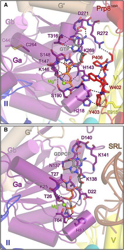
Nucleic Acids Research, 2020, Vol. 48, No. 8 4573

modeling of the resulting catalytic post-branching complex        tial for splicing of at least a subset of pre-mRNAs, but that
C yields the catalytic pre-exon ligation complex C*, which        Snu114-mediated GTP hydrolysis may not be required for
mediates the second transesterification step. The ensuing         splicing.
post-splicing P complex releases the mRNA product as an
mRNP, giving rise to the intron-lariat spliceosome (ILS),
from which the remaining subunits are recycled.                   MATERIALS AND METHODS
   The spliceosomal assembly, activation, catalysis and dis-
                                                                  Cloning, expression and protein purification
assembly cycle is driven and controlled by eight highly con-
served superfamily 2 RNA-dependent NTPases/RNA he-                The MultiBac system (19) was used to express pro-
licases and a single G protein, Snu114 (7,8). While specific      teins of interest in insect cells. Synthetic genes encod-
functions have by now been attributed to the NTPases, the         ing yeast Snu11472-1008 , Prp8132-2413 and Aar2 were cloned
role of the Snu114 GTPase remains enigmatic. Snu114 bears         into modified pIDS-C-Strep, pFL-C-Strep and pIDK-C-
striking resemblance to the prokaryotic/eukaryotic ribo-          Strep vectors, respectively. After Cre-mediated recombina-

                                                                                                                                     Downloaded from https://academic.oup.com/nar/article/48/8/4572/5810484 by guest on 13 November 2020
somal translocases EF-G/eEF2, exhibiting the same five-           tion of the three plasmids, virus and the Snu11472-1008 –
domain arrangement preceded by a Snu114-specific, ca. 125         Prp8132-2413 –Aar2 complex were produced in insect cells
residue, acidic N-terminal region (9). Removal of the N-          as described previously (20). A DNA fragment encod-
terminal region or mutations in other regions of Snu114           ing yeast Prp8SBR was sub-cloned to pFL-N-Strep vector.
in yeast led to a block in splicing before the first catalytic    pIDS-C-Strep-SNU11472-1008 and pFL-N-Strep-PRP8SBR
step (10,11), implicating the protein in spliceosome activa-      were Cre-recombined and used to produce virus and the
tion. Consistent with this notion and with GTP hydrolysis         Snu11472-1008 –Prp8SBR complex in insect cells as described
by Snu114 being important for this process, a D271N muta-         before (20). Site-directed mutagenesis was performed using
tion in the G domain of Snu114, which renders the protein         the QuikChange II XL Site-Directed Mutagenesis Kit (Ag-
XTP-specific, also led to a block of spliceosome activation,      ilent). All constructs were confirmed by Sanger sequencing.
which was partially overcome by addition of XTP and ATP              For purification of yeast Snu11472-1008 –Prp8132-2413 –Aar2
(12). Furthermore, mutations in all EF-G/eEF2-like do-            complex, the cell pellet was re-suspended in lysis buffer I
mains have been identified that exhibit growth defects, led to    (20 mM HEPES-NaOH, pH 8.0, 400 mM NaCl, 2 mM
accumulation of pre-catalytic spliceosomes and/or showed          DTT, 5% (v/v) glycerol, 0.03% (v/v) Triton X-100) sup-
genetic interactions with factors involved in snRNP biogen-       plemented with DNase I, RNase A and avidin (Thermo
esis, snRNP stability, B complex formation or spliceosome         Fisher Scientific) to final concentrations of 0.01 mg/ml,
activation (11–13). Moreover, mutations in the G domain           0.1 mg/ml and 0.01 mg/ml, respectively. Cells were lysed
of Snu114 led to U5 snRNP and U4/U6•U5 tri-snRNP                  by sonication using a Sonopuls Ultrasonic Homogenizer
assembly defects (12,13). Based on these studies and the          (Bandelin), cleared by centrifugation and the supernatant
similarities to EF-G/eEF2, Snu114 has been proposed to            was loaded on a 5 ml StrepTactin sepharose column (GE
act as a mechano-chemical motor that drives RNA–RNA               Healthcare). After washing with lysis buffer, the protein
or RNA-protein rearrangements in the spliceosome (7,11–           complex was eluted using lysis buffer supplemented with 2.5
12). Snu114 has also been implicated in spliceosome dis-          mM D-desthiobiotin (Iris Biotech GmbH). The eluted pro-
assembly (14). However, while spliceosome activation and          tein complex was passed through 5 ml Heparin sepharose
disassembly seem to require GTP-bound Snu114, they did            and Mono Q columns (GE Healthcare). The pooled pro-
not depend on GTP hydrolysis, suggesting that Snu114 may          tein complex was further purified by SEC on a Superose 6
rather act like a classic regulatory G protein that controls      16/300GL column (GE Healthcare) in 50 mM TRIS–HCl,
the activity of the spliceosomal helicase Brr2 depending on       pH 8.5, 300 mM NaCl, 2 mM DTT.
its nucleotide-bound state (14).                                     For purification of yeast Snu11472-1008 –Prp8SBR complex,
   Based on the latter findings, spliceosomal Snu114 regu-        the cell pellet was re-suspended in lysis buffer II (50 mM
latory factors, such as a GTPase activating protein (GAP),        TRIS–HCl, pH 8.0, 300 mM NaCl, 2 mM DTT, 0.05%
a guanine nucleotide exchange factor (GEF) and/or a gua-          (v/v) Triton X-100) supplemented with protease inhibitors
nine nucleotide dissociation inhibitor (GDI), have been pos-      (Roche) and avidin, and lysed by sonication. After centrifu-
tulated (14), but presently the identity of such putative reg-    gation and filtration, the extract was mixed with StrepTactin
ulators is unclear. A prime candidate for such functions is       sepharose beads (Iris Biotech GmbH) in a gravity flow col-
the Prp8 protein, which forms a salt-stable complex with          umn and incubated for 1 hour at 4◦ C. After washing with ly-
Snu114 (15), extensively interacts with Snu114 G and G’ do-       sis buffer II, the complex was eluted using lysis buffer II con-
mains in structures of spliceosomal complexes (16,17) and         taining 2.5 mM D-desthiobiotin and subjected to SEC on
is generally considered a master regulator of the spliceo-        a Superdex S200 16/600 (GE Healthcare) in 10 mM TRIS-
some (18). Here, we have determined the crystal structure         HCl, pH 8.0, 300 mM NaCl, 2 mM DTT.
of yeast Snu114 in complex with an N-terminal fragment               For purification of isolated yeast Snu11472-1008 , cells were
of Prp8 (Prp8 Snu114-binding region, Prp8SBR ) and GTP.           lysed in lysis buffer III (100 mM TRIS–HCl/100 mM Na-
Biochemical analyses showed that Prp8SBR completely ab-           citrate, pH 6.8, 100 mM KCl, 5 mM MgCl2 , 2 mM DTT)
rogated the very low intrinsic GTPase activity of Snu114.         supplemented with protease inhibitors and avidin. The pro-
Based on the structure, we identified Snu114 and Prp8 mu-         tein was captured on StrepTactin sepharose beads and
tations that led to yeast growth defects and affect splicing of   eluted in 100 mM Na-citrate, pH 5.9, 100 mM KCl, 5 mM
the same sub-set of genes in yeast. Our results suggest that      MgCl2 , 2 mM DTT, 2.5 mM D-desthiobiotin. Pooled frac-
stable Snu114 GTP binding supported by Prp8SBR is essen-          tions were subjected to SEC on a Superdex S200 16/600 col-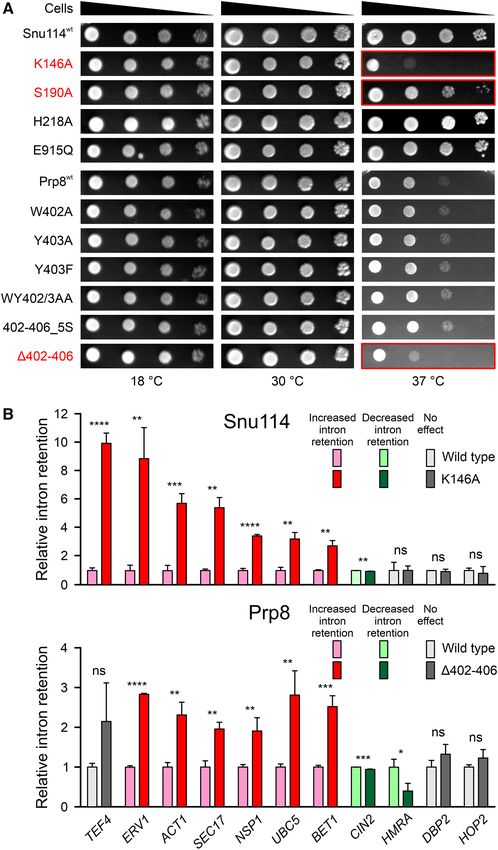
4574 Nucleic Acids Research, 2020, Vol. 48, No. 8
umn in 100 mM Na-citrate, pH 5.9, 100 mM KCl, 5 mM Table 1. Crystallographic data
MgCl2 , 2 mM DTT. Data collection
Wavelength [Å] 0.91841
Limited proteolysis Space group C2
Unit cell parameters
A total of 100 g of purified Snu11472-1008 –Prp8132-2413 – a, b, c [Å] 173.1, 158.6, 110.7
Aar2 complex in 40 l buffer (50 mM TRIS–HCl, pH 8.0,  [◦ ] 116.5
300 mM NaCl, 2 mM DTT) were incubated with 10 l Resolution [Å]a 50–3.1 (3.18–3.10)
chymotrypsin at 0.05 g/l at room temperature for 35 Reflections
Total 207 277 (15 200)
min, and the reaction was stopped by adding PMSF to a Unique 48 253 (3 522)
final concentration of 2 mM. The sample was separated by Multiplicity 4.3 (4.3)
SEC on a Superose 6 Increase 3.2/300 column (GE Health- Completeness [%] 99.3 (99.2)
care) in 20 mM TRIS–HCl, pH 8.5, 500 mM NaCl, 2 mM Mean I/(I) 12.32 (0.92)
Downloaded from https://academic.oup.com/nar/article/48/8/4572/5810484 by guest on 13 November 2020
Rmerge (I) [%](b) 9.7 (151.7)
DTT. Half of the eluted fractions were inspected by sodium
Rmeas (I) [%](c) 11.0 (173.1)
dodecyl sulphate-polyacrylamide gelelectrophoresis (SDS- CC1/2 [%](d) 99.8 (51.6)
PAGE), and bands of interest were analyzed by tryptic mass Refinement
spectrometric fingerprinting. The remainder of the fractions Resolution [Å]a 47.5–3.1 (3.16–3.10)
was separated by SDS-PAGE and blotted on a PVDF mem- Reflections
brane, stained with Ponceau S and fragments of interest Unique 48 232 (4 752)
Test set [%] 5.0 (5.0)
were subjected to N-terminal sequencing. Rwork [%](e) 23.0 (47.7)
Rfree [%](f) 27.1 (50.4)
Crystallographic analysis Contents of A.U.(g)
Non-H atoms 16 330
For crystallization, the Snu11472-1008 –Prp8SBR complex was Protein residues/atoms 2 025/16 249
concentrated to 26 mg/ml. Crystallization was conducted GTP, Mg2+ , SO4 2− atoms 76
Water oxygens 5
by sitting-drop vapor diffusion in 24-well plates. The best
Mean B factors [Å2 ]
crystals grew upon mixing 1 l of protein solution with Wilson 87.2
1 l of reservoir solution containing 50 mM MgSO4 , 200 Model atoms 102.3
mM LiCl, 10% (w/v) PEG 8000. For cryo-protection, the Rmsd(h) from ideal geometry
crystals were transferred to reservoir solution supplemented Bond lengths [Å] 0.003
with 20% (v/v) ethylene glycol, and flash-cooled in liquid Bond angles [◦ ] 0.74
Model quality(i)
nitrogen. Overall score 1.98
Diffraction data were collected on beamline 14.2 of the Clash score 12.1
BESSY II storage ring (Berlin, Germany) at 100 K. All data Ramachandran favored [%] 94.3
were processed with XDS (21,22). The structure was solved Ramachandran outliers [%] 0.8
PDB ID 6TEO
by molecular replacement, using the coordinates of Snu114
derived from the cryo-electron microscopy (cryoEM) struc- a Values in parentheses refer to the highest resolution shells.
ture of a yeast spliceosome (PDB ID: 3JB9) (16). The struc- bR
merge (I) = h i Iih − / h i Iih , in which is the mean
ture was refined by alternating rounds of model building in intensity of symmetry-equivalent reflections h and Iih is the intensity of a
Coot (23) and automated maximum-likelihood restrained particular observation
of h (46).
meas (I) = i Iih − / h i Iih , in which
cR 1/2
refinement in PHENIX (24). Model quality was evaluated h [N/(N-1)]
is the mean intensity of symmetry-equivalent reflections h, Iih is the inten-
with MolProbity (25). Figures were prepared using PyMOL sity of a particular observation of h and N is the number of redundant
(26). Data collection and refinement statistics are provided observations of reflection h (46).
1/2 = ( − ) / ( − ) + ε , in which ε is the
d CC 2 2 2 2 2 2
in Table 1.
mean error within a half-dataset
(46).
work = h Fo − Fc / Fo (working set, no cut-off applied).
eR
fR
Analysis of Snu114-bound nucleotides free is the same as Rwork , but calculated on the test set of reflections ex-
cluded from refinement.
A total of 50 l of purified Snu11472-1008 or -Prp8SBR g A.U.––asymmetric unit.
complex variants at 50 M were incubated for 3 min h Rmsd––root-mean-square deviation.
h Calculated with MolProbity (25).
at 95◦ C, centrifuged at 17 000 g for 5 min, and 20 l
of the supernatants were loaded on a Poroshell 120 EC-
C18 RP-HPLC column (Agilent), equilibrated in 100 mM
K2 HPO4 /KH2 PO4 , pH 6.5, 10 mM tetrabutylammonium incubated in 25 mM TRIS–HCl pH 8.0, 300 mM NaCl,
bromide, 7.5% (v/v) acetonitrile. The samples were chro- 5 mM MgCl2 , 5 mM DTT, 12 mM [␣-32 P] GTP (6000
matographed at 1.5 ml/min. Sample buffer, GDP and GTP mCi/mmol) at 30◦ C for up to 60 min. Reactions were termi-
served as references. nated at various time points by adding 10 l of 40% formic
acid, and 0.5 l of each sample were spotted on a thin-
layer chromatography (TLC) plate and dried. TLC plates
GTPase assays
were placed in a TLC chamber with 0.5 M LiCl in 80%
To monitor steady-state GTPase, 10 l of purified (v/v) 1 M acetic acid/20% (v/v) ethanol. Developed TLC
Snu11472-1008 or -Prp8SBR complex variants at 10 M were plates were air-dried and used to expose phosphorimager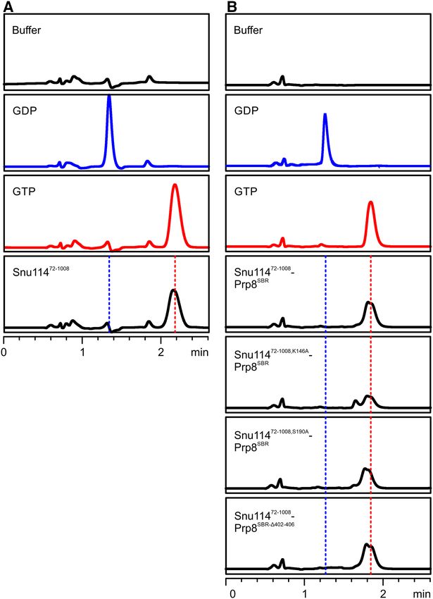
Nucleic Acids Research, 2020, Vol. 48, No. 8 4575
screens for 3 h. Screens were scanned on a Storm phospho- thermo-cycler (Stratagene) according to the manufacturer’s
rimager (GE Healthcare), and spots corresponding to GDP instructions. For qRT-PCR, one or two forward primers
and GTP were quantified using ImageQuant software (GE pairing to the intron or the 5 -exon and reverse primers
Healthcare). pairing to the intron or the 3 -exon were employed (Sup-
plementary Table S1). All experiments were performed as
biological triplicates, qRT-PCRs were performed as techni-
Yeast growth assays
cal duplicates. Intron retention ratios were calculated as the
Yeast growth was assessed after plasmid shuffling. In the amount of intron-containing transcripts versus the amount
YPF8 strain (MAT␣, trp1-Δ1; his3-Δ; ura3-52; lys2-801; of all transcripts, normalized to the wt.
ade2-101; snu114Δ::HIS3 [pRS316/SNU114, ARS, CEN6,
URA3]) (9), the chromosomal SNU114 gene is replaced
by a HIS3 marker, and the strain carries a wild-type (wt) RESULTS
SNU114 gene on a counter-selectable pRS316 plasmid
Structure of a Snu114–Prp8SBR complex
Downloaded from https://academic.oup.com/nar/article/48/8/4572/5810484 by guest on 13 November 2020
with a URA3 marker. YPF8 cells were transformed with
wt SNU114 or mutant snu114 genes on plasmid pRS314 Snu114 adopts virtually identical, GTP-bound confor-
(TRP1), and transformants were selected on complete min- mations in all cryoEM structures of spliceosomes and
imal (CM)-agar plates lacking histidine, tryptophan and of U4/U6•U5 tri-snRNPs available to date. As Snu114
uracil. In the JDY8.06 strain (ura3-52; leu2-3,-112; ade2; bears close resemblance to the ribosomal translocases,
his3-A1; trpl-289; prp8::LEU2 [pY8500/PRP8, ARS, CEN, EF-G/eEF2 (Figure 1A), which undergo large-scale con-
URA3]) (27), the chromosomal PRP8 gene is replaced by a formational changes upon binding to ribosomes, and as
LEU2 marker, and the strain carries a wt PRP8 gene on the Snu114 GTPase has been implicated in U5 snRNP
a counter-selectable pY8500 plasmid with URA3 marker. or U4/U6•U5 tri-snRNP assembly (12,13), we wondered
JDY8.06 cells were transformed with wt PRP8 or mutant whether a wider conformational spectrum may be acces-
prp8 genes on plasmid pJU186 (HIS), and transformants sible to the protein outside its spliceosomal environments.
were selected on CM-agar plates lacking histidine, leucine However, we were unable to crystallize isolated, full-length,
and uracil. Five colonies of each were picked and streaked recombinant yeast Snu114 or a deletion variant lacking
out on 5-fluoro-orotic acid (5-FOA; 0.1% [w/v]) CM-agar the intrinsically unstructured, acidic, N-terminal region
plates lacking histidine and tryptophan or histidine and (Snu11472-1008 ). We thus turned to a Snu114–Prp8–Aar2
leucine. Mutations in the snu114 or prp8 genes of the complex, comprised of Snu11472-1008 , a large fragment of
colonies growing on 5-FOA were confirmed by colony poly- the Prp8 protein lacking the first 131 residues (Prp8132-2413 )
merase chain reaction (PCR) and sequencing. Strains that and Aar2. Aar2 is a U5 snRNP assembly factor in yeast,
survived 5-FOA selection and carried the desired snu114 or which associates with pre-U5 snRNP particles during U5
prp8 mutations were grown overnight in CM medium, di- snRNP biogenesis in the cytoplasm and is replaced by the
luted to an OD600 of 1.0 and 5 l of serial dilutions (1:1, Brr2 RNA helicase in mature U5 in the nucleus (28–32). To
1:10, 1:100, 1:1000) were spotted on CM-agar plates lacking experimentally delineate interacting regions from binding
histidine and tryptophan or histidine and leucine, followed partners that may aid in crystallization, we subjected this
by incubation at 18◦ C, 30◦ C or 37◦ C for 4 days. trimeric complex, which we obtained by co-production of
the proteins via a recombinant baculovirus in insect cells, to
limited proteolysis (Figure 1B and C). Chymotrypsin treat-
In vivo splicing assays
ment gave rise to two sub-complexes, a large C-terminal
Relevant yeast strains were grown in 5 ml of CM medium region of Prp8 (Prp8CTR ) that remained stably bound to
for 24 h, diluted to OD600 of 0.1 in fresh CM medium Aar2 and a region encompassing Prp8 residues 315–555
and grown at 30 or 37◦ C to an OD600 of 2.5. Cells were (Prp8 Snu114-binding region, Prp8SBR ) that formed a com-
harvested by centrifugation (10 min, 3500 × g, 4◦ C). Cell plex with Snu11472-1008 , as revealed by mass spectromet-
pellets were re-suspended in denaturing solution (4 M ric fingerprinting and N-terminal sequencing (Figure 1C).
guanidine thiocyanate, 0.5% (w/v) sarkosyl, 750 mM Na- Prp8CTR -Aar2 most likely corresponds to a complex of
citrate, pH 7.0, 100 mM -mercaptoethanol). Total RNA the two proteins whose structure has previously been de-
was phenol/chloroform/isoamyl alcohol-extracted, precip- termined (32). Prp8SBR is largely congruent with the N-
itated with isopropanol and pelleted by centrifugation (1 h, terminal three quarters of a Prp8 element previously char-
14 000 rpm, room temperature). Extraction and precipita- acterized as a Snu114/Cwc21-interacting domain (SCwid;
tion were repeated once and RNA was washed with 75% Prp8 residues 253–543) (33).
(v/v) ethanol. The RNA pellet was dissolved in water and Based on these observations, we prepared a recombi-
treated with DNase I at 37◦ C for 2 h. After another round of nant Snu11472-1008 –Prp8SBR complex by co-production of
precipitation and washing, the RNA was dissolved in water Snu11472-1008 and Prp8SBR in insect cells (Figure 1D), and
at 1 mg/ml. determined its crystal structure at 3.1 Å resolution. Crys-
Specific cDNA libraries were produced by reverse tran- tals contained two independent copies of the Snu11472-1008 –
scription with primers pairing in the 3 -exons of TEF4, Prp8SBR complex, which were virtually identical (root-
ERV1, ACT1, SEC17, NSP1, UBC5, BET1, HMRA, CIN2, mean-square deviation [rmsd] of 0.66 Å for 997 pairs of C␣
DBP2 orHOP2 (pre-)mRNAs (Supplementary Table S1). atoms). The following discussion refers to both complexes
qRT-PCR was performed using the ABsolute QPCR SYBR in a crystallographic asymmetric unit. For Snu11472-1008 ,
Green Mix (Thermo Fisher Scientific) on a Mx3000P we could trace residues 103–1008, with regions 72–102,4576 Nucleic Acids Research, 2020, Vol. 48, No. 8
Downloaded from https://academic.oup.com/nar/article/48/8/4572/5810484 by guest on 13 November 2020
Figure 1. Structural overview. (A) Domain organization of yeast Snu114, eEF2 and the N-terminal portion of Prp8 (drawn to scale). NTR, N-terminal
region; Ga, first part of G domain, G’, G’ domain; Gb, second part of G domain; II/III/V, domains II/III/V; IVa, first part of domain IV; IVb, sec-
ond part of domain IV; NLS, nuclear localization signal; SCwid, Snu114/Cwc21-interaction domain; SBR, Snu114-binding region. Domain coloring is
maintained in the following figures. (B) SDS-PAGE analysis of an analytical gel filtration run of a recombinant Prp8132-2013 –Snu11472-1008 –Aar2 com-
plex. (C) SDS-PAGE analysis of an analytical gel filtration run of chymotrypsin-treated, recombinant Prp8132-2013 –Snu11472-1008 –Aar2 complex. Elution
positions of two sub-complexes (Prp8CTR –Aar2 and Snu11472-1008 –Prp8SBR ) are indicated below the gel. CTR, C-terminal region. (D) SDS-PAGE anal-
ysis of an analytical gel filtration run of a recombinant Snu11472-1008 –Prp8SBR complex. (E) Comparison of the overall structures of a Snu11472-1008 –
GTP–Prp8SBR complex (left) and of yeast eEF2 (right; PDB ID: 1N0V; (47)) after superposition of the G domains. Proteins are shown as cartoons
with helices as cylinders and sheets as arrows. Mg2+ -GTP in the Snu11472-1008 –GTP–Prp8SBR complex is shown as spheres colored by atom type. Car-
bon, gray; nitrogen, blue; oxygen, red; phosphorus, orange; magnesium, green. N/C, N-/C-termini of Prp8SBR . (F) Ribbon plots comparing the con-
formation of Snu11472-1008 in the isolated Snu11472-1008 –GTP–Prp8SBR complex (blue) with corresponding Snu114 regions in structures of yeast (left)
or human (right) spliceosomal complexes. Yeast complexes/PDB IDs/references: U4/U6•U5 tri-snRNP/5GAN/(17); pre-B complex/5ZWM/(48); B
complex/5ZWO/(48); Bact complex/5GM6/(49); B* complex/6J6G/(50); C complex/5GMK/(51); C* complex/5WSG/(52); P complex/6BK8/(53);
ILS/5Y88/(54). Human complexes/PDB IDs/references: U4/U6•U5 tri-snRNP/6Q6W/(55); pre-B complex/6QX9/(55); B complex/6AHD/(56); Bact
complex/6FF4/(57); C complex/5YZG/(58); C* complex/5MQF/(59); P complex/6ICZ/(60); ILS/6ID1/(60).Nucleic Acids Research, 2020, Vol. 48, No. 8 4577
520–531, 691–704 and 977–984 lacking interpretable elec-
tron density. The modeled portion of Prp8SBR encompasses
residues 367–533, with 52 N-terminal, 26 C-terminal and in-
ternal residues 416–419 and 435–449 lacking clear electron
density.
Snu11472-1008 contains five EF-G/eEF2-like domains, i.e.
G, G’, II, III, IV and V, with the G’ domain inserted into
the G domain, and domain V intervening between the IVa
and IVb sub-domains (Figures 1A and E). It adopts a com-
pact conformation, in which the N-terminal G and G’ do-
mains form a globular head that is cradled in an array of
domains II, III and V, and in which the split domain IV
forms a pedestal at the bottom (Figure 1E). Cys264 and
Downloaded from https://academic.oup.com/nar/article/48/8/4572/5810484 by guest on 13 November 2020
Cys442 in the G domain of one Snu11472-1008 molecule in an
asymmetric unit are partially engaged in a disulfide bridge
that links the N- and C-terminal parts of the G domain
(Figure 2A); the corresponding disulfide bridge is broken
in the other Snu11472-1008 molecule, most likely due to radi-
ation damage. Prp8SBR exhibits an extended, loosely twisted
conformation that lacks a globular fold, suggesting that the
fragment would be intrinsically disordered in isolation (Fig-
ure 1E). The N-terminus of Prp8SBR resides on one side of
the IVb sub-domain of Snu114. The protein then meanders
along the bottom part of domain V and along domain III
toward the G nucleotide-binding pocket of the Snu114 G
domain. Residues 402–410 stretches along the open side of
the nucleotide-binding pocket (Figure 2A) and the follow-
ing residues 411–471 form a long loop, previously referred
to as the Prp8 lasso (16), which encircles a protruding re-
gion of the G’ domain (Figure 1E). Prp8SBR then returns
toward domain V of Snu114, forming a small helical bundle
at one tip of the G’ domain and ends in an ␣ helix that runs
along the top of domain V (Figure 1E). The C-terminal 43
residues of Prp8SBR adopt a different conformation in the
U4/U6•U5 tri-snRNP and in spliceosomes due to alterna-
tive interactions with other parts of Prp8 (Supplementary
Figure S1). Whether the conformation seen in the present
crystal structure is an artifact due to the crystallization of
a proteolyzed fragment or whether it may exist in another
context, such as during U5 snRNP assembly, remains to be
seen.
Contrary to our initial expectation, Snu11472-1008 in com-
plex with Prp8SBR closely resembles the structures of the
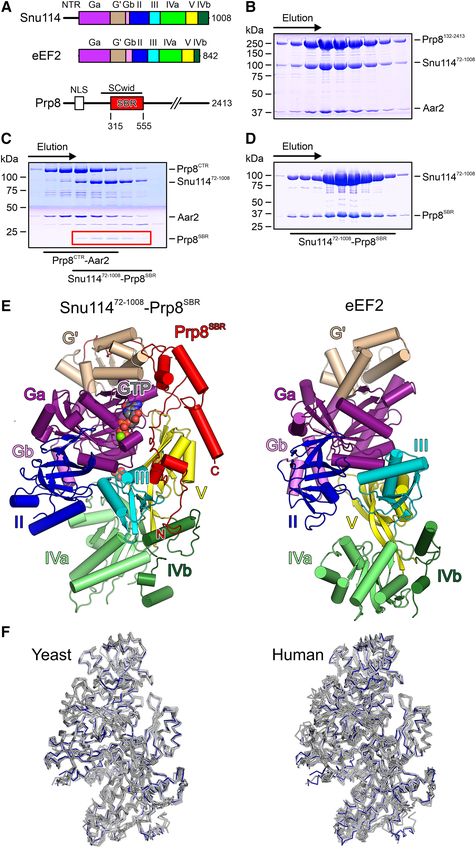
corresponding Snu114 regions in available cryoEM struc- Figure 2. GTP binding pocket. (A) Mg2+ -GTP bound at Snu11472-1008 in
tures of yeast or human spliceosomal complexes, (rmsd val- complex with Prp8SBR . (B) Mg2+ -GDPCP bound at EF-G on the ribo-
some (PDB ID: 4WPO; (34)). Domains are labeled as in Figure 1A. GTP
ues of about 1 Å for about 900 pairs of C␣ atoms; Figure and relevant protein residues are shown as sticks colored by atom type
1F). Thus, Snu11472-1008 in complex with Prp8SBR seems to (as in Figure 1E; except carbon, as the respective protein/domain). Green
represent a rigid structural building block of the spliceo- spheres, Mg2+ ions. Dashed lines, hydrogen bonds or salt bridges. SRL,
some. sarcin-ricin loop.
Prp8SBR stabilizes Snu11472-1008 in a non-hydrolytic confor-
(34) (Figure 2A and B). G proteins employ up to five con-
mation
served sequence motifs (G1–G4) for G nucleotide binding
In the crystal structure of the isolated Snu11472-1008 – (35). In Snu114, the Watson–Crick flank of the nucleobase
Prp8SBR complex, the electron density clearly indicated that is recognized by the side chains of D271 (G4 motif) and
both Snu114 molecules in an asymmetric unit were bound T316 (G5 motif), and T316 also forms a hydrogen bond
to GTP and a metal ion, most likely Mg2+ , although no nu- with the N7 position of GTP. The ribose O4 is bound by
cleotide had been added during purification or crystalliza- K269 (G4 motif). The phosphate groups are predominantly
tion. Mg2+ -GTP is bound in a similar manner as seen for recognized by residues from the G1 motif/P-loop, with the
Mg2+ -GDPCP bound to the bacterial ribosomal translo- backbone NH groups of H143, S144, G145, T147 and S148
case EF-G on the 70S ribosome in the pre-translocated state forming hydrogen bonds to the ␣,  and ␥ -phosphates. Ad-4578 Nucleic Acids Research, 2020, Vol. 48, No. 8
ditionally, the S144 side chain hydrogen bonds with the - the wt Snu11472-1008 –Prp8SBR complex (Figure 3A and B).
phosphate, the side chain of K146 engages in ionic inter- These observations corroborate the idea that Snu11472-1008
actions with the  and ␥ -phosphate groups, the side chain has a strong intrinsic tendency to adopt a conformation that
hydroxyl of T147 hydrogen bonds with the -phosphate and stably traps bound GTP. Based on our structure, stable GTP
the side chain of S148 forms a hydrogen bond with the binding by Snu11472-1008 seems to be further supported by
␣-phosphate. The ␥ -phosphate is additionally hydrogen- Prp8SBR .
bonded to the backbone NH group G217 (G3 motif/switch To test the effect of Prp8 on Snu114 GTPase activity,
2) as well as the backbone NH of S190 (G2/switch 1). The we monitored Snu11472-1008 GTPase activity in isolation
Mg2+ ion is coordinated by the  and ␥ -phosphates, as well or in complex with Prp8SBR . To this end, we incubated
as by the side chains of T147 (G1 motif/P-loop) and S190 Snu11472-1008 alone or in complex with Prp8SBP for ex-
(G2 motif/switch 1). tended times at 30◦ C in the presence of ␣-[32 P]-GTP, and
Previous studies had established that a conserved histi- monitored product nucleotides by TLC. Consistent with
dine in translation factor GTPases (H87 in EF-G) serves to non-hydrolyzed GTP co-purifying with Snu11472-1008 or
Downloaded from https://academic.oup.com/nar/article/48/8/4572/5810484 by guest on 13 November 2020
position and polarize a catalytic water molecule for GTP hy- Snu11472-1008 –Prp8SBR complexes, very weak GTP hydroly-
drolysis (36). On the ribosome, EF-G H87 is brought into sis was detectable under these conditions with Snu11472-1008
its hydrolysis-supporting conformation by the sarcin/ricin alone (Figure 4A). The weak intrinsic GTPase activity was
loop, a conserved element of 23S ribosomal RNA that completely abrogated by Prp8SBR (Figure 4B). Michaelis–
forms part of the ribosome’s GTPase-activating center (Fig- Menten titrations revealed a Km of 578.4 M and a
ure 2B). H218 is the equivalent residue in Snu114. Simi- kcat of 1.012 * 10−3 s−1 for GTP hydrolysis by isolated
lar to the situation in structures of spliceosomal complexes Snu11472-1008 (Figure 4C).
(17), H218 in the isolated Snu11472-1008 –Prp8SBR complex is Conversion of Prp8 Y403 to a phenylalanine or ala-
rotated away from the GTP ␥ -phosphate, hydrogen bond- nine, exchange of Snu114 E915 (contacting Prp8 Y403)
ing with the hydroxyl group of Prp8SBR Y403 (Figure 2A). to a glutamine, asparagine or alanine, deleting the
W402 of Prp8SBR lies on top of the Snu11472-1008 switch I Prp8 segment spanning the Snu114 nucleotide binding
region, helping to anchor the neighboring Prp8SBR Y403 in pocket (Prp8402–406 ) or replacing it with five serines
front of the nucleotide-binding pocket. The conformation (Prp8402–406 5S ), weakening the anchoring of this segment on
of Y403 is additionally stabilized by E915 from domain V Snu114 by exchange of Prp8 W402 (Prp8W402A ) or of W402
of Snu11472-1008 (Figure 2A) Thus, Prp8SBR may stabilize a and Y403 (Prp8WY402/3AA ), or short-circuiting the neigh-
non-hydrolytic conformation in Snu11472-1008 . boring Prp8 lasso structure (Prp8421–468 ) would all be ex-
pected to interfere with the apparent Prp8SBR -mediated in-
Prp8SBR stabilizes GTP-bound Snu11472-1008 and inhibits its hibitory mechanism targeting Snu11472-1008 H218, either by
low, intrinsic GTPase activity directly modulating Prp8 contacts to H218 or by weaken-
ing neighboring Snu11472-1008 –Prp8SBR interactions. How-
To investigate the importance of Snu11472-1008 and Prp8SBR ever, none of the Snu11472-1008 or Prp8SBR variants in the
residues in stable GTP anchoring, we used structure-guided, context of the Snu11472-1008 –Prp8SBR complex led to in-
site-directed mutagenesis to alter residues in Snu11472-1008 creased GTP hydrolysis (Figure 4D). These results suggest
or the Snu11472-1008 –Prp8SBR complex that potentially af- that Prp8SBR inhibits Snu11472-1008 GTPase activity on sev-
fect Snu11472-1008 –GTP or Snu11472-1008 –Prp8SBR interac- eral levels, including by sequestering H218 but also pre-
tions. While isolated wt Snu11472-1008 could be produced sumably by restricting hydrolysis-relevant conformational
soluble in insect cells and purified, all tested Snu11472-1008 changes in Snu11472-1008 .
variants (H218A, S190A, K146A, T147V, E915Q/A/D)
formed insoluble aggregates when produced alone in in-
Effects of Snu114 and Prp8 variants on splicing
sect cells. This observation is consistent with the idea that
the affected residues are required for stable GTP binding We further investigated the possible functional role of
to Snu11472-1008 and that bound GTP is required to main- the Snu114–GTP–Prp8 interaction network characterized
tain a stable fold in Snu11472-1008 . Interestingly, several of above. To this end, we introduced plasmids that guided the
the tested Snu11472-1008 variants (K146A, S190A) could expression of full-length Snu114 or Prp8 variants into yeast
be produced in soluble form together with Prp8SBR , in- strains, carrying a sole copy of the SNU114 or PRP8 wt
dicating that Prp8SBR stabilizes the fold of Snu11472-1008 . genes on counter-selectable plasmids, and monitored effects
Similarly, Prp8SBR variants (wt, Y403A, Y403F, WY402- on yeast growth and splicing after eliminating the wt pro-
403AA, 402–406, 402–406 5S and 421–468) could not teins. Snu114T147A (exchange of a residue contacting the
be produced alone, but could be made in complex with GTP -phosphate) and Prp8421–468 (bearing a deletion of
Snu114. Thus, Snu114 presumably protects Prp8SBR and the lasso-like region interacting with Snu114) did not sup-
variants from degradation. port cell viability. Snu114K146A (exchange of a residue con-
We purified wt Snu11472-1008 alone and Snu11472-1008 tacting the GTP  and ␥ -phosphates), Snu114S190A (ex-
variants in complex with Prp8SBR variants and identi- change of a residue that coordinates Mg2+ and contacts the
fied the bound nucleotide by reverse-phase ion-pair high- GTP ␥ -phosphate) and Prp8402–406 (Snu114 H218-binding
performance liquid chromatography (RP-HPLC). In all region) variants led to growth defects at 37◦ C (Figure 5A).
preparations, GTP was the only nucleotide detectable (Fig- Strains expressing the Snu114H218A variant, in which the
ure 3A and B). Furthermore, all mutant complexes con- potential catalytic H218 is exchanged, or Snu114E915Q , in
tained GTP at comparable levels as wt Snu11472-1008 or which a residue in the H218 interaction network is al-Nucleic Acids Research, 2020, Vol. 48, No. 8 4579
Downloaded from https://academic.oup.com/nar/article/48/8/4572/5810484 by guest on 13 November 2020
Figure 3. Snu114-bound G nucleotides. (A and B) RP-HPLC analysis of nucleotides bound to Snu11472-1008 (A) and to the indicated Snu11472-1008 –
Prp8SBR complexes (B). Snu11472-1008 and all complexes co-purify with GTP. Buffer, GDP (blue) and GTP (red) control runs are shown on the top.
Different retention times for nucleotides in (A) and (B) are due to different buffers used for the preparation of isolated Snu11472-1008 and for Snu11472-1008 –
Prp8SBR complexes.
tered, showed no obvious growth differences compared to the Brr2 helicase (38). Besides canonical introns, the col-
the parent strain. Consistently, lack of a growth pheno- lection includes pre-mRNAs with unusual 5 SS (HOP2),
type upon H218 exchanges was noted before (17). Simi- BP (ERV1, CIN2) or 3 SS (SEC17, UBC5), a pre-mRNA
larly, Prp8Y403A , Prp8Y403F , Prp8W402A , Prp8WY402/3AA or with an unusually short distance between the BS and 3 SS
Prp8402–406 5S variants, in which residues contacting Snu114 (HMRA) and a pre-mRNA with an unusually long intron
H218 or the region running along the GTP-binding pocket (DBP2). Relative to the parent strains, intron retention of
of Snu114 are altered, did not lead to altered growth under seven pre-mRNAs (TEF4, ERV1, ACT1, SEC17, NSP1,
the conditions tested. UBC5, BET1) was strongly increased in the snu114 K146A
To further delineate the basis of the growth defects ob- strain, intron retention was to a small but significant ex-
served at 37◦ C with Snu114K146A and Prp8402–406 , we tent decreased in CIN2, while splicing of the other pre-
tested the splicing of a set of 11 pre-mRNAs (TEF4, mRNAs was unaffected (Figure 5B, top). Strikingly, in the
ERV1, ACT1, SEC17, NSP1, UBC5, BET1, HMRA, CIN2, prp8 402–406 strain, intron retention for the same subset
DBP2 and HOP2) by quantitative real time (qRT) PCR of pre-mRNAs was also increased, albeit to a smaller extent
in snu114 K146A and prp8 402–406 strains, grown at and intron retention was decreased again for CIN2 and, in
the non-permissive temperature, compared to the parent addition, for HMRA pre-mRNA (Figure 5B, bottom). For
strains. These pre-mRNAs have previously been used to the same strains grown at a permissive temperature (30◦ C),
assess splicing defects originating from yeast prp8 muta- or for strains expressing Snu114H218A or Prp8Y403A , which
tions that are linked to retinitis pigmentosa in humans (37) did not show any growth defects, no effect on splicing of
or from mutations that lead to N-terminal truncations in selected pre-mRNAs (ERV, NSP1) was observed (Supple-4580 Nucleic Acids Research, 2020, Vol. 48, No. 8
of splicing events, possibly affecting all splicing events that
involve introns with canonical features.
DISCUSSION
Here, we have delineated a crystal structure of a large
portion of the yeast Snu114 protein, containing all EF-
G/eEF2-homologous regions, in complex with GTP and an
intrinsically disordered Snu114-binding region of the Prp8
protein, investigated the nucleotide binding and hydrolysis
activities of wt Snu114 alone and in complex with Prp8SBR ,
and of variants of this complex bearing exchanges in Snu114
Downloaded from https://academic.oup.com/nar/article/48/8/4572/5810484 by guest on 13 November 2020
or Prp8 residues that are expected to weaken Snu114–GTP
or Snu114–Prp8 interactions. Moreover, we have monitored
growth and splicing in yeast strains that harbored corre-
sponding Snu114 or Prp8 variants as the only variants of
the proteins.
Our structural analysis showed that Snu114 only com-
plexed to Prp8SBR adopts the same GTP-bound confor-
mation as has so far been observed in all structures
of Snu114/Prp8-containing spliceosomal complexes. Thus,
Snu114–GTP–Prp8SBR seems to constitute a rigid build-
ing block of the spliceosome, except perhaps for the C-
terminal 43 residues of Prp8SBR , which change conforma-
tion in the isolated complex compared to the situation in
spliceosomes. The conformational rigidity of the Snu114–
GTP–Prp8SBR unit may be additionally supported by a
disulfide bridge that connects two parts of the Snu114 G do-
main and thereby provides intra-molecular cross-strutting.
Although the reducing environment of the nucleus disfa-
vors disulfide bridge formation, disulfide bridges have been
observed in structures of nuclear proteins (39) and have
been implicated in the function of some nuclear factors (40).
The lack of major structural changes in the Snu11472-1008 –
Prp8SBR sub-complex when studied outside the spliceosome
is in stark contrast to conformational changes observed in
the closely related translation factors EF-G/eEF2 (41,42),
suggesting that Snu114 in the spliceosome exhibits a differ-
Figure 4. GTPase activities. (A and B) TLC monitoring time courses of ent mode of action or function compared to EF-G/eEF2
GTP hydrolysis by Snu11472-1008 (A) or by the Snu11472-1008 –Prp8SBR on the ribosome.
complex (B). (C) Michaelis–Menten titrations of GTP hydrolysis by
Snu11472-1008 or in the Snu11472-1008 –Prp8SBR complex. Inset––Km and We find very low intrinsic GTPase activity associated
vmax of GTP hydrolysis by Snu11472-1008 . Prp8SBR leads to complete inhi- with Snu114, as is the case for many G proteins includ-
bition of the low intrinsic GTPase activity of Snu11472-1008 . (D) Relative ing EF-G/eEF2 (43). Still, this activity could be reliably
GTPase rates of Snu11472-1008 and of the indicated Snu11472-1008 –Prp8SBR quantified and we unequivocally showed that it is com-
complexes. Values in (C and D) represent means ± SD for three indepen-
dent experiments.
pletely abrogated in the presence of Prp8SBR . The lat-
ter observation formally establishes Prp8 as a GTPase-
inhibiting protein, and thus to the best of our knowledge
as the first GTPase-regulatory factor, of Snu114. As the
mentary Figure S2). While we did not observe a particular conformation and nucleotide-bound state of the Snu114–
feature in the introns that correlates with increased reten- GTP–Prp8SBR sub-complex remains constant in all Snu114-
tion upon altering Snu114 or Prp8, the four targets that do containing yeast or human spliceosomal complexes struc-
not show increased intron retention (CIN2, HMRA, DBP2, turally analyzed to date, Prp8 may constitute the sole
HOP2) all exhibit unusual, albeit diverse (unusual BS, short Snu114 GTPase-regulatory protein in the spliceosome. Pro-
BS-3 SS distance, long intron, unusual 5 SS, respectively) teins or RNAs that serve as Snu114 GTPase-activating
features. On the one hand, these analyses indicate that al- protein, G-nucleotide exchange factor of G-nucleotide ex-
tered pre-mRNA splicing elicited by certain Snu114 or Prp8 change inhibitor, as have been found to regulate the activ-
variants underlies growth phenotypes observed in corre- ity cycles of small G proteins, may not exist in the spliceo-
sponding mutant strains. On the other hand, they clearly some. It remains to be seen whether other proteins or RNAs
suggest that the Snu114–GTP–Prp8 interaction network might intermittently activate Snu114 GTPase, e.g. during
characterized here is important for at least a large subset U5 snRNP or U4/U6•U5 tri-snRNP assembly.Nucleic Acids Research, 2020, Vol. 48, No. 8 4581
Downloaded from https://academic.oup.com/nar/article/48/8/4572/5810484 by guest on 13 November 2020
Figure 5. Yeast growth and in vivo splicing assays. (A) Serial dilutions (1, 10−1 , 10−2 , 10−3 ) of the indicated yeast strains, incubated at the indicated
temperatures. Strains producing Snu114K146A or Prp8402–406 as the only Snu114 or Prp8 variants show mild temperature-sensitive growth (red). Colonies
for each combination of target protein variants and temperature were grown on the same plate, and whole-plate images were uniformly adjusted for
brightness and contrast. Serial dilutions were afterward separated into individual panels for display purposes. (B) Intron retention observed for the pre-
mRNAs indicated at the bottom in strains producing Snu114K146A (top) or Prp8402–406 (bottom) relative to strains expression the respective wt protein.
Snu114K146A and Prp8402–406 (dark colors) lead to increased intron retention (red) or decreased intron retention (green) in almost the same sets of genes
relative to strains producing wt Snu114 or Prp8 (light colors). Values represent means ± SD for biological triplicates and technical duplicates. Significance
indicators: ****, P ≤ 0.0001; ***, P ≤ 0.001; **, P ≤ 0.01; *, P ≤ 0.05; ns, not significant (P-values were calculated using Student’s unpaired t-test).4582 Nucleic Acids Research, 2020, Vol. 48, No. 8
While presently, we have no evidence that Prp8-mediated cific transitions in a splicing cycle. The suggested mech-
shutdown of Snu114 GTPase activity per se has major con- anism of Prp8 converting Snu114 into a stable scaffold,
sequences for splicing, we interpret our findings as another on which other factors can assemble, which involves fix-
indication for Prp8SBR stabilizing the Snu114 conforma- ing GTP in its binding site while preventing hydrolysis, is
tion, exerted by Prp8SBR inter-connecting several domains reminiscent of the core of the exon junction complex. In
of Snu114 and by locking GTP in a hydrolysis-resistant the latter case, the MAGOH and Y14 proteins lock the
fashion inside of the Snu114 G domain. This notion is fur- ATP-bound DEAD-box RNA helicase eIF4AIII in a pre-
ther supported by our observation that Snu114 variants, in hydrolytic state on RNA (44,45). eIF4AIII is thereby tran-
which GTP-contacting residues are exchanged, are not ex- siently reprogrammed from an RNA/RNP remodeling en-
pressed as soluble proteins in insect cells, but that their solu- zyme to a scaffold protein that allows the build-up of a
ble expression can be rescued by co-production of Prp8SBR . larger RNA-protein complex.
We did not discern obvious structural features of Snu114
that could explain why it requires a bound GTP for stabil-
Downloaded from https://academic.oup.com/nar/article/48/8/4572/5810484 by guest on 13 November 2020
DATA AVAILABILITY
ity when related G proteins do not. Answering this ques-
tion would require, e.g. elaborate, comparative molecular Structure factors and coordinates have been deposited in
dynamics simulations. the RCSB Protein Data Bank (https://www.rcsb.org/) with
Also the magnitudes of the growth defects we observed accession code 6TEO and will be released upon publication.
upon mutating Snu114 or Prp8 in a manner that is expected
to affect GTP binding or Prp8-Snu114 interactions are in
SUPPLEMENTARY DATA
line with the notion of a stable Snu114–GTP–Prp8SBR sub-
complex as a functional unit in the spliceosome. Only the Supplementary Data are available at NAR Online.
deletion of the entire Prp8SBR lasso region led to loss of cell
viability. Other tested Snu114 or Prp8 variants either did
ACKNOWLEDGEMENTS
not elicit a growth defect or led to mild growth defects at
an elevated temperature. In these variants individual con- We acknowledge access to beamline BL14.2 of the BESSY
tact points or a small portion of the large Snu114–GTP or II storage ring (Berlin, Germany) via the Joint Berlin
Snu114–Prp8SBR contact regions were altered, and our co- MX Laboratory sponsored by Helmholtz Zentrum Berlin
purification studies clearly indicate that these variants do für Materialien und Energie, Freie Universität Berlin,
not lead to complete disintegration of the Snu11478-1008 – Humboldt-Universität zu Berlin, Max-Delbrück Cen-
GTP–Prp8SBR complex. Rather, we suggest that the muta- trum, Leibniz-Institut für Molekulare Pharmakologie and
tions destabilize local interactions with GTP or Prp8, ren- Charité––Universitätsmedizin Berlin. We thank the Bio-
dering Snu114 more malleable, in particular at increased logical Mass Spectromety Unit (Dr Christoph Weise) at
temperature. the DFG-funded core facility BioSupraMol of Freie Uni-
Contrary to previous hypotheses that Snu114 might act versität for mass spectrometric fingerprinting, Dr Werner
as a molecular motor or as a regulatory G proteins dur- Schröder, Freie Universität Berlin, for N-terminal pep-
ing splicing, our results are consistent with the idea that tide sequencing, Karen Vester, Freie Universität Berlin, for
Snu114 binds but does not hydrolyze GTP during a splicing help in limited proteolysis and crystallization screening and
cycle, in line with a similar previous suggestion (17). Rather, Christian M. Stegmann, Bayer AG, for help with Snu114
our findings suggest that the Snu114–GTP–Prp8SBR sub- purification.
complex represents a stable building block of the spliceo- Author contributions: J.J. and O.M.G. performed experi-
some. This building block may serve as a binding platform ments with help by M.P., E.A., B.L. and C.A. J.J. and
that supports factor exchange or repositioning during a M.C.W. wrote the manuscript with input from the other au-
splicing cycle. Indeed, focusing on the Snu114–Prp8SBR re- thors. F.H. and M.C.W. coordinated the studies. All authors
gion, a number of other proteins transiently bind to this participated in data interpretation.
region during a splicing process (Supplementary Figure
S3). Strikingly, the sets of proteins bound at the Snu114–
FUNDING
Prp8SBR region change from the B to the Bact complex and
again in the P and ILS complexes (Supplementary Figures Chinese Scholarship Council Fellowship (to J.J.); Peter and
S3), i.e. precisely during the stages of a splicing cycle (acti- Traudl Engelhorn Foundation Post-doctoral Fellowship (to
vation and disassembly, respectively), in which Snu114 has M.P.); Deutsche Forschungsgemeinschaft [TRR186-A15/1
been implicated (14). As the Snu114–GTP–Prp8SBR sub- to F.H., M.C.W.]. Funding for open access charge: Institu-
complex clearly provides a key landing pad for transiently tional.
integrated spliceosomal factors, and as the newly incoming Conflict of interest statement. None declared.
factors help propel the spliceosome along the splicing path-
way, we consider Snu114–Prp8 as a ‘relay station’ that en-
ables efficient splicing. REFERENCES
Taken together, our results suggest that in the context of 1. Will,C.L. and Lührmann,R. (2011) Spliceosome structure and
the spliceosome, Snu114 has been converted into a pseudo- function. Cold Spring Harb. Perspect. Biol., 3, 1–24.
2. Wahl,M.C., Will,C.L. and Lührmann,R. (2009) The spliceosome:
GTPase, at least in part due to stable interaction with design principles of a dynamic RNP machine. Cell, 136, 701–718.
Prp8SBR , which now serves as a rigid landing pad for other 3. Will,C.L. and Lührmann,R. (2001) Spliceosomal UsnRNP
splicing factors, and which thereby might facilitate spe- biogenesis, structure and function. Curr. Opin. Cell Biol., 13, 290–301.Nucleic Acids Research, 2020, Vol. 48, No. 8 4583
4. Kornblihtt,A.R., Schor,I.E., Allo,M., Dujardin,G., Petrillo,E. and 29. Boon,K.L., Grainger,R.J., Ehsani,P., Barrass,J.D., Auchynnikava,T.,
Munoz,M.J. (2013) Alternative splicing: a pivotal step between Inglehearn,C.F. and Beggs,J.D. (2007) prp8 mutations that cause
eukaryotic transcription and translation. Nat. Rev. Mol. Cell Biol., human retinitis pigmentosa lead to a U5 snRNP maturation defect in
14, 153–165. yeast. Nat. Struct. Mol. Biol., 14, 1077–1083.
5. Fica,S.M. and Nagai,K. (2017) Cryo-electron microscopy snapshots 30. Weber,G., Cristao,V.F., de,L., Alves,F., Santos,K.F., Holton,N.,
of the spliceosome: structural insights into a dynamic Rappsilber,J., Beggs,J.D. and Wahl,M.C. (2011) Mechanism for
ribonucleoprotein machine. Nat. Struct. Mol. Biol., 24, 791–799. Aar2p function as a U5 snRNP assembly factor. Genes Dev., 25,
6. Shi,Y. (2017) Mechanistic insights into precursor messenger RNA 1601–1612.
splicing by the spliceosome. Nat. Rev. Mol. Cell Biol., 18, 655–670. 31. Weber,G., Cristao,V.F., Santos,K.F., Mozaffari-Jovin,S.,
7. Staley,J.P. and Guthrie,C. (1998) Mechanical devices of the Heroven,A.C., Holton,N., Luhrmann,R., Beggs,J.D. and Wahl,M.C.
spliceosome: motors, clocks, springs, and things. Cell, 92, 315–326. (2013) Structural basis for dual roles of Aar2p in U5 snRNP
8. Cordin,O. and Beggs,J.D. (2013) RNA helicases in splicing. RNA assembly. Genes Dev., 27, 525–540.
Biol., 10, 83–95. 32. Galej,W.P., Oubridge,C., Newman,A.J. and Nagai,K. (2013) Crystal
9. Fabrizio,P., Laggerbauer,B., Lauber,J., Lane,W.S. and Lührmann,R. structure of Prp8 reveals active site cavity of the spliceosome. Nature,
(1997) An evolutionarily conserved U5 snRNP-specific protein is a 493, 638–643.
GTP-binding factor closely related to the ribosomal translocase 33. Grainger,R.J., Barrass,J.D., Jacquier,A., Rain,J.C. and Beggs,J.D.
Downloaded from https://academic.oup.com/nar/article/48/8/4572/5810484 by guest on 13 November 2020
EF-2. EMBO J., 16, 4092–4106. (2009) Physical and genetic interactions of yeast Cwc21p, an ortholog
10. Bartels,C., Klatt,C., Lührmann,R. and Fabrizio,P. (2002) The of human SRm300/SRRM2, suggest a role at the catalytic center of
ribosomal translocase homologue Snu114p is involved in unwinding the spliceosome. RNA, 15, 2161–2173.
U4/U6 RNA during activation of the spliceosome. EMBO Rep., 3, 34. Lin,J., Gagnon,M.G., Bulkley,D. and Steitz,T.A. (2015)
875–880. Conformational changes of elongation factor G on the ribosome
11. Brenner,T.J. and Guthrie,C. (2005) Genetic analysis reveals a role for during tRNA translocation. Cell, 160, 219–227.
the C terminus of the Saccharomyces cerevisiae GTPase Snu114 35. Wittinghofer,A. and Vetter,I.R. (2011) Structure-function
during spliceosome activation. Genetics, 170, 1063–1080. relationships of the G domain, a canonical switch motif. Annu. Rev.
12. Bartels,C., Urlaub,H., Lührmann,R. and Fabrizio,P. (2003) Biochem., 80, 943–971.
Mutagenesis suggests several roles of Snu114p in pre-mRNA splicing. 36. Li,W., Liu,Z., Koripella,R.K., Langlois,R., Sanyal,S. and Frank,J.
J. Biol. Chem., 278, 28324–28334. (2015) Activation of GTP hydrolysis in mRNA-tRNA translocation
13. Brenner,T.J. and Guthrie,C. (2006) Assembly of Snu114 into U5 by elongation factor G. Sci. Adv., 1, e1500169.
snRNP requires Prp8 and a functional GTPase domain. RNA, 12, 37. Mayerle,M. and Guthrie,C. (2016) Prp8 retinitis pigmentosa mutants
862–871. cause defects in the transition between the catalytic steps of splicing.
14. Small,E.C., Leggett,S.R., Winans,A.A. and Staley,J.P. (2006) The RNA, 22, 793–809.
EF-G-like GTPase Snu114p regulates spliceosome dynamics 38. Absmeier,E., Wollenhaupt,J., Mozaffari-Jovin,S., Becke,C., Lee,C.T.,
mediated by Brr2p, a DExD/H box ATPase. Mol. Cell, 23, 389–399. Preussner,M., Heyd,F., Urlaub,H., Luhrmann,R., Santos,K.F. et al.
15. Achsel,T., Ahrens,K., Brahms,H., Teigelkamp,S. and Luhrmann,R. (2015) The large N-terminal region of the Brr2 RNA helicase guides
(1998) The human U5-220kD protein (hPrp8) forms a stable productive spliceosome activation. Genes Dev., 29, 2576–2587.
RNA-free complex with several U5-specific proteins, including an 39. Bosnjak,I., Bojovic,V., Segvic-Bubic,T. and Bielen,A. (2014)
RNA unwindase, a homologue of ribosomal elongation factor EF-2, Occurrence of protein disulfide bonds in different domains of life: a
and a novel WD-40 protein. Mol. Cell Biol., 18, 6756–6766. comparison of proteins from the Protein Data Bank. Protein Eng.
16. Yan,C., Hang,J., Wan,R., Huang,M., Wong,C.C. and Shi,Y. (2015) Des. Sel., 27, 65–72.
Structure of a yeast spliceosome at 3.6-angstrom resolution. Science, 40. Koutroumani,M., Papadopoulos,G.E., Vlassi,M., Nikolakaki,E. and
349, 1182–1191. Giannakouros,T. (2017) Evidence for disulfide bonds in SR Protein
17. Nguyen,T.H., Galej,W.P., Bai,X.C., Oubridge,C., Newman,A.J., Kinase 1 (SRPK1) that are required for activity and nuclear
Scheres,S.H. and Nagai,K. (2016) Cryo-EM structure of the yeast localization. PLoS One, 12, e0171328.
U4/U6.U5 tri-snRNP at 3.7 A resolution. Nature, 530, 298–302. 41. Stark,H., Rodnina,M.V., Wieden,H.J., van Heel,M. and
18. Grainger,R.J. and Beggs,J.D. (2005) Prp8 protein: at the heart of the Wintermeyer,W. (2000) Large-scale movement of elongation factor G
spliceosome. RNA, 11, 533–557. and extensive conformational change of the ribosome during
19. Gupta,K., Tolzer,C., Sari-Ak,D., Fitzgerald,D.J., Schaffitzel,C. and translocation. Cell, 100, 301–309.
Berger,I. (2019) MultiBac: Baculovirus-Mediated Multigene DNA 42. Agrawal,R.K., Penczek,P., Grassucci,R.A. and Frank,J. (1998)
cargo delivery in insect and mammalian cells. Viruses, 11, 198. Visualization of elongation factor G on the Escherichia coli 70S
20. Santos,K.F., Mozaffari-Jovin,S., Weber,G., Pena,V., Lührmann,R. ribosome: the mechanism of translocation. Proc. Natl. Acad. Sci.
and Wahl,M.C. (2012) Structural basis for functional cooperation U.S.A., 95, 6134–6138.
between tandem helicase cassettes in Brr2-mediated remodeling of 43. Rodnina,M.V., Stark,H., Savelsbergh,A., Wieden,H.J., Mohr,D.,
the spliceosome. Proc. Natl. Acad. Sci. U.S.A., 109, 17418–17423. Matassova,N.B., Peske,F., Daviter,T., Gualerzi,C.O. and
21. Kabsch,W. (2010) XDS. Acta Crystallogr. D, 66, 125–132. Wintermeyer,W. (2000) GTPases mechanisms and functions of
22. Sparta,K.M., Krug,M., Heinemann,U., Mueller,U. and Weiss,M.S. translation factors on the ribosome. Biol. Chem., 381, 377–387.
(2016) Xdsapp2.0. J. Appl. Crystallogr., 49, 1085–1092. 44. Bono,F., Ebert,J., Lorentzen,E. and Conti,E. (2006) The crystal
23. Emsley,P. and Cowtan,K. (2004) Coot: model-building tools for structure of the exon junction complex reveals how it maintains a
molecular graphics. Acta Crystallogr. D, 60, 2126–2132. stable grip on mRNA. Cell, 126, 713–725.
24. Afonine,P.V., Grosse-Kunstleve,R.W., Echols,N., Headd,J.J., 45. Andersen,C.B., Ballut,L., Johansen,J.S., Chamieh,H., Nielsen,K.H.,
Moriarty,N.W., Mustyakimov,M., Terwilliger,T.C., Urzhumtsev,A., Oliveira,C.L., Pedersen,J.S., Seraphin,B., Le Hir,H. and
Zwart,P.H. and Adams,P.D. (2012) Towards automated Andersen,G.R. (2006) Structure of the exon junction core complex
crystallographic structure refinement with phenix.refine. Acta with a trapped DEAD-box ATPase bound to RNA. Science, 313,
Crystallogr. D, 68, 352–367. 1968–1972.
25. Chen,V.B., Wedell,J.R., Wenger,R.K., Ulrich,E.L. and Markley,J.L. 46. Karplus,P.A. and Diederichs,K. (2012) Linking crystallographic
(2015) MolProbity for the masses-of data. J. Biomol. NMR, 63, 77–83. model and data quality. Science, 336, 1030–1033.
26. DeLano,W.L. (2002) In: The PyMOL Molecular Graphics System. 47. Jorgensen,R., Ortiz,P.A., Carr-Schmid,A., Nissen,P., Kinzy,T.G. and
DeLano Scientific, San Carlos. Andersen,G.R. (2003) Two crystal structures demonstrate large
27. Brown,J.D. and Beggs,J.D. (1992) Roles of PRP8 protein in the conformational changes in the eukaryotic ribosomal translocase. Nat.
assembly of splicing complexes. EMBO J., 11, 3721–3729. Struct. Biol., 10, 379–385.
28. Gottschalk,A., Kastner,B., Lührmann,R. and Fabrizio,P. (2001) The 48. Bai,R., Wan,R., Yan,C., Lei,J. and Shi,Y. (2018) Structures of the
yeast U5 snRNP coisolated with the U1 snRNP has an unexpected fully assembled Saccharomyces cerevisiae spliceosome before
protein composition and includes the splicing factor Aar2p. RNA, 7, activation. Science, 360, 1423–1429.
1554–1565. 49. Yan,C., Wan,R., Bai,R., Huang,G. and Shi,Y. (2016) Structure of a
yeast activated spliceosome at 3.5 A resolution. Science, 353, 904–911.You can also read