Oxidant/Antioxidant Profile in the Thoracic Aneurysm of Patients with the Loeys-Dietz Syndrome
←
→
Page content transcription
If your browser does not render page correctly, please read the page content below
Hindawi
Oxidative Medicine and Cellular Longevity
Volume 2020, Article ID 5392454, 17 pages
https://doi.org/10.1155/2020/5392454

Research Article
Oxidant/Antioxidant Profile in the Thoracic Aneurysm of
Patients with the Loeys-Dietz Syndrome

 Maria Elena Soto ,1 Lináloe G. Manzano-Pech,2 Verónica Guarner-Lans ,3
 Jorge A. Díaz-Galindo,2 Xicoténcatl Vásquez,4 Vicente Castrejón-Tellez,3
 Ricardo Gamboa ,3 Claudia Huesca,3 Giovanny Fuentevilla-Alvárez,4
 and Israel Pérez-Torres 2
 1
 Immunology Department, Instituto Nacional de Cardiología “Ignacio Chávez”, Juan Badiano 1, Sección XVI, Tlalpan,
 14080 México City, Mexico
 2
 Cardiovascular Biomedicine Department, Instituto Nacional de Cardiología “Ignacio Chávez”, Juan Badiano 1, Sección XVI,
 Tlalpan, 14080 México City, Mexico
 3
 Physiology Department, Instituto Nacional de Cardiología “Ignacio Chávez”, Juan Badiano 1, Sección XVI, Tlalpan,
 14080 México City, Mexico
 4
 Cardiothoracic Surgery Department, Instituto Nacional de Cardiología “Ignacio Chávez”, Juan Badiano 1, Sección XVI, Tlalpan,
 14080 México City, Mexico

 Correspondence should be addressed to Israel Pérez-Torres; pertorisr@yahoo.com.mx

 Received 19 December 2019; Revised 25 February 2020; Accepted 11 March 2020; Published 23 March 2020

 Guest Editor: Ayman M. Mahmoud

 Copyright © 2020 Maria Elena Soto et al. This is an open access article distributed under the Creative Commons Attribution License,
 which permits unrestricted use, distribution, and reproduction in any medium, provided the original work is properly cited.

 Patients with the Loeys-Dietz syndrome (LDS) have mutations in the TGF-βR1, TGF-βR2, and SMAD3 genes. However, little is
 known about the redox homeostasis in the thoracic aortic aneurysms (TAA) they develop. Here, we evaluate the
 oxidant/antioxidant profile in the TAA tissue from LDS patients and compare it with that in nondamaged aortic tissue from
 control (C) subjects. We evaluate the enzymatic activities of glutathione peroxidase (GPx), glutathione S-transferase (GST),
 glutathione reductase (GR), catalase (CAT), superoxide dismutase (SOD) isoforms, and thioredoxin reductase (TrxR). We also
 analyze some antioxidants from a nonenzymatic system such as selenium (Se), glutathione (GSH), and total antioxidant
 capacity (TAC). Oxidative stress markers such as lipid peroxidation and carbonylation, as well as xanthine oxidase (ORX) and
 nuclear factor erythroid 2-related factor 2 (Nrf2) expressions, were also evaluated. TAA from LDS patients showed a decrease in
 GSH, Se, TAC, GPx, GST, CAT, and TrxR. The SOD activity and ORX expressions were increased, but the Nrf2 expression was
 decreased. The results suggest that the redox homeostasis is altered in the TAA from LDS patients, favoring ROS
 overproduction that contributes to the decrease in GSH and TAC and leads to LPO and carbonylation. The decrease in Se and
 Nrf2 alters the activity and/or expression of some antioxidant enzymes, thus favoring a positive feedback oxidative background
 that contributes to the TAA formation.

1. Introduction I receptor of transforming growth factor (TGF-βR1), which
 is located on chromosome 9q22.33; mutations in the TGF-
The Loeys-Dietz syndrome (LDS) is a variant of the Marfan βR2 gene, located on chromosome 3p24.1; and mutations
syndrome (MS), and in it, as in all complex genetic diseases, in the SMAD3 gene located on chromosome 15q22.33. The
there is damage to different organs and systems. Bart Loeys LDS is characterized by unusual facies, dolichocephaly, cra-
and Harry Dietz described the LDS in 2005 [1]. This syn- niosynostosis, hypertelorism, malar hypoplasia, arched pal-
drome is an autosomal dominant disease due to mutations ate, wide or bifid uvula, retrognathia, sternum deformity,
in several genes including the gene that encodes for the beta scoliosis, joint laxity, dural ectasia, long fingers without bone
2 Oxidative Medicine and Cellular Longevity

growth, contractures, equine foot, translucent skin, and ties of the enzymes that use GSH are decreased [14], and
learning problems [1]. The patients with LDS show compli- iNOS expression and the nitrate and nitrite ratio
cations during pregnancy, such as rupture of the uterus and (NO3-/NO2-) are increased. These changes are associated
death [2]. The patients also have a strong predisposition to with a decrease in the total antioxidant capacity (TAC) and
arterial tortuosity [3] and a high frequency in the appearance an increase in LPO and carbonylation [16]. These changes
of thoracic aortic aneurysms (TAA) at an early age [4]. The contribute to a chronic inflammatory process [10], which
TAA in LDS are characterized by a high risk of dissection may lead to the development of the aneurysm in the aortic
or rupture, even when diameters are small (less than 5 cm). root [17, 18]. Although there are several studies on the role
Up to 53% of patients also develop aneurysms in other aortic of OS in TAA in MS, there are no reports showing the partic-
locations [5]. The first histological alterations described in ipation or presence of OS in the development of TAA in LDS.
TAA from LDS showed similarities with lesions in aortas Therefore, the goal of this investigation was to analyze the
from MS. In both syndromes, a loss of content of elastic fibers oxidant/antioxidant profile in the TAA of LDS patients and
and their disordered disposition were found [5]. There was to assess the participation of OS in this syndrome.
also an increase in the collagen deposition. However, in
LDS, these changes are more aggressive than those in MS 2. Material and Methods
[6], resulting in a faster growth of the aneurysm (growing
rate of 1.0 cm/year), that may lead to premature death [5]. 2.1. Ethical Considerations. The research protocol was
At present, 5 different types of LDS have been reported each approved by the research and ethics committee of the
having different clinical characteristics. For example, the National Institute of Cardiology “Ignacio Chávez” (protocol
characteristics of type 2 LDS may not be easily recognized number 09654). An informed consent form was presented
from those of MS, since in this form of LDS, there are some and signed by each LDS patient and control individual
musculoskeletal criteria similar to those of MS that can be in accordance with the Helsinki declaration, as amended
easily confused or lead to a late diagnosis of the disease [7]. by the congress of Tokyo, Japan [19]. This was an obser-
 In the development of the TAA in MS and other related vational and comparative study that was performed in a
diseases, there is a presence of endothelial dysfunction (ED) prospective cohort of patients that attended our institution
and oxidative stress (OS) [8]. The endothelium directly and that were assisted to the aorta clinic of the National
responds to the physical forces of blood flow losing the Institute of Cardiology “Ignacio Chávez” between March
inflammatory/anti-inflammatory balance and leading to the 2011 and March 2019.
release of mediators that participate in the regulation of the Based on the inclusion criteria, 4 men with ages ranging
vascular tone. As an important part of this imbalance, there from 13 to 46 years and 6 women with ages between 14 and
are alterations in the NO produced by eNOS [9]. 35 years were included. The patients with LDS were incorpo-
 Regarding OS, overproduction of ROS can contribute to rated when they had been evaluated and diagnosed by an
arterial wall degeneration [9–11], as has been described in expert rheumatologist (M.E.S.) according to the criteria pro-
animal models and in humans with MS [12, 13]. ROS pro- posed by Ghent [20]. When the patients had other character-
duction is linked to ED. When the substrate in the synthesis istics that suggested a diagnosis more of LDS than of MS,
of the NO is insufficient, the transfer of electrons is diverted genetic tests were performed to unmask mutations in the
from O2 to highly reactive substances such as superoxide FBN-1, TGF-BR2, and COL3A1 genes. In patients in whom
(O2-) and hydrogen peroxide (H2O2). These products dam- LDS was diagnosed, there were hyperelasticity and hypermo-
age the lipids of the cell membrane, the proteins, and the bility similar to those in the Ehlers-Danlos syndrome. How-
DNA. They also inactivate antioxidant enzymes, increase ever, nowadays there is no international consensus on
the proinflammatory gene expression, and induce cell dam- diagnostic criteria of LDS. Patients were programmed for
age and apoptosis [14]. However, not only do reactive oxygen surgery, when they had ≥4.5 cm dilation, shown by magnetic
species (ROS) come from the NO synthesis pathway but also resonance or computerized tomography, and had been previ-
they originated from xanthine oxidase (ORX), NADPH ously presented and discussed in an interdisciplinary meeting
dehydrogenase, cytochrome p450, and the mitochondria. with surgeons and the heart team (HT) or when they had
 Antioxidant systems are also present in cells, and they attended the institute for the first time with aortic dissection
constitute a compensatory mechanism, which are activated and/or dilation. The surgical techniques employed were Ben-
to reduce the damage caused by ROS and are capable of sta- tall and Bono and David type 5 according to their arterial
bilizing and neutralizing the oxidative effects [15]. These complication and the final decision by the HT [21].
antioxidant systems are divided into the enzymatic and non- The control (C) subjects included had no aortic damage
enzymatic systems. Superoxide dismutase (SOD) isoforms, and did not undergo surgery for aortic stenosis, having had
glutathione S-transferase (GST), catalase (CAT), glutathione no syndrome pathology diagnosed. In these patients, there
reductase (GR), glutathione peroxidase (GPx), and thiore- was no suspicion of inflammatory disease or presence of
doxin reductase (TrxR) form part of the enzymatic antioxi- degenerative disorders such as thyroid and autoimmune
dant system [14]. The nonenzymatic system includes diseases, diabetes mellitus, and arterial hypertension. Med-
antioxidant substances such as selenium (Se), vitamins E and ications that could interfere with the outcome of the study
C, and the glutathione tripeptide (GSH) among others [15]. such as lipid-lowering drugs or NSAIDs were suspended
 In studies on the role of OS in MS, the enzymatic and in the perioperative period. Cases were dealt with caution,
nonenzymatic systems show disequilibrium [14]. The activi- to avoid including patients undertaking treatment with
Oxidative Medicine and Cellular Longevity 3

allopurinol, antioxidants, or probable inhibitors of ROS the protein of the TAA homogenate was utilized according
production pathways. Warfarin, aspirin, clopidogrel, anti- to previously described methods [24, 25]. The GPx activity
coagulant, and antiplatelet medications and other drugs is expressed as nmol of NADPH oxidized/min/mg protein,
were suspended. with an extinction coefficient of 6220 M–1 cm–1 at 340 nm
 In patients with LDS and C subjects, laboratory tests were for NADPH. The GST activity is expressed as units of GS-
made to determine acute-phase reactants, high-density lipo- TNB mol/min/mg protein with an extinction coefficient of
protein (HDL), low-density lipoprotein (LDL), cholesterol 14150 M−1 cm−1. The GR activity is expressed as μmol of
(CT), and triglycerides (TG). Additionally, image studies by reduced GSSG/min/mg protein, with an extinction coeffi-
magnetic resonance echocardiography or computerized cient of 6220 M–1 cm–1.
tomography were done to discard aortic damage in addition
to valvular damage. We assessed the presence of congenital 2.5. Thioredoxin Reductase. The TrxR activity was deter-
heart anomalies (CHA) including tetralogy of Fallot, atrial mined indirectly by the amount of DTNB in the presence
septal defect, patent ductus arteriosus, or bicuspid aortic of NADPH to form 2 moles of TNB. The DTNB oxidation
valve based on echocardiographic images or institutional is monitored at 412 nm at 37°C for 6 min with an extinction
MR angiographies because in these patients, the CHA are coefficient of 13600 M-1 cm-1. 100 μg of TAA homogenate
related and support the clinical diagnosis. protein suspended in 3 ml of 0.1 mM phosphate buffer
 Based on the exclusion criteria, we excluded two patients (KH2PO4, pH 7.0) was added to 0.2 mM NADPH, 1 mM
because the integral genetic study was not possible. EDTA, and 0.1 mg/ml bovine serum albumin. The samples
 The Bentall technique consists in the replacement of the were read in the presence of 20 μl of the specific TrxR inhib-
aortic valve root and ascending aorta with a Dacron tube, itor (10 μM auranofin) and together with a duplicate of the
to which both coronary arteries are anastomosed on the lat- sample without the inhibitor [26].
eral faces and at one of its ends to a valvular prosthesis.
The difference between the Bentall technique and the David 2.6. Evaluation of Oxidative Stress Markers. For the measure-
type 5 techniques is that in the latter, the native aortic valve ment of the protein carbonylation, 1 mg of protein from the
and valve commissures are preserved and are reimplanted TAA homogenate was processed according to the method
within the Dacron tube [21]. described by Pech et al. [27]. Absorbance was read at
 A segment of the TAA was taken on the surgery; the tis- 370 nm, using air as the blank and a molar absorption coeffi-
sue was placed in LN2 and was kept at -70°C until used. cient of 22000 M-1 cm-1.

 2.7. Lipid Peroxidation. 1 mg of protein from the TAA
2.2. Thoracic Aortic Aneurysm Tissue Homogenization. The
 homogenate was measured by the TBARS method that has
segment from the TAA was homogenized in LN2, according
 been previously reported [23]. In this assay, the product mal-
to the methods that were previously described by Soto et al.
 ondialdehyde (MDA), which is a reactive aldehyde produced
[16]. The Bradford method was utilized to determine the pro-
 by lipid peroxidation (LPO) of polyunsaturated fatty acids
tein concentration in the homogenates [22]. All assays on
 that form a 1 : 2 adduct with thiobarbituric acid, is deter-
biochemical variables (except for those in the native gels
 mined. The absorbance was read at 532 nm and is propor-
and western blot analysis) were made in duplicate.
 tional to the concentration of MDA and the calibration
 curve obtained using tetraethoxypropane as the standard.
2.3. Superoxide Dismutase and Catalase Activities in Native
Gels. The measurement of the activity of the SOD isoforms 2.8. The Total Antioxidant Capacity. The total antioxidant
and CAT was performed by electrophoresis in 10% poly- capacity (TAC) of the nonenzymatic system was determined
acrylamide native gels. CAT and SOD isoforms were revealed in 1 mg of protein from the TAA homogenate according to
according to the methods that were previously described by the method that was previously described by Benzie and
Pérez-Torres et al. [23]. Purified SOD from bovine erythro- Strain [28]. The absorbance was measured at 593 nm. The
cytes with a specific activity of 112 U/mg of protein (Sigma- calibration curve was obtained using 6-hydroxy-2,5,7,8-tet-
Aldrich, St. Louis, MO, USA) and purified CAT from a ramethylchroman-2-carboxylic acid, which is an analogue
bovine liver having a specific activity of 60 U/mg (Sigma- of vitamin E, and it is commonly used as a positive control
Aldrich) were used as positive controls. The previously men- in antioxidant assays.
tioned antioxidant enzyme activity determinations were per-
formed according to the manufacturer’s instructions. 2.9. Quantification of GSH Concentrations. In 1 mg of protein
Samples were placed in a separate lane of the gel and run in from the TAA homogenate, GSH levels were measured by
parallel with the biological samples. The intensity of the sig- the Ellman reagent (5,5-dithiobis-2-nitrobenzoic), according
nal from the controls was used as a reference to measure the to the method described by Ellman [29]. The absorbance was
enzymatic activity in the tissue samples. Therefore, the read at 412 nm. The calibration curve was determined using
results are expressed as U of activity per mg of protein. The GSH at concentrations ranging from 5 to 25 μM.
gels were analyzed by densitometry with image SigmaScan
Pro 5.1 software (Systat Software, Inc., San Jose, CA, USA). 2.10. Quantification of Vitamin C. 1 mg of the protein from
 the TAA homogenate was mixed with C2HCl3O2 (20%)
2.4. Enzymes That Use Glutathione and/or Oxidized and was kept in an ice bath for 5 min and centrifuged at
Glutathione. For the GPx, GST, and GR activities, 100 μg of 5000 rpm for 5 min. The supernatant was recuperated and
4 Oxidative Medicine and Cellular Longevity

 Table 1: Oligonucleotide primer sequences to the FBN-1 and TGF-βR2 coding regions.

 Annealing
Gene Exon Forward primer Reverse primer
 temperature (°C)
FBN-1 14 5 ′ -TGGCCGGATCTGCAATAATG-3 ′ 5 ′ -ACAGTTCTTCCCATCTCGTGT-3 ′ 56
FBN-1 19 5 ′ -TGGTGCAGATATAAATGAATGTGC-3 ′ 5 ′ -GAAAATGGGTAAAACTTCTCACCA-3 ′ 64
FBN-1 28 5 ′ -AGATCCTCTCCTATGCCGAG-3 ′ 5 ′ -GATACACGCGGAGATGTTGG-3 ′ 57
FBN-1 42 5 ′ -GCATCACCAACCCTCCAATC-3 ′ 5 ′ -CACCTGTACTTGGGATGGGA-3 ′ 56
TGF-βR2 6 5 ′ -ATGGGCCTCACTGTCTGTTT-3 ′ 5 ′ -CACAATGATGCTGGTCCACA-3 ′ 55

added to 200 μl of Folin-Ciocalteu reagent (0.20 mM) and One software from Bio-Rad). The values of the density of
incubated for 10 min. The absorbance was measured at each band are expressed as arbitrary units (AU).
760 nm [30].
 2.13. Histology. A segment of 2 mm from the TAA was proc-
 essed according to conventional histological techniques and
2.11. Selenium. The technique used was previously described
 stained with hematoxylin and eosin (HE) staining, Weigert’s
by Pérez Ruiz et al. [31], with some adaptation and modifica-
 staining for elastic fibers, and Masson’s trichrome staining.
tion described by Rodríguez et al. [32] and with some modi-
 Histological sections were acquired according to the method
fications and adaptations in our laboratory. All solutions
 and equipment described by Zúñiga-Muñoz et al. [14] and
were made with tridistilled H2O and are only used for the
 examined at a 40x magnification.
assay and discarded. In brief, in new Corning sterile polypro-
pylene centrifuge tubes, 250 μg of TAA homogenate protein
 2.14. Assay Design and the HRMA Technique. High-resolu-
and 500 μl of acid mixture (4 : 1 vol/vol of HNO3+HCl) plus
 tion melting analysis (HRMA) was performed on the Bio-
500 μl of 10% H2O2 were added and incubated in a sand bath
 Rad Real-Time CFX96 PCR System, using precision melt
at 120°C for 4 hours. After incubation, 100 μl of tridistilled
 super mix (Bio-Rad Laboratories, Hercules, CA, USA). Spe-
H2O, 150 μl of 0.5 N NaOH, 200 μl of 30% formaldehyde,
 cific pairs of oligonucleotide primers (forward and reverse)
200 μl of a mixture containing 0.5 N N2S and 0.5 N Na2SO3,
 were designed using Primer3 v4 (http://frodo.wi.mit.edu) to
plus 250 μl of 0.01 M EDTA (pH 10.2), and 300 μl of 4 mM
 flank FBN-1 exons 14, 19, 28, and 42 and TGF-βR2 exon 6
toluidine blue were added. Samples were incubated for
 (Table 1). PCR was performed in a 20 μl reaction volume
15 min at 25°C. At the end of the incubation, they were
 containing 10 μM of each primer, 25 ng genomic DNA, and
centrifuged at 448 rcf for 2 min and the absorbance was read
 master mix high resolution. Cycling conditions were set as
at 600 nm. The calibration curve was determined with
 follows: an initial denaturation step of 95°C for 10 min,
100 ng/ml Na2SeO3 which was treated under conditions sim-
 followed by 40 cycles of 95°C for 10 s, annealing T (according
ilar to the experimental samples.
 to each primer) for 30 s, and 72°C for 30 s. After completion
 of amplification, DNA was heated at 95°C for 30 s, kept at
2.12. Western Blotting of eNOS, Nrf2, Cu/Zn-SOD, and ORX. 50°C for 30 s, and then melted from 65 to 95°C (increment
50 μg of protein from the homogenate was run on 12% 0.2°C, dwell time 10 s). The results were analyzed using the
SDS-PAGE (bis-acrylamide-laemmli gel) and blotted onto Bio-Rad Precision Melt Analysis software. Melting profiles
a polyvinylidene difluoride (PVDF) membrane (0.22 μm, were normalized, grouped, and displayed as fluorescence-
Millipore, Billerica, MA, USA) and then blocked 1 h at versus-temperature plots or subtractive difference plots
room temperature with Tris buffer solution-0.01% Tween (−df/dt vs. T). All samples with distinguished melting curves
(TBS-T 0.01%) plus 5% nonfat milk. The membranes were from wild type were confirmed by a duplicate study. Oligo-
incubated overnight at 4°C with mouse primary monoclonal nucleotide primer sequences to carry out HRMA of the
antibodies eNOS (sc-376751), Nrf2 (sc-365949), Cu/Zn-SOD FBN-1 and TGF-βR2 coding regions are shown (Table 1).
(sc-101523), and ORX (sc-398548) from Santa Cruz Biotech- For the study of COL3A1, the molecular analysis was per-
nology, Santa Cruz, CA, USA, with 1 : 1000 dilution. After formed on genomic DNA by polymerase chain reaction
that, the membranes were incubated overnight at 4°C with amplification of all coding exons and the flanking intron
a secondary antibody that is conjugated with horseradish regions. The amplicons were analyzed by the sequencing-
peroxidase with 1 : 10000 dilutions (Santa Cruz Biotechnol- by-synthesis (SBS) technology (MiSeq Personal Sequencer,
ogy, Santa Cruz, CA, USA). All the blots were incubated with Illumina, San Diego, California, United States). The gene
an α-actin antibody as a load control. The protein was panel including COL3A1, NOTCH1, MYH11, and
detected by the chemiluminescence assay (Clarity Western SMAD3, but not all, was identified in these groups of
ECL Substrate, Bio-Rad Laboratories, Inc., Hercules, CA, patients. The presence of the variants was confirmed by
USA). Chemiluminescence that was emitted in this process Sanger sequencing.
was detected in X-ray films (AGFA, Ortho CP-GU, Agfa
HealthCare NV, Belgium). Images from each film were 2.15. Statistical Analysis. The SigmaPlot 14 program (Jendel
acquired with a GS-800 densitometer (including Quantity Corporation, 1986-2017) was used to generate the graphs
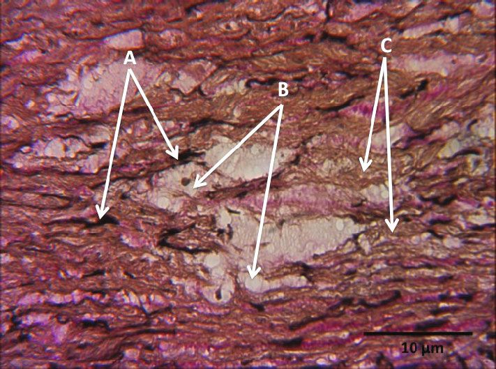
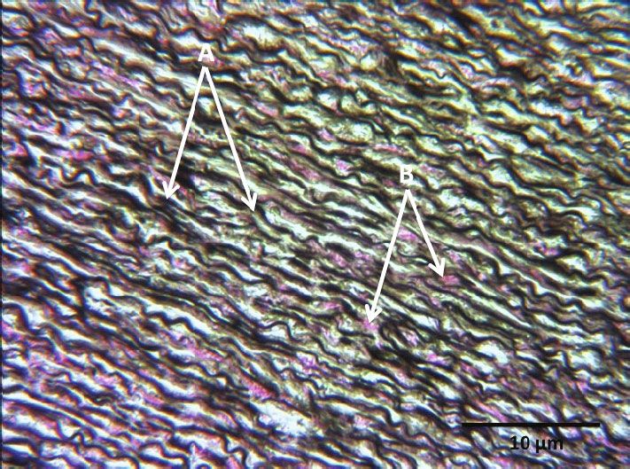
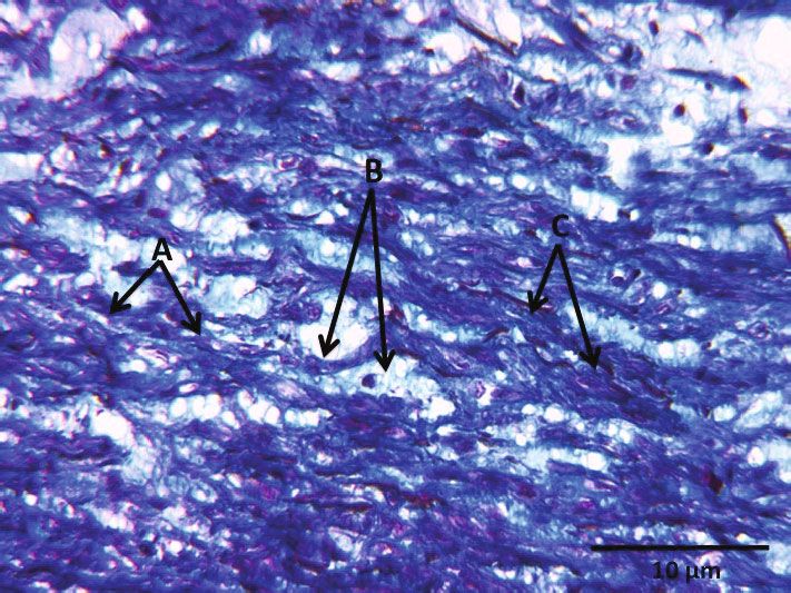
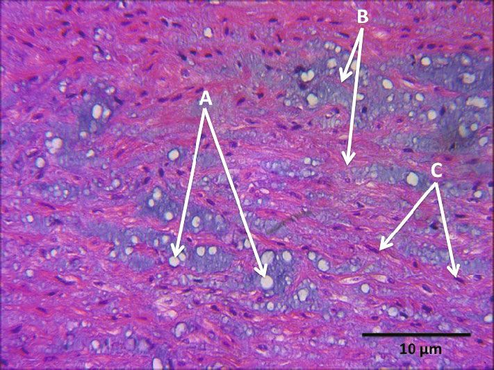
Oxidative Medicine and Cellular Longevity 5

and to perform the statistical analyses. Statistical significance LDS was significantly decreased (p = 0:01, Table 6) when
was determined by the Mann-Whitney rank sum test compared to that from the C subjects. However, the
followed by the normality test (Shapiro-Wilk). The correla- LPO index and NO3-/NO2- ratio tended to rise without
tion was made with Spearman’s test. The data are presented reaching significance in patients with LDS compared to
as the median and Min–Max range. Differences were consid- the C subjects (Table 6).
ered statistically significant when p ≤ 0:05.
 3.9. Histology
3. Results 3.9.1. Hematoxylin-Eosin Staining. Figure 5(a) shows the
3.1. General Characteristics. Out of the 10 LDS patients, 4 representative photomicrograph of the aortic middle wall
were men and 6 were women. The demographical character- of a C subject, showing black nuclei of fibrocytes arranged
istics of the patients are shown in Table 2. in fascicles, red elastic fibers, and pink collagen fibers,
 without cystic necrosis. Figure 5(b) shows the representa-
3.2. Mutations in LDS Patients. The percentage of positive tive photomicrograph of the middle aortic wall of a patient
mutations considering the mutation in any of the 4 exons with LDS, showing blue nuclei of fibrocytes arranged in
of FBN-1 was of 80%. It was 100% for TGF-βR2 and 10% fascicles, in red elastic fibers, and in pink collagen fibers;
for COL3A1. Also, 90% of the patients shared mutations in the presence of cystic necrosis is observed in white.
all genes.
 3.9.2. Masson’s Trichrome Stain. Figure 6(a) shows the repre-
3.3. Correlations. The correlations between the LPO and sentative photomicrograph of the aortic middle wall of a C
aortic diameters and TAC and aortic diameters are shown subject; elastic fibers are observed in red, collagen fibers are
in Table 3. observed in blue, and no cystic necrosis is observed.
 Figure 6(b) shows the representative photomicrograph of
3.4. General Variables. Table 4 shows the different general the aortic middle wall of a patient with LDS; elastic fibers
variables of both C subjects and patients with LDS. These are observed in red, collagen fibers are observed in blue,
are demographic variables and serum biochemistry; in this and cystic necrosis is observed in white with the presence of
table, the results are expressed as median with the Min– amorphous material in faint blue.
Max values.
 3.9.3. Weigert’s Staining. Figure 7(a) shows the representative
3.5. Enzymatic Activity and Expression. Table 5 shows the
 photomicrograph of the aortic middle wall of a C subject;
enzymatic activity and expression in the homogenate of the
 elastic fibers are observed in black, collagen fibers are
thoracic aortic aneurysms in both C subjects and patients
 observed in brown-yellow, and cystic necrosis is not distin-
with LDS.
 guished. Figure 7(b) shows the representative photomicro-
3.6. Enzymatic Activity. The Mn- and Cu/Zn-SOD iso- graph of the aortic middle wall of a patient with LDS;
forms showed a significant increase in their activities com- fragmented elastic fibers are observed in black, collagen fibers
pared to the C group (p = 0:04 and p = 0:03, respectively) are observed in brown-yellow, and cystic necrosis is distin-
(Figures 1(a) and 1(b)). The enzymatic activities of CAT guished in white.
(p = 0:01), GPx (p = 0:02), GST (p = 0:04), and TrxR
(p = 0:01) in patients with LDS were significantly 4. Discussion
decreased when compared to those in the C subjects
(Figures 2(a)–2(d), respectively). The GR activity showed In the MS, there is an imbalance of the redox homeostasis
a significant increase (p = 0:006) in patients with LDS that contributes to the alteration and development of the
when compared to the C subjects (Figure 3). TAA [33]. However, in LDS which is a rare variant of
 the MS [5], the role of the imbalance in the redox homeo-
3.7. eNOS, Cu/Zn-SOD, ORX, and Nrf2 Expressions. The stasis has not been evaluated. The main LDS characteristic
eNOS and Cu/Zn-SOD expressions did not show a signifi- is the presence of TAA with rapid progression that
cant difference in patients with LDS when compared to the decreases the survival of the patients [3]. Therefore, the
C subjects (Figures 4(a) and 4(b)). The ORX and Nrf2 aim of this work was to analyze the oxidant/antioxidant
expressions showed significant increases and decreases, profile in the TAA from patients with LDS. An imbalance
respectively, in patients with LDS when compared to the C similar to that found in MS with a positive feedback oxi-
subjects (p ≤ 0:05 and p = 0:02, respectively; Figures 4(c) dizing background was found.
and 4(d)).
 4.1. The FBN-1, TGF-βR2, and COL3A1 Coding Regions. The
3.8. Nonenzymatic Antioxidant System. The TAC levels, molecular testing in LDS is crucial to assess the diagnosis and
GSH, and vitamin C concentration showed a significant management of the patients. We analyzed the mutations in
decrease (p = 0:006, p = 0:006, and p = 0:01, respectively) in the FBN-1, TGF-βR2, and COL3A1 genes. The percentage
patients with LDS when compared to the C subjects of positive mutations considering the mutation in any of
(Table 6). The protein carbonylation in patients with LDS the 4 exons of FBN-1 was of 80%. It was 100% for TGF-
showed a significant increase (p = 0:01, Table 6). The Se βR2 and 10% for COL3A1. 90% of the patients also shared
concentration in the TAA homogenate from patients with mutations in all genes. The fact that these mutations were

6
 Table 2: Demographic characteristics of Loeys-Dietz syndrome patients.

Sex Age MS family history Clinical characteristics Gene Dao (mm) Z score Surgery type Evolution over time
 Dolichocephaly, malar hypoplasia,
 retrognathia, ogival palate, enlarged
 uvula, hypertelorism, low implantation
 He died a year after the
 The father died at of atrial pavilion, milia in the malar Aortic root replacement
 FBN-1 exon 28 and cardiovascular postoperative
M 15 age 26 and was region, arm stroke ratio size > 1:06, 29 4.07 by David’s procedure
 TGF-βR2 exon 6 period, due to an acute
 diagnosed with LDS stretch marks, pectus carinatum, (09/06/2013)
 cholecystitis complication
 scoliosis, equine foot varus. Steinberg
 and Walker-Murdoch positive,
 without ectopia lentis
 Hypertelorism, ogival-type palate,
 The father and sister dental overlap, uvula, widened millia, Satisfactory evolution
 Aortic root replacement
 died at 27 and 17 pectus carinatum, scoliosis, Steinberg FBN-1 exon 15 and currently in functional class 1
F 22 48 5.2 by David’s procedure
 years ago with and Walker-Murdoch, positive flat feet TGF-βR2 exon 6 (NYHA) was evaluated in
 (02/07/2013)
 LDS diagnosis and valgus. Arm/height atio rgreater consultation in May 2019
 than 1.04, without ectopia lentis
 Dolichocephaly, hypertelorism, Since 11/11/2013, he has had
 His mother was ogival palate, uvula bifida, micrognathia, dyskinetic and ballism that
 diagnosed with low implantation of atrial pavilion, pectus Aortic surgery by improved with anticonvulsive,
 LDS, died at 22 due carinatum of right hemithorax TGF-βR2 exon David’s procedure and the CT scan did not show
M 13 24 2.98
 to aortic rupture, and (asymmetric), stretch marks, Steinberg 6 and COL3A1 he had mitral plasty aneurysms, he is
 he has a sister sign and Walker-Murdoch positive, flat (11/01/2013) hemodynamically stable,
 with LDS foot, contractures in eyes, scoliosis, and and he remains alive
 mitral valve prolapse until 2019
 She showed dolichocephaly,
 hypertelorism, micrognathia, ogival
 palate and overlap, dental bifida tooth,
 arm reaction/height > 1:06 hyperelasticity,
 Aortic surgery by Stable evolution,
 No history of MS pectus carinatum, contractural FBN-I exon 28 and
F 18 46 9.5 David’s procedure with functional class 1. Last
 or LDS arachnodactyly in feet and hands, flatfoot TGF-βR2 exon 6
 a 30 mm graft (04/10/2012) consultation July 2019
 and varus Steinberg- and Walker
 Murdoch-positive scoliosis ecstasy. In
 her study of magnetic resonance, she
 had artificial tortuosity
 Hypertelorism, ogival palate, dental Hemodynamically
 Mother, one brother,
 overlap, bifida uvula, scoliosis, stretch FBN-1 exon 60+ Aortic surgery by David’s asymptomatically stable.
M 38 and 7 cousins with MS, 48 NA
 marks. Without lens dislocation, size TGF-βR2 exon 6 procedure (20/01/2014) In 2019, it was found in
 1 cousin with LDS
 and stroke 1.98 and height 1.90 functional class 1
 FBN-1 exon 28
 Hypertelorism, uvula Aortic surgery by Bentall In 2019, he went to
F 14 Father with LDS and TGF-βR2 54 4.30
 bifid, scoliosis pectus carinatum and De Bono (17/12/2018) evaluation and was stable
 exon 6
 Oxidative Medicine and Cellular Longevity
Table 2: Continued.
 Oxidative Medicine and Cellular Longevity

Sex Age MS family history Clinical characteristics Gene Dao (mm) Z score Surgery type Evolution over time
 Hypertelorism, ogival palate, wide
 Satisfactory evolution.
 The background uvula, pectus carinatum valgus, foot Aortic surgery by David’s
F 21 TGF-βR2 exon 6 25 3.47 The last revision was
 is unknown without lens dislocation. procedure (07/09/2015)
 in 2019
 Tortuous arteries
 FBN-1 exon 28 Aortic surgery by
 The background With hypertelorism, uvula bifida, He came to the office
M 46 and TGF-βR2 63 NA Bentall and De Bono
 is unknown scoliosis, flat feet until 2019
 exon 6 (09/09/2015)
 Her father died of Hypertelorism, millia in the malar
 FBN-1 exon 42 Aortic surgery by She came to the
 acute aortic dissection region, pectus carinatum, normal
F 25 and TGF-βR2 46 NA Bentall and De Bono hospital in 2019, and
 at age 24 and was uvula flat foot. By imaging, she had
 exon 6 (18/12/2015) she was stable
 diagnosed with MS tortuous arteries
 Hypertelorism, pectus carinatum, FBN-1 exon 28 Aortic surgery by
 The background She visited the
F 35 flat feet, tortuous arteries. Height and TGF-βR2 58 NA Bentall and De Bono
 is unknown hospital in 2019
 1.59 weight 64 exon 6 (06/04/2017)
Abbreviations: M = male; F = female; NYHA: New York Health Association; MS = Marfan syndrome; LDS = Loeys-Dietz syndrome; Dao = diameter aortic.
 7
8 Oxidative Medicine and Cellular Longevity

 Table 3: Correlations of LPO with TAC and aortic diameters.

 Total C LDS
 R2 p R2 p R2 p
LPO/TAC -0.59 0.008 -0.55 NS -0.45 NS
Carbonylation/TAC -0.55 0.01 -0.49 NS -0.32 NS
VBD/LPO -0.01 0.97 0.40 NS -0.75 NS
SUD/LPO 0.07 0.83 0.02 NS -0.52 NS
VBD/TAC -0.02 0.93 -0.47 NS 0.74 NS
SUD/TAC -0.14 0.69 -0.29 NS 0.82 NS
Abbreviations: LDS = Loeys-Dietz syndrome; LPO = lipid peroxidation; TAC = total activity antioxidant; VBD = Valsalva breast diameter; SUD = sinotubular
union diameter.

 Table 4: Demographic variables and serum biochemistry in C subjects and patients with LDS.

 C LDS
 p
 Median (Min–Max) Median (Min–Max)
General characteristics
 Age (range) 62 (37–77) 23 (14–46) 0.001
 Size (m) 1.65 (1.45–1.76) 1.67 (1.58–1.77) NS
 Weight (kg) 67 (56–105) 57 (33–71.5) 0.03
BMI (range) 27.8 (22.2–36.3) 20.8 (12.7 – 25.3) 0.005
Serum biochemistry
 CT (mg/dl) 125 (102–191) 129.5 (33–171) NS
 HDL (mg/dl) 39 (32–54) 48 (8.4–61) NS
 LDL (mg/dl) 73.5 (50–143) 84 (11.4–122) NS
 TG (mg/dl) 97.5 (67–148) 69 (57–118) 0.09
Abbreviations: BMI=body mass index; CT = cholesterol; HDL = high-density lipoproteins; LDL = low-density lipoproteins; TG = triglycerides.

Table 5: Enzymatic activity and expression in the homogenate of the thoracic aortic aneurysms in both C subjects and patients with LDS.

 C LDS
 p
 Median (Min–Max) Median (Min-Max)
Enzymatic activity
 Mn-SOD (U/mg/protein) 107.9 (93.3–117.8) 118.5 (103.9–125.2) 0.04
 Zn/Cu-SOD (U/mg protein) 101.2 (90.9–122) 123.4 (103.5–132.23) 0.03
 CAT (μM/mg/protein) 74.7 (45.9–82.4) 40.2 (14.9–77.5) 0.01
 GPx (nmol/mg/protein) 0.23 (0.003–0.69) 0.04 (0.01–0.06) 0.02
 GST (μM/mg/protein) 7 × 10⁻⁵ (2 × 10⁻⁵ –4 × 10⁻⁴ ) 1 × 10⁻⁵ (1 × 10⁻⁴ –8 × 10⁻⁵ ) 0.04
 TRx (μM/mg/protein) 3.30 (1.8–7.5) 2.1 (0.23–3.7) 0.01
 GR (μM/mg/protein) 0.32 (-1.1–0.71) 0.86 (0.30–1.56) 0.006
Enzymatic expression
 eNOS 0.99 (0.48–1.13) 0.87 (0.17–1.08) NS
 Nrf2 1.13 (0.98–1.35) 0.95 (0.87–1.08) 0.02
 ORX 0.85 (0.54–0.97) 1.00 (0.77–1.19) 0.05
Mn-SOD = manganese superoxide dismutase; Zn/Cu-SOD = zinc copper superoxide dismutase; CAT = catalase; GPx = glutathione peroxidase;
GST = glutathione reductase; TRx = thioredoxin reductase; GR = glutathione reductase; eNOS = endothelial nitric oxide synthase; Nrf2 = nuclear factor
erythroid 2-related factor 2; ORX = xanthine reductase.

not found in the FBN-1 gene in all patients is due to the size unavailable and expensive. The same happens for the TGF-
of the gene which is greater than 200 k. The coding sequence βR2 gene. Therefore, we only examined those exons that
is divided into 65 exons, and more than 800 mutations are have the highest frequency in this disease and mutations in
known within it. This makes the complete study of the gene the FBN-1 gene.
Oxidative Medicine and Cellular Longevity 9

 140 140

 130 130

 Cu/Zn−SOD activity
 (U/mg of protein)

 (U/mg of protein)
 Mn-SOD activity

 120 120

 110 p = 0.04 110 p = 0.03

 100 100

 90 90

 80 80
 C LDS C LDS

 Mn-SOD
 Cu/Zn-SOD

 Bovine C LDS
 CAT
 (a) (b)

Figure 1: (a) Average activity of the Mn-SOD and (b) average activity of the Cu/Zn-SOD in LDS patients (n = 10) and C subjects (n = 9).
Values expressed represent the median and Min–Max range. The image of the center is a representative gel of the electrophoresis of the
SOD isoforms. Abbreviations: LDS = Loeys-Dietz syndrome; C = control subjects.

 Most investigations on LDS focus on the study of muta- express MMPs that may be altered by the GPx that is dynam-
tions [34]. Mutations in FBN-1 are associated with 60-90% ically regulated in the blood vessels [39]. They are the result
of the diagnosis [35]. In addition, mutations in the genes that of the TGF-βR1 and TGF-βR2 increases and ROS accumula-
encode for TGF-β1 and TGF-β2 have been identified in tion that contribute to the cystic necrosis of the aortic wall,
patients having this disease. TGFβ is involved in the develop- vasomotor dysfunction, and ED, thus favoring the aneurysm
ment and maintenance of blood vessels and craniofacial formation [6, 9].
growth [34]. In patients in whom no mutations in FBN-1
and TGFβ were found, homozygous deletions in the 4.2. Antioxidant Enzymatic Profile in TAA from Patients
COL3A1 gene have been identified, and these mutations lead with LDS. Several studies in MS mouse models have
to structural alterations of the collagen that could cause aortic described vasomotor dysfunction in the TAA associated
dissection [36]. Mutations affecting the intracellular kinase with ROS overproduction. This has been related to alter-
domain of this protein can disturb TGF-β signaling, which ations in antioxidant enzymes such as SOD isoforms [9,
subsequently leads to features of LDS patients. In fact, 18] and to several diseases such as Turner syndrome, arte-
TGF-βR2 mutations have been related to skeletal abnormal- rial hypertension, and some MS variants [16]. However,
ities, aortic dilation, and ED. Mutations of the FBN-1 gene another investigation has shown a decrease in SOD iso-
have also been found in MS, and they produce a decon- forms due to the increase in O2- in the TAA in a mouse
structed protein product which cannot bind to the latent model of MS. This decrease was related to the deteriora-
TGF-βR1 [4, 9, 10]. Activation of the latent TGF-βR1 by tion of aortic function [10].
ROS could be associated with the decrease in GPx activity Our results suggest that the alteration of the activity of
[37]. Therefore, an increase in the TGF-βR1 concentration Cu/Zn- and Mn-SOD can be due to the overproduction of
in the TAA could be expected, and an increase in the TGF- O2- through ORX and NADPH oxidase overexpression in
βR1 and TGF-βR2 receptor expressions could upregulate tissue from the TAA [10]. An increase in ORX expression
the noncanonical and canonical TGF-β pathways which are could increase the O2- concentration and stimulate Cu/Zn-
increased in myocytes from TAA [9]. and Mn-SOD activity, increasing the H2O2 concentration.
 TGF-βR1 and TGF-βR2 can upregulate the expression of This ROS is more harmful than others due to its capacity to
elastases and metalloproteinases (MMPs), which may diffuse through the cell membrane [40]. However, in our
enhance elastin degradation. MMPs participate in the elastic determinations, the expression of the Cu/Zn-SOD did not
fiber disintegration, which is observed in the representative show significant changes. These results can be explained con-
microphotographs of the patients with LDS. In these sidering that the enzyme expression is not necessarily related
patients, there is a reduction of connective tissue elasticity to an increase in its activity since the expressed protein may
that results in weakness of the aneurysm aortic wall [9, 16, be inactive or have a lower expression while having a greater
38]. These breakings in structure could be associated with activity. Thus, the degree of phosphorylation of the active site
decreases in GSH and GPx. ROS can affect the migration, dif- of SOD could increase its activity [41]. The increase in SOD
ferentiation, and proliferation of the myocyte cells which activity in the patients with LDS suggests that the SOD
10 Oxidative Medicine and Cellular Longevity

 0.8

 (nmol/min/mg of oxidized NADPH)
 90
 0.6
 ( M H2O2/min/mg of protein)
 80

 GPx activity
 70 0.4
 CAT activity

 60 p = 0.01
 50 0.2
 40 p = 0.02
 30 0.0
 20
 10
 C LDS

 Bovine C LDS
 CAT
 (a) (b)
 0.0005 8

 (TNB mol/min/mg of protein)
 (GS-DNB mol/mg of protein)

 0.0004
 6
 0.0003
 TrxR activity
 GST activity

 0.0002 4

 0.0001 p = 0.01
 p = 0.04 2
 0.0000

 −0.0001 0
 C LDS C LDS
 (c) (d)

Figure 2: (a) Average activity of CAT. The image below the graph is a representative native gel of the electrophoresis. (b) GPx activity, (c)
GST activity, and (d) TrxR activity in LDS patients (n = 10) and C subjects (n = 9). Values are expressed as the median and Min–Max
range. Abbreviations: LDS = Loeys-Dietz syndrome; C = control subjects.

 CAT is responsible for the H2O2 elimination, and its
 2.0
 overexpression can decrease OS in the TAA [12]. CAT activ-
 1.5 ity is altered in aortas of patients with MS through continued
 H2O2 exposure [10]. Our results show that CAT activity is
 (U/min/mg of protein)

 1.0 significantly decreased in TAA of patients with LDS. This
 may be due to an H2O2 overproduction by the SOD isoforms
 GR activity

 0.5 p = 0.006
 [42, 43]. Also, the excessive O2- concentration can cause
 0.0 glucose-6-phosphate inhibition that catalyzes the formation
 of NADPH, which serves as an electron donor to CAT [44].
 −0.5
 GPx is among the enzymes responsible for the H2O2
 −1.0 detoxification. It is more efficient than CAT when removing
 intracellular H2O2 and organic peroxides [15]. The GPx
 −1.5 activity was decreased in patients with LDS, and this may
 C LDS
 be due to the low availability of GSH in these patients. The
Figure 3: Average activity of GR in LDS patients (n = 10) and C GPx has a Se atom in the form of selenocysteine in its cata-
subjects (n = 9). Values are expressed as the median and Min–Max lytic site, and Se is a limiting factor of the antioxidant defense
range. Abbreviations: LDS = Loeys-Dietz syndrome; C = control [45]. The deficiency of Se leads to cardiovascular pathologies
subjects. [46] and diseases such as Kashin-Beck and Keshan [47–49].
 In the patients with Keshan disease who have dilated cardio-
isoforms try to counteract the O2- overproduction. However, myopathy, the Se deficiency decreases the GPx activity [49].
this seems counterproductive since it results in H2O2 This suggests that selenoprotein expression or activity is reg-
overproduction. ulated by the concentration of this trace element [50]. Our
Oxidative Medicine and Cellular Longevity 11

 1.2 2.2
 2.0
 1.0

 Cu/Zn-SOD/ –actin
 1.8
 eNOS/ –actin

 0.8
 1.6
 0.6 1.4
 1.2
 0.4
 1.0
 0.2 0.8

 0.0 0.0
 eNOS Cu/Zn-SOD
 140 kDa 95 kDa

 –Actin –Actin

 C LDS C LDS
 (a) (b)
 1.2 1.4

 1.1 1.3
 1.0
 ORX/ –actin

 Nrf2/ –actin 1.2
 0.9 p ≤ 0.05
 1.1
 0.8
 1.0 p = 0.02
 0.7

 0.6 0.9

 0.5 0.8
 ORX 66 kDa Nrf2
 300 kDa
 –Actin –Actin
 C LDS C LDS
 (c) (d)

Figure 4: (a) Representative histograms of eNOS/α-actin expression. (b) Cu/Zn-SOD/α-actin expression, (c) ORX/α-actin expression, and
(d) Nrf2/α-actin expression in LDS patients (n = 10) vs. C subjects (n = 9). Values are expressed as the median and Min–Max range.
Abbreviations: LDS = Loeys-Dietz syndrome; C = control subjects.

Table 6: Redox biomarkers of the nonenzymatic system in the homogenate of the thoracic aortic aneurysm patients with LDS and C subjects.

 C LDS
Parameters (mg/protein) p
 Median (Min–Max) Median (Min–Max)
TAC (nmol Trolox) 119.6 (35–220) 15.09 (0.38–145) 0.006
LPO index (nmol MDA) 2.4 (0.53–7.4) 5.7 (2.3–15) 0.04
Carbonylation (ng carbonyls) 5.5 (2.04–11.8) 10.4 (4.3–21.1) 0.01
GSH (nM) 9 × 10⁻² (1 × 10⁻³ –6 × 10⁻² ) 8 × 10⁻³ (3 × 10⁻³ –1 × 10⁻² ) 0.006
NO3-/NO2- (nM) 2.6 (1.97–4.3) 3.37 (2.14–7.8) NS
Vitamin C (μM) 0.03 (0.02–0.04) 0.02 (0.01–0.04) 0.01
Se (pg) 119.6 (35–220) 15.09 (0.38–145) 0.01
Abbreviations: TAC: total antioxidant capacity; LPO: lipid peroxidation; GSH: glutathione; Se: selenium; LDS: Loeys-Dietz syndrome; C: control subjects. The
data are presented as the median and Min–Max range.

results show that the Se concentrations are decreased in ence of vitamins A, D, and E. Patients with LDS probably
patients with LDS, and this may impact on the activity of have an intestinal malabsorption syndrome which could
Se-dependent enzymes such as GPx and TrxR. Also, Se is cause the decrease in the Se [51]. Our results also showed a
absorbed in the form of organic compounds and in the pres- decrease in the concentration of vitamin C, which is
12 Oxidative Medicine and Cellular Longevity

 (a) (b)

Figure 5: (a) Representative photomicrograph at 40x with hematoxylin-eosin staining of the aortic middle wall of a control subject. (A)
Elastic fibers in deep pink. (B) Dark-colored fibrocyte nucleus. (b) Representative photomicrograph of the aortic middle wall of an LDS
patient. (A) Cystic necrosis, with accumulation of amorphous material. (B) Elastic and collagen fibers in reddish and deep pink. (C) Blue-
stained fibrocyte nucleus.

 (a) (b)

Figure 6: (a) Representative photomicrograph at 40x with Masson’s trichrome staining of the aortic wall of a control subject. (A) Elastic fibers
in red. (B) Collagen fibers in blue. (b) Representative photomicrograph of the aortic middle wall of an LDS patient. (A) Collagen fibers in deep
blue color. (B) Cystic necrosis with the presence of amorphous material in faint blue color. (C) Elastic fibers in red color.

 (a) (b)

Figure 7: (a) Representative photomicrograph at 40x with Weigert’s staining of the aortic wall of a control subject. (A) Elastic fibers in black
color. (B) Collagen and elastic fibers in pink and light brown color. (b) Representative photomicrograph of the aortic wall of an LDS patient.
(A) Elastic fibers. (B) Cystic necrosis. (C) Collagen fibers.

important to regenerate vitamin E [15]. The decrease in both used by the GST are formed as a result of ROS modification
may affect the Se absorption. of macromolecules [52]. A decrease in GST in aortas of
 GST is responsible for conjugating GSH with xenobiotics, patients with MS [14] and in patients with other pathologies
peroxidized lipids, hydroperoxides, and H2O2 among other that affect the cardiovascular system has been associated with
molecules favoring their elimination [15]. The substrates OS and with an increase in LPO [16]. Our results show that
Oxidative Medicine and Cellular Longevity 13

the GST activity is decreased in patients with LDS. This sug- numerous antioxidant enzyme genes such as GPx, SOD iso-
gests that the GST activity is compromised by the low avail- forms, GST, and TrxR [62]. The low levels of Nrf2 expression
ability of GSH. This contributes to the tendency to increase in patients with LDS suggest that it can be responsible, in
LPO. In addition, the increase in protein carbonylation may part, for the decrease in the activity of the antioxidant
depend on the decrease in the TrxR activity, which would enzymes. Our results showed a tendency to decrease eNOS
be reflected in a smaller reduction of thiol groups in proteins. expression in the patients with LDS. Other studies have
TrxR is another Se-dependent protein possessing a seleno- shown that the iNOS activity is increased and the activity of
cysteine in its catalytic site [53]. OS induces a compensatory eNOS is decreased in the homogenate of TAA from patients
increase in TrxR to reduce the oxidative modification of pro- with MS [63]. This result also suggests that the NO produc-
teins present in several pathologies [53]. However, TrxR is tion mostly comes from the iNOS pathway and to a lesser
decreased in pathologies with severe OS and metabolic dis- extent from the eNOS [39]. Furthermore, in MS animal
turbances [54]. The low TrxR activity in patients with LDS models, O2- overproduction by the NADPH oxidase and
could increase the carbonylation and be associated with the ORX pathway is associated with increase in NO production
OS present in this syndrome. by iNOS and is involved in inflammatory processes [14, 18,
 63]. This promotes the ONOO- formation that contributes
4.3. Antioxidant Nonenzymatic Profile in TAA from LDS to OS in the TAA [64]. High concentrations of ONOO- can
Patients: Glutathione Levels and Glutathione Reductase. lead to apoptosis in endothelial cells and may favor the acti-
GSH is the main defense mechanism against ROS and elec- vation of MMPs 1 and 2 in vascular smooth muscle cells
trophiles [55]. It is formed by γ-ι-glutamyl-ι-cysteinyl-gly- degrading essential proteins to maintain the anatomical
cine. GSH can also be a substrate for metabolic pathways integrity of the aorta [64].
such as the one mediated by the SOD isoforms and ORX.
GSH homeostasis occurs mainly through the balance of con- 4.5. Histological Characteristics. The observation of the his-
sumption and supply [34]. When GSH reduces target mole- tological cuts from the TAA segments of patients with LDS
cules, electrophiles, or ROS, the thiol group of the cysteine showed cystic necrosis and accumulation of amorphous
is oxidized through the GPx activity. Excessive GSH oxida- material. This suggests the development of fibrosis in the
tion by GPx might be due to chronic OS in patients with TAA that may be associated with chronic OS. The partici-
LDS. GSH is then reduced to GSSG by GR to restore GSH pation ROS in the formation of the abdominal aortic aneu-
levels [55, 56]. GR requires NADPH consumption for its rysm was associated with the overexpression of the MMPs
activity [40, 56]. A decrease in the GR activity will cause a 2 and 9 [65]. The results with Weigert’s staining showed
decrease in the reduced GSH concentrations resulting in an fragmentation, thickening, and a smaller number of elastic
increase in the level of the ROS. In several pathological pro- fibers in the LDS group. This could be related to aortic stiff-
cesses, an alteration of the GR activity and in the GSH levels ness. The elastic fiber fragmentation in the aorta is related
has been found, which is associated with an increased risk of to the interaction between OS, MMPs, and the TGF-β
OS [57]. Our results show that the GR activity was increased deregulation [66, 70]. Our results show a collagen increase
in patients with LDS, but despite this, the GSH values were in photomicrographs from the patients with LDS. This is
not restored. This may be due to low levels of GSH which probably due to deregulation in TGF-β signaling.
may stimulate the GR activity [58]. A low GSH level limits
the activities of GPx and GST, and in turn, the low activities 5. Conclusion
of GPX and GST may contribute less to the scavenging of
H2O2 and O2− and this can favor and contribute to chronic The antioxidant enzyme activities, including those of SOD
OS in patients with LDS. It may also be due to a compensa- isoforms, CAT, TrxR, and GST, decrease in the TAA of
tory protective mechanism of the cells against oxidant agents patients with LDS. These decreases in the enzyme activities
[59]. Moreover, the GSH produced by GR may be oxidized favor the accumulation of ROS that contributes to GSH
and therefore be insufficient to compensate for the high pro- decrease and favor LPO and carbonylation. The decrease in
duction of ROS [60]. Se and Nrf2 also impacts on the activity and/or expression
 Another possible cause of the GSH decrease could be of some of these antioxidant enzymes. The GR increase does
the alteration of the enzyme γ-glutamyl-cysteine synthase not completely restore the GSH concentration, which is
which is responsible for the GSH synthesis or for a deficient reflected in the decrease in the TAC and in the enzymes that
amount of the amino acids that constitute GSH [56]. In use it which contributes to and favors ROS production. This
addition, the TAC results which include the GSH level leads to a positive feedback oxidizing background which con-
show a decrease in the LDS patients. This confirms the tributes to the TAA formation.
presence of OS in the TAA of tissues from patients with
this disease. Likewise, the LPO index showed a tendency 5.1. Perspectives. The goal of the medical treatment in LDS is
to increase, and this could lead to an increase in prooxidant to delay the progression of aortic dilation to avoid cata-
compounds such as 4-hydroxinonenal and MDA [61]. strophic complications. This study proposes that the use of
 antioxidants together with the current treatments could help
4.4. Expressions of Nrf2, Xanthine Oxidase, and Endothelial patients with LDS. The use of β blockers, as well as the use of
Nitric Oxide Synthase. The Nrf2 transcription factor is angiotensin 1 antagonists, constitutes the first-line therapy to
responsible for the regulation of the inducible expression of this day for LSD and MS [67]. However, the scientific
14 Oxidative Medicine and Cellular Longevity

evidence supporting these therapies is limited since it has lethal development of the TAA and increase the quality of
been obtained from simple randomized trials with a life and survival in patients with LDS.
restricted sample number [68]. In 2007, a meta-analysis of
6 studies that included 802 patients with MS showed no dif- 5.2. Limitations. The main limitation of this study was the
ference in the results of β blocker therapy in relation to mor- obtainment of TAA fragments from patients with LDS. As
tality and morbidity [69]. described above, this disease is a variant of MS with unknown
 The excessive activity of MMPs resulting in an excessive incidence that may be of around 1/10000. Only 10 patient
deposition of matrix elements such as proteoglycans and samples could be obtained in a period of approximately 9
collagen which are organized in a random manner and years. Another limitation was the improbability of having
fragmentation of elastic fibers is similar to that seen in ath- matched C subjects for gender and age. Although may there
eromatous plaques, and therefore, the use of statins such as be a correlation of TAC with age, it is not possible to obtain
pravastatin at a dose of 40 mg/day might also be useful for aortic tissue from healthy subjects at specific ages. The only
the treatment of TAA in LDS and MS. Statins are inhibitors way to obtain these samples is finding subjects having a sur-
of HMG-CoA reductase and are used as CT-reducing gical indication where there is a possibility to ethically with-
agents with proven efficacy to reduce morbidity. Moreover, draw a small sample. This depends on the treatment applied
a study concluded that statins could have a similar effect to and/or the surgical technique employed and on the obtain-
that of losartan in their capability to attenuate aortic root ment of their informed consent. However, even if there are
dilation in a mouse model with MS. Although these drugs age differences in C subjects and patients with LDS, there is
reduced excessive protein manufacturing by vascular certainty that there were no comorbidities or aortic damage
smooth muscle cells [70], inhibition of MMPs by these as shown in the photomicrographs of the C subjects.
drugs may alter fibrillin-1 which is the component of the
microfibrillins that provides a connection between elastin Data Availability
fibers and vascular smooth muscle cells [70, 71]. The struc-
tural homology between fibrillins and latent TGF-β binding The datasets generated and analyzed during the current
proteins suggests an additional role for FBN-1 to direct study are available from the corresponding author on reason-
TGF-β to the extracellular matrix and to keep it in an inac- able request.
tive state. Therefore, an FBN-1 deficiency would lead to
excessive TGF-β signaling [71] which would not benefit Conflicts of Interest
LDS patients.
 Deregulation of TGF-β signaling contributes to aortic All the authors declare that there are no competing interests
and mitral valve pathology in mouse models with MS [72]. regarding the publication of this paper.
This is supported by the observation of a Marfan phenotype
in humans with mutations in the TGF-β receptor [73]. Acknowledgments
Fibrillin-1 deficiency that leads to deregulation of TGF-β sig-
naling causes vascular smooth muscle cells to reshape the We thank Benito Chávez Rentería for histology technical
extracellular matrix through the SMAD pathway [73]. How- support and Alberto Guevara Flores, Héctor Vázquez Meza,
ever, other mechanisms such as redox status also importantly and José de Jesús Martínez González for providing auranofin
participate in the expansion of TAA in LDS together with the and the Department of Biochemistry of the Faculty of Medi-
genetic predisposition, according to our findings, and there- cine, UNAM.
fore, therapeutics in this regard should be evaluated.
 It is a very important state that during the evolution References
and progression of aneurysms, patients are typically asymp-
tomatic and it is not until aortic dissection occurs, generat- [1] J. J. J. Aalberts, M. P. van den Berg, J. E. H. Bergman et al., “The
ing severe pain, that the doctor suspects the diagnosis. At many faces of aggressive aortic pathology: Loeys-Dietz syn-
that stage of the disease, it is too late for many therapeutic drome,” Netherlands Heart Journal, vol. 16, no. 9, pp. 299–
 304, 2008.
interventions and sudden death can happen in up to 50% of
patients. Since it is clear that OS plays an important role in [2] H. Bashari, A. Brooks, O. O’Brien, S. Brennecke, and
 D. Zentner, “Maternal Loeys-Dietz syndrome (transforming
the development of aneurysms, OS should be monitored in
 growth factor ligand 2) in a twin pregnancy: case report and
clinical practice and aimed during treatment since diagno- discussion,” SAGE Open Medical Case Reports, vol. 7, 2019.
sis. The execution of randomized clinical trials in this
 [3] J. J. Maleszewski, D. V. Miller, J. Lu, H. C. Dietz, and M. K.
regard should be addressed. Finally, it is possible that the Halushka, “Histopathologic findings in ascending aortas from
use of a combination of antioxidants or antioxidants individuals with Loeys-Dietz syndrome (LDS),” The American
together with AT-1 inhibitors and statins may have an Journal of Surgical Pathology, vol. 33, no. 2, pp. 194–201, 2009.
impact on the control of aortic dilation. Their beneficial [4] G. Kuzmik, A. Sang, and J. Elefteriandes, “Natural history of
effect could be monitored by various avant-garde imaging thoracic aortic aneurysms,” Journal of Vascular Surgery,
studies. In this sense, the treatment with antioxidant sup- vol. 56, no. 2, pp. 561–575, 2012.
plements such as N-acetylcysteine, a GSH donor, vitamins [5] J. A. N. Meester, A. Verstraeten, D. Schepers, M. Alaerts,
C and E, or combination of these or other antioxidants L. Van Laer, and B. L. Loeys, “Differences in manifestations
could contribute to the deceleration of the aggressive and of Marfan syndrome, Ehlers-Danlos syndrome, and Loeys-
You can also read