FMRI Neurofeedback-Enhanced Cognitive Reappraisal Training in Depression: A Double-Blind Comparison of Left and Right vlPFC Regulation
←
→
Page content transcription
If your browser does not render page correctly, please read the page content below
ORIGINAL RESEARCH
                                                                                                                                             published: 23 August 2021
                                                                                                                                       doi: 10.3389/fpsyt.2021.715898

                                                fMRI Neurofeedback-Enhanced
                                                Cognitive Reappraisal Training in
                                                Depression: A Double-Blind
                                                Comparison of Left and Right vlPFC
                                                Regulation
                                                Micha Keller 1*, Jana Zweerings 1 , Martin Klasen 1,2 , Mikhail Zvyagintsev 1 , Jorge Iglesias 3 ,
                                                Raul Mendoza Quiñones 3 and Klaus Mathiak 1,4
                                                1
                                                 Department of Psychiatry, Psychotherapy and Psychosomatics, School of Medicine, RWTH Aachen University, Aachen,
                                                Germany, 2 Interdisciplinary Training Centre for Medical Education and Patient Safety—AIXTRA, Medical Faculty, RWTH
                                                Aachen University, Aachen, Germany, 3 Department of Cognitive Neuroscience, Cuban Center for Neuroscience, Havana,
                                                Cuba, 4 JARA-Brain, Research Center Jülich, Jülich, Germany

                            Edited by:
                          Hongming Li,          Affective disorders are associated with maladaptive emotion regulation strategies. In
            University of Pennsylvania,         particular, the left more than the right ventrolateral prefrontal cortex (vlPFC) may
                          United States
                                                insufficiently regulate emotion processing, e.g., in the amygdala. A double-blind
                     Reviewed by:
                     Dandan Zhang,              cross-over study investigated NF-supported cognitive reappraisal training in major
          Shenzhen University, China            depression (n = 42) and age- and gender-matched controls (n = 39). In a randomized
                   Jennifer Barredo,
                                                order, participants trained to upregulate either the left or the right vlPFC during cognitive
       Providence VA Medical Center,
                      United States             reappraisal of negative images on two separate days. We wanted to confirm regional
                 *Correspondence:               specific NF effects with improved learning for left compared to right vlPFC (ClinicalTrials.
                         Micha Keller           gov NCT03183947). Brain responses and connectivity were studied with respect to
                mkeller@ukaachen.de;
               kmathiak@ukaachen.de             training progress, gender, and clinical outcomes in a 4-week follow-up. Increase of vlPFC
                                                activity was stronger after NF training from the left- than the right-hemispheric ROI.
                  Specialty section:            This regional-specific NF effect during cognitive reappraisal was present across patients
        This article was submitted to
       Neuroimaging and Stimulation,
                                                with depression and controls and supports a central role of the left vlPFC for cognitive
              a section of the journal          reappraisal. Further, the activity in the left target region was associated with increased use
               Frontiers in Psychiatry
                                                of cognitive reappraisal strategies (r = 0.48). In the 4-week follow-up, 75% of patients
            Received: 27 May 2021               with depression reported a successful application of learned strategies in everyday life
            Accepted: 29 July 2021
          Published: 23 August 2021             and 55% a clinically meaningful symptom improvement suggesting clinical usability.
                             Citation:          Keywords: real-time fMRI neurofeedback, depression, emotion regulation, cognitive reappraisal, lateral PFC
    Keller M, Zweerings J, Klasen M,
  Zvyagintsev M, Iglesias J, Mendoza
   Quiñones R and Mathiak K (2021)
      fMRI Neurofeedback-Enhanced
                                                INTRODUCTION
     Cognitive Reappraisal Training in
          Depression: A Double-Blind
                                                In everyday life we are frequently challenged by situations that evoke negative emotions. As part
  Comparison of Left and Right vlPFC            of an adaptive response to these encounters we may change the experience and expression of
                          Regulation.           our emotions by using emotion regulation (1). However, the success of our emotional response
        Front. Psychiatry 12:715898.            modulation may depend on our mental condition. For instance, emotion dysregulation is a
    doi: 10.3389/fpsyt.2021.715898              characteristic symptom of major depressive disorder (MDD). Increased selective attention and

Frontiers in Psychiatry | www.frontiersin.org                                        1                                        August 2021 | Volume 12 | Article 715898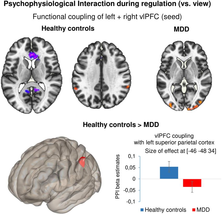
Keller et al.                                                                                     fMRI Neurofeedback-Enhanced Reappraisal Training

processing of negative mood-congruent stimuli as well as                  the vlPFC have been found in patients with depression (39).
maladaptive emotional responses may propel the development                Especially reinterpretation (compared to distancing) of negative
and recurrence of depressive episodes (2–4). A recent meta-               affective content has been related to peak activation in the left
analysis indicates that patients with depression show abnormal            vlPFC and the left STG (40). The vlPFC relays the need to regulate
recruitment of the emotion regulation brain network during                to a fronto-parietal cognitive control network (dlPFC, pre-SMA,
cognitive reappraisal of negative images (5). Furthermore,                STG, posterior parietal cortex) which is subsequently involved in
patients with depression often find it difficult to spontaneously         the execution of regulation (37). Cognitive reappraisal or related
utilize these strategies in everyday situations (6, 7) and methods        emotion regulation strategies that are applied in response to
are needed to bridge the gap between theory and everyday life             emotional provocation, modulate the semantic representation
application. A recent study by (8) has shown that even a single           of an emotional stimulus and the emotional responding
session of real-time functional magnetic imaging neurofeedback            through subcortical pathways (36, 41). Interestingly, gender
(rtfMRI-NF) could enhance the transfer of skills learned by               differences in cognitive regulation such as more rumination and
patients with depression during CBT to real-world situations.             catastrophizing in females (42) and alcohol for coping in men
RtfMRI-NF is a novel technique by which individuals with                  (43) have also been observed on the neural level (44). Meta-
psychiatric disorders can learn to voluntarily self-regulate their        analyses have investigated differences during emotion regulation
brain signal in areas amongst others involved in the neural               between healthy controls and individuals with depression (5, 40).
circuitry of emotion regulation and thereby induce changes in             Hyperactivity in the amygdala during downregulation of negative
neural plasticity (9–13). Therefore, providing NF can inform              stimuli has consistently been reported to be specific for affective
neuroscience-based interventions for emotion dysregulation and            disorders which indicates increased bottom-up responding or
may offer a more specific clinical tool for augmenting self-              ineffective modulatory capacity of regulatory networks during
regulation in patients with depression by strengthening the               emotion appraisals (5). Furthermore, the vlPFC and dlPFC (5)
monitoring within the emotion regulation process (7, 14).                 as well as the left STG (40) show less activation in patients
    Cognitive reappraisal is essential for psychological                  with mood disorders which may be related to a dysfunctional
functioning and well-being and has been linked to lower                   management of attentional and inhibitory resources and make
levels of psychopathology (15). This strategy is focused on               these areas potential targets for NF training.
lowering the valence of negative situations by reinterpreting its             Neuromodulation approaches of emotion regulation
meaning in a more positive way (1). This process is associated            networks not only underline the causal role of the lPFC for
with a decrease in negative and an increase in positive affect            cognitive reappraisal but also show promise for improving
(16). It mediates the relationship between stress and depressive          depressive      symptomatology.       For    instance,   repetitive
symptoms (17) and may be beneficial on the long run by                    transcranical magnetic stimulation (rTMS) over the lPFC is
decreasing the impact of recurrent negative stimuli (18). Yet,            an evidence-based treatment for depression which is successfully
patients with depression tend to overuse maladaptive emotion              applied in treatment refractory depression (45, 46). In a
regulation strategies such as rumination as well as suppression           meta-analysis of therapeutic responses to brain stimulation
of emotional experiences (19–21) which adds to the negativity             in depression (47), excitatory rTMS seemed to perform better
bias of attention, information processing and memory formation            at the left than the right lPFC (odds ratio: 1.89) and the
(22–24). Emotion dysregulation may facilitate the development             reversed pattern was observed after inhibitory stimulation (OR:
and recurrence of symptoms of depression (25, 26). As cognitive           3.29). These comparisons, however, did not encompass direct
reappraisal strategies are not frequently used by patients                comparison within studies and failed to show a statistically
with depression (20, 27–29), training reappraisal ability is an           significant difference. The causal role of the vlPFC in specific
established component of cognitive behavioral therapy (CBT)               is supported by experimental TMS studies that demonstrated a
(30, 31). CBT modulates the neural circuitry of emotions (32).            facilitation of reappraisal after vlPFC stimulation (48, 49). In a
Furthermore, self-reported reappraisal success following CBT              similar vein, transcranial direct current stimulation (tDCS) of the
in patients with social anxiety disorder predicted symptom                vlPFC may influence emotion regulation. For instance, excitatory
reductions (33). Despite of promising effects, patients often             tDCS stimulation of the vlPFC facilitated the downregulation
struggle to transfer these cognitive strategies from theoretical to       of negative emotions using cognitive reappraisal compared
real-world applications (34). This can be supported by web-based          to dlPFC or sham stimulation (50–52). Taken together, these
interventions that train cognitive reappraisal on a regular basis         findings further support a key role of the lPFC for mood
by receiving feedback from peers (35) or instead by rtfMRI NF             disorders and show that especially the vlPFC is a suitable target
training (8).                                                             for NF-supported emotion regulation training.
    On the neurobiological level, the downregulation of emotional             EEG and fMRI studies indicate that the laterality of neural
reactivity in healthy participants is associated with a widespread        activation may be indicative of affective style. According to the
network including frontal, parietal as well as subcortical regions        frontal asymmetry model, relatively higher right frontal alpha
(36–38). In this network, the vlPFC seems to play a pivotal role          power (more left PFC neural activity) suggests approach-related
for the process of emotion regulation due to its dense structural         emotions while higher left frontal alpha power (more right PFC
and functional connections to other prefrontal, somatosensory,            neural activity) may relate to withdrawal-related emotions (53,
motor and language areas and its link to response selection and           54). Furthermore, electrophysiological studies have suggested
inhibition (37). Furthermore, gray matter volume reductions in            that left-sided frontal alpha asymmetry (FAA; higher alpha

Frontiers in Psychiatry | www.frontiersin.org                         2                                    August 2021 | Volume 12 | Article 715898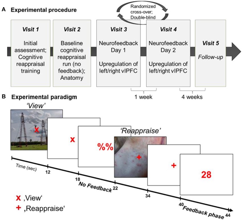
Keller et al.                                                                                    fMRI Neurofeedback-Enhanced Reappraisal Training

power in the left compared to right frontal channels) may be a            provides an objective neural indicator of regulation success that
biomarker of depression [e.g., (55)]. Even though the relation            may improve identification of successful regulation strategies and
of FAA and depression may be only of small magnitude and                  strengthen experienced self-efficacy.
use of FAA as a biomarker of depression seems too farfetched                  In the current study, we investigated the feasibility of a NF-
(56), frontal EEG asymmetry may be used as index of emotion               guided cognitive reappraisal training in patients with depression
regulation capability (57). For instance, a shift toward left             using a double-blind cross-over design. On two separate NF
asymmetry induced by mindfulness training was associated with             training days, participants upregulated either the left or right
improved responses during emotional challenges (58). In an EEG            vlPFC in response to negative pictures by applying strategies
emotion regulation paradigm, individuals with higher capacity             of cognitive reappraisal. Based on the reviewed fMRI and EEG
for reappraisal showed more left-lateralized (ventro) lateral PFC         literature, we hypothesized that the left compared to right vlPFC
activation (59). Consequently, recruitment of left-lateralized            may be more important for the emotion regulation process.
PFC areas was associated with creating alternative appraisals of          Therefore, the right vlPFC was chosen as active control condition
negative situations. Furthermore, in an fMRI investigation of             as it is known to be involved in cognitive reappraisal, however,
laterality, less activation of the left relative to right IFG was         it is less consistently observed and activation levels tend to be
associated with poor performance on an emotion perception task            lower (37, 70, 71). An active control condition can circumvent
(60). Lastly, in a combined rtfMRI-EEG-NF paradigm, patients              the problem of causing frustration as regulation is expected to
with depression achieved a shift toward left frontal activation           be possible in both conditions. Accordingly, motivation levels
as well as simultaneous changes in amygdala neural activity               between conditions are likely comparable. Following this line of
laterality, indicating an enhancement of approach motivation              interpretation, we hypothesized that (1) learning of regulation
(61). These studies indicate that training left relative to right         would be enhanced by feedback from the left compared to
hemispheric IFG activity may be beneficial for patients with              the right vlPFC. This hypothesis reflected the double-blind
depression. Nevertheless, the right vlPFC seems to exhibit similar        randomization condition and was registered a-priori as primary
effects as the left hemispheric cognate in social perception (48,         outcome (NCT03183947). To further explore effects of the
50).                                                                      training, we (2) investigated neurofeedback effects on the whole-
    So far, several rtfMRI NF studies training self-regulation of         brain level as well as (3) task-dependent changes in connectivity
emotion processing areas suggest this method may have an added            patterns. Lastly, we (4) hypothesized that successful regulation
benefit for the treatment of depression (62, 63). A reduction             during NF would be accompanied by changes in measures of
of depressive symptomatology has been achieved by training                mood, emotion regulation and depressive symptomatology.
upregulation of the amygdala (64, 65) and of areas responsive
to positive mental imagery (66, 67). To date, only one NF                 METHOD
study trained patients with depression to downregulate brain
activity in response to negative stimuli. Hamilton et al. (68)            Participants
observed that providing patients with real [salience network              Forty-two patients with major depressive disorder (MDD) and
(SN) node] compared to yoked feedback during an emotion                   39 age and gender matched healthy individuals completed the
regulation task led to decreased SN responses and greater                 rt-fMRI NF training. All participants had adequate knowledge
reduction of emotional responses to negative stimuli. Successful          of the German language, normal or corrected to normal vision
downregulation of subcortical emotional processing areas such             and were right-handed. Exclusion criteria were contraindications
as the amygdala has also been shown in healthy individuals                to MRI, traumatic brain injury, neurological illness, serious
(69). Furthermore, limbic activity can indirectly be influenced           suicidal ideation, or inability for informed consent. Furthermore,
by increasing top-down regulation. For instance, Sarkheil et              healthy participants were not included if they had a history
al. (70) showed that attenuation of amygdala responses during             of psychiatric illness assessed with the screening questions of
emotion regulation training in healthy individuals could be               the German version of the Structured Clinical Interview for
enhanced by receiving feedback from the lateral PFC. In a                 assessment of DSM-IV-TR criteria [SCID-I; (73)]. All patients
similar NF-supported emotion regulation paradigm, Zweerings               fulfilled the formal criteria of a diagnosis of an acute MDD
et al. (71) found reduced amygdala responses when receiving               such as established by a psychiatrist. We included patients
NF vs. not receiving NF in patients with posttraumatic stress             meeting criteria for comorbid disorders in addition to MDD.
disorder. Furthermore, the level of amygdala attenuation could            The diagnosis was confirmed according to the Diagnostic
be associated with improved symptomatology and negative affect            and Statistical Manual of Mental Disorders (DSM-IV-TR;
4 weeks later. In a preliminary report by Takamura et al. (72),           American Psychiatric Association) (73) criteria by an experienced
rtfMRI NF of the left dlPFC was associated with clinical measures         psychologist. Average number of MDEs was 4.5 (± 5.7) and
of depression. Patients with depression show deficient top-down           average duration of illness was 8.4 years (± 8.0). Patients had a
regulation and may profit from NF-guided cognitive reappraisal            stable level of medication for at least 1 week prior to inclusion
training which may ease the transition from laboratory settings           and during the time of the study. For the analysis, three patients
to the application in daily life. Emotion regulation strategies           were excluded due to remission at the time of NF training
such as cognitive reappraisal are difficult to apply in emotionally       and two control participants were excluded due to excessive
demanding (high stress) situations and ways to train cognitive            head movement during measurements and lost data (technical
reappraisal more effectively may be beneficial. RtfMRI NF                 problems in real-time processing). Accordingly, the analyzed

Frontiers in Psychiatry | www.frontiersin.org                         3                                   August 2021 | Volume 12 | Article 715898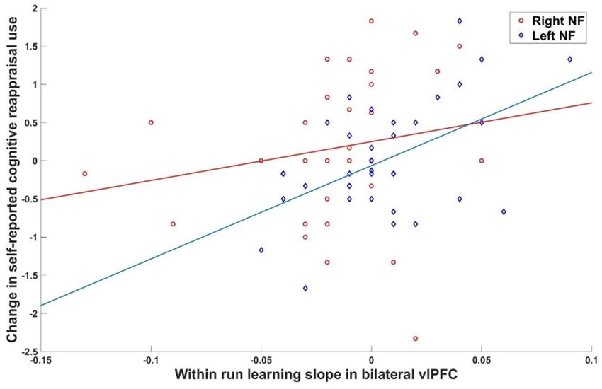
Keller et al.                                                                                       fMRI Neurofeedback-Enhanced Reappraisal Training

sample consisted of 39 patients (35.2 ± 2.2 years; 17 female)               more successful cognitive reappraisal could be associated with
and 37 healthy controls (32.3 ± 2.1 years; 15 female). Groups               increased activation in the target region and higher NF scores.
did not differ with respect to age [t (74) = −0.96, p = 0.34] and
years of education [t (74) = 0.78, p = 0.44] (Table 1 for more              NF Task
information). The study was pre-registered at clinicaltrials.gov            The NF task was adapted from our previous study in patients
(NCT7171). Due to constraints in recruitment procedures the                 with PTSD (71) and consisted of 18 blocks of picture presentation
subsample of patients with schizophrenia has not been completed             (12 s each; see Figure 1B). The task was either to passively view
until now. The study was approved by the local Ethics Committee             a picture and to allow spontaneous thoughts and emotions to
of the RWTH Aachen (EK 050/17) and all participants provided                occur (“view” condition) or to upregulate the BOLD signal in
written informed consent.                                                   the respective target region by using a cognitive reappraisal
                                                                            strategy to reduce negative feelings associated with the presented
                                                                            picture (“reappraise” condition). The condition was indicated by
                                                                            a fixation cross (“x” = view; “+” = reappraise). Each picture
Experimental Procedure                                                      presentation (12 s) was followed by a 4 s rest period (display of
In a randomized, double-blind cross-over design (see Figure 1A),
                                                                            “x” or “+”) and 4 s presentation of either a numerical feedback
participants were trained to upregulate their brain activation
                                                                            value (1–99) after “reappraise” condition or a placeholder (“%%”)
in the anatomically defined region of interest (ROI) (left or
                                                                            after the “view” condition, respectively. During the “reappraise”
right ventrolateral prefrontal cortex, vlPFC). At the first visit,
                                                                            condition participants were free to try strategies related to
patients underwent the SCID interview and were assessed on
                                                                            cognitive reappraisal. They were, however, asked to stick to
the items of the Hamilton Rating Scale for Depression (HAM-
                                                                            the same strategy during a regulation block. Prior to and
D) (74). Furthermore, all participants completed questionnaires
                                                                            following each NF run, participants indicated their emotional
and received a standardized cognitive reappraisal training and
                                                                            state using a Self-Assessment Manikin (SAM) from 1 to 9
instructions about the NF training. The second visit entailed two
                                                                            (valence and arousal).
baseline cognitive reappraisal runs without NF (1st run: decrease
negative feelings in response to aversive pictures; 2nd run:                Stimuli
increase positive feelings in response to pleasant pictures) as well        For the NF training, 72 pictures with negative valence
as anatomical recordings. However, second day data is not part of           were selected from the International Affective Picture System
the current analyses. The NF training was completed on the third            (IAPS). The IAPS contains pictures validated on valence (1–9;
and fourth visit with random allocation to the order of left vs.            1 = extremely negative) and arousal (1–9; 1 = no arousal) (75).
right vlPFC regulation. This was done in a double-blind manner              From these 72 pictures (see Appendix A), two sets of 36 pictures
as investigators saw the time-courses of both left and right vlPFC          were selected which were matched for overall valence and arousal
ROIs during the NF training, however, were blind to which ROI               (Set1: MeanValence = 2.55 ± 0.31, MeanArousal = 5.82 ±
was used for feedback computation. Trainings were separated by              0.53; Set2: MeanValence = 2.54 ± 0.36, MeanArousal = 5.81
at least 1 week. On each NF day, participants completed four                ± 0.48) and the two semi-randomized versions of these sets
NF runs (∼7 min each), each comprised of 9 regulation blocks.               were randomized over days. Each complete set consisted of two
Participants received intermittent numerical feedback, signaling            subsets of 18 pictures. One subset was used for Run 1 and 3
the increase in brain activation within the target ROI. Each NF             whereas the other was used for Run 2 and 4. The order of
training was preceded and followed by a resting state (RS) fMRI             “view” and “reappraise” was switched within each subset for each
measurement. In a follow-up telephone interview 4 weeks after               repetition. Within subsets “view” and “reappraise” pictures were
completing the last NF training, the change of symptomatology,              also matched for valence and arousal. All chosen pictures were
affect, and emotion regulation strategy use were assessed.                  related to one of four categories (“accident,” “assault,” “sadness,”
                                                                            “other”) and each “view-reappraise” cycle showed two pictures of
Cognitive Reappraisal Training for Emotion                                  the same category.
Regulation
A 30-min training entailed instructions and information about               Questionnaires and Neuropsychological
the NF procedure and training on the content and application of             Assessment
reappraisal strategies. Participants were told that the goal of the         Symptom severity was assessed at different time points using
NF training was to temporarily enhance activation in a region               well-established measures of depression and related features. At
of the brain that has been associated with the regulation of                baseline, the patients completed the German version of the 21-
emotions. Emotional reactions to negative stimuli were discussed            item Hamilton Rating Scale for Depression (74) as well as the
and participants learned to apply reappraisal strategies. In                Becks Depression Inventory (BDI-II) (76), the Hospital Anxiety
specific, participants saw negative stimuli and were instructed             and Depression Scale (HADS) (77), the Chapman Anhedonia
to use reappraisal strategies to reduce their negative affect.              Scale (78), the Emotion Regulation Questionnaire (ERQ) (73),
Suggested strategies were to think (1) the situation will change in         and the Heidelberg Form for Emotion Regulation Strategies
the future, (2) the situation is not as bad as it looks. Participants       (HFERST) (79). The BDI-II as well as the ERQ and HFERST
could also imagine that the situation is not real or change their           were repeated on the day of the first MRI measurement if the
perspective (e.g., professional). Lastly, it was explained that a           time between baseline and first fMRI measurement was >1 week

Frontiers in Psychiatry | www.frontiersin.org                           4                                    August 2021 | Volume 12 | Article 715898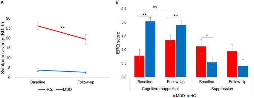
Keller et al.                                                                                                                  fMRI Neurofeedback-Enhanced Reappraisal Training

TABLE 1 | Demographic and clinical data.

                                                                       MDD                                        HC                                                Comparison
                                                                     (n = 39)                                  (n = 37)

                                                                 Mean                 SD               Mean                  SD                Df               t                   p

Age (years)                                                       35.2                13.6              32.3                12.8               74            −0.96                0.34
Education (years)                                                 14.3                2.5               14.8                 2.6               74             0.78                0.44
Parental education (years)                                        13.7                2.7               13.6                 3.2               67            −0.16                0.88
Socioeconomic status (monthly income in Euro)                    1,372               1,191             1,115               1,034               72            −0.99                0.33
Clinical characteristics
ERQa -
Reappraisal                                                       3.8                 1.3                5.0                 0.8               71              4.7
Keller et al. fMRI Neurofeedback-Enhanced Reappraisal Training
FIGURE 1 | (A) Experimental procedure. The NF training entailed 5 visits to the lab, starting with initial assessment and cognitive reappraisal training (Visit 1), followed
by baseline cognitive reappraisal runs and anatomical recordings (Visit 2), two NF training days (Visit 3 and 4) and a follow-up assessment (Visit 5). (B) Experimental
paradigm during NF training. All participants completed 4 NF runs on each of the two NF days. Each NF run entailed 9 view-regulate cycles. During “view” trials (“x”),
participants passively viewed the picture and could allow spontaneous thoughts and emotions. On “regulate” trials (“+”) participants reappraised the picture to reduce
the negative affect and upregulate the BOLD signal in the target area. Each picture was presented for 12 s, followed by a 6-s fixation cross and 4-s (pseudo-)
neurofeedback interval.
NF training were rated on valence and arousal after each training Online Real-Time fMRI Analysis
day and the applied regulation strategies were noted. To assess Online analysis of functional data during real-time fMRI was
cognitive performance, a verbal intelligence test (Wortschatztest performed using Turbo-BrainVoyagerTM (TBV) Version 3.2
– WST) (81), a working memory test measuring the capacity (Brain Innovation, Maastricht, NL) as elsewhere described in
to store numbers (digit-span task) as well as the Digit Symbol more detail (82). Online preprocessing included 3D motion
Substitution Test were administered. detection and correction as well as intra session alignment
for subsequent NF runs (alignment to reference volume of
fMRI Data Acquisition the first run), linear trend removal, spatial smoothing with
A 3.0 T whole body scanner (Magnetom TRIO, Siemens Medical 3 mm Gaussian smoothing kernel and temporal filtering (drift
Systems, Erlangen, Germany) with a standard 20-channel head removal). Statistics were computed incrementally using a general
coil was used to acquire the fMRI data. For baseline cognitive linear model (GLM) based on the predefined stimulation
reappraisal runs as well as NF runs, 230 T2∗ - weighted protocol. The BOLD percentage signal change within the ROI
whole-brain functional images were recorded using echo-planar was calculated using the “reappraise > view” contrast and
imaging (TR = 2,000 ms, TE = 28 ms, flip angle = 77◦ , fed back as a positive number between 1 and 99 reflecting
voxel size = 3 × 3 × 3 mm, matric size = 64 × 64, 0–1% BOLD signal change. Feedback was computed and
ascending interleaved acquisition of 34 transverse slices, 3 mm presented with custom scripts running under Matlab R2014a
slice thickness, 0.75 mm gap). Furthermore, high resolution T1- (The MathWorks Inc., Natick, MA).
weighted images were acquired using a MPRAGE sequence
(TE = 3.03 ms, inversion time TI = 900 ms, TR = 2,000 ms, flip ROI Definition
angle = 9◦ , FOV = 256 × 256 mm2, 1 mm isotropic voxels, Predefined anatomical ROIs, namely the left and right vlPFC,
0.5 mm gap, 176 sagittal slices). were used for NF training. The chosen ROIs are major hubs
Frontiers in Psychiatry | www.frontiersin.org 6 August 2021 | Volume 12 | Article 715898Keller et al. fMRI Neurofeedback-Enhanced Reappraisal Training
of the cognitive reappraisal network and were based on peak signal change values below 5%. Three participants (MDD: n = 1)
coordinates provided by a meta-analysis about the neural had single runs exceeding this threshold. However, the first level
correlates of cognitive reappraisal in healthy individuals (37). fMRI results of these participants did not show significant motion
The lateral PFC has been shown to be consistently recruited Artifacts upon visual inspection and were therefore included.
during cognitive reappraisal of negative stimuli and activation Average PSC values did not differ between groups (HC: 2.48 ±
in the vlPFC is altered in depression (5). For a comparable 0.7; MDD: 2.48 ± 0.5; t (74) = −0.02, p > 0.05) and indicate
selection of left and right vlPFC, the mirrored center of the MNI adequate data quality. Movement parameters did not exceed
coordinates of left and right vlPFC (left vlPFC: −42,22, −6; right 3 mm within any NF run.
vlPFC: 50,30, −8) (37) were selected as respective ROIs (± 46,
26, −7) and transformed to Tailarach space (± 44, 22, 3) using Preprocessing
the “mni2tal” web application (https://bioimagesuiteweb.github. fMRI data was preprocessed in Matlab R2018b (Mathworks
io/webapp/mni2tal.html). Based on these coordinates, ROIs (10 Inc., Natick, MA) using the Statistical Parametric Mapping
× 10× 10 mm) were created for the left and right vlPFC 12 toolbox (SPM12; https://www.fil.ion.ucl.ac.uk/spm/software/
using BrainVoyager QX 2.8 (Brain Innovation, Maastricht, NL). spm12/). To minimize T1-saturation effects, the first five volumes
These standardized ROIs were applied using coregistration of of each NF run were discarded for data analysis. Functional fMRI
individual anatomical scans to the Tailarach template. data were realigned to the first volume (6 movement parameters).
Furthermore, fMRI data was co-registered to the participant’s
TBV Based ROI Analysis structural T1 image, smoothed with an 8 mm FWHM Gaussian
To test our primary hypothesis concerning a learning effect of kernel and normalized to the T1-weighted ICBM152 brain
self-control over the neuronal activity within the target ROIs template of the Montreal Neurological Institute (MNI). Data
we examined the feedback data recorded during NF trainings. from all participants was visually inspected after preprocessing
Investigation of the learning effect is in line with the consensus to ensure adequate coregistration and normalization.
paper by Ros et al. (83). To avoid confounds such as order
effects, only the first day of NF training was used for this analysis. Whole Brain Analysis
The ROI data created by TBV during online processing was Brain mapping analyses were performed using SPM12. On the
exported to and analyzed in Matlab R2018. Similar to the online first level, the six movement parameters were added as covariates
FB calculation, differences between regulation and view blocks of no interest. The main contrast of interest (reappraise > view)
were calculated for each regulation trial (4 runs each with 9 from the first level analysis was used in a 2 × 2 × 2 × 4 full
reappraise-view trials) to investigate the learning effect within factorial model [group (HC, MDD) × gender (female, male)
runs. Learning within runs was defined as a linear increase as × condition (Left, Right), run (NF1, NF2, NF3, NF4)]. Results
suggested by the “consensus” (83), computed as linear regression were evaluated after application of a voxel-wise threshold of p <
slope across trials for each NF run. Separate independent t-tests 0.001 and family-wise error (FWE) correction of pFWE < 0.05 at
investigated differences in learning in the left and right ROI voxel level.
between groups and between receiving left vs. right feedback. Generalized Psychophysiological Interaction
Associations between learning success (average learning slope A generalized psychophysiological interaction (gPPI) was
in vlPFC ROI) with changes in self-rating of depressive severity computed using the functional connectivity toolbox CONN
[BDI-II Total score (Post NF1) – BDI-II Total score (baseline)] (www.nitrc.org/projects/conn, RRID:SCR_009550). Signal
and cognitive reappraisal [ERQ-CR Total score (Post NF1) – variance that correlated with the seed region during the
ERQ-CR Total score (baseline)] were calculated. Accordingly, regulation compared to view condition (“reappraise – view”) was
improvement of depressive symptomatology was indicated by investigated. The bilateral vlPFC was chosen as a seed and was
a negative change score whereas improvement on cognitive created based on the target regions. Second-level results were
reappraisal use was linked to a positive change score. One-tailed evaluated at p < 0.001 uncorrected voxel level and with p < 0.05
testing was chosen based on the assumption of a negative relation FDR-correction at the cluster level.
between learning and symptoms of depression and a positive
relation between learning and cognitive reappraisal use. Statistical Analyses
Statistical analyses were performed in IBM SPSS Statistics
Offline Data Processing and Analysis (version 26). Independent samples t-tests were computed to
Quality Assurance of MRI Data investigate differences in baseline questionnaires (e.g., BDI-II,
To ensure high quality functional and structural MRI data, all ERQ, neuropsychological tests, PANAS) as well as demographic
data sets were examined within 48 h following recording using measures (age, educational level) between groups. To investigate
a standardized quality assurance pipeline developed and used by changes in symptom severity (BDI-II) and use of cognitive
the Psychiatric Imaging Network Germany (PING; ping-rwth- reappraisal strategies from baseline to follow-up measurement,
aachen.de). Quality of structural data was assured by the quality two 2 × 2 × 2 [Time (NF1, NF2) × Group (MDD, controls)
parameters of the Computational Anatomy Toolbox (CAT) (84). × Condition (L-R, R-L)] repeated measures ANOVAs were
Further, the procedure entailed the assessment of functional data computed. Furthermore, 2 × 2 × 2 repeated measures ANOVAS
within the Automated Quality Assurance toolbox (AQuA) (85). [Time × Condition × Group] were calculated separately for
All fMRI data used for further analyses had (on average) percent SAM valence and arousal ratings. Lastly, to investigate the
Frontiers in Psychiatry | www.frontiersin.org 7 August 2021 | Volume 12 | Article 715898Keller et al. fMRI Neurofeedback-Enhanced Reappraisal Training
subjective experience during NF trainings, three 2 × 2 × 2 Different metacognitive parameters of self-control were
repeated measures ANOVAs (Time x Condition × Group) of assessed before and after each NF session (also see Appendix B).
metacognitive parameters of self-control (perceived intensity Before each NF training, participants were asked whether they
of general control, perceived intensity to control brain signal, think they are generally able to control their brain activity (yes/no)
perceived success to use cognitive reappraisal strategies) were and asked for the intensity of control. On the first day, more
computed. Post-hoc t-tests were performed whenever suitable. A healthy individuals than patients with MDD indicated that they
p-value of < 0.05 was considered statistically significant. generally have control over their brain activity [HC: 90%, MDD:
53%; χ 2(1, 67) = 11.2, p = 0.001] whereas this difference was not
significant anymore at the second training [HC: 90%, MDD: 71%;
RESULTS χ 2(1, 67) = 3.5, p = 0.06]. A 2 × 2 × 2 repeated measures ANOVA
[Time (NF1, NF2) × Condition (Left, Right) × Group (MDD,
Demographic and Clinical Data control); Table 2] of perceived intensity of general control (1–10)
Groups did not significantly differ regarding age, (parental) revealed a significant main effect of time [Day1: 4.99 ± 2.1, Day2:
education, socioeconomic status, or basic cognitive functioning 5.61 ± 1.9; F (1, 62) = 7.7, p = 0.007] as well as a significant group
such as working memory, verbal IQ, and attention (all p > difference [MDD: 4.71 ± 1.7, HC: 6.03 ± 1.7; F (1, 62) = 9.97,
0.2, see Table 1) as well as in gender ratio [χ 2 (1, 76) = 0.07, p = 0.002]. The increase of patients’ positive evaluations of the
p > 0.2]. However, there were significantly more smokers in ability to control one’s brain activity combined with increasing
the patient group [HC: 6, MDD: 18, χ 2 (1, 74) = 8.9, p < 0.01]. perceived intensity of control indicates an increase of self-efficacy
As expected, patients with depression showed elevated baseline across NF days.
scores on depression [BDI-II: t (73) = −10.8, p < 0.001; HADS-
depression: t (74) = −9.5, p < 0.001] as well as HADS-anxiety NF Effects
scores [t (74) = −6.9, p < 0.001] compared to HCs (see Table 1). To test whether learning success was specific to the left vs.
On the 21-item HAM-D, patients had average scores of 16.5 (± right vlPFC NF condition, ROI data of the first NF day was
7.5) indicating mild to moderate depressive symptoms. Prior to investigated. Within run learning slopes were steeper when
the first fMRI measurement, patients showed higher negative receiving left as compared to right ROI feedback for both vlPFC
affect [MDD: 20.0 ± 9.2, HC: 12.4 ± 4.3; t (74) = −4.6, p < ROIs [left vlPFC: t (74) = 2.55, p = 0.01; right vlPFC: t (74) = 3.73,
0.001] and lower positive affect [MDD: 26.9 ± 6.6, HC: 32.5 p < 0.001]. This confirmed the primary hypothesis of a regional
± 7.4; t (74) = 3.5, p = 0.001] than healthy individuals assessed specific NF effect meaning that receiving feedback from the left
through the PANAS. Furthermore, HCs indicated to use more ROI was advantageous over feedback from the right ROI. MDD
cognitive reappraisal strategies [t (71) = 4.7, p < 0.001] and less and controls did not differ at either ROI [left vlPFC: t (74) = 0.51,
suppression [t (71) = −2.4, p < 0.05] than patients with MDD p > 0.2; right vlPFC: t (74) = 0.96, p > 0.2]. Learning slopes
(ERQ). The HFERST subscales further supported the clinical within NF runs were significantly correlated with the change in
picture with lower scores of patients on reappraisal, acceptance, self-reported cognitive reappraisal use from baseline to after the
problem solving (all p < 0.001), and social support (p < 0.01) and first NF training when receiving feedback from the left vlPFC
higher scores on rumination (p < 0.001), avoidance (p < 0.01), (bilateral vlPFC ROI: r = 0.484, p = 0.002) but not from the
experience suppression (p < 0.05), and expressive suppression right vlPFC (bilateral vlPFC ROI: r = 0.170, p = 0.150; Figure 2).
(p = 0.08) compared to HCs. However, there was no association between learning slopes on
Two separate 2 × 2 × 2 repeated measures ANOVAs (Time the first day and change of severity of symptoms of depression
× Group × Condition) investigated mean SAM valence and (left feedback: r = 0.09, p = 0.33; right feedback: r = 0.04,
arousal ratings across NF days, conditions, and groups (Table 2). p = 0.41). This indicates that receiving feedback from the left
The average SAM valence ratings were similar across NF days as compared to the right vlPFC did not have a detectable effect
[F (1, 72) = 0.05, p > 0.2) and conditions [F (1, 72) = 0.1, p > 0.2], on depressive symptomatology, however, left over right vlPFC
however, significantly different between groups [F (1,72) = 24.9, p feedback showed an advantage for increasing the subjective use
< 0.001] with higher (more positive) valence ratings for healthy of cognitive reappraisal strategies. Values fed back to participants
individuals (6.7 ± 1.3) than patients with depression (5.4 ± during NF training are shown in Appendix C.
1.25). Furthermore, there was a significant group x condition
interaction [F (1, 72) = 6.4, p < 0.05] and post-hoc tests indicated Offline Analysis
that HCs showed a significant difference between conditions on Whole-Brain Analysis
the second [t (35) = −2.1, p < 0.05] but not first day of NF Whole-brain activations related to NF training with cognitive
[t (35) = −1.1, p = 0.30] whereas patients with depression showed reappraisal were investigated with a 2 × 2 × 2 × 4 full factorial
a significant difference between conditions on day 1 [t (37) = 2.1, model [Group (MDD, control) × Gender (Female, Male) ×
p < 0.05] but not on day 2 [t (37) = −1.1, p = 0.14] of NF Condition (L-R, R-L) × Run (NF1, NF2, NF3, NF4)] which
training. A repeated measures ANOVA of mean arousal ratings revealed a main effect of group showing involvement of a dorsal
showed no significant difference between NF days [F (1, 72) = 1.6, fronto-parietal network during NF training (Figure 3A; top;
p > 0.2], between conditions [F (1, 72) = 0.2, p > 0.2] or groups Table 3). Healthy controls exhibited more activation in the right
[F (1, 72) = 2.5, p = 0.12] indicating similar arousal throughout (and to some extent left) opercular and triangular part of the
NF trainings, across groups and conditions. inferior frontal gyrus (IFG), the right middle temporal gyrus,
Frontiers in Psychiatry | www.frontiersin.org 8 August 2021 | Volume 12 | Article 715898Keller et al. fMRI Neurofeedback-Enhanced Reappraisal Training
TABLE 2 | Repeated-measures ANOVAs of change from baseline to follow-up (BDI-II and ERQ) and change of general perceived control across neurofeedback trainings.
Parameter Source F p
Mean SAM valence ratings at neurofeedback day 1 and 2 Time 0.05 0.83
Group 24.9Keller et al. fMRI Neurofeedback-Enhanced Reappraisal Training FIGURE 2 | Brain behavior relationship. Association between within run learning during the first NF day and change in cognitive reappraisal use was significant for left (r = 0.484, p = 0.002) but not right (r = 0.170, p = 0.150) vlPFC feedback. FIGURE 3 | Neural correlates of emotion regulation. (A) The main effect of group revealed a widespread network active during NF including the bilateral prefrontal cortex, precentral gyrus, SMA, MCC, bilateral occipital and superior parietal areas, thalamus, and cerebellum. Healthy individuals showed a stronger engagement of prefrontal areas while patients showed more activation in cingulate areas. (B) The main effect of gender revealed engagement of an extensive network during emotion regulation in males including the bilateral prefrontal cortex, SMA, dmPFC, bilateral precentral and occipital gyrus and bilateral thalamus. Female participants showed stronger activation in the bilateral angular gyrus. (C) Learning and reduction across NF runs 1 to 4. Overall, participants showed an increase in the bilateral fusiform gyrus and occipital lobe as well as a decrease of activation in the right insula and the MCC. bilateral occipital lobe (Figure 3B; top; Table 3) and female an increase in bilateral fusiform gyrus and occipital lobe as participants showing more activation in the bilateral angular well as a decrease of activation in the right insula (Figure 3C; gyrus (Figure 3B; bottom; Table 3). Across NF runs there was Table 3). Frontiers in Psychiatry | www.frontiersin.org 10 August 2021 | Volume 12 | Article 715898
Keller et al. fMRI Neurofeedback-Enhanced Reappraisal Training
TABLE 3 | Activation peaks associated with NF-guided cognitive reappraisal.
Cluster Brain region MNI coordinates T kE
x y z
All (regulate > view)
1 Bilateral IFG, dorsal ACC, MCC, dmPFC, −4 6 62 52.84 67,733
SMA, SFG, STG, angular gyrus, thalamus,
striatum, occipital gyrus, pre-/postcentral
gyrus, cerebellum, superior parietal lobe
Healthy controls > MDD (regulate > view)
1 Left MFG −36 56 20 9.07 124
2 Right IFG 62 20 10 8.76 321
3 Right MFG 36 2 44 8.14 393
4 Right SFG 10 60 34 8.04 102
5 Left superior occipital gyrus −24 −92 30 7.60 223
6 Right MTG 44 −42 4 6.81 82
7 Left SMA −18 12 68 6.76 202
8 Right superior occipital gyrus 22 0.94 28 6.19 82
MDD > Healthy controls (regulate > view)
1 Left anterior insula extending into frontal −34 28 10 9.02 181
operculum
2 Right pre-/postcentral gyrus 62 2 22 8.06 506
3 Left pre-/postcentral gyrus −60 −8 42 7.45 486
4 MCC −8 −4 42 7.05 893
5 ACC 10 38 22 6.79 441
6 Supplementary motor cortex −6 −20 74 5.79 116
7 PCC −12 −50 32 5.77 124
Male > Female (regulate > view)
1 Bilateral IFG, pre-/postcentral gyrus, SMA, −4 6 62 52.84 78,444
dmPFC, SFG, thalamus, striatum, occipital
gyrus, left STG
Female > Male (regulate > view)
1 Right angular gyrus 44 −62 56 9.41 1,632
2 Left angular gyrus −48 −70 44 6.50 386
3 ACC extending into left MFG −12 38 −4 5.78 1,122
Learning over time (regulate > view)
1 Left superior occipital gyrus −12 −90 2 6.32 121
2 Left fusiform gyrus −26 −44 −8 6.31 173
3 Right fusiform gyrus 24 −40 −12 5.96 249
Reduction over time (regulate > view)
1 Right anterior insula 20 30 14 5.20 97
p < 0.001 voxel threshold and p < 0.05 FWE-corrected at voxel level.
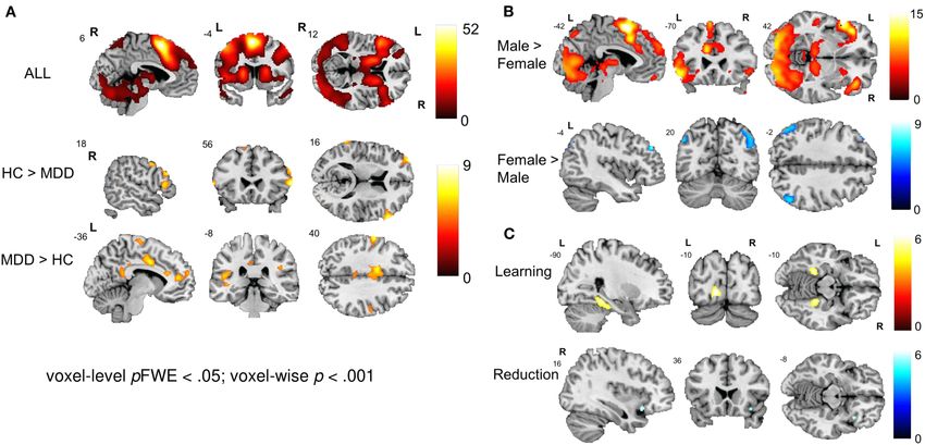
Connectivity Analysis gyrus (x = −34, y = −94, z = 0, T = 5.03; x = 44, y = −82, z = 10,
Task-dependent changes in functional connectivity of the T = 4.56). Direct group comparison revealed significantly lower
bilateral vlPFC during neurofeedback was studied with a functional connectivity during NF in the left superior parietal
generalized Psychophysiological Interaction analysis in the cortex in patients compared with healthy controls (x = −46,
contrast reappraise vs. view. In healthy controls, coupling y = −48, z = 34, T = −6.69) (Figure 4). In a post-hoc analysis,
increased with the left (x = −58, y = −52, z = 46, T = 4.64) baseline BDI scores were negative linear predictors for the
and right superior parietal cortex (x = 60, y = −42, z = 32, connectivity measure at this location [T (72) = −2.99, p = 0.004].
T = 4.68) and decreased with the ACC (x = 12, y = 18, z = 0,
T = −6.69), precuneus/PCC (x = 4, y = −38, z = 8, T = −5.27), Follow-Up Assessments
right superior frontal gyrus (x = 24, y = 30, z = 44, T = −4.81). Four weeks after the last NF training, all participants were
In the patients, neurofeedback enhanced coupling with the left contacted to fill out questionnaires on their current depressive
(x = 10, y = −102, z = 2, T = 5.14) and right inferior occipital symptomatology, their state of affect, their use of emotion
Frontiers in Psychiatry | www.frontiersin.org 11 August 2021 | Volume 12 | Article 715898Keller et al. fMRI Neurofeedback-Enhanced Reappraisal Training
that patients on average showed a meaningful reduction of BDI-
II scores from baseline to follow-up. Furthermore, investigation
of MCID on an individual level indicated a clinically meaningful
change (change ≥5 or 17.5%) in 55% of patients who completed
the follow-up interview.
Change of Emotion Regulation (ERQ)
A 2 × 2 × 2 [Time (Baseline, Follow-up) ×Group (HC, MDD)
× Condition (L-R, R-L)] repeated measures ANOVA of cognitive
reappraisal use at baseline and follow-up showed non-significant
main effects of time [F (1,59) = 2.2, p = 0.14] and condition
[F (1, 59) = 0.85, p = 0.36], but a significant main effect of group
[F (1,59) = 14.3, p < 0.001] as well as significant time∗ group
interaction [F (1, 59) = 5.9, p = 0.02]. Post-hoc tests (see Figure 5B)
indicated that patients had a significant increase in cognitive
reappraisal use [t (30) = −2.5, p = 0.02, d = 0.43] from baseline
to follow-up, whereas healthy individuals showed a stable use
of cognitive reappraisal [t (31) = 0.80, p > 0.2]. The difference
between groups remained significant at follow-up [t (64) = 2.1, p
< 0.05]. A similar repeated measures ANOVA of self-reported
suppression revealed only a marginally significant main effect of
FIGURE 4 | Psychophysiological interaction during regulation. Results of the group [F (1, 59) = 3.9, p = 0.05]. Post-hoc tests indicated that the
generalized psychophysiological interaction analysis for the contrast ‘regulate
difference between groups for suppression that was significant
– view’ with bilateral vlPFC as seed. Healthy controls showed increased
coupling between the seed region and bilateral superior parietal cortex and
at baseline [t (71) = 2.1, p = 0.02] was not significant at follow-
negative coupling between the seed and ACC as well as PCC. Patients up [t (64) = −1.4, p > 0.1], indicating that use of suppression in
showed positive coupling between the seed and bilateral occipital cortex. patients equalized with that of healthy individuals. Interestingly,
Compared with healthy controls, patients with depression showed significantly male participants used more suppression compared to female
reduced coupling between the bilateral vlPFC and left superior parietal cortex.
participants at baseline [t (71) = −2.6, p < 0.01] and follow-up
All results are reported at p < 0.001 voxel threshold and p < 0.05
FDR-corrected at cluster level. Bar plots indicate size of effect (beta values) at [t (64) = −2.3, p = 0.02] whereas there were no gender differences
the voxel showing the maximum coupling effect. for cognitive reappraisal use at baseline [t (71) = 0.72, p > 0.2] or
follow-up [t (64) = 0.14, p > 0.2].
Subjective Experience
regulation strategies and experience with the NF training as At follow-up, the application of learned strategies in everyday
well as applications of learned strategies in everyday life. Not life was assessed. Seventy-five percentage of patients with MDD
all participants could be recontacted for follow-up assessments and 57% of healthy individuals indicated that they had used the
(MDD: 32, HC: 35) and the following analyses are based on the strategies over the past month. Of these, all patients and 89%
available subset of data. Please see Table 2 for detailed results. of controls experienced the applied strategies as helpful. Patients
reported that reappraisal strategies made them feel less frustrated,
Symptom Change more relaxed, more optimistic, more aware of the situation, and
A 2 × 2 × 2 [Time (Baseline, Follow-up) × Group (HC, MDD) × led to improved mood which indicates that generalization of
Condition (L-R, R-L)] repeated measures ANOVA of depressive the training to negative situations in everyday life was high.
symptom severity (BDI-II) showed a significant main effect of Furthermore, 89% of controls and 91% of patients were willing
time [F (1, 60) = 23.7, p < 0.001], a significant effect of group to repeat such a NF training which indicates high acceptance of
[F (1, 60) = 100.3, p < 0.001] whereas the main effect of condition the NF training.
did not reach significance [F (1, 60) =.12, p = 0.73]. Furthermore,
the time∗ group interaction was significant [F (1, 60) = 15.0, DISCUSSION
p < 0.001] while the time∗ condition interaction did not show
significance [F (1,60) = 1.1, p = 0.29]. Post-hoc paired t-tests In this double-blind cross-over rtfMRI study we tested the
showed that the reduction of symptom severity from baseline to feasibility and clinical efficacy of NF-supported cognitive
follow-up was significant for patients [9.2 ± 11.8; t (28) = 4.2, reappraisal training in patients with depression and a matched
p < 0.001, d = 0.77; Figure 5A] but not for HCs [1.0 ± 3.3; group of healthy individuals. In specific, we investigated whether
t (34) = 1.9, p = 0.07, d = 0.24]. Previous studies have shown left- or right hemispheric ventrolateral prefrontal cortex (vlPFC)
that the minimal clinically important difference (MCID) of BDI- NF would be a neurologically and clinically more suitable
II scores should be a change of at least 5 points (86) or 17.5% region for NF during cognitive reappraisal. During 2 days
(87) depending on the baseline severity of depression. The mean of NF training, participants trained to regulate their brain
change of 9.2 points (27.7% reduction) in our sample indicates signal in the vlPFC using reappraisal strategies in response to
Frontiers in Psychiatry | www.frontiersin.org 12 August 2021 | Volume 12 | Article 715898Keller et al. fMRI Neurofeedback-Enhanced Reappraisal Training
FIGURE 5 | Change of severity of depression and emotion regulation ability from baseline to follow-up. (A) Symptom severity measured by BDI-II showing a significant
decrease of symptom scores from baseline to follow-up only in patients with depression [9.2 ± 11.8; t(28) = 4.2, p < 0.001]. (B) Patients showed a significant increase
of cognitive reappraisal and a stable level of suppression strategies. Healthy individuals had stable levels of cognitive reappraisal and suppression. Error bars indicate
standard errors. **p < 0.001, *p < 0.05.
negative pictures and guided by intermittent NF (“reappraise > (especially left) vlPFC a promising target for future NF-guided
view”). This paradigm has already been applied successfully in rt-fMRI studies.
healthy individuals (70) and patients with post-traumatic stress Behaviorally, patients with depression can achieve
disorder (PTSD) (71). Overall, the cognitive reappraisal evoked downregulation of negative emotions by applying reappraisal
predicted responses within the emotion regulation network strategies within clear laboratory boundaries. However,
such as activations in prefrontal, motor, and subcortical areas. Zilverstand et al. (5) have shown that patients with depression
Furthermore, our ROI analysis revealed that NF of left compared may – despite similar behavioral regulation success – display
to right vlPFC activity specifically enhanced bilateral prefrontal dysfunctions in a cognitive control network for negative
activation during reappraisal in patients with depression as well emotions of which the vlPFC offers a promising NF target.
as in age and gender matched healthy individuals. Such laterality Our findings from the first NF day suggest a beneficial effect
effect did not survive the correction for multiple testing in the of receiving left compared to right vlPFC feedback reflected by
whole-brain analysis. First, it has to be taken into account that enhanced bilateral vlPFC activation and a significant increase
the size of lateralization effects is usually limited, e.g., about in reappraisal strategy use specific for left vlPFC feedback. The
30% lower responses at the non-dominant hemisphere to speech vlPFC has repeatedly been found to support the selection of
stimuli (88). Secondly, recent data show a relevant contribution appropriate reappraisals (37, 38) and left vlPFC activity during
of the right vlPFC [e.g., (50)]. However, a larger learning effect reappraisal differentiates healthy individuals from patients with
in response to left vs. right vlPFC feedback in the ROI analysis depression (5, 40). Furthermore, more evidence for a superior
suggests a regional specificity of our rt-fMRI-based NF paradigm role of the left compared to right vlPFC for cognitive reappraisal
and may further support a central causal role of this region for has been provided by previous studies (52, 59).
cognitive reappraisal (41, 52, 89). Further investigation on the whole-brain level showed that
NF learning on the first day of training was related to healthy individuals and patients with depression recruited
improvements in self-rated reappraisal use only when different regions of the emotion regulation network during NF-
receiving NF from the left rather than right vlPFC but was supported reappraisal training. The effect of interest showed
not associated with change of depressive symptomatology. extensive activation in the bilateral vlPFC, dlPFC, dACC, (pre-
Nevertheless, 55% of patients showed a clinically meaningful ) SMA, dmPFC, SFG, MFG, STG, angular gyrus, thalamus,
change in depression scores (BDI-II) from baseline to striatum, occipital gyrus, pre- and postcentral gyrus, cerebellum
follow-up and 75% of patients reported that they had and superior parietal lobe during cognitive reappraisal. This is
successfully applied the learned cognitive strategy in everyday consistent with the emotion regulation network areas commonly
life. Our study supports a positive effect of rt-fMRI NF recruited during reappraisal (36–38). Furthermore, in the group
for the enhancement of emotion regulation for patients comparison, healthy individuals displayed more recruitment of
with depression (90). The combination of specific NF cortical areas including the right IFG, left SMA, bilateral middle
effects, reappraisal skill learning, reduction of depressive frontal gyrus, right superior frontal gyrus, right middle temporal
symptomatology and a high acceptance of training yield the gyrus and the bilateral superior occipital gyrus. Deficient
Frontiers in Psychiatry | www.frontiersin.org 13 August 2021 | Volume 12 | Article 715898You can also read