Peptides from Cauliflower By-Products, Obtained by an Efficient, Ecosustainable, and Semi-Industrial Method, Exert Protective Effects on ...
←
→
Page content transcription
If your browser does not render page correctly, please read the page content below
Hindawi
Oxidative Medicine and Cellular Longevity
Volume 2019, Article ID 1046504, 13 pages
https://doi.org/10.1155/2019/1046504

Research Article
Peptides from Cauliflower By-Products, Obtained by an Efficient,
Ecosustainable, and Semi-Industrial Method, Exert Protective
Effects on Endothelial Function

          C. Caliceti ,1 A. L. Capriotti,2 D. Calabria,1 F. Bonvicini,3 R. Zenezini Chiozzi,2
          C. M. Montone,2 S. Piovesana,2 M. Zangheri,1 M. Mirasoli,1 P. Simoni,4 A. Laganà,2
          and A. Roda 1
          1
            Department of Chemistry “Giacomo Ciamician”, Alma Mater Studiorum-University of Bologna, Bologna, Italy
          2
            Department of Chemistry, Università di Roma “La Sapienza”, Rome, Italy
          3
            Department of Pharmacy and Biotechnology, Alma Mater Studiorum-University of Bologna, Bologna, Italy
          4
            Department of Medical and Surgical Sciences, Alma Mater Studiorum-University of Bologna, Bologna, Italy

          Correspondence should be addressed to A. Roda; aldo.roda@unibo.it

          Received 20 July 2018; Revised 4 November 2018; Accepted 12 November 2018; Published 6 February 2019

          Academic Editor: Tullia Maraldi

          Copyright © 2019 C. Caliceti et al. This is an open access article distributed under the Creative Commons Attribution License,
          which permits unrestricted use, distribution, and reproduction in any medium, provided the original work is properly cited.

          The large amount of cauliflower industry waste represents an unexplored source of bioactive compounds. In this work, peptide
          hydrolysates from cauliflower leaves were characterized by combined bioanalytical approaches. Twelve peptide fractions were
          studied to evaluate unexplored biological activities by effect-based cellular bioassays. A potent inhibition of intracellular
          xanthine oxidase activity was observed in human vascular endothelial cells treated with one fraction, with an
          IC50 = 8 3 ± 0 6 μg/ml. A different fraction significantly induced the antioxidant enzyme superoxide dismutase 1 and decreased
          the tumor necrosis factor α-induced VCAM-1 expression, thus leading to a significant improvement in the viability of human
          vascular endothelial cells. Shotgun peptidomics and bioinformatics were used to retrieve the most probable bioactive peptide
          sequences. Our study shows that peptides from cauliflower waste should be recycled for producing valuable products useful for
          the prevention of endothelial dysfunction linked to atherogenesis progression.

1. Introduction                                                           In the last decade, an increasing attention has been
                                                                      devoted to the recycling of protein or other functional ingre-
Agricultural and food waste management is a great challenge           dients from fruit and vegetable by-products. In the perspec-
for global security and environmental governance, directly            tive of biosustainable development and renewable resource
linked with global competitiveness, increasing population             technologies, by-products and waste represent a relatively
and other economic related factors. Under the European                cheap source of material suitable for bioactive molecules
2020 growth strategy launched in 2010, Europe has set itself          production [2], which would reduce both the amount of
the goal of shifting from linear to circular models of produc-        waste and the related costs of disposal, while producing
tion and consumption. This important issue needs advanced             value-added nutritional products [3]. Indeed, leaf protein
efficient alternatives other that landfilling or composting, in          has been considered as a supplementary protein source since
order to maximize the value derived from such an important            the 1960s [4, 5]. In particular, food processing wastes and
waste source. The food waste, including both edible food and          by-products have been considered for the production of
inedible parts, has been estimated in Europe of 88 million            antioxidant and ACE inhibitor peptides [3]. These peptides
tons (9 million tons comes from primary production)                   are often functionally inactive within the native proteins
directly associated with around 143 billion euros of costs [1].       and must be released by proteolysis (in vivo digestion,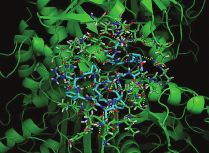
2                                                                                    Oxidative Medicine and Cellular Longevity

in vitro enzymatic hydrolysis, or bacterial fermentation) to        Mini Kit was from QIAGEN (Hilden, Germany). Primers
achieve their potential “bioactive” roles.                          for RT-PCR were purchased from IDT (Coralville, IA,
    As a representative example, the cultivation and con-           USA). Cell counting kit-8 (CCK8) and LDH assay kit were
sumption of cauliflower (Brassica oleracea L. ssp botrytis)          purchased from Dojindo Molecular Technologies (Rock-
have increased rapidly over the last few years with a large         ville, MD, USA). SuperScript® III First-Strand Synthesis
waste production, except for cauliflower curd (the sole edible       SuperMix and EXPRESS SYBR® GreenER™ qPCR Super-
part of cauliflower). Tons of cauliflower by-products (stems          Mix were purchased from Life Technologies (Carlsbad,
and leaves) are also generated during the harvest every year.       CA, USA). All the other chemicals and solvents were of
Cauliflower is well known to contain various beneficial mol-          the highest analytical grade.
ecules, such as vitamin C, glucosinolates, carotenoid, and
leaf protein [6, 7]. Numerous extraction techniques have            2.2. Peptidomic Workflow. The entire peptidomic workflow
been developed for bioactive compound extraction, such as           was performed as previously reported [17] with some
supercritical fluid extraction [8], microwave-assisted extrac-       modifications. The procedure is reported in Supplementary
tion [9], and ultrasonic-assisted extraction [7], in order to       Material S1. Briefly, 1 kg of lyophilized cauliflower by-
treat larger quantities at the industrial scale still controlling   products was extracted using an ecofriendly saline buffer
the cost of the entire process.                                     consisting of 50 mmol L-1 Tris-HCl (pH 8.8) and 15 mmol
    Protein hydrolysates from cauliflower by-products have           L-1 KCl. The extracted proteins were digested by Alcalase®
shown antioxidant [10] and angiotensin I-converting                 enzyme and the whole obtained hydrolysate was purified
enzyme (ACE) inhibitory [11] activities in cell-free systems;       by a semipreparative reverse phase high-performance liquid
therefore, they may be potential complementary to antihy-           chromatography (SP-RP-HPLC) in order to simplify the
pertensive drugs [12]. It has also been reported that they          complex mixture. Twelve fractions were collected and subse-
regulate the glucose consumption and glycogen content in            quently tested for specific and less unexplored bioactivities.
HepG2 cells, indicating an important role also in glucose           The fractions with positive bioactivity were further analyzed
metabolism [7]. In addition, several authors studied                by nano-HPLC coupled to high-resolution mass spectrome-
numerous antimicrobial peptides from plants, such as                try. The peptides in the most active fractions were identified
thionins, defensins, proline-rich peptides, lipid transfer          by peptidomic technologies and screened for bioactivity by
proteins, cyclotides, and snakins [13, 14] that are also            the use of bioinformatics, to retrieve most probable bioactive
found in Brassicaceae species [15].                                 peptide candidates.
    However, few researchers have focused on the study of
                                                                    2.3. Sample Preparation for Analysis. Stock solutions were
protein fractions and preparation of their hydrolysates from
                                                                    prepared solubilizing cauliflower lyophilized fractions
cauliflower by-products and its biological activities [7, 11,
                                                                    derived from HPLC separation in 1 ml of PBS buffer 0.1 M
16, 17] in order to exploit them as preventive biomolecules
                                                                    pH 7.4 by sonication. The protein content for each stock
for people genetically predisposed to diseases or within the
                                                                    solutions was determined by absorbance spectroscopy, at
framework of a healthy lifestyle.
                                                                    280 nm by NanoDrop 2000c (Thermo Fisher Scientific Inc.,
    Therefore, the aim of this paper was the development of
                                                                    Massachusetts, USA). Stock solutions were diluted in PBS
a combined “ad hoc” bioanalytical approach based on an
                                                                    buffer 0.1 M pH 7.4 to obtain a final concentration of
efficient recovery of peptides from cauliflower leaves, a char-
                                                                    10 mg/ml in protein content. After filtration, fractions were
acterization of their functional properties as potential nutra-
                                                                    sampled and stored at -20°C for further analysis.
ceuticals with highly predictive effect-based bioassays in cells
and an in silico identification of the most active peptides.         2.4. Cell Culture. In order to study the protective effect of
    The study of peptide bioactivity, with highly predictive        peptide fractions on endothelial dysfunction, experiments
cell models, is an efficient and reliable tool to reproduce           were performed in human umbilical vein endothelial cells
in vivo physiological conditions avoiding the use of animal         (HUVECs), a robust in vitro model for the study of endothe-
experiments to observe their effects on a wide range of              lial cell physiology and function [18].
biological activities, from endothelial dysfunction to antimi-           HUVECs pools, purchased from Life Technologies, were
crobial properties.                                                 plated on gelatin-coated tissue culture dishes and main-
                                                                    tained in phenol red-free basal medium M200 (Life Technol-
2. Materials and Methods                                            ogies) containing 10% FBS and growth factors (LSGS, Life
                                                                    Technologies) at 37°C with 5% CO2. Cells from passages 3
2.1. Materials/Chemicals. Xanthine oxidase from bovine              to 7 were actively proliferating (70–90% confluent) when
milk, luminol sodium salt, xanthine, oxypurinol, PBS tabs,          samples were harvested and analyzed [19].
Na-EDTA salt, gelatin from bovine skin, penicillin/strepto-
mycin, trypsin-EDTA, Trolox, and 2′,7′-dichlorodihydro-             2.5. Cell Viability Bioassay. The cell viability was assessed
fluorescein diacetate (H2DCFDA) were purchased from                  by WST8 [2-(2-methoxy-4-nitrophenyl)-3-(4-nitrophenyl)-
Sigma-Aldrich (St. Louis, MO, USA). Sodium perborate,               5-(2,4-disulfophenyl)-2H-tetrazolium, monosodium salt]
boric acid, NaOH, and FeCl2 were from Carlo Erba (Milan,            (Dojindo Molecular Technologies, Japan) that, in the pres-
Italy). M200 medium, low serum growth supplements, and              ence of an electron mediator, is reduced by dehydrogenases
fetal bovine serum, RNaseOUT, were purchased from                   in cells (as a vitality biomarker) to formazan dye which is
Thermo Fisher Scientific (Waltham, MA, USA). RNeasy                  soluble in the tissue culture medium. The amount of the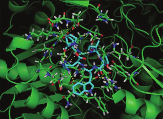
Oxidative Medicine and Cellular Longevity                                                                                        3

formazan dye generated by dehydrogenases in cells is directly      2.9. Real-Time PCR. RNA concentration and purity were
proportional to the number of living cells [20]. The decrease      determined by NanoDrop 2000 spectrophotometer (Thermo
in absorbance between the treatment after 24 h (representing       Fisher Scientific, Waltham, MA). 25 ng of total RNA was
t 1) and the control (representing t 0) was monitored at 37°C at   reverse transcribed using the SuperScript® III First-Strand
450 nm using a Varioskan™ flash multimode reader.                   Synthesis SuperMix (Life Technologies, Carlsbad, CA,
      HUVEC cells were seeded in a transparent 96-well plate       USA) and amplified using the EXPRESS SYBR® GreenER™
at a density of 5 × 10 cells/well [3]. The next day, cells were    qPCR SuperMix (Life Technologies, Carlsbad, CA, USA)
treated with stock dilutions (1 × 100 -1 × 10⁻² mg/ml in pro-      according to the manufacturer’s protocol in a final volume
tein content) in complete culture medium for 24 h.                 of 20 μl. Real-time PCR reactions were conducted on a
                                                                   Rotor-Gene Q QIAGEN Real-Time PCR System (QIAGEN
2.6. Cell Cytotoxicity: Lactate Dehydrogenase Release. Lactate     GmbH, QIAGEN Strasse 1, D-40724, Hilden), with an initial
dehydrogenase (LDH) release from HUVECs was moni-                  5 min incubation at 60°C, then 2 min at 95°C, followed by 40
tored by collecting aliquots of medium at different times,          cycles of amplification: 95°C for 15 s and 60°C for 1 min and
using a standard spectrophotometric method [16]. The               examined on by Rotor-Gene Real-Time Analysis Software
method is based on a coupled enzymatic reaction in which           6.0 (QIAGEN GmbH, QIAGEN Strasse 1, D-40724, Hilden).
LDH catalyzes the conversion of lactate to pyruvate via            Primer concentration was 500 nM. The following primers were
NAD+ reduction to NADH. Diaphorase reduces tetrazo-                used: LOX1: forward 5′-TCGGGCTCATTTAACTGGGAA-3′
lium salt, oxidizing NADH, to a red formazan product               , reverse 5′-TTGCTGGATGAAGTCCAGATCA-3′; NOX2:
that can be measured at 490 nm. Medium derived from                forward 5′-GTCTCAGGCCAATCACTTTGC-3′, reverse 5′-C
HUVECs treated with stock dilutions of protein fractions           ATTATCCCAGTTGGGCCGT-3′; NOX-4: forward 5′-TCTG
(1 × 100 -1 × 10⁻² mg/ml in protein content) for 24 h was
                                                                   GCTCTCCATGAATGTCC-3′, reverse 5′-GACACAATCCT
collected and the increase in absorbance between the treat-
ment after 24 h (representing t 1) and the control (repre-         AGCCCCAACA-3′; VCAM: forward 5′-GGTATCTGCATC
senting t 0) was monitored at 37°C using a Varioskan™              GGGCCTC-3′, reverse 5′-TAAAAGCTTGAGAAGCTGCAA
flash multimode reader.                                             ACA-3′; ICAM: forward 5′-AGCTTCGTGTCCTGTATGG
                                                                   C-3′, reverse 5′-TTTTCTGGCCACGTCCAGTT-3′; eNOS:
2.7. Intracellular Total Oxidant Fluorescent Detection. Intra-     forward 5′-ATCTTCAGCCCCAAACGGAG-3′, reverse 5′-G
cellular oxidant levels were evaluated by using the oxidant-       ATCAGACCTGGCAGCAACT-3′; SOD: forward 5′-AGGCA
sensitive fluorescent probe 2′,7′-dichlorodihydrofluorescein         TGTTGGAGACTTGGG-3′, reverse 5′-TGCTTTTTCATGG
diacetate (H2DCFDA).                                               ACCACCAG-3′; HO-1: forward 5′-CAACAAAGTGCAAGA
    Briefly, the probe is not fluorescent until the acetate
                                                                   TTCTG-3′, reverse 5′-TGATTCACATGGCATAAAG-3′; XO
groups are removed by intracellular esterases; in the
presence of oxidants, the probe is oxidized within the cells       D: forward 5′-CTACAGCTTTGAGACTAACTC-3′, reverse
producing a fluorescent signal related to intracellular             5′-TCTTATGATCTCCTGTTAGGC-3′; p65: forward 5′-TG
oxidant levels that was measured using a microtiter plate          GGGACTACGACCTGAATG-3′, reverse 5′-GGGGGCACG
reader (Varioskan™ flash multimode reader, Thermo                   ATTGTCAAAGA-3′; p52: forward 5′-CCGTTGTACAAAGA
Fisher Scientific). Excitation wavelength was 485 nm and            TACGCGG-3′, reverse 5′-CATCCAGACCTGGGTTGTAGC-
emission wavelength was 535 nm. HUVECs were treated                3′; p50: forward 5′-AATGGGCTACACCGAAGCAA3′, reverse
with cauliflower peptide fractions (1 × 100 -1 × 10⁻² mg/ml)        5′-AGCTCGTCTATTTGCTGCCT-3′; SOD-2: forward 5′-G
for 24 h and Trolox (100 μM) was used as reference. After
                                                                   CTCCCCGCGCTTTCTTA-3′, reverse 5′-GCTGGTGCCGC
treatment, cells were incubated with 5 μM H2DCFDA for
20 min at 37°C and then subjected or not to oxidative              ACACT-3′; GPx1: forward 5′-TATCGAGAATGTGGCGTCC
stress generated by 50 μM H2O2 for 30 min. The decrease            C-3′, reverse 5′-TCTTGGCGTTCTCCTGATGC-3′; catalase:
of fluorescence signal between cells treated with cauliflower        forward 5′-CTCCGGAACAACAGCCTTCT-3′, reverse 5′-AT
peptide fractions and control was reported as the                  AGAATGCCCGCACCTGAG-3′; and RPL13A: forward 5′-C
percentage of intracellular reactive oxygen species (ROS)          ACCCTGGAGGAGAAGAGGA-3′, reverse 5′-CCGTAGCCT
normalized with H2O2 treatment alone [21].                         CATGAGCTGTT-3′. Changes in gene expression were calcu-
    H2DCFDA can be used as a redox indicator probe for             lated by the 2−ΔΔCt formula using RPL13A as reference gene.
detecting intracellular oxidant formation caused by changes
in iron or heme signaling or peroxynitrite (ONOO-) forma-
tion. The fluorescent response based on the oxidation of            2.10. Chemiluminescent Intracellular Xanthine Oxidase Assay.
DCFH provides an index for the total oxidants present in           To monitor xanthine oxidase activity, 5 × 10³ cells/well were
biological systems, not for cell-derived H2O2. This limitation     plated in a 96-black well microtiter plate; the day after, cells
determines a low selectivity toward H2O2 [22–24].                  were incubated at 37°C with CL reaction cocktail solution
                                                                   containing different amounts of cauliflower peptide fractions
2.8. RNA Extraction. HUVECs were preincubated with cauli-          ranging from 1 × 100 -1 × 10⁻² mg/ml and the CL emission
flower peptide fractions (1 mg/ml) for 8 hours at 37°C before       produced after the addition of xanthine (2.0 mM) was moni-
24 h of exposure to TNF-α (10 ng/ml). Total RNA was extracted      tored for 20 min using the Luminoskan™ Ascent lumin-
using a commercial RNA extraction kit (QIAGEN) [20].               ometer automatic plate reader (Thermo Fisher Scientific,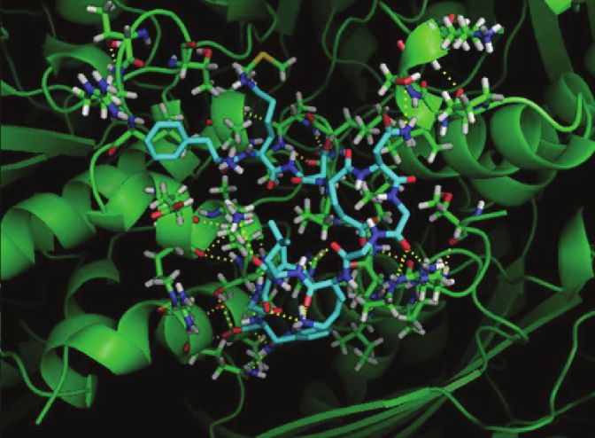
4                                                                                    Oxidative Medicine and Cellular Longevity

Roskilde, Denmark). The detailed procedure is reported in a       GYNPSYGARPL, and KWAGGKPEKPILR from fraction 8)
previous paper [25].                                              were generated using PEP-FOLD3 server that provides a gen-
                                                                  eral framework for the structural characterization of peptides
2.11. Antioxidant Capacity Using a Chemiluminescent (CL)          and returns in a few minutes the five best models. The models
Method. The chemiluminescence method for measurement              with the lowest energy conformations were selected for dock-
of antioxidant effect is based on the competition between          ing runs [31–33]. The xanthine oxidase model from bovine
the reaction of peroxyl radicals with luminol, giving rise to     milk source (1FIQ) was the most used for docking simulation
light emission, and the scavenging of peroxyl radicals by anti-   as reported in literature [34, 35] because of its suitable crystal-
oxidants. Indeed, the addition of a solution of known antiox-     lographic resolution (~2 Å) assuring best docking results. The
idants to a glowing steady-state chemiluminescent reaction        xanthine oxidase model from bovine milk source (1FIQ) was
temporarily quenches light output. The extent of light emis-      downloaded from the RCSB protein data bank (http://www
sion quenching is related to the amount and the strength of       .rcsb.org/) and refined by molecular graphic PyMOL software
antioxidant added. The procedure is reported in [26].             (The PyMOL Molecular Graphics System, Version 2.0 Schrö-
                                                                  dinger, LLC). Then, ADT was used to create the necessary
2.12. Antimicrobial Activity. The in vitro antimicrobial activ-   .pdbqt files of both peptides and xanthine oxidase (XOD)
ity of the cauliflower peptide fractions was evaluated towards     structure that are read by Vina. The identification of candidate
a panel of reference bacterial strains from the American          regions of the protein surface likely to be involved in the inter-
Type Culture Collection (ATCC) including Staphylococcus           action with a peptide sequence required to assist in silico
aureus ATCC 25923, Staphylococcus epidermidis ATCC                experiments was obtained using PEP-SiteFinder server [36].
12228, Enterococcus faecalis ATCC 29212, Escherichia coli
ATCC 25922, Pseudomonas aeruginosa ATCC 27853, and                2.14. Statistical Analysis. Results are expressed as mean ± SD
Klebsiella pneumoniae ATCC 9591 and the yeast Candida             of at least three independent experiments. Differences
albicans ATCC 10231.                                              between the means were determined by one-way ANOVA
     The peptide fractions were assayed by means of a broth       followed by the Bonferroni multiple comparison test using
microdilution method as previously described, with minor          the GraphPad Prism software, version 6.0 (GraphPad Soft-
modifications [27, 28]. Briefly, for antibacterial determina-       ware Inc., La Jolla, CA), and a P value < 0.05 was considered
tions, a suspension at 0.5 McFarland of each reference strain     statistically significant.
was diluted 1 : 200 in Mueller-Hinton broth (Sigma-Aldrich)
or in Brain heart infusion broth (Biolife) for E. faecalis and
incubated with tenfold dilutions of cauliflower peptide frac-      3. Results and Discussion
tions starting from 1 mg/ml and with gentamicin as refer-
ence drug. For antifungal determinations, yeast suspension        3.1. Peptide Hydrolysate Safety. Peptide hydrolysates from
was diluted 1 : 20 in RPMI-1640 medium (Gibco®, Thermo            cauliflower by-products were obtained by Alcalase®, a
Fisher Scientific Inc., Waltham, USA), containing glucose          low-cost enzyme compatible with large-scale applications
2%, 0.3% levoglutamine, and 0.165 M 3-(N-morpholino)--            [37]. Alcalase® displayed a greater degree of hydrolysis over
propanesulfonic acid (MOPS), pH 7.0, and then incubated           the other common used enzymes, but it was employed in
with tenfold dilutions of peptide fractions starting from         most cases just to obtain antioxidant and ACE inhibitory
1 mg/ml and with fluconazole, as reference drug. As addi-          peptides [3]. It is certainly interesting to test hydrolysates
tional control, cells were incubated in regular medium in         for less-studied bioactivity since it was suggested that this
the absence of fractions to check both background turbidity       kind of sample could be a promising source of understudied
and the sterility of the procedure. Following 24 h of incuba-     bioactive peptides [3]; therefore, hydrolysates were subjected
tion at 37°C, microbial growth was determined by adding in        to dose-effect safety experiments in HUVECs. Fractions 1, 2,
each well the WST-8 dye (Microbial Viability Assay                and 3 showed a reduction of cell viability after a 24 h treat-
kit-WST, Dojindo Laboratories) and measuring the absor-           ment with 1 mg/ml while the others did not affect cell viabil-
bance at 450 nm using the Multiskan Ascent microplate             ity. Then, lactate dehydrogenase (LDH), a marker for cell
reader (Thermo Fisher Scientific Inc., Waltham, USA). Per-         death both in vitro and in vivo, was quantified in cell culture
centage values of samples at the different experimental con-       medium and we did not observe any toxic effect (Figures 1(a)
ditions were determined as relative to the positive growth        and (b)).
controls. Determinations were performed in triplicate and              Fractions 4-12 were subsequently investigated to evaluate
in two independent experiments.                                   several unexplored biological activities, such as the protection
                                                                  against endothelial dysfunction and antimicrobial effects.
2.13. Computational Methods. Molecular docking simula-
tions were performed using the open-source program                3.2. Peptides from Fractions 8 and 12 Reduce Oxidative Stress
AutoDock Vina [29] along with AutoDockTools (ADT)                 through Intracellular Endogenous Antioxidant Enzyme
[30], a graphical user interface compliment to the AutoDock       Modulation in HUVEC Cells. Fractions 8 and 12 decreased
software suite. In order to run the Vina docking program, both    intracellular ROS levels after acute exposure to H2O2
peptidic ligands and protein structure must be first refined and    (P < 0 05 and P < 0 01, respectively) (Figure 2(a)), suggest-
then prepared in a specific file format (.pdbqt). The peptide       ing they exert a protective effect against oxidative stress in
models of sequences (FKDENGGKLIGF, GNIFDGIQRPL,                   the vasculature, process involved in endothelial dysfunction.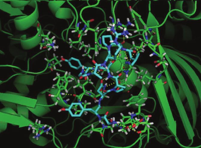
Oxidative Medicine and Cellular Longevity                                                                                                                                   5

                        150                                                                                       150
  (a.u. % vs control)

                                                                                            (a.u. % vs control)
    Cell variability

                        100                                                                                       100

                                                                                               LDH activity
                                 ⁎⁎⁎         ⁎⁎⁎
                         50            ⁎⁎⁎                                                                         50

                         0                                                                                          0
                              ctrl 1    2     3    4   5     6   7   8   9   10 11 12 TOT                               ctrl   4   5   6    7    8   9   10   11   12 TOT
                                                       (a)                                                                                 (b)

Figure 1: HUVECS were treated with peptide fractions (1-12) and the whole hydrolysate [13], the total (1 mg/ml) for 24 hours. (a) Cell
viability was spectrophotometrically detected through formazan production in the presence of dehydrogenases in cells. (b) LDH activity
was spectrophotometrically quantified in cellular medium as index of cytotoxicity.

    Even if H2DCFDA is still used to detect intracellular                                      antiatherogenic enzymes that counteract oxidative damage
oxidant species from the scientific community, this probe                                       in the vascular endothelium [39, 40].
suffers of some limitations as artefactual amplification of                                          Among the antioxidant enzymes, SOD-1 is the most
the fluorescence intensity via a redox cycling mechanism                                        abundant and ubiquitous isoform, with a great physiological
involving an intermediate radical, DCF⋅−, and responds                                         significance and therapeutic potential in CV diseases
to changes in intracellular iron signaling or enhanced per-                                    because the endothelium is particularly sensitive to oxidant
oxidase activity [24]. In fact, DCFH does not directly react                                   injury [39], so we investigated its expression in HUVECs
with superoxide, H2O2, or nitric oxide. Instead, DCF fluo-                                      upon treatment with peptide fractions in the presence of
rescence results from oxidation by potent oxidants, such                                       TNF-α. Numerous studies suggest that SOD-2 is perhaps
those produced from metal ion- and peroxidase-catalyzed                                        one of the most famous NF-κB targets with antioxidant
reactions and from decomposition of ONOO-. Moreover,                                           activity in the vascular endothelium [41–44], at least in part
DCF-dependent fluorescence can be self-amplified by                                              via nuclear transcription factor p65 [45]. We investigated
redox cycling of the one-electron oxidized dye [23].                                           SOD-2 gene expression, but we did not observe any signifi-
Indeed, we observed that DCFH probe showed a                                                   cant changes cells upon treatment with peptide fraction 12
dose-dependent increase in fluorescence intensity propor-                                       in the presence of TNF-α.
tional to increase amount of Fe2+ (0.3-10 μM) in the pres-                                         GPx-1 is the most abundant selenoperoxidase form in
ence of 50 μM H2O2 (Figure S1) in a cell-free system, with                                     mammalian tissues and a key antioxidant enzyme in many
a limit of detection (LOD) = 0.6 ± 0.3 μM and a limit of                                       cell types including endothelial cells. GPx-1 consumes
quantification (LOQ) = 2.4 ± 0.3 μM, while we did not                                           reduced glutathione to convert H2O2 to water and lipid per-
observe a direct correlation of DCFH-related fluorescence                                       oxides to their respective alcohols [46]; it also acts as an
as a function of increase amount of H2O2 (0.5-100 μM)                                          ONOO- reductase [47]. Mice with a disrupted GPx1 gene
(data not shown).                                                                              exhibit increased susceptibility to oxidative stress-inducing
    Most peptides derived from hydrolysis of food proteins                                     agents [48], while induction of this isozyme has been shown
such as those from milk, egg, meat, wheat, and soy were                                        to provide protection against oxidative damage in endothelial
characterized with chemical assays in cell-free in vitro condi-                                cells [49] GPx1 deficiency causes endothelial dysfunction [50,
tions, generally for radical-scavenging activity or for metal                                  51] and endothelial progenitor cell dysfunction in mice [52].
chelating activity [38]. However, these methods do not allow                                   Furthermore, transgenic GPx1 expression was observed to
evaluating the bioactivity of antioxidant peptides under                                       impair endothelial dysfunction [51].
physiological conditions, in order to establish their real                                         As some authors observed [53], food-derived peptides
protective roles in diseases. We demonstrated that peptide                                     can display protective effects by induction of gene expression
form cauliflower fractions 8 and 12 reduced intracellular                                       of proteins that protect cellular components from oxidative
oxidant species, acting as good antioxidants in HUVECs.                                        stress-induced deterioration; however, to the best of our
    To clarify the possible mechanisms of action, we investi-                                  knowledge, this is the first time that was reported an induc-
gated the expression of several prooxidant (NADPH oxi-                                         tion of SOD-1 and GPx-1 expression caused by peptides
dases 2 and 4, lectin-type-oxidized LDL receptor 1,                                            from cauliflower leaves.
endothelial nitric oxide synthetase, and xanthine oxidase)                                         In the endothelium, ROS predominantly arise from the
and antioxidant biomarkers (superoxide dismutases 1 and                                        isoforms of NAPDH oxidases 2 and 4 [54]; however, XOD
2, heme oxygenase 1, catalase, and glutathione peroxidase                                      and endothelial nitric oxide synthase (eNOS) play a physio-
1). Peptide fraction 12 at the higher concentration (1 mg/ml)                                  logic role in inflammatory signaling regulation of NO
significantly increased superoxide dismutase- (SOD-) 1 and                                      production and vascular function [55]. The oxidative stress
glutathione peroxidase- (GPx-) 1 expression (Figure 2(b))                                      generated by these enzymes induces endothelial dysfunction,
(P < 0 01 and P < 0 05, respectively), important intracellular                                 leading to atherosclerosis, cardiovascular diseases, and
6 Oxidative Medicine and Cellular Longevity
200
50 휇M H2O2 for 30 minutes SOD 1
4 TNF 훼 (10 ng/ml 24 h)
⁎⁎
(% vs V with H2O2)
Relative gene expression
150
Flourescence (%)
3
fold change
100 ⁎ ⁎
⁎⁎
2
⁎
50
1
⁎⁎⁎
0 0
ctrl v 4 5 6 7 8 9 10 11 12 TOT T ctrl v 8 12 TOT
(a) (b)
GPx-1
TNF 훼 (10 ng/ml 24 h)
4 ⁎⁎
Relative gene expression
3
fold change
2
1
0
ctrl v 8 12 TOT
(c)
Figure 2: (a) HUVECs were treated with peptide fractions (4-12) and the whole hydrolysate at a concentration of 1 mg/ml for 24 hours and
then exposed to oxidative stress generated by 50 μM H2O2 for 30 min. Treatment with 100 μM Trolox (T) for 24 hours was used as reference.
Intracellular ROS levels were measured by means of H2DCFDA assay as described in Materials and Methods. HUVECs were pretreated with
cauliflower fractions (1 mg/ml) for 8 h before 24 h of exposure to TNF-α (10 ng/ml). Total RNA was extracted, and qRT-PCR analysis was
performed to determine (b) SOD-1 gene expression and (c) GPx-1 expression. Relative changes in mRNA expression levels were
calculated according to the 2−ΔΔCt method using RPL13A as reference gene. Results are expressed as mean ± SEM of three independent
experiments. ∗ P < 0 05, ∗∗ P < 0 01, and ∗∗∗ P < 0 001 significantly different from the vehicle (V, DMSO).
metabolic syndrome. Indeed, XOD activity is inversely intracellular CL reaction, exploiting an assay based on
related to endothelium-dependent vasodilation, since it was enhanced chemiluminescent (ECL) detection method, able
located primarily in cells derived from the vasculature and to reveal different species of ROS [20], demonstrating that
especially in endothelial cells [56]; elevation of XOD activity fractions 8 and 12 cannot be considered direct ROS scaven-
is associated to poor clinical outcomes [57]. Therefore, to ger (data not shown). Moreover, to confirm that peptide
date, XOD is recognized as an important biomarker, incen- fractions 8 and 12 act as intracellular antioxidant, we detect
tivizing extensive exploration of inhibition strategies to intracellular H2O2 level exploiting cell-based assays with a
address disease processes where oxidative stress is contribu- bioluminescent detection using a boronic probe selective
tory, such as cardiovascular disorders [58]. for H2O2. We treated cells with menadione, one of the sim-
Peptide fraction treatment did not modulate XOD plest quinones, widely used for evaluating the cellular effects
expression in HUVECs (data not shown) while fraction 8 of oxidative stress in endothelial cells [59, 60]. The major
inhibited intracellular XOD activity (Figures 3(a) and (b)). mechanism caused by menadione is the intracellular produc-
To quantify intracellular XOD activity, we previously tion of ROS by redox cycling, where one-electron reduction
developed an ultrasensitive cell-based biosensor reporting of O2 by the semiquinone form of menadione generates super-
that the xanthine oxidase activity in living endothelial cells oxide (O2⋅−). O2⋅− is an extremely unstable ROS that rapidly
(HUVECs) was 6 ± 1 × 10⁻⁷ mU/ml/cell and the IC50 of dismutates in the cells to H2O2 either spontaneously or enzy-
oxypurinol, the active metabolite of allopurinol, was 152 ± matically catalyzed by SOD [61]. As it is shown in Figure S2,
76 ng/ml [25]. After 20 minutes of incubation, the intracellu- peptide fractions 8 and 12 reduced menadione-derived
lar IC50 of fractions 4-12 was evaluated by a dose-response intracellular H2O2. These results confirmed the intracellular
curve, obtaining that fraction 8 has an IC50 = 8 3 ± 0 6 μg/ antioxidant activities of fractions 8 and 12.
ml (Figures 3(a) and (b)). This cell-based biosensor utilizing
whole cells takes into consideration also the bioavailability of 3.3. Peptides from Fraction 12 Ameliorate TNF-α-Triggered
the compound, especially the ability to cross cell plasma Endothelial Dysfunction in HUVEC Cells. To better clarify
membranes, so it is more representative and predictive to the protective effect of fractions 8 and 12 in respect with
human situation. Moreover, we previously excluded the pos- TNF-α-induced endothelial dysfunction, we evaluated the
sible interferences of all the peptide fractions in the cell viability in inflammatory conditions. As shown in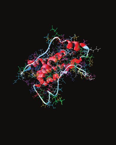
Oxidative Medicine and Cellular Longevity                                                                                                                                  7

                                                         Peptide fraction 8                                                               Whole hydrolysate
                                     0.08                                                                             0.08

                                     0.06                                                                             0.06
         CL signal at 30 min (rlu)

                                                                                          CL signal at 30 min (rlu)
                                     0.04                                                                             0.04

                                     0.02                                                                             0.02

                                     0.00                                                                             0.00
                                            −5   −4        −3         −2         −1   0                                      −5   −4        −3         −2         −1   0
                                                      Log [fraction 8] (mg/ml)                                                    Log [whole hydrolysate] (mg/ml)

                                                      IC50 = 8.3 ± 0.6 휇g/ml                                                          IC50 = 11.8 ± 2.8 휇g/ml
                                                           (a)                                                                              (b)

Figure 3: Concentration-response plot of intracellular XOD inhibition obtained by analyzing CL signals after 20 min of incubation with
Fe2+-EDTA-luminol reaction cocktail in HUVECs treated with peptide (a) fraction 8 and (b) whole hydrolysate (range 1–0.0001 mg/ml).

Figures 4(a) and (b), we observed that TNF-α treatment                                                  NF-κB activation is regulated by reactive species [63],
decreased cell viability in HUVECs; the addition of fractions                                       and ROS derived from intracellular XOD activity is impli-
8 and 12 (1 mg/ml for 24 hours) significantly counteracted                                           cated in heart failure [66] possibly through NF-κB-related
the effects induced by TNF-α (P < 0 05), while lower doses                                           p50 modulation.
had slight, not significant effect (data not shown). NF-κB                                                Therefore, a decrease in intracellular oxidative stress by
signaling is an attractive target for the development of novel                                      fraction 8 and 12 treatment can inhibit NF-κB signaling in
anti-inflammatory drugs and the ability of certain small                                             endothelial cells, which can suppress its downstream effects.
cell-penetrating peptides to enter cells inhibiting NF-κB sig-                                      This notion was supported by the effects of fractions 8 and
naling offer exciting potential also in the clinical setting.                                        12 on VCAM-1 mRNA level measured by qPCR.
Classical NF-κB activity regulates the expression of many                                               Notably, the anti-inflammatory activity of a peptide
genes involved in inflammatory and survival responses,                                               derived from ovotransferrin, which is present in the albumen
including those encoding cytokines (e.g., IL-1, IL-2, and                                           of eggs, was recently explained through the NF-κB-related
IL-6), leukocyte adhesion molecules (e.g., E-selectin,                                              p50 and p65 inhibitions [67].
ICAM-1, and VCAM-1), and antiapoptotic proteins (e.g.,                                                  Increased reactive oxygen species (ROS) production
Bcl2, Bcl-XL, and XIAP) [62]. Therefore, the expression of                                          together with increased adhesion molecules and thrombo-
the cellular adhesion proteins VCAM-1 and ICAM-1 was                                                genic tissue factor expression on endothelial cells has a key
investigated; as it is represented in Figure 4(b), fractions 8                                      role in proatherogenic mechanisms [68]. Therefore, peptides
and 12 significantly decreased TNF-α-induced VCAM-1                                                  able to decrease the expression of inflammatory bio-
expression (P < 0 001) in HUVECs while had no effect in                                              markers could be useful for reducing the severity of
ICAM-1 expression (not shown). To clarify the mechanism                                             atherosclerosis progression.
of action, the expression of NF-κB-related nuclear transcrip-
tion factors p65, p50, and p52 was also investigated. As                                            3.4. Peptides from All of the Fractions Do Not Show Any
shown in Figures 4(c) and (d), fraction 12 treatment signifi-                                        Antimicrobial Activity. The characterization of several plant
cantly decreased p65 expression while peptide fraction 8                                            protein hydrolysates demonstrated that they have antimicro-
decreased p50 expression (P < 0 001 and P < 0 01, respec-                                           bial activity, thus qualifying as functional foods [69], but we
tively). p65 protein binds to the promoter of SOD-2 gene                                            did not observe inhibitory activity of the cauliflower peptide
[45], so a decrease in p65 expression induced by fraction                                           fractions towards the human pathogenic bacteria and yeast
12 explains results obtained in SOD-2 expression. NF-κB                                             strains selected in the present study.
was the first transcription factor shown to be redox-
regulated [63] and it has been demonstrated that overex-                                            3.5. Identification of Bioactive Peptides from Fractions 8 and
pression of SOD-1 suppressed ischemia-induced activation                                            12. Fractions 8 and 12, the most active ones, were analyzed
of NF-κB through a decrease in nuclear translocation pro-                                           by nano-HPLC-MS/MS method. The obtained MS/MS raw
tein levels [64]. It has been also shown that transfection of                                       files were searched by the Proteome Discoverer software to
endothelial cells with SOD-1, but not catalase, inhibited                                           obtain peptide sequences. The identified peptides were man-
NF-κB signaling and expression of VCAM-1 induced by                                                 ually validated taking into consideration only the most
TNF-α [65].                                                                                         abundant peptides in both fractions; peptide abundance is
8 Oxidative Medicine and Cellular Longevity
50 ng/ml TNF 훼 for 24 hours
150
°
° VCAM-1
40
⁎⁎ 10 ng/ml TNF 훼 for 24 hours
Relative gene expression
(a.u. % vs vehicle)
100 ⁎⁎⁎
Cell viability
30 ⁎⁎⁎
fold change
20
50
10 °°°
°°°
0 0
ctrl v 4 5 6 7 8 9 10 11 12 13 ctrl v 8 12 TOT
(a) (b)
p 65
TNF 훼 24 hours p 50
3.0 ⁎⁎⁎ 2.0 TNF 훼 24 hours
Relative gene expression
Relative gene expression
2.5 ⁎⁎⁎
°°° 1.5 °°°
fold change
fold change
2.0
1.5 1.0
1.0
0.5
0.5
0.0 0.0
ctrl v 8 12 ctrl v 8 12
(c) (d)
Figure 4: HUVECs were pretreated with peptide fractions (1 mg/ml) for 8 h before 24 h of exposure to TNF-α (50 ng/ml). (a) Cell viability
was spectrophotometrically detected through formazan production in the presence of dehydrogenases in cells. Total RNA was extracted and
qRT-PCR analysis was performed to determine (b) VCAM-1 gene expression, (c) p65 gene expression, and (d) p50 gene expression. Relative
changes in mRNA expression levels were calculated according to the 2−ΔΔCt method using RPL13A as reference gene. Results are expressed as
mean ± SEM of three independent experiments. ∗∗ P < 0 01 and ∗∗∗ P < 0 001 significantly different from the control; ° P < 0 05, °° P < 0 01, and
°°°
P < 0 001 significantly different from the vehicle (V, DMSO).
related to their area; thus, peptides were filtered according to than 0.9, namely, IDNIFRF. As it is possible to see in Supple-
it and only the ones with an area larger than 107 and higher mentary Material S2, only four potentially bioactive peptides
score were accepted. A total of 181 peptides were identified come from fraction 8, and all the others belong to fraction
after this manual validation. The complete list of identified 12. The treatment with fraction 12 increased SOD-1 and
peptides coming from the two most active fractions from reduced VCAM-1 expression in the presence of TNF-α.
the first chromatographic dimension, with sequence and Both genes are modulated by NF-κB signaling [43, 71], sug-
related data, is reported in Supplementary Material S2. gesting that one or more peptides in fraction 12 could act as
After that, an in silico analysis using PeptideRanker [70] inhibitor of NF-κB pathway machinery. Several bioactive
was carried out to further mine data. In this way, each pep- peptides have been described in literature as inhibitors of
tide was assigned a score based on the probability of being classical NF-κB signaling by either disrupting the IKK com-
bioactive, probability that the built-in N-to-1 neural network plex or by inhibiting critical events downstream of IKKβ
computed on the basis of the peptide primary sequence (the [72]. In addition, several peptides that target upstream inter-
complete list of probability scores assigned to each identified mediates in the NF-κB pathway or other signaling mecha-
peptide is reported in Supplementary Material S2). Such nisms, such as the ERK and JNK pathways, have also been
algorithm is capable of predicting the bioactivity of peptides developed. IDNIFRF seems the most promising candidate
because of the general features that different bioactive pep- due to the presence of both the cationic (arginine) and hydro-
tide functional classes have in common; therefore, PeptideR- phobic side chains (isoleucine) that can facilitate its uptake
anker represented a useful tool to select the most probable across the plasma membrane. An in silico determination of
bioactive peptides. Since the software labelled as “bioactive” the hydrophobic character of this peptide was performed
any peptide possessing a score above the 0.5 threshold, we exploiting Peptide 2.0 ProtParam tool of ExPASy bioinformat-
applied a higher 0.7 threshold in order to reduce the number ics resource portal and peptide synthesis and proteotypic pep-
of false positive hits. After such filtering of peptide scores, tide analyzing tool of Thermo Scientific confirmed a
most peptide sequences were rejected. Twenty-three pep- hydrophobicity of 30.90, with an aliphatic index of 111.43
tides have shown a score higher than 0.7 in fraction 12, and a grand average of hydrophobicity (GRAVY) of 0.443
and among these, only one has shown a probability higher [73]. These results suggested that IDNIFR is an adequate
Oxidative Medicine and Cellular Longevity 9
(a) (b)
(c) (d)
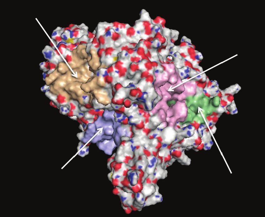
Figure 5: Picture of peptides docked in XOD enzyme (1FIQ), showing polar interactions within active sites for (a) GDSNPSNPKPRFGAY,
(b) FKDENGGKLIGF, (c) GYNPSYGARPL, and (d) PDSITWR sequences, calculated by AutoDock Vina and elaborated by PyMOL software.
Table 1: Comparison between inhibition constant values (K i ) derived from cell-based assay and in silico molecular docking simulations of
bioactive peptides in cauliflower fraction 8.
K i calculated from in silico
K i calculated from cell-based assay Peptide sequence Energy affinity
simulation results
GDSNPSNPKPRFGAY -28.03 kJ/mol 20 μg ml-1
PDSITWR -25.10 kJ/mol 34 μg ml-1
8.6 μg ml-1
GYNPSYGARPL -25.52 kJ/mol 40 μg ml-1
FKDENGGKLIGF -25.10 kJ/mol 52 μg ml-1
hydrophobic peptide able to cross plasma membranes. How- from 20 to 52 μg ml-1) for peptide sequences were calculated
ever, to the best of our knowledge, more studies are needed from docking outputs and compared with experimental K i ,
to determine its efficacy as NF-κB signaling modulator. derived from cell-based assay results. The in silico simulations
referred only to a single isolated peptide and did not consider
3.6. Molecular Docking Simulations for Xanthine Oxidase. To cell permeability of inhibitors. Moreover, the discrepancy
validate the experimental work conducted to determine the between experimental and computational K i values can be
intracellular XOD inhibition, molecular docking simulations explained considering a positive synergic effect of the four
have been used as a tool to augment the molecular level inter- inhibitors [74]. Indeed, Figure 5(b) shows four different regions
pretation of the data. In particular, to seek the molecular expla- of the protein surface involved in the interaction with each pep-
nation of the inhibition behavior of fraction 8 against XOD tide, respectively. The synergistically interaction of the four
activity in the cell-based assay, we undertook a series of molec- sequences to different sites of XOD can justify the increased
ular docking studies using AutoDock Vina. The in silico calcu- inhibitory effect, as observed in in vitro assays. Molecular dock-
lations provided nine best output results for each docking run. ing simulations demonstrated that the trend of in silico predic-
From the analysis of docking study, the peptide sequence tion of peptide effects is in agreement with experimental
GDSNPSNPKPRFGAY showed the best docked conformation in vitro results and confirmed inhibitory effect of cauliflower
(Figure 5) with the lowest energy affinity of -28.03 kJ/mol than fraction 8 on XOD activity as obtained from cell-based assay,
other peptides (Table 1). The docking results indicated that providing a theoretical explanation of molecular inhibition
medium-weak interactions between protein surface and pep- mechanism by the identification of protein-peptide interac-
tides exist. Then, theoretical dissociation constants K i (ranged tions (Figures 5 and 6).
10                                                                                      Oxidative Medicine and Cellular Longevity

                               GDSNPSPKPRFGAY

                                                                                              PDSITWR

                                       FKDENGGKLIGF
                                                                                         GYNPSYGARPL

Figure 6: Picture of XOD enzyme polar surface, showing that each sequence (GDSNPSNPKPRFGAY, PDSITWR, FKDENGGKLIGF, and
GYNPSYGARPL) interacts with a different site of XOD, calculated by PEP-SiteFinder server and elaborated by PyMOL software.

4. Conclusions                                                        on the possibility to find new uses of waste, even outside the
                                                                      agricultural field, contributing to the creation of sustainable
We developed an innovative combined (bio)analytical                   value chains in the farming and processing sectors.
approach, based on a rationale recovery of bioactive peptides
from cauliflower waste through an advance peptidomic-based             Data Availability
strategy integrated with an in vitro activity characterization uti-
lizing highly predictive cell-based bioassays. Our study suggests     The in vitro data obtained in human umbilical vein endothe-
that cauliflower peptides from two fractions possess antioxi-          lial cells used to support the findings of this study are
dant and anti-inflammatory effects in the vasculature, at               included within the article. The description of the in vitro
least in part through inhibition of intracellular XOD activity        method to determine antimicrobial activity is included
and modulation of SOD-1 and VCAM-1 expression. In silico              within the article; however, no data are available since pep-
analysis showed that four peptides from fraction 8 and one            tide fractions did not show any significant antimicrobial
from fraction 12 could be the most probable bioactive can-            activity. Previously reported data regarding the multidimen-
didates exerting protective effects against endothelial dys-           sional liquid chromatography characterization of peptide
function. Moreover, one peptide from fraction 8 able to               fractions were used to support this study and are available
synergistically inhibit XOD was detected through a detail             at DOI: 10.1016/j.jff.2018.02.022. These prior studies are
in silico docking analysis.                                           cited at relevant places within the text as references [17].
     Advancements in the biopharmaceutical industry have              The data obtained by peptidomic analysis and bioinformat-
resulted in the development of several new peptide-based              ics used to support the findings of this study are included
therapeutics; to the best of our knowledge, this is the first          within the article and in supplementary information files 1
attempt to determine biological effects of peptides from cau-          and 2.
liflower waste, in order to evaluate their possible application
into valuable functional components in nutraceutics and               Conflicts of Interest
pharmaceutics as well as in animal feed.
     Oral administration is most preferred because of                 The authors have declared no conflict of interest.
patient compliance and acceptability; however, to exercise
their effects in the target organ, peptides need to remain             Acknowledgments
intact during the digestive process. To solve this crucial
issue, several approaches including chemical modifications             This work was supported by the PRIN 2015 with project
(lipidation), physical methods (microencapsulation), use of           number 2015FFY97L_002.
mucoadhesive polymers, formulation design, and use of
enzyme inhibitors have been developed to improve their                Authors’ Contributions
bioavailability after oral ingestion [58].
     This translational (bio)analytical approach represents a         CC had substantial contributions to the conception and
smart and powerful tool that allows to open new perspectives          design of the work; the acquisition, analysis, and interpretation
Oxidative Medicine and Cellular Longevity 11
of data for the work; and drafting the work. ALC did mass potential biological activities of protein hydrolysates,” Food
spectrometry analysis and revised for important intellectual Chemistry, vol. 221, pp. 114–122, 2017.
content. DC did the acquisition, analysis, and interpretation [8] P. Sookwong and S. Mahatheeranont, “Supercritical CO2
of data for the work and molecular docking simulations using extraction of rice bran oil –the technology, manufacture, and
AutoDock software. FB did the acquisition, analysis, and applications,” Journal of Oleo Science, vol. 66, no. 6, pp. 557–
interpretation of data for the work and revised the paper crit- 564, 2017.
ically for important intellectual content. RCC did the mass [9] I. G. Zigoneanu, L. Williams, Z. Xu, and C. M. Sabliov, “Deter-
spectrometry analysis and computational analysis and revised mination of antioxidant components in rice bran oil extracted
the paper critically for important intellectual content. CMM by microwave-assisted method,” Bioresource Technology,
did the acquisition and analysis of data for the work. SP did vol. 99, no. 11, pp. 4910–4918, 2008.
the acquisition and analysis of data for the work. MZ revised [10] R. Zenezini Chiozzi, A. L. Capriotti, C. Cavaliere, G. la Barbera,
the paper critically for important intellectual content. MM S. Piovesana, and A. Laganà, “Identification of three novel
had an agreement to be accountable for all aspects of the work angiotensin-converting enzyme inhibitory peptides derived
from cauliflower by-products by multidimensional liquid
in ensuring that questions related to the accuracy or integrity
chromatography and bioinformatics,” Journal of Functional
of any part of the work are appropriately investigated and Foods, vol. 27, pp. 262–273, 2016.
resolved. PS did the acquisition and analysis of data for the
[11] Y. Xu, T. Bao, W. Han, W. Chen, X. Zheng, and J. Wang,
work. AL revised the paper critically for important intellectual
“Purification and identification of an angiotensin
content and final approval of the version to be published. AR I-converting enzyme inhibitory peptide from cauliflower
revised the paper critically for important intellectual content by-products protein hydrolysate,” Process Biochemistry,
and final approval of the version to be published. vol. 51, no. 9, pp. 1299–1305, 2016.
[12] N. Yamamoto, A. Akino, and T. Takano, “Antihypertensive
effect of the peptides derived from casein by an extracellular
Supplementary Materials proteinase from Lactobacillus helveticus CP790,” Journal of
Dairy Science, vol. 77, no. 4, pp. 917–922, 1994.
Supplementary 1. Supplementary Material S1: a detail
[13] L. Padovan, M. Scocchi, and A. Tossi, “Structural aspects of
description of the entire peptidomic workflow, procedures, plant antimicrobial peptides,” Current Protein & Peptide Sci-
and results regarding the intracellular oxidants measurement ence, vol. 11, no. 3, pp. 210–219, 2010.
were reported. [14] G. Wu, X. Li, X. Fan et al., “The activity of antimicrobial pep-
Supplementary 2. Supplementary Material S2: the complete tide S-thanatin is independent on multidrug-resistant spec-
list of identified peptides coming from the two most active trum of bacteria,” Peptides, vol. 32, no. 6, pp. 1139–1145, 2011.
fractions from the first chromatographic dimension, with [15] H. Cao, T. Ke, R. Liu et al., “Identification of a novel
sequence and related data, was reported. proline-rich antimicrobial peptide from Brassica napus,” PLoS
One, vol. 10, no. 9, article e0137414, 2015.
References [16] J.-E. Lee, I. Y. Bae, H. G. Lee, and C.-B. Yang, “Tyr-Pro-Lys, an
angiotensin I-converting enzyme inhibitory peptide derived
[1] Å. Stenmark, C. Jensen, T. Quested, and G. Moates, “Estimates from broccoli (Brassica oleracea italica),” Food Chemistry,
of European food waste levels,” IVL-Report C, vol. 186, pp. 1– vol. 99, no. 1, pp. 143–148, 2006.
80, 2016. [17] C. M. Montone, A. L. Capriotti, C. Cavaliere et al., “Character-
[2] C. M. Montone, A. L. Capriotti, C. Cavaliere et al., “Peptidomic ization of antioxidant and angiotensin-converting enzyme
strategy for purification and identification of potential inhibitory peptides derived from cauliflower by-products
ACE-inhibitory and antioxidant peptides in Tetradesmus obli- by multidimensional liquid chromatography and bioinfor-
quus microalgae,” Analytical and Bioanalytical Chemistry, matics,” Journal of Functional Foods, vol. 44, pp. 40–47,
vol. 410, no. 15, pp. 3573–3586, 2018. 2018.
[3] S. Piovesana, A. L. Capriotti, C. Cavaliere et al., “Recent trends [18] B. C. Berk, J. I. Abe, W. Min, J. Surapisitchat, and C. Yan,
and analytical challenges in plant bioactive peptide separation, “Endothelial atheroprotective and anti-inflammatory mecha-
identification and validation,” Analytical and Bioanalytical nisms,” Annals of the New York Academy of Sciences,
Chemistry, vol. 410, no. 15, pp. 3425–3444, 2018. vol. 947, no. 1, pp. 93–111, 2001.
[4] P. G. Righetti and E. Boschetti, “Global proteome analysis in [19] C. Caliceti, G. Aquila, M. Pannella et al., “17β-estradiol
plants by means of peptide libraries and applications,” Journal enhances signalling mediated by VEGF-A-delta-like ligand
of Proteomics, vol. 143, pp. 3–14, 2016. 4-notch1 axis in human endothelial cells,” PLoS One, vol. 8,
[5] K. V. Badar and A. U. Kulkarni, “LPC is novel source of pro- no. 8, article e71440, 2013.
tein for human health and nutrition: a review,” Current Bot- [20] C. Caliceti, P. Rizzo, R. Ferrari et al., “Novel role of the nutra-
any, vol. 2, no. 1, pp. 5–7, 2011. ceutical bioactive compound berberine in lectin-like OxLDL
[6] J. Volden, G. B. Bengtsson, and T. Wicklund, “Glucosinolates, receptor 1-mediated endothelial dysfunction in comparison
L-ascorbic acid, total phenols, anthocyanins, antioxidant to lovastatin,” Nutrition, Metabolism and Cardiovascular Dis-
capacities and colour in cauliflower (Brassica oleracea L. Ssp. eases, vol. 27, no. 6, pp. 552–563, 2017.
botrytis); effects of long-term freezer storage,” Food Chemistry, [21] B. Rizzo, L. Zambonin, C. Angeloni et al., “Steviol glycosides
vol. 112, no. 4, pp. 967–976, 2009. modulate glucose transport in different cell types,” Oxidative
[7] Y. Xu, Y. Li, T. Bao, X. Zheng, W. Chen, and J. Wang, “A recy- Medicine and Cellular Longevity, vol. 2013, Article ID
clable protein resource derived from cauliflower by-products: 348169, 11 pages, 2013.12 Oxidative Medicine and Cellular Longevity
[22] H. Maeda, Y. Fukuyasu, S. Yoshida et al., “Fluorescent probes [36] A. Saladin, J. Rey, P. Thévenet, M. Zacharias, G. Moroy,
for hydrogen peroxide based on a non-oxidative mechanism,” and P. Tufféry, “PEP-SiteFinder: a tool for the blind identi-
Angewandte Chemie International Edition, vol. 43, no. 18, fication of peptide binding sites on protein surfaces,”
pp. 2389–2391, 2004. Nucleic Acids Research, vol. 42, no. W1, pp. W221–W226,
[23] H. J. Forman, O. Augusto, R. Brigelius-Flohe et al., “Even free 2014.
radicals should follow some rules: a guide to free radical [37] C. G. Rizzello, D. Tagliazucchi, E. Babini, G. Sefora Rutella,
research terminology and methodology,” Free Radical Biology D. L. Taneyo Saa, and A. Gianotti, “Bioactive peptides from
and Medicine, vol. 78, pp. 233–235, 2015. vegetable food matrices: research trends and novel biotechnol-
[24] B. Kalyanaraman, V. Darley-Usmar, K. J. A. Davies et al., ogies for synthesis and recovery,” Journal of Functional Foods,
“Measuring reactive oxygen and nitrogen species with fluores- vol. 27, pp. 549–569, 2016.
cent probes: challenges and limitations,” Free Radical Biology [38] S. Chakrabarti and J. Wu, “Bioactive peptides on endothelial
and Medicine, vol. 52, no. 1, pp. 1–6, 2012. function,” Food Science and Human Wellness, vol. 5, no. 1,
[25] C. Caliceti, D. Calabria, and A. Roda, “A new sensitive and pp. 1–7, 2016.
quantitative chemiluminescent assay to monitor intracellular [39] C. M. Sena, A. M. Pereira, and R. Seiça, “Endothelial dysfunc-
xanthine oxidase activity for rapid screening of inhibitors in tion — a major mediator of diabetic vascular disease,” Biochi-
living endothelial cells,” Analytical and Bioanalytical Chemis- mica et Biophysica Acta (BBA) - Molecular Basis of Disease,
try, vol. 408, no. 30, pp. 8755–8760, 2016. vol. 1832, no. 12, pp. 2216–2231, 2013.
[26] A. Roda, C. Russo, P. Pasini et al., “Antioxidant properties of [40] L. He, T. He, S. Farrar, L. Ji, T. Liu, and X. Ma, “Antioxidants
bile salt micelles evaluated with different chemiluminescent maintain cellular redox homeostasis by elimination of reactive
assays: a possible physiological role,” Journal of Biolumines- oxygen species,” Cellular Physiology and Biochemistry, vol. 44,
cence and Chemiluminescence, vol. 13, no. 6, pp. 327–337, no. 2, pp. 532–553, 2017.
1998. [41] M. R. ABID, J. C. TSAI, K. C. SPOKES, S. S. DESHPANDE,
[27] F. Bonvicini, F. Antognoni, M. Mandrone et al., “Phytochemical K. IRANI, and W. C. AIRD, “Vascular endothelial growth fac-
analysis and antibacterial activity towards methicillin-resistant tor induces manganese-superoxide dismutase expression in
Staphylococcus aureus of leaf extracts from Argania spinosa endothelial cells by a Rac1-regulated NADPH oxidase-
(L.) Skeels,” Plant Biosystems - An International Journal Dealing dependent mechanism,” The FASEB Journal, vol. 15, no. 13,
with all Aspects of Plant Biology, vol. 151, no. 4, pp. 649–656, pp. 2548–2550, 2001.
2016. [42] M. Kairisalo, L. Korhonen, K. Blomgren, and D. Lindholm,
“X-linked inhibitor of apoptosis protein increases mitochon-
[28] F. Bonvicini, F. Antognoni, C. Iannello, A. Maxia, F. Poli, and
drial antioxidants through NF-κB activation,” Biochemical
G. A. Gentilomi, “Relevant and selective activity of Pancratium
and Biophysical Research Communications, vol. 364, no. 1,
illyricum L. against Candida albicans clinical isolates: a com-
pp. 138–144, 2007.
bined effect on yeast growth and virulence,” BMC Complemen-
tary and Alternative Medicine, vol. 14, no. 1, p. 409, 2014. [43] K. C. Das, Y. Lewis-Molock, and C. W. White, “Activation of
NF-kappa B and elevation of MnSOD gene expression by thiol
[29] O. Trott and A. J. Olson, “AutoDock Vina: improving the
reducing agents in lung adenocarcinoma (A549) cells,” Amer-
speed and accuracy of docking with a new scoring function,
ican Journal of Physiology: Lung Cellular and Molecular Phys-
efficient optimization, and multithreading,” Journal of Compu-
iology, vol. 269, no. 5, pp. L588–L602, 1995.
tational Chemistry, vol. 31, no. 2, pp. 455–461, 2009.
[44] L. A. Macmillan-Crow and D. L. Cruthirds, “Manganese
[30] G. M. Morris, R. Huey, W. Lindstrom et al., “AutoDock4 and superoxide dismutase in disease,” Free Radical Research,
AutoDockTools4: automated docking with selective receptor vol. 34, no. 4, pp. 325–336, 2009.
flexibility,” Journal of Computational Chemistry, vol. 30,
[45] Q. Zhong and R. A. Kowluru, “Epigenetic changes in mito-
no. 16, pp. 2785–2791, 2010.
chondrial superoxide dismutase in the retina and the develop-
[31] A. Lamiable, P. Thévenet, J. Rey, M. Vavrusa, P. Derreumaux, ment of diabetic retinopathy,” Diabetes, vol. 60, no. 4,
and P. Tufféry, “PEP-FOLD3: faster de novo structure predic- pp. 1304–1313, 2011.
tion for linear peptides in solution and in complex,” Nucleic
[46] X. G. Lei, W.-H. Cheng, and J. P. McClung, “Metabolic regula-
Acids Research, vol. 44, no. W1, pp. W449–W454, 2016.
tion and function of glutathione peroxidase-1,” Annual Review
[32] Y. Shen, J. Maupetit, P. Derreumaux, and P. Tufféry, of Nutrition, vol. 27, no. 1, pp. 41–61, 2007.
“Improved PEP-FOLD approach for peptide and miniprotein [47] H. Sies, V. S. Sharov, L. O. Klotz, and K. Briviba, “Glutathione
structure prediction,” Journal of Chemical Theory and Compu- peroxidase protects against peroxynitrite-mediated oxidations:
tation, vol. 10, no. 10, pp. 4745–4758, 2014. a new function for selenoproteins as peroxynitrite reductase,”
[33] P. Thevenet, Y. Shen, J. Maupetit, F. Guyon, P. Derreumaux, The Journal of Biological Chemistry, vol. 272, no. 44,
and P. Tuffery, “PEP-FOLD: an updated de novo structure pp. 27812–27817, 1997.
prediction server for both linear and disulfide bonded cyclic [48] J. B. de Haan, C. Bladier, P. Griffiths et al., “Mice with a homo-
peptides,” Nucleic Acids Research, vol. 40, no. W1, zygous null mutation for the most abundant glutathione per-
pp. W288–W293, 2012. oxidase, Gpx1, show increased susceptibility to the oxidative
[34] A. B. Nongonierma, C. Mooney, D. C. Shields, and R. J. Fitz- stress-inducing agents paraquat and hydrogen peroxide,” The
gerald, “Inhibition of dipeptidyl peptidase IV and xanthine Journal of Biological Chemistry, vol. 273, no. 35, pp. 22528–
oxidase by amino acids and dipeptides,” Food Chemistry, 22536, 1998.
vol. 141, no. 1, pp. 644–653, 2013. [49] Y. Zhang, D. E. Handy, and J. Loscalzo, “Adenosine-depen-
[35] L. Shen and H.-F. Ji, “Insights into the inhibition of xanthine dent induction of glutathione peroxidase 1 in human primary
oxidase by curcumin,” Bioorganic & Medicinal Chemistry Let- endothelial cells and protection against oxidative stress,” Cir-
ters, vol. 19, no. 21, pp. 5990–5993, 2009. culation Research, vol. 96, no. 8, pp. 831–837, 2005.You can also read