MicroRNA-27b shuttled by mesenchymal stem cell-derived exosomes prevents sepsis by targeting JMJD3 and downregulating NF-κB signaling pathway
←
→
Page content transcription
If your browser does not render page correctly, please read the page content below
Sun et al. Stem Cell Research & Therapy    (2021) 12:14
https://doi.org/10.1186/s13287-020-02068-w

 RESEARCH                                                                                                                                          Open Access

microRNA-27b shuttled by mesenchymal
stem cell-derived exosomes prevents sepsis
by targeting JMJD3 and downregulating
NF-κB signaling pathway
Jia Sun1,2†, Xuan Sun3†, Junhui Chen2, Xin Liao1, Yixuan He1, Jinsong Wang1, Rui Chen1, Sean Hu1,4* and
Chen Qiu5*

  Abstract
  Background: Exosomal microRNAs (miRs) derived from mesenchymal stem cells (MSCs) have been shown to play
  roles in the pathophysiological processes of sepsis. Moreover, miR-27b is highly enriched in MSC-derived exosomes.
  Herein, we aimed to investigate the potential role and downstream molecular mechanism of exosomal miR-27b in
  sepsis.
  Methods: Inflammation was induced in bone marrow-derived macrophages (BMDMs) by lipopolysaccharide (LPS),
  and mice were made septic by cecal ligation and puncture (CLP). The expression pattern of miR-27b in MSC-
  derived exosomes was characterized using RT-qPCR, and its downstream gene was predicted by in silico analysis.
  The binding affinity between miR-27b, Jumonji D3 (JMJD3), or nuclear factor κB (NF-κB) was characterized to
  identify the underlying mechanism. We induced miR-27b overexpression or downregulation, along with silencing of
  JMJD3 or NF-κB to examine their effects on sepsis. The production of pro-inflammatory cytokines TNF-α, IL-1β, and
  IL-6 was detected by ELISA.
  Results: miR-27b was highly expressed in MSC-derived exosomes. Mechanistic investigations showed that miR-27b
  targeted JMJD3. miR-27b decreased expression of pro-inflammatory genes by inhibiting the recruitment of JMJD3
  and NF-κB at gene promoter region. Through this, MSC-derived exosomal miR-27b diminished production of pro-
  inflammatory cytokines in LPS-treated BMDMs and septic mice, which could be rescued by upregulation of JMJD3
  and NF-κB. Besides, in vitro findings were reproduced by in vivo findings.
  Conclusion: These data demonstrated that exosomal miR-27b derived from MSCs inhibited the development of
  sepsis by downregulating JMJD3 and inactivating the NF-κB signaling pathway.
  Keywords: Sepsis, Mesenchymal stem cells, Exosome, MicroRNA-27b, Jumonji D3, Nuclear factor κB/p65

* Correspondence: junyuanhu0507res@163.com; szchester@163.com
†
 Jia Sun and Xuan Sun contributed equally to this work.
1
 ShenZhen Beike Biotechnology Research Institute, No. 59, Gaoxin South 9th
Road, Nanshan District, Shenzhen 518057, Guangdong Province, People’s
Republic of China
5
 Respiratory and Critical Care Medicine Department, Shenzhen People’s
Hospital, No. 1017, Dongmen North Road, Luohu District, Shenzhen 518020,
Guangdong Province, People’s Republic of China
Full list of author information is available at the end of the article

                                       © The Author(s). 2021 Open Access This article is licensed under a Creative Commons Attribution 4.0 International License,
                                       which permits use, sharing, adaptation, distribution and reproduction in any medium or format, as long as you give
                                       appropriate credit to the original author(s) and the source, provide a link to the Creative Commons licence, and indicate if
                                       changes were made. The images or other third party material in this article are included in the article's Creative Commons
                                       licence, unless indicated otherwise in a credit line to the material. If material is not included in the article's Creative Commons
                                       licence and your intended use is not permitted by statutory regulation or exceeds the permitted use, you will need to obtain
                                       permission directly from the copyright holder. To view a copy of this licence, visit http://creativecommons.org/licenses/by/4.0/.
                                       The Creative Commons Public Domain Dedication waiver (http://creativecommons.org/publicdomain/zero/1.0/) applies to the
                                       data made available in this article, unless otherwise stated in a credit line to the data.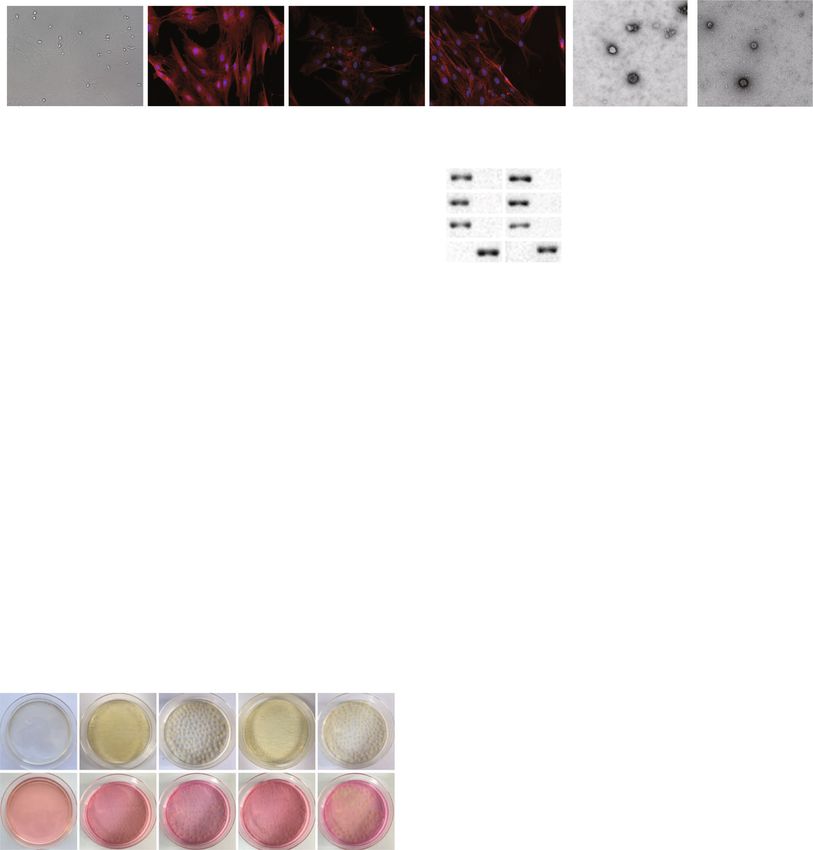
Sun et al. Stem Cell Research & Therapy   (2021) 12:14                                                            Page 2 of 15

Background                                                         Based on the bioinformatics analysis and the dual-
Sepsis is considered as a global health care problem and        luciferase reporter gene assay in the present study, miR-
remains the leading cause of infectious death [1]. Sepsis is    27b could target Jumonji D3 (JMJD3), a histone lysine
characterized by life-threatening organ dysfunction attrib-     demethylase, regulates transcription and actives the ex-
uted to a dysregulated widespread activation of host re-        pression of genes via demethylating H3K27me3 [22–24].
sponse to infection, which is known as systemic                 More importantly, JMJD3 has been reported to play a crit-
inflammatory response syndrome, leading to large human          ical role in the epigenetic regulation during sepsis [25, 26].
morbidity and mortality rates each year [2, 3]. Sepsis can      Furthermore, an interaction between JMJD3 and the acti-
trigger complex interactions between the pro-                   vation of nuclear factor-κB (NF-κB) has been highlighted
inflammatory and anti-inflammatory processes of the host.       [27]. NF-κB participates a typical pro-inflammatory signal-
A better understanding of inflammatory response-                ing pathway and plays important role in regulating pro-
associated mechanisms will provide a new therapeutic ap-        inflammatory gene expression [28], whose dysregulation is
proach for the treatment of sepsis [4]. Because therapeutic     linked to many inflammatory diseases including sepsis [29,
regimens to ameliorate sepsis are not available, infection is   30]. Therefore, we hypothesized that MSC-derived exoso-
mainly controlled through source control, antibiotics, and      mal miR-27b could potentially affect the development of
organ function support [5]. Thus, identification of bio-        sepsis by regulating JMJD3 and NF-κB, which may provide
markers based on the molecular mechanism underlying             novel therapeutic approaches for the treatment of sepsis.
sepsis is important to diagnose sepsis, which may also en-
lighten innovative approaches to treat this disease [6].        Materials and methods
   Exosomes belong to a class of membrane bound extra-          Ethics statement
cellular vesicles that are released by all cells, with a size   The current study was approved by the Ethics Committee of
range from 40 to 150 nm and a composition of bilayer            Shenzhen People’s Hospital (ethical approval number: 2020-
lipid membrane. Exosomes are capable of facilitating            099) and performed according to the Guide for the Care and
multiple intercellular activities, such as communication        Use of Laboratory Animals published by the US National In-
between cells and activation of signaling pathway [7].          stitutes of Health. Extensive efforts were made to ensure
Moreover, it has been reported that exosome-mediated            minimal suffering of the animals used in the study.
transportation of microRNAs (miRNAs or miRs) may
play an important role in sepsis treatment [8].                 Cell culture
   miRNAs are a class of small, endogenous, non-coding          Isolation and identification of MSCs: mouse bone
RNAs capable of negatively regulating gene expression           marrow-derived mesenchymal stem cells (BMMSCs)
by repressing translation or inducing degradation of tar-       were isolated from the tibia and femur bone marrow
get mRNA [9]. Dysregulation of miRNAs has been dem-             compartments (n = 5) and cultured in a Dulbecco’s
onstrated to contribute to development and progression          modified Eagle’s medium (DMEM) containing 100 μg/
of human diseases [10]. Recently, many studies have sug-        mL penicillin and 100 μg/mL streptomycin, 2-mm glu-
gested that abnormally expressed miRNAs play pivotal            tamine, and 15% fetal bovine serum (FBS) in an incuba-
roles in the initiation and development of sepsis, which        tor at 37 °C with 5% CO2. The medium was renewed
suggests that miRNAs may act as potential therapeutic           every 3–4 days, and non-adhesive hematopoietic cells
targets of sepsis [11–13]. A previous study reported that       were removed. Cells were passaged after treatment with
exosomes from endothelial progenitor cells confer pro-          0.025% trypsin containing 0.02% ethylenediamine tetraa-
tection against sepsis by transporting miR-126 [14]. In         cetic acid (EDTA) for 10 min. MSCs at passage 3 were
addition, mesenchymal stem cell (MSC)-derived exoso-            used in this study. Immunofluorescence was applied to
mal miR-223 has been shown to confer cardio-                    identify MSC phenotypes. Briefly, MSCs were incubated
protection in sepsis [15]. More importantly, MSC-based          with primary anti-mouse antibodies to CD29, Sca-1, and
cell therapy showed beneficial effects on sepsis treatment      CD34 (1: 100, Affymetrix-eBiosciece, CA, USA),
[16]. Several studies have highlighted miR-27b as an            followed by incubation with secondary fluorescent-
anti-inflammatory miRNA in the context of infection             labeled Alexa Fluor® 488 goat anti-mouse immunoglobu-
[17–19]. Meanwhile, miR-27b has been shown to be sig-           lin G (IgG) antibodies (H + L) (1: 500, Gaithersburg,
nificantly decreased in mice with sepsis [20]. Interest-        MD, USA). MiR-27b-mimic, negative control (NC)-
ingly, miR-27b was found enriched in exosomes isolated          mimic, miR-27b-inhibitor, NC-inhibitor, small interfer-
from the serum of septic patients [21]. These results           ing RNA (si)-JMJD3, si-p65, and si-NC (RiboBio,
suggest that MSC-derived exosomes may play a thera-             Guangzhou, China) were transfected into MSCs using
peutic role in sepsis by transferring miR-27b. However,         RNAiMAX (Invitrogen, Carlsbad, CA, USA).
how MSC-derived exosomal miR-27b function in sepsis                Isolation and culture of bone marrow-derived macro-
still remains exclusive.                                        phage (BMDMs): BMDMs were isolated from the bone
Sun et al. Stem Cell Research & Therapy   (2021) 12:14                                                         Page 3 of 15

marrow of 6–8-week-old C57BL/6 male mice (n = 5).              Establishment of cell and mouse sepsis models
The femur and tibia were separated with removal of ad-         Inflammation was induced in BMDMs (1 × 10⁵ cells/
herent tissues. The two ends of the bone were cut, and         well) as a cell sepsis model with lipopolysaccharide (LPS;
the bone marrow was washed with DMEM supple-                   100 ng/mL, Cat#2630, Sigma-Aldrich Chemical Com-
mented with 20% FBS, 100 μg/mL penicillin, 10 μg/mL            pany, St Louis, MO, USA, purified from Escherichia coli
streptomycin, and 30% L929 cell culture supernatant            [O111: B4]) for 24 h. The supernatant was collected for
(containing macrophage and macrophage colony-                  cytokine measurement to identify the inflammatory
stimulating factor [M-CSF]), followed by culturing in          model [15].
flask for 7 days. Flow cytometry was used to analyze cell        A sepsis mouse model was established by ligation at
markers CD14 and F4/80 in order to identify BMDMs.             75% of the distal end of the cecum from the base.
   Culture of human embryonic kidney 293T (HEK-                C57BL/6 male mice (aged 6–8 weeks, weighing 16–22 g)
293T) cells: HEK-293T cells (CL-0005, Procell, Wuhan,          were used for cecal ligation and puncture (CLP). Mice
Hubei, China) were cultured in high-glucose DMEM               were derived of food for 12 h before operation and then
containing 10% FBS, 100 μg/mL penicillin, and 10 μg/mL         anesthetized by intraperitoneal injection of 2.5% pento-
streptomycin, and L929 cells were cultured in regular          barbital at a dose of 2 mL/kg.
DMEM at 37 °C with 5% CO2.                                       Mice used as control underwent open surgery to sep-
                                                               arate the distal cecum from mesentery and the abdomen
Isolation, culture, and differentiation of BMMSCs              was closed. For sepsis mouse model establishment, the
Following isolation, BMMSCs were cultured in DMEM-             abdomen of mice was routinely disinfected and cut open
F12 medium (Hyclone Laboratories, Logan, UT, USA)              in the middle to expose the abdominal cavity. Next, the
containing 10% FBS (10099141, Gibco, Grand Island, NY,         cecum was found, and the mesenteric vessels were sepa-
USA), 0.2% penicillin and streptomycin (Hyclone Labora-        rated from the mesenteric vessels carefully to avoid
tories, Logan, UT, USA), and sub-cultured every 3 days.        bruising the mesenteric vessels. At the distal end of the
The cells at passages 3–7 were used for follow-up experi-      cecum, 3/4 was ligated with a sterile No. 4 thread, and a
ments. Then, cells were cultured in the medium of osteo-       sterile 7-gauge needle was used to pierce the center of
genic, adipogenic, or chondrogenic differentiation (all        the distal end of the occlusal cecum. The cecum was put
purchased from Cyagen Biosciences Inc., Guangzhou,             back into the abdominal cavity, which was closed and
China) and stained with 0.5% oil red O solution, 5% silver     sutured layer by layer. After being subjected to CLP,
nitrate solution (Von Kossa staining), or 1% alcian blue so-   mice were injected 4 h later with MSC-EXO-miR-27b-
lution, respectively, to evaluate the accumulation of lipid    mimic (30 μg/mouse, exosomes isolated from MSCs
droplets, calcium deposition, or proteoglycan in cells.        transfected with miR-27b-mimic), Ad-overexpression
                                                               (oe)-JMJD3 (1.5 × 10⁹/mouse, adenovirus expressing oe-
Flow cytometry for BMMSC identification                        JMJD3 plasmids), Ad-oe-p65 (1.5 × 10⁹/mouse, adeno-
BMMSCs at passage 3 with 80% confluence were se-               virus expressing oe-p65 plasmids), and Ad-oe-NC
lected for surface identification. After discarding the cul-   (adenovirus expressing oe-NC plasmids). Ad-oe-JMJD3
ture medium, cells were digested and centrifuged. The          and Ad-oe-p65 were purchased from Hanbio Biotech-
pellets were washed twice with phosphate-buffered sa-          nology Co., Ltd. (Shanghai, China) via intravenous tail
line (PBS) buffer, counted with the concentration ad-          injection [31, 32]. Tramadol [33] was used to relieve the
justed to 1 × 10⁶ cells/mL, and transferred into a 15-mL       pain of mice after operation. The mice were divided into
centrifuge tube containing 100 μL of PBS buffer contain-       two groups: one was used for the survival rate monitor
ing 2% FBS. According to the instructions, cells were in-      in 7 days, and the other was used for pathological detec-
cubated with specific fluorescent flow cytometric              tion. The whole blood and tissues were collected 48 h
antibodies against CD90, CD105, CD73, CD45, and                after CLP treatment for follow-up experiments, with 10
CD11b (rat anti-mouse, 1:100, labeled by fluorescein iso-      animals in each group.
thiocyanate [FITC], BD Biosciences, San Jose, CA, USA)
at 4 °C in the dark for 30 min. Thereafter, cells were re-     Extraction of exosomes
suspended with 3 mL of PBS buffer, centrifuged, and            Mouse MSCs were cultured in Roswell Park Memorial
added with 300 μL of PBS buffer. In the control group,         Institute 1640 (RPMI-1640) medium containing FBS
the background marker was determined by homotype               without exosomes (removal by centrifugation at 100,000
monoclonal antibody, and the fluorescence cells were           g for 18 h), followed by centrifugation at 2000 g for 10
analyzed by a flow cytometer (BD Biosciences, San Jose,        min and then at 10,000 g for 30 min to remove debris
CA, USA). The positive rate of surface antigen (%) was         and apoptotic bodies (Avanti-J-26XP, Beckman Coulter,
calculated using the FlowJo software (Tree Star, Ashland,      CA, USA). The supernatant was centrifuged at 110,000 g
OR, USA).                                                      for 70 min (Optima L-80XP, 70 Ti rotor, Beckman
Sun et al. Stem Cell Research & Therapy           (2021) 12:14                                                              Page 4 of 15

Coulter, CA, USA). After centrifugation at 110,000 for                        Other primers are listed in Table 1. The relative expres-
10 min, the precipitate was purified by washing with                          sion of target genes was quantified by 2-ΔΔCt method
PBS. All centrifugation was carried out at 4 °C. The pel-                     normalized to U6. The experiment was repeated 3 times
let was resuspended in PBS and sterilized by filtration                       independently.
through a 0.22-um filter (Millipore, Bedford, MA, USA).
  About 10 μL of the exosomal suspension was dropped                          Enzyme-linked immunosorbent assay (ELISA)
on the cling film. A 200 mesh copper coating was placed                       BMDMs were cultured for 48 h after inoculation. The
down on the suspension for 60 s. The coating was re-                          supernatant was collected, centrifuged to remove cells
moved and adsorbed at room temperature for 3 min                              and cell debris, and frozen at − 20 °C to avoid repeated
with the grid facing down. The coating was stained with                       freezing and thawing cycles or directly subjected to de-
2% uranyl acetate for 5 min with the grid facing up.                          tection. The whole blood of the mice was centrifuged at
After dried for 5 min, exosome particle size was visual-                      2000 g for 20 min, and the serum was collected and fro-
ized and quantified under a transmission electron mi-                         zen at − 20 °C to avoid repeated freezing and thawing or
croscopy (TEM) at 80 kV.                                                      directly subjected to detection. The expression of tumor
  Nanoparticle tracking analysis (NTA) was performed                          necrosis factor-α (TNF-α; MTA00B), inflammatory cyto-
to examine exosomes by means of a 405-nm monochro-                            kine interleukin-1β (IL-1β; MLB00C), and IL-6
matic laser of the NanoSight NS500 instrument. Exo-                           (M6000B) proteins in cell culture supernatant and
somes were recorded using NTA software (Malvern                               mouse serum was analyzed according to the ELISA kit
Instruments GmbH, Malvern, UK) for 5 times with 30 s                          (R&D System, Minneapolis, MN, USA) instructions. The
at a time. The frame rate was recorded with 25 frames/s.                      experiment was repeated 3 times independently.
The size of exosome was calculated by Stokes-Einstein
equation. Western blot was employed to analyze exoso-                         Western blot
mal markers TAPA-1 (CD81, TA343281, 1: 1000),                                 Total cell proteins were extracted using radio-
tumor susceptibility gene 101 (TSG101, TA343598, 1:                           immunoprecipitation assay (RIPA) lysis buffer (Cat#
500), and syntenin-1 (ABIN1881779, 1: 10000).                                 R0020, Beijing Solarbio Science & Technology Co., Ltd.,
                                                                              Beijing, China). Protein concentration was quantified
Reverse transcription quantitative polymerase chain                           using a bicinchoninic acid (BCA) protein assay kit (Cat#
reaction (RT-qPCR)                                                            ab102536, Abcam Inc., Cambridge, UK) and adjusted to
Total RNA from cells and exosomes was extracted with                          the same concentration. Proteins were loaded and sepa-
TRIzol reagents (Invitrogen, Carlsbad, CA, USA) using                         rated by sodium dodecyl sulfate-polyacrylamide gel elec-
RNeasy Mini Kit (Qiagen, Valencia, CA, USA). Reverse                          trophoresis (SDS-PAGE). After separation, proteins were
transcription was performed according to the instruc-                         transferred to a nitrocellulose membrane, followed by
tions of reverse transcription kit (RR047A, Takara,                           blocking with 5% skim milk for 1–2 h. Diluted primary
Japan) to generate complementary DNA (cDNA) as the                            antibodies JMJD3 (Abcam, Cambridge, UK; 1: 1000),
template for qPCR. cDNA of miRNA was synthesized                              H3K27me3 (Abcam, Cambridge, UK; 1: 2000), H3 (Cell
based on the instructions of First-Strand cDNA Synthe-                        Signaling Technologies, Danvers, MA, USA; 1: 1000),
sis (Tailing Reaction) kit (B532451-0020, Sangon Biotech                      and β-actin (Santa Cruz, CA, USA; 1: 2000) were added
Co., Ltd., Shanghai, China). Quantitative PCR was per-                        and incubated with NC membranes overnight at 37 °C.
formed with SYBR® Premix Ex Taq™ II (Perfect Real                             After washed three times with Tris-buffered saline
Time) kit (DRR081, Takara, Japan) and processed on                            Tween-20 (TBST), the membranes were incubated with
qPCR machine (ABI 7500, Foster City, CA, USA). The                            corresponding horseradish peroxidase (HRP)-labeled
random negative primer of miRNA and the upstream                              secondary rabbit anti-mouse IgG (West Grove, PA,
primer of U6 internal control were provided by miRNA                          USA; 1: 10000) for 2 h. The immunocomplexes on the
First-Strand cDNA Synthesis (Tailing Reaction) kit.                           membrane were visualized using enhanced chemilumin-
                                                                              escence (ECL) solution (Thermo Fisher Scientific Inc.,
                                                                              Waltham, MA, USA), and band intensities were quanti-
Table 1 Primer sequences for RT-qPCR
                                                                              fied using ImageJ software. The experiment was repeated
Gene                                   Sequences
                                                                              3 times independently.
miR-27b                                F: GGGGTTCACAGTGGCTAA
                                       R: CAGTGCGTGTCGTGGAGT                  Endocytosis of exosomes by BMDMs
U6                                     F: GCTTCGGCAGCACATATACTAAAAT           Exosomes were pre-labeled with PKH-67 (a new dye for
                                       R: CGCTTCACGAATTTGCGTGTCAT             fluorescent labeling of living cells which can be used to
Note: RT-qPCR reverse transcription quantitative polymerase chain reaction,
                                                                              label living cells by binding to lipid molecules of mem-
miR-27b microRNA-27b, F forward, R reverse                                    brane structure). PKH-67 has less cytotoxicity to cells,
Sun et al. Stem Cell Research & Therapy (2021) 12:14 Page 5 of 15
low fluorescence background, high-lipid solubility, and Bacterial colony experiment on sepsis
can easily penetrate the cell membrane with strong and Solid Luria-Bertani (LB) culture plates were prepared. A
stable green fluorescence. PKH-67-labeled cells can be serial dilution of 1: 10, 1: 100, 1: 1000, 1: 10,000, and 1:
used in vitro and in vivo proliferation studies and have 100,000 was made using peritoneal fluid and serum of
the function of not staining adjacent cells (Sigma-Al- mice, respectively. Next, 1 mL of each dilution was uni-
drich Chemical Company, St Louis, MO, USA), followed formly streaked on LB plates, which were incubated at
by centrifugation at 110,000 g for 70 min and washed 37 °C for 48 h. Colonies were photographed and the
with PBS to remove the excess staining solution. PKH- number was counted. The appropriate dilution ratio was
67-labeled exosomes (10 mg/mL) were incubated with selected for subsequent experiments.
BMDMs for 24 h. Cells were fixed with 4% paraformal-
dehyde (PFA) for 10 min and permeabilized with ice cold Liver and kidney function determination
methanol for 15 min. The nuclei were stained with 4′,6- Forty-eight hours following CLP, 0.1 mL of fresh whole
diamidino-2-phenylindole (DAPI). Images were taken blood was collected via tail vein [34]. The serum was ob-
under a laser scanning confocal microscope (LSCM, tained by centrifugation at 2000 g for 20 min aliquoted
LSM 510, Carl Zeiss, Germany). Then, the rate of endo- and stored at − 20 °C. The levels of liver function indica-
cytosis was analyzed by flow cytometry. tors, aspartate aminotransferase (AST), and alanine ami-
notransferase (ALT) and renal function indicator serum
creatinine (SCr) were routinely measured using auto-
Dual-luciferase reporter gene assay analyzers (Sysmex XT-4000i, Japan; Hitachi 7600-100,
Dual-luciferase reporter gene assay was performed to Japan; hemagglutination analyzers PUN-2048B, Sysmex,
study the interaction between miR-27b and JMJD3. The Japan; blood gas analyzer, GEM Premier 3500) [35].
potential target genes of miR-27b were predicted via bio-
informatics analysis using Starbase (http://starbase.sysu. Hematoxylin-eosin (HE) staining
edu.cn/). The wild-type (wt) 3′untranslated region (UTR) Forty-eight hours following CLP, the liver, lung, and kid-
of JMJD3 mRNA sequence containing the predicted target ney tissues were collected, fixed with 4% PFA at room
sites of miR-27b was synthesized. The reporter vectors temperature for more than 16 h, embedded in paraffin,
containing JMJD3 wt (pmirGLO-Pygo2-wt) and JMJD3 and sectioned at a 3-um thickness. The sections were
mutant miR-27b-binding sequence (pmirGLO-JMJD3- immersed in xylene I for 20 min xylene II 20 min, abso-
mut) or the NC sequence (pmirGLO-NC) (GenePharma, lute ethanol I 5 min, absolute ethanol II 5 min, and 75%
Shanghai, China) were co-transfected into BMDMs with alcohol 5 min to be dewaxed and rehydrated. After rins-
miR-27b-mimic or NC-mimic. After 24-h transfection, ing with tap water, the sections were stained with
activities of firefly luciferase and Renilla luciferase were hematoxylin for 3–5 min, blued, dehydrated in increas-
detected according to the manufacturer’s instruction for ing concentrations of alcohol (85% and 95%) for 5 min,
the dual-luciferase reporter assay system (Promega, and counterstained with eosin for 5 min. Thereafter, the
Madison, WI, USA). sections were cleared with absolute ethanol I for 5 min,
absolute ethanol II for 5 min, absolute ethanol III for 5
min, and xylene I and xylene II for 5 min, respectively.
Chromatin immunoprecipitation (ChIP)-qPCR The sections were mounted with neutral gum and
To study the enrichment of JMJD3 and NF-κB in the graphed under a light microscope. At least 10 random
promoter region of TNF-α, IL-1β, and IL-6, ChIP was fields were examined for each mouse. Lung and kidney
performed using a ChIP assay kit (Millipore, Billerica, injuries were assessed and scored by pathologists blinded
MA, USA). Briefly, PBMCs were crosslinked with 1% to the experiment [14, 31].
formaldehyde, washed, and re-suspended in SDS (so-
dium dodecyl sulfate) lysis buffer. Chromatin was frag- Statistical analysis
mented by sonication. Chromatin fractions were The SPSS 21.0 statistical software (IBM Corp., Armonk,
precleared with protein A-agarose beads followed by im- NY, USA) was used to analyze statistical data. Measure-
munoprecipitation overnight at 4 °C with anti-JMJD3, ment data were presented as mean ± standard deviation.
anti-H3K27me3, anti-NF-κB/p65 antibodies, or with IgG Unpaired data in compliance with normal distribution
(rabbit IgG-ChIP, ab171870, Abcam, Cambridge, UK) and equal variance between two groups were analyzed
for JMJD3, H3K27me3, and NF-κB; mouse IgG1 using unpaired t test. Comparisons among multiple
(ab81032). All antibodies were obtained from Abcam groups were conducted by one-way analysis of variance
(Cambridge, UK). Crosslinking was reversed followed by (ANOVA) with Tukey’s post hoc test. Data at different
proteinase K incubation. Immunoprecipitated DNA was time points were analyzed by repeated measures
subjected to qPCR. ANOVA, followed by Bonferroni post hoc test. SurvivalSun et al. Stem Cell Research & Therapy (2021) 12:14 Page 6 of 15
curves were calculated using Kaplan-Meier’s method. than that in mice treated with PBS (p < 0.01), but the
p < 0.05 indicated significant difference. number of colonies in mice injected with MSC-miR-
27b-inhibitor-EXO was larger than that in mice injected
Results with MSC-EXO (p < 0.01; Fig. 1h).
Exosomal miR-27b derived from mouse BMMSCs inhibits The liver, kidney, and lung tissue sections of mice
CLP-induced sepsis were observed with HE staining. The results revealed
MSC-derived exosomes have protective roles in sepsis that the liver, kidney, and lung injury scores of MSC-
[15]. miR-27b is downregulated in the serum sample of EXO-treated CLP-induced mice were decreased. Com-
sepsis patients [21]. In addition, miR-27b has been found pared with MSC-NC-inhibitor-EXO-treated mice, the
in MSC-derived exosomes [36]. Therefore, it is proposed injury scores of MSC-miR-27b-inhibitor-EXO-treated
that exosomal miR-27b may be involved in the develop- mice were increased (p < 0.01; Supplementary Figure
ment of sepsis. BMMSCs were isolated and cultured, 2B). Taken together, knockdown of miR-27b could re-
followed by the identification by examining the expression verse the inhibitory effect of MSC-EXO on sepsis.
of CD29, Sca-1, and CD34 by immunofluorescence
(Fig. 1a). At the same time, we detected MSC surface miR-27b attenuates LPS-mediated BMDM inflammation
markers and the potential of MSC differentiation using by inhibiting JMJD3
flow cytometry (Supplementary Figure 1A, B). Exosomes The molecular mechanism of how miR-27b inhibits sep-
were isolated from the supernatants of mouse MSCs sis was explored. The target genes of miR-27b were pre-
(MSC-EXO) and mouse fibroblasts L929 (L929-EXO) and dicted using the Starbase database, which revealed that
examined by TEM and NTA (Fig. 1b, c). The expression miR-27b targeted the JMJD3 mRNA 3′UTR in both
of miR-27b in MSC-EXO and L929-EXO was quantified mice and humans (Fig. 2a). Subsequently, dual-luciferase
by RT-qPCR, and the results of which showed that miR- reporter gene assay was applied to verify their inter-
27b was highly expressed in MSC-EXO compared to L929 action. pmirGLO-JMJD3-wt and pmirGLO-JMJD3-mut
control (p < 0.05; Fig. 1d). MSC-EXO can inhibit the oc- plasmid were constructed (Fig. 2b), which were co-
currence of sepsis [15]. The above results showed that transfected with miR-27b-mimic into the BMDMs. The
miR-27b expression was increased in MSC-EXO, and results displayed that miR-27b increased the activity of
thus, it was hypothesized that MSCs might be involved luciferase in BMDMs transfected with JMJD3-wt (p <
with the initiation of sepsis through exosomal miR-27b. 0.01; Fig. 2c).
The miR-27b-inhibitor and NC-inhibitor were trans- To verify whether exosomes derived from MSCs could
fected into BMMSCs for 48 h followed by exosome isola- be taken up by BMDMs, PKH-67-labeled MSCs and un-
tion. RT-qPCR data showed that miR-27b was knocked labeled exosomes were incubated with BMDMs for 24 h.
down in exosomes (Supplementary Figure 2A). The re- Images were taken under a LSCM (Fig. 2d), and the
sults of 7-day survival rate demonstrated that the sur- endocytosis of exosomes by BMDMs was found to be
vival rate of mice injected with MSC-EXO was higher more than 90%. BMDMs were treated with LPS (100 ng/
than that (0%) of CLP-treated mice (p < 0.05), while the mL) to establish a sepsis model. miR-27b-mimic was
mice injected with MSC-miR-27b-inhibitor-EXO showed transfected into MSCs, and MSC-derived exosomes
decreased survival rate compared with those injected (MSC-miR-27b-mimic-EXO) were isolated. RT-qPCR
with MSC-NC-inhibitor-EXO (p < 0.05; Fig. 1e). Further- was used to analyze miR-27b expression in BMDMs, the
more, as detected by ELISA, the production of TNF-α, results of which showed that the expression of miR-27b
IL-1β, IL-10, and IL-6 was increased in the serum of in BMDMs was reduced under LPS treatment compared
CLP-induced mice, compared to sham-operated mice. to control group, which was rescued by the treatment of
However, TNF-α, IL-1β, and IL-6 presented a decreased MSC-miR-27b-mimic-EXO (p < 0.05; Fig. 2e).
production and IL-10 production was increased in re- The expression of JMJD3 and H3K27me3 in BMDMs
sponse to treatment with MSC-EXO. Mice injected with was analyzed by Western blot. Results showed that the
MSC-miR-27b-inhibitor-EXO had higher expression of expression of JMJD3 was increased while that of
TNF-α, IL-1β, and IL-6, yet lower IL-10 expression than H3K27me3 was decreased in the LPS-treated BMDMs in
that with MSC-EXO (p < 0.05; Fig. 1f). Additionally, the comparison with PBS-treated BMDMs (p < 0.05), which
levels of ALT, AST, and SCr were lower in MSC-EXO- was abolished by MSC-NC-mimic-EXO or MSC-miR-
treated CLP-induced mice than that in PBS-treated 27b-mimic-EXO treatment, as evidence by reduced ex-
CLP-induced mice (p < 0.05), which was reversed when pression of JMJD3 (p < 0.05) and increased H3K27me3
comparing MSC-miR-27b-inhibitor-EXO-treated mice expression (p < 0.01) (Fig. 2f).
with SC-EXO-treated mice (p < 0.05; Fig. 1g). ELISA assay was applied to examine the expression of
The number of colonies was smaller in peritoneal fluid inflammatory cytokines including TNF-α, IL-1β, and IL-
and serum of CLP-induced mice treated with MSC-EXO 6 and anti-inflammatory factor IL-10 in the supernatantSun et al. Stem Cell Research & Therapy (2021) 12:14 Page 7 of 15 Fig. 1 Exosomal miR-27b in mouse BMMSCs inhibits CLP-induced sepsis. a Isolation and identification of mouse MSCs, bright field (BF), CD29, Sca-1 and CD34 (green), DAPI (blue). b TEM micrographs of mouse MSC-derived exosomes. c Size distribution and concentration of particles as quantified by NTA. d The expression of miR-27b in mouse MSC-derived exosomes analyzed by RT-qPCR. *p < 0.05 vs. exosomes derived from mouse fibroblasts L929. e Measurements of a 7-day survival of mice by Kaplan-Meier method. n = 10 for mice following each treatment. f The production of TNF-α, IL-1β, IL-10, and IL-6 in serum of CLP- induced septic mice injected with MSC-EXO, MSC-miR-27b-inhibitor-EXO, and MSC-NC-inhibitor-EXO examined by ELISA. g Detection of ALT, AST, and SCr levels in the serum of mice. h Bacterial colonies in peritoneal fluid and serum of mice. *p < 0.01 vs. CLP-induced mice; #p < 0.001 vs. mice with sham operation; &p < 0.01 vs. CLP-induced septic mice treated with MSC-miR-27b-inhibitor-EXO. n = 10 for mice following each treatment. Quantitative data were presented as mean ± standard deviation. Unpaired data in compliance with normal distribution and equal variance between two groups were compared using unpaired t test. Comparisons among multiple groups were analyzed by one-way ANOVA with Tukey’s post hoc test. p < 0.05 indicated significant difference
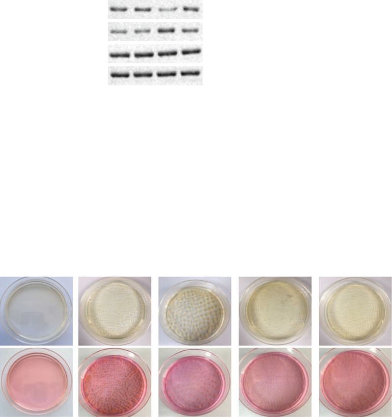
Sun et al. Stem Cell Research & Therapy     (2021) 12:14                                                                             Page 8 of 15

 Fig. 2 miR-27b prevents LPS-mediated BMDM inflammation through suppression of JMJD3. a Target genes of miR-27b predicted by the Starbase
 website. b, c Binding sites (b) of miR-27b to JMJD3 and verification (c) by dual-luciferase reporter gene assay. *p < 0.01 vs. BMDMs transfected
 with NC-mimic. d Images of BMDMs uptaking exosomes visualized under a LSCM. e The expression of miR-27b in BMDMs treated with LPS or
 MSC-NC-mimic-EXO and MSC-miR-27b-mimic-EXO analyzed by RT-qPCR. f The expression of JMJD3 and H3K27me3 in BMDMs treated with LPS or
 MSC-NC-mimic-EXO and MSC-miR-27b-mimic-EXO analyzed by western blot. In panel 2 e, f, *p < 0.05 vs. PBS-treated BMDMs; #p < 0.05 vs. LPS-
 treated BMDMs; &p < 0.05 vs. BMDMs treated with MSC-NC-mimic-EXO. The experiment was repeated 3 times independently. Quantitative data
 were presented as mean ± standard deviation. Unpaired data in compliance with normal distribution and equal variance between two groups
 were compared using unpaired t test. Comparisons among multiple groups were analyzed by one-way ANOVA with Tukey’s post hoc test. p <
 0.05 indicated significant difference

of BMDMs. Expression of TNF-α, IL-1β, and IL-6 in-                         27b repressed the effect of MSC-EXO on inflammation
duced by LPS was downregulated while that of IL-10                         (Supplementary Figure 3B). In summary, high expression
was increased in the presence of MSC-miR-27b-mimic-                        of miR-27b in MSC-derived exosomes could inhibit the
EXO (p < 0.05) (Supplementary Figure 3A).                                  expression and function of JMJD3 on LPS-induced in-
  Whether the therapeutic effects of MSC-EXO on LPS-                       flammation leading to the repression of sepsis.
induced inflammation as cell sepsis model were associ-
ated with miR-27b was further investigated. miR-27b-                       JMJD3 interacting with NF-κB/p65 increases expression of
inhibitor was transfected into MSCs and exosomes                           pro-inflammatory cytokines in LPS-induced BMDMs
(MSC-miR-27b-inhibitor-EXO) were isolated. MSC-                            The aforementioned results demonstrated that miR-27b
miR-27b-inhibitor-EXO was co-incubated with LPS-                           targeted and downregulated JMJD3 to decrease the pro-
treated BMDMs. ELISA was used to determine the ex-                         duction of pro-inflammatory cytokines in BMDMs so as
pression of TNF-α, IL-1β, IL-6, and IL-10 in cell super-                   to inhibit the inflammation in sepsis. So, how JMJD3
natant. The results demonstrated that inhibition of miR-                   regulated the expression of pro-inflammatory cytokines
Sun et al. Stem Cell Research & Therapy   (2021) 12:14                                                           Page 9 of 15

was further investigated. JMJD3 is known as a demethy-          derived exosomal miR-27b inhibited LPS-induced pro-
lase that regulates the transcription of target genes, and      inflammatory effects on BMDMs by inhibiting JMJD3
meanwhile, it has been shown to coordinate with NF-             and NF-κB/p65 axis.
κB/p65 to regulate transcription of target genes [27].
Moreover, NF-κB/p65 is involved in the initiation of sep-       MSC-derived exosomal miR-27b attenuates CLP-induced
sis [37]. Therefore, we wanted to study whether JMJD3           sepsis in mice via inhibition of JMJD3/NF-κB/p65 axis
and NF-κB/p65 co-regulated the expression of pro-               Mice subjected to CLP could simulate sepsis, which was
inflammatory cytokines.                                         utilized to validate our results in vivo. Western blot was
  The recruitment of JMJD3 and NF-κB/p65 in the pro-            used to measure expression of JMJD3 and H3K27me3 in
moter region of pro-inflammatory factor was analyzed by         BMDMs of CLP-treated mice or CLP-treated mice in re-
CHIP-qPCR. The promoter region of pro-inflammatory              sponse to MSC-miR-27b-mimic-EXO. The results re-
cytokines TNF-α, IL-1β, and IL-6 in LPS-treated BMDMs           vealed that CLP-treated mice in response to MSC-miR-
showed increased enrichment of JMJD3 and NF-κB/p65,             27b-mimic-EXO showed a significant increase in JMJD3
decreased H3K27me3, increased H3K27me1, and upregu-             expression but a decrease in H3K27me3 expression (p <
lated expression of pro-inflammatory cytokines (p < 0.05;       0.05; Fig. 5a). Analysis of 7 days of survival curve demon-
Fig. 3a–c).                                                     strated that the survival rate of the mice was decreased
  To further validate the effects of JMJD3 and p65 on the ex-   in the presence of MSC-miR-27b-mimic-EXO and up-
pression of pro-inflammatory cytokines, LPS-induced pro-        regulation of JMJD3 and p65 (p < 0.05; Fig. 5b).
inflammatory factor expression was inhibited after knock-          Ad-oe-JMJD3 and Ad-oe-p65 were injected into CLP-
down of JMJD3 and p65 by siRNA (p < 0.05; Fig. 3d–f). After     treated mice. The production of pro-inflammatory cyto-
treatment with GSK-J4 (4 μmol/L, ab144395, Abcam Inc.,          kines TNF-α, IL-1β, IL-10, and IL-6 in serum was
Cambridge, UK), a pharmacological inhibitor of histone          detected by ELISA, which demonstrated that upregulation
demethylase JMJD3, inflammatory response of LPS-induced         of JMJD3 and p65 inhibited effect of MSC-miR-27b-
BMDMs was reduced (p < 0.05; Fig. 3g–i). These results indi-    mimic-EXO on inflammation in CLP-treated mice (p <
cated that LPS induced the recruitment of JMJD3 and NF-         0.05; Fig. 5c).
κB in the promoter region of pro-inflammatory cytokines            The number of colonies in the peritoneal fluid and
and H3K27me3 demethylation, leading to the regulation of        blood of mice was quantified, which was found to be in-
gene transcription.                                             creased after overexpression of JMJD3 and p65 (p < 0.05;
                                                                Fig. 5d).
MSC-EXO suppresses LPS-induced inflammation in                     Analysis of the liver and kidney function showed over-
BMDMs through regulating miR-27b and inhibiting JMJD3           expressing of JMJD3 and p65 promoted the ALT, AST,
and NF-κB/p65                                                   and SCr indexes and indicated liver and kidney dysfunc-
The molecular mechanism of anti-inflammatory effect             tion (p < 0.05; Fig. 5e). HE staining showed that the liver,
of MSC-EXO on LPS-induced BMDMs was further                     lung, and kidney tissue injury scores were increased and
investigated. The inhibitory effect of miR-27b in               tissue injury was worse after overexpression of JMJD3
MSC-EXO on the recruitment of JMJD3 and NF-κB/                  and p65 (p < 0.05; Supplementary Figure 4). In summary,
p65 in the pro-inflammatory cytokine promoter re-               the CLP mouse model confirmed that overexpression of
gions in LPS-induced BMDMs was analyzed by ChIP-                miR-27b in MSC-EXO could suppress the JMJD3/NF-
qPCR. The MSCs were transfected with miR-27b-                   κB/p65 axis to inhibit the development of sepsis in vivo.
inhibitor and NC-inhibitor for 48 h, after which the
exosomes were isolated and the exosomes with low                Discussion
expression of miR-27b were incubated with LPS-                  Sepsis is a syndrome attributed to infections of body im-
treated BMDMs. ChIP-qPCR was then performed to                  mune and coagulation systems leading to approximately
detect the recruitment of JMJD3 and NF-κB/p65 in                44,000 deaths each year in UK, which is likely to cause
the pro-inflammatory cytokine promoter regions, and             shock, multiple organ failure, and even death without
the results of which showed that knockdown of miR-              early detection and proper treatment [38]. Currently, no
27b reversed the inhibitory effect of MSC-EXO on                specific treatment to sepsis is available [5]. Thus, novel
LPS-mediated BMDM inflammatory response (p <                    therapies tailored with the aid of molecular biomarkers
0.05). Meantime, knockdown of miR-27b obviously                 are urgently needed to diagnose and prevent sepsis.
changed the expression of H3K27 in the promoter re-             MSCs have been reported to improve the survival of
gion of inflammatory factors (p < 0.05; Fig. 4a–c).             Gram-negative diabetic peritoneal sepsis [39–41]. How-
Overexpression of JMJD3 and p65 also inhibited the              ever, the specific mechanism of how MSCs function in
effect of MSC-EXO on LPS-mediated inflammation of               sepsis remains poorly understood. Our data demon-
BMDMs (p < 0.05; Fig. 4d–f). In summary, MSC-                   strated that MSCs were able to ameliorate sepsis via the
Sun et al. Stem Cell Research & Therapy (2021) 12:14 Page 10 of 15 Fig. 3 JMJD3 interacting with NF-κB/p65 elevates expression of the pro-inflammatory cytokines in LPS-induced BMDMs. a–c The recruitment of JMJD3, p65, H3K27me3, and H3K27me1 in the promoter region of pro-inflammatory cytokines TNF-α (a), IL-1β (b), and IL-6 (c) in BMDMs analyzed by CHIP-qPCR. #p < 0.05 vs. PBS-treated BMDMs. d–f The expression of LPS-induced pro-inflammatory factors TNF-α (d), IL-1β (e), and IL-6 (f) after knockdown of JMJD3 and p65. *p < 0.05 vs. PBS-treated BMDMs in response to si-NC; #p < 0.05 vs. BMDMs in response to si-NC. g–i The expression of pro-inflammatory factors TNF-α (g), IL-1β (h), and IL-6 (i) in BMDMs after 1-h treatment with GSK-J4 (4 umol/L, pharmacological inhibitor of JMJD3). *p < 0.05 vs. LPS-treated BMDMs in response to GSK-J4; #p < 0.05 vs. BMDMs in response to GSK-J4. The experiment was repeated 3 times independently. Quantitative data were presented as mean ± standard deviation. Unpaired data in compliance with normal distribution and equal variance between two groups were compared using unpaired t test. Comparisons among multiple groups were analyzed by one-way ANOVA with Tukey’s post hoc test. p < 0.05 indicated significant difference exosome-mediated transportation of miR-27b through [15]. In addition, the anti-inflammatory effect of regulating the JMJD3/NF-κB/p65 axis. MSC-derived exosomes was also presented in sepsis In fact, the therapeutic and regenerative effects of by carrying miR-146a [31]. miR-27b has been demon- MSCs may not only depend on local mechanisms but strated to be poorly expressed in the serum of pa- through the secretion of paracrine cytokines such as tients with sepsis [21] but highly expressed in MSC- exosomes [42, 43]. MSCs could exert anti- derived exosomes. The present data revealed that exo- inflammatory effects through the exosome-mediated somal miR-27b from MSCs showed an inhibitory ef- transportation of miR-223 in polymicrobial sepsis fect on the development of sepsis as evidenced by
Sun et al. Stem Cell Research & Therapy       (2021) 12:14                                                                              Page 11 of 15

 Fig. 4 MSC-derived exosomal miR-27b inhibits JMJD3 and NF-κB/p65 axis to restrain LPS-induced pro-inflammatory response. a–c The recruitment of
 JMJD3, p65, H3K27me3, and H3K27me1 in the promoter region of pro-inflammatory cytokines TNF-α (a), IL-1β (b), and IL-6 (c) in LPS-treated BMDMs
 transfected with MSC-miR-27b-inhibitor-EXO analyzed by CHIP-qPCR. *p < 0.05 vs. LPS-treated BMDMs transfected with MSC-NC-inhibitor-EXO. d–f The
 expression of pro-inflammatory cytokines TNF-α (d), IL-1β (e), and IL-6 (f) in BMDMs after overexpression of JMJD3 or p65. *p < 0.05 vs. PBS-treated
 BMDMs. The experiment was repeated 3 times independently. Quantitative data were presented as mean ± standard deviation. Unpaired data in
 compliance with normal distribution and equal variance between two groups were compared using unpaired t test. Comparisons among multiple
 groups were analyzed by one-way ANOVA with Tukey’s post hoc test. p < 0.05 indicated significant difference

decreased injuries in the liver, kidney, and lung in                         multiplication of hepatitis B virus [44]. Additionally, miR-
CLP-treated mice.                                                            146a could suppress osteogenesis in MSCs by downregu-
   In a mouse model of sepsis, miR-27a was found to be                       lating JMJD3 expression [45]. Some papers have shown
significantly decreased and its overexpression can alleviate                 that JMJD3 can facilitate great effects on several inflam-
inflammatory response, thus preventing the LPS-induced                       matory diseases including atherosclerosis [46], rheumatoid
liver injury following sepsis [20]. Bioinformatics analysis                  arthritis [47], and sepsis [48]. JMJD3 was demonstrated to
and dual-luciferase reporter gene assay together identified                  be upregulated in cells treated with LPS, along with a re-
that miR-27b targeted JMJD3 and downregulated its ex-                        duced expression of H3K27me3- and LPS-mediated in-
pression. This contributed to the reduction of LPS-                          flammation [26]. The immune system responds by
mediated BMDM inflammation, accompanied by de-                               secreted inflammatory molecules, whose dysregulation re-
creased expression of LPS-stimulated inflammatory cyto-                      sults in chronic inflammation disease and tissue damage
kines TNF-α, IL-1β, and IL-6 as well as increased                            [49]. Pro-inflammatory cytokines, including TNF-α, IL-1β,
expression of anti-inflammatory factor IL-10. Partially in                   and IL-6 play an important role in the development of
agreement with our results, miR-939 has been reported to                     chronic inflammation [50, 51]. JMJD3 demonstrates pro-
target and downregulate JMJD3 to inhibit the                                 moting properties in sepsis by upregulating pro-
Sun et al. Stem Cell Research & Therapy        (2021) 12:14                                                                                Page 12 of 15

 Fig. 5 Exosomal miR-27b from MSCs suppresses the JMJD3/NF-κB/p65 axis to inhibit the development of CLP-induced sepsis. a The expression of
 JMJD3 and H3K27me3 in mice subjected to sham operation or CLP in response to MSC-miR-27b-mimic-EXO analyzed by Western blot. *p < 0.05 vs.
 mice subjected to CLP; #p < 0.05 vs. mice subjected to sham operation. b Results of a 7-day survival in mice. n = 10 for mice following each treatment.
 c The production level of TNF-α, IL-1β, IL-10, and IL-6 in mouse serum examined by ELISA. d Bacterial colonies in peritoneal fluid and serum of mice.
 e Detection of the liver and kidney function in mice. *p < 0.05 vs. mice subjected to CLP. n = 10 for mice following each treatment. Cellular
 experiments were repeated 3 times independently. Quantitative data were presented as mean ± standard deviation. Comparisons among multiple
 groups were analyzed by one-way ANOVA with Tukey’s post hoc test. p < 0.05 indicated significant difference

inflammatory cytokines, IL-1β, and TNF-α expression                            suppression of NF-κB contributed to a reduction of
[51]. Furthermore, miR-27-3p has the capacity to increase                      release of LPS-induced pro-inflammatory cytokines
the production of IL-10 in dendritic cells [52]. Taken to-                     such as TNF-α, IL-1β, and IL-6 in sepsis [37, 56].
gether, we concluded that exosomal miR-27b derived                             Furthermore, a previous study highlighted the ability
from MSCs could target and downregulate JMJD3 to                               of miR-27b to suppress the activity of NF-κB, thus ar-
reduce the inflammatory responses in sepsis.                                   resting an excessive inflammation during infection
  JMJD3 is known as a H3K27 demethylase, which                                 [18]. In consistent with these results, we demon-
regulates the transcription of target genes through                            strated that the recruitment of JMJD3 and NF-κB in
H3K27me3 demethylation [53]. A previous study                                  the promoter regions of TNF-α, IL-1β, and IL-6 con-
showed that JMJD3 synergistically regulated the tran-                          tributed to the upregulation of these pro-
scription of target genes with the transcription regula-                       inflammatory cytokines via H3K27me3 demethylation,
tor NF-κB/p65 [27] and emerging evidences                                      thus promoting inflammatory response in sepsis. Our
demonstrated that NF-κB/p65 was associated with the                            work also revealed that MSC-derived exosomes
development of sepsis [54, 55]. More importantly,                              through the transportation of miR-27b regulated
Sun et al. Stem Cell Research & Therapy (2021) 12:14 Page 13 of 15
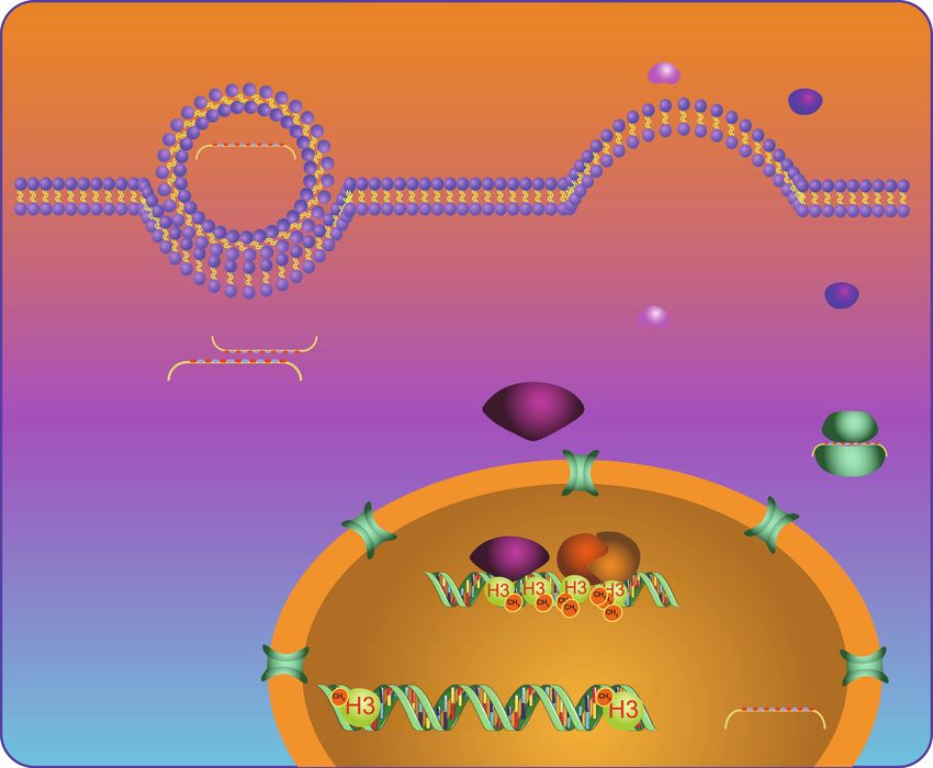
Fig. 6 Schematic diagram of the function and mechanism of exosomal miR-27b in sepsis. JMJD3 can be transferred into nucleus through
cytoplasm and nucleus and promote the demethylation of H3K27me3 by recruiting NF-κB, thus activating the transcription of TNF-α/IL-β/IL-6 to
increase the expression of these target proteins. However, miR-27b loaded in the exosomes secreted by MSCs binds to the mRNA of JMJD3 in
the target cells after membrane fusion and inhibits the expression of JMJD3 protein, thus suppressing the expression of inflammatory factors.
GSK-J4, a pharmacological inhibitor of JMJD3, has similar effects
JMJD3/NF-κB axis to inhibit LPS-induced BMDM [18, 19] and the specific mechanism involving the miR-
pro-inflammatory response as well as CLP-induced 27b/JMJD3 axis thus needs to be further identified
sepsis in mice. through additional studies. Additionally, it would be ne-
cessary for future studies to investigate specific mechan-
Conclusions istic basis by which overexpression of miR-27b as well as
In summary, miR-27b delivered by MSC-derived exo- inhibition of JMJD3 and NF-κB/p65 could impede the
somes could downregulate JMJD3 and NF-κB/p65 to in- progression of sepsis.
hibit inflammatory response and suppress sepsis (Fig. 6).
Therefore, investigation of the miR-27b/JMJD3/NF-κB/
Supplementary Information
p65 axis and their functions presents new mechanistic The online version contains supplementary material available at https://doi.
insights for an understanding of sepsis initiation and de- org/10.1186/s13287-020-02068-w.
velopment and provides potential novel therapeutic tar-
gets against sepsis. Nevertheless, in addition to JMJD3, Additional file 1: Supplementary Figure 1. Characterization of
BMMSCs. A, adipogenic (the left panel), osteogenic (the middle panel)
there are many other genes that are targeted by miR-27bSun et al. Stem Cell Research & Therapy (2021) 12:14 Page 14 of 15
and chondrogenic (the right panel) differentiation of MSCs. B, MSC the manuscript. Jinsong Wang contributed to revising the manuscript. The
surface makers CD105, CD73, CD90, CD45 and D11b analyzed by flow authors have read and approved the final submitted manuscript.
cytometry.
Funding
Additional file 2: Supplementary Figure 2. Inhibition of miR-27b re-
This study is supported by the General Program of National Nature Science
verses the inhibitory effect of MSC-EXO on CLP-induced liver, kidney and
Foundation of China (81770028).
lung injuries. .A, miR-27b expression determined by RT-qPCR in exosomes
derived from MSCs transfected with miR-27b inhibitor. B, HE staining of
liver, kidney and lung tissues of mice treated with MSC-EXO or MSC-miR- Availability of data and materials
27b-inhibitor-EXO (400 ×). *, p < 0.01 vs. CLP-induced septic mice; #, p < The datasets generated/analyzed during the current study are available.
0.001 vs. mice with sham operations; &, p < 0.01 vs. CLP-induced septic
mice treated with MSC-NC-inhibitor-EXO. n = 10 for mice following each Ethics approval and consent to participate
treatment. Quantitative data were presented as mean ± standard devi- The current study was approved by the Ethics Committee of Shenzhen
ation. Comparisons among multiple groups were analyzed by one-way People’s Hospital (ethical approval number: 2020-099) and performed accord-
ANOVA with Tukey’s post hoc test. p < 0.05 indicated significant ing to the Guide for the Care and Use of Laboratory Animals published by
difference. the US National Institutes of Health. Extensive efforts were made to ensure
Additional file 3: Supplementary Figure 3. Inhibition of miR-27b re- minimal suffering of the animals used in the study.
presses the effect of MSC-EXO on LPS-induced inflammation. A, The ex-
pression levels of inflammatory cytokines TNF-α, IL-1β, IL-10 and IL-6 in Consent for publication
the supernatant of BMDMs in response to LPS, MSC-NC-mimic-EXO and Not applicable.
MSC-miR-27b-mimic-EXO measured by ELISA assay. *, p < 0.05 vs. PBS-
treated BMDMs; #, p < 0.05 vs. LPS-treated BMDMs; &, p < 0.05 vs. BMDMs Competing interests
treated with MSC-NC-mimic-EXO. B, The expression of inflammatory cyto- The authors declare that they have no competing interests.
kines TNF-α, IL-1β, IL-6 and IL-10 in the supernatant of BMDMs treated
with MSCs-miR-27b-inhibitor-EXO. #, p < 0.05 vs. LPS-treated BMDMs; &, p Author details
< 0.05 vs. BMDMs treated with MSC-NC-mimic-EXO. The experiment was 1
ShenZhen Beike Biotechnology Research Institute, No. 59, Gaoxin South 9th
repeated 3 times independently. Quantitative data were presented as Road, Nanshan District, Shenzhen 518057, Guangdong Province, People’s
mean ± standard deviation. Comparisons among multiple groups were Republic of China. 2Intervention and Cell Therapy Center, Shenzhen Hospital
analyzed by one-way ANOVA with Tukey’s post hoc test. p < 0.05 indi- of Peking University, Shenzhen 518057, People’s Republic of China.
cated significant difference. 3
Hematology Department, Shenzhen People’s Hospital, Shenzhen 518020,
Additional file 4: Supplementary Figure 4. HE staining of liver, kidney People’s Republic of China. 4Clinical Medical Research Center, Shenzhen
and lung tissues of mice treated with MSC-miR-27b-mimic-EXO, MSC- People’s Hospital, Shenzhen 518020, People’s Republic of China. 5Respiratory
miR-27b-mimic-EXO + Ad-oe-p65 or MSC-miR-27b-mimic-EXO + Ad-oe- and Critical Care Medicine Department, Shenzhen People’s Hospital, No.
JMJD3 (400 ×). *, p < 0.05 vs. mice subjected to CLP. n = 10 for mice fol- 1017, Dongmen North Road, Luohu District, Shenzhen 518020, Guangdong
lowing each treatment. Quantitative data were presented as mean ± Province, People’s Republic of China.
standard deviation. Comparisons among multiple groups were analyzed
by one-way ANOVA with Tukey’s post hoc test. p < 0.05 indicated signifi- Received: 26 April 2020 Accepted: 2 December 2020
cant difference.
References
Abbreviations
1. Napolitano LM. Sepsis 2018: definitions and guideline changes. Surg Infect.
MSCs: Mesenchymal stem cells; miRNAs or miRs: MicroRNAs; miR-
2018;19:117–25.
27b: MicroRNA-27b; BMMSCs: Bone marrow-derived mesenchymal stem cells;
2. Iskander KN, Osuchowski MF, Stearns-Kurosawa DJ, Kurosawa S, Stepien D,
BMDMs: Bone marrow macrophages; MSC-EXO: MSC-derived exosomes;
Valentine C, et al. Sepsis: multiple abnormalities, heterogeneous responses,
CLP: Cecal ligation and puncture; NF-κB: Nuclear factor κB; JMJD3: Jumonji
and evolving understanding. Physiol Rev. 2013;93:1247–88.
D3; DMEM: Dulbecco’s modified Eagle’s medium; HEK-293T: Human
3. Pinsky MR. Pathophysiology of sepsis and multiple organ failure: pro- versus
embryonic kidney 293T; TEM: Transmission electron microscopy;
anti-inflammatory aspects. Contrib Nephrol. 2004;144:31–43.
NTA: Nanoparticle tracking analysis; LSCM: Laser scanning confocal
4. Hotchkiss RS, Monneret G, Payen D. Sepsis-induced immunosuppression:
microscope; PFA: Paraformaldehyde; FBS: Fetal bovine serum;
from cellular dysfunctions to immunotherapy. Nat Rev Immunol. 2013;13:
oe: Overexpression; siRNA: Small interfering RNA; NC: Negative control;
862–74.
EDTA: Ethylenediaminetetraacetic acid; LPS: Lipopolysaccharide; 3′UTR: 3′
5. Rello J, Valenzuela-Sanchez F, Ruiz-Rodriguez M, Moyano S. Sepsis: a review
untranslated region; PBS: Phosphate-buffered saline; IL-6: Interleukin-6; TNF-
of advances in management. Adv Ther. 2017;34:2393–411.
α: Tumor necrosis factor-α; FITC: Fluorescein isothiocyanate;
6. Faix JD. Biomarkers of sepsis. Crit Rev Clin Lab Sci. 2013;50:23–36.
cDNA: Complementary DNA; RT-qPCR: Reverse transcription quantitative
7. Kalluri R. The biology and function of exosomes in cancer. J Clin Invest.
polymerase chain reaction; RIPA: Radio-immunoprecipitation assay;
2016;126:1208–15.
BCA: Bicinchoninic acid; SDA-PAGE: Sodium dodecyl sulfate polyacrylamide
8. Real JM, Ferreira LRP, Esteves GH, Koyama FC, Dias MVS, Bezerra-Neto JE,
gel electrophoresis; IgG: Immunoglobulin G; TBST: Tris-buffered saline with
et al. Exosomes from patients with septic shock convey miRNAs related to
Tween-20; HRP: Horseradish peroxidase; ECL: Enhanced chemiluminescence;
inflammation and cell cycle regulation: new signaling pathways in sepsis?
ChIP: Chromatin immunoprecipitation; AST: Aspartate aminotransferase;
Crit Care. 2018;22:68.
ALT: Aminotransferase; SCr: Serum creatinine; HE: Hematoxylin and eosin;
9. Liu Q, Chen YQ. A new mechanism in plant engineering: the potential roles
DAPI: 4′,6-Diamidino-2-phenylindole; wt: Wild type; mut: Mutant;
of microRNAs in molecular breeding for crop improvement. Biotechnol Adv.
ELISA: Enzyme-linked immunosorbent assay; ANOVA: Analysis of variance
2010;28:301–7.
10. Orellana EA, Tenneti S, Rangasamy L, Lyle LT, Low PS, Kasinski AL. FolamiRs:
Acknowledgements ligand-targeted, vehicle-free delivery of microRNAs for the treatment of
The authors would like to acknowledge the helpful suggestions concerning cancer. Sci Transl Med. 20179(401):eaam9327.
this study received from their colleagues. 11. Goodwin AJ, Guo C, Cook JA, Wolf B, Halushka PV, Fan H. Plasma levels of
microRNA are altered with the development of shock in human sepsis: an
Authors’ contributions observational study. Crit Care. 2015;19:440.
Jia Sun and Xuan Sun designed the study. Junhui Chen and Xin Liao 12. Tudor S, Giza DE, Lin HY, Fabris L, Yoshiaki K, D'Abundo L, et al. Cellular and
collated the data, and Yixuan He and Rui Chen analyzed and produced the Kaposi's sarcoma-associated herpes virus microRNAs in sepsis and surgical
initial draft of the manuscript. Sean Hu and Chen Qiu contributed to drafting trauma. Cell Death Dis. 2014;5:e1559.You can also read