Coordinated interactions between endothelial cells and macrophages in the islet microenvironment promote β cell regeneration
←
→
Page content transcription
If your browser does not render page correctly, please read the page content below
www.nature.com/npjregenmed

                  ARTICLE                  OPEN

                  Coordinated interactions between endothelial cells and
                  macrophages in the islet microenvironment promote
                  β cell regeneration
                  Diane C. Saunders 1,2,6, Kristie I. Aamodt 1,6, Tiffany M. Richardson 1, Alexander J. Hopkirk2, Radhika Aramandla2,
                  Greg Poffenberger2, Regina Jenkins2, David K. Flaherty3, Nripesh Prasad4, Shawn E. Levy4, Alvin C. Powers1,2,5 ✉ and
                  Marcela Brissova 2 ✉

                      Endogenous β cell regeneration could alleviate diabetes, but proliferative stimuli within the islet microenvironment are
                      incompletely understood. We previously found that β cell recovery following hypervascularization-induced β cell loss involves
                      interactions with endothelial cells (ECs) and macrophages (MΦs). Here we show that proliferative ECs modulate MΦ infiltration and
                      phenotype during β cell loss, and recruited MΦs are essential for β cell recovery. Furthermore, VEGFR2 inactivation in quiescent
                      ECs accelerates islet vascular regression during β cell recovery and leads to increased β cell proliferation without changes in MΦ
                      phenotype or number. Transcriptome analysis of β cells, ECs, and MΦs reveals that β cell proliferation coincides with elevated
                      expression of extracellular matrix remodeling molecules and growth factors likely driving activation of proliferative signaling
1234567890():,;

                      pathways in β cells. Collectively, these findings suggest a new β cell regeneration paradigm whereby coordinated interactions
                      between intra-islet MΦs, ECs, and extracellular matrix mediate β cell self-renewal.
                      npj Regenerative Medicine (2021)6:22 ; https://doi.org/10.1038/s41536-021-00129-z

                  INTRODUCTION                                                                             understanding how MΦs contribute to β cell proliferation and
                  While investigating how vascular endothelial growth factor-A                             further defining MΦ phenotype and function in the βVEGF-
                  (VEGF-A) regulates islet vascularization, our group previously                           A model.
                  characterized a mouse model in which signals from the local                                 In addition to MΦs, ECs are known to participate in tissue
                  microenvironment stimulate β cell self-renewal1. In this model,                          repair via activation of the VEGF-A–VEGFR2 pathway, which
                  transiently increasing VEGF-A production in β cells (βVEGF-A)                            mediates angiocrine factor production and promotes local cell
                  induces endothelial cell (EC) expansion and hypervascularization                         renewal and regeneration13–16. There is a precedent for ECs
                  that causes β cell loss; remarkably, islet morphology, capillary                         facilitating tissue repair by influencing MΦ activation toward a
                  network, β cell mass, and function normalize 6 weeks after                               restorative, M2-like phenotype4. Because vasculature is essential
                  withdrawal (WD) of the VEGF-A stimulus. This regenerative                                for normal islet function, understanding signals that govern
                  response is the result of a transient but robust burst in β cell                         EC homeostasis and the effects of ECs on neighboring cell
                  proliferation, which is dependent on VEGF-A-mediated recruit-                            populations is crucial for maintaining and restoring islet health.
                  ment of macrophages (MΦs). These recruited cells express                                 Signaling between ECs and the pancreatic epithelium is critical
                  markers of both pro-inflammatory (M1) and restorative (M2)                                for establishing islet vasculature and β cell mass during
                  activation, suggesting a unique regenerative phenotype1–5.                               development, and in mature islets ongoing signaling between
                  Further investigation into the role of various microenvironmental                        endocrine and ECs is required to maintain the capillary network
                  components on β cell proliferation in this model is needed given                         through which endocrine cells receive adequate nutrition and
                  that this regenerative microenvironment promotes proliferation of                        oxygen and can rapidly sense and secrete hormones17–19. The
                  human in addition to mouse β cells1.                                                     vascular basement membrane is also the primary component of
                     MΦs are often perceived as damaging to islets due to their role                       the intra-islet extracellular matrix (ECM) and acts as a reservoir
                  in β cell loss during diabetes6–8, but it is becoming increasingly                       for growth factors and other signaling molecules important for
                  evident that tissue-resident MΦs play important roles in immune                          β cell differentiation, function, and proliferation20–23. In the
                  surveillance and tissue homeostasis and function in the islet. Mice                      βVEGF-A model, ECs may affect the regenerative process
                  with compromised MΦ populations during pancreatic develop-                               indirectly by promoting MΦ recruitment and activation, altering
                  ment exhibit reduced β cell proliferation, β cell mass, and                              ECM composition and signaling, and/or by directly influencing
                  impaired islet morphogenesis9,10. Furthermore, recent studies                            β cell proliferation.
                  have supported the notion that MΦs contribute to β cell                                     Here we deconstructed the complex in vivo islet microenviron-
                  regeneration after several types of injury, including surgically                         ment in the βVEGF-A model and show that β cell self-renewal is
                  induced pancreatitis11 and diphtheria toxin (DT)-mediated                                mediated by coordinated interactions between recruited MΦs,
                  apoptosis12. This recent work highlights the importance of                               intra-islet ECs, and the ECM (Fig. 1, Supplementary Fig. 1).

                  1
                   Department of Molecular Physiology and Biophysics, Vanderbilt University, Nashville, TN, USA. 2Department of Medicine, Division of Diabetes, Endocrinology, and
                  Metabolism, Vanderbilt University Medical Center, Nashville, TN, USA. 3Flow Cytometry Shared Resource, Vanderbilt University Medical Center, Nashville, TN, USA. 4Hudson
                  Alpha Institute of Biotechnology, Huntsville, AL, USA. 5VA Tennessee Valley Healthcare, Nashville, TN, USA. 6These authors contributed equally: Diane C. Saunders, Kristie I.
                  Aamodt. ✉email: al.powers@vumc.org; marcela.brissova@vumc.org

                  Published in partnership with the Australian Regenerative Medicine Institute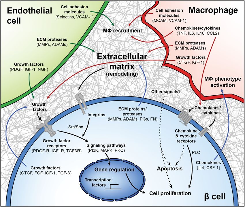
D.C. Saunders et al.
                  2

                      Fig. 1 The RIP-rtTA; Tet-O-VEGF-A (βVEGF-A) model of β cell regeneration permits specific modulation of the islet microenvironment.
                      Induction of VEGF-A overexpression in β cells with doxycycline (Dox) causes rapid endothelial cell (EC) expansion, β cell death, and
                      recruitment of circulating monocytes increasing the number of intra-islet macrophages (MΦs). Once the Dox stimulus is removed, VEGF-A
                      levels normalize and β cells undergo self-renewal1. To establish the role of MΦs in β cell proliferation, monocytes and MΦs were depleted with
                      clodronate liposomes (scheme A). To determine whether proliferative intra-islet ECs were required for β cell loss, MΦ recruitment, and MΦ
                      phenotype activation, an additional genetic construct was introduced to knockdown key signaling receptor VEGFR2 in ECs prior to VEGF-A
                      induction (scheme B). To determine if VEGFR2 signaling in quiescent ECs contributes to β cell proliferation, VEGFR2 was inactivated in ECs
                      during β cell recovery (scheme C). To identify cell-specific transcriptome changes during β cell loss and recovery, populations of ECs, MΦs, and
                      β cells were isolated prior to and during VEGF-A induction, and after VEGF normalization (scheme D). For additional details, including mouse
1234567890():,;

                      models utilized, see Supplementary Fig. 1.

                      To isolate the roles of MΦs and ECs in this model we removed                leads to β cell mass restoration (Fig. 1). In addition, Dox dosing
                      MΦs from the islet microenvironment (Fig. 1 and Supplementary               and treatment duration were previously optimized so βVEGF-A
                      Fig. 1; experimental scheme A) and inactivated VEGFR2 signaling             mice maintained normal glucose clearance, random blood
                      in ECs to discern the effects of proliferative or quiescent ECs on          glucose levels, and body weight during the brief 1 week Dox
                      β cell proliferation (Fig. 1 and Supplementary Fig. 1; experimental         treatment and subsequent period of Dox withdrawal used in
                      schemes B and C). Since the βVEGF-A islet microenvironment                  these studies1.
                      is a complex in vivo system involving dynamic changes in islet
                      cell composition, we also identified regenerative signals by                 Chemical depletion of macrophages in βVEGF-A islets inhibits
                      performing transcriptome analysis (Fig. 1 and Supplementary                 β cell proliferation
                      Fig. 1; experimental scheme D) of purified islet cell populations
                      including β cells, ECs, and MΦs over the course of β cell loss and          To dissect pathways and signaling molecules in the islet
                      recovery. Based on previous work1,16 we predicted that either MΦ            microenvironment required for β cell recovery, we first employed
                      depletion (clodronate) or loss of VEGFR2 signaling in ECs would             clodronate-mediated MΦ depletion which impacts both infiltrat-
                      perturb MΦ recruitment and polarization and impair β cell                   ing and islet resident MΦs. Clodronate is an ATP/ADP translocase
                      regeneration. Indeed, we found that MΦ depletion suppressed                 inhibitor, and when packaged into liposomes it is selectively
                      the transient burst in β cell proliferation leading to reduced              taken up by circulating monocytes and differentiated MΦs due
                      recovery of β cell mass. In addition, VEGFR2-mediated signaling in          to their phagocytic properties and causes apoptosis. We treated
                      intra-islet ECs was necessary for maximal MΦ recruitment and                βVEGF-A mice with either control or clodronate liposomes
                      M2-like phenotype activation. Surprisingly, VEGFR2 ablation in              starting one day before VEGF-A induction and continuing for
                      quiescent ECs during the period of β cell recovery accelerated islet        1 week after VEGF-A normalization (Fig. 2a). Compared to
                      vascular regression leading to increased β cell proliferation while         control, clodronate treatment reduced circulating monocytes
                      MΦ phenotype and number was unchanged. Transcriptome                        (CD11b+ Ly6G− cells) by 50% within 24 h (Fig. 2b, No Dox, and
                      analysis during β cell death and recovery revealed intricate                Supplementary Fig. 2) and reduced the MΦ population in islets
                      changes in expression of growth factors, integrins, and matrix              (Iba1+) by 94% 1 week after VEGF-A induction (Fig. 2c and d, 1wk
                      remodeling enzymes in all three cell types, suggesting that ECM             Dox). MΦ depletion was maintained during VEGF-A normal-
                      remodeling and activation of ECM-associated molecules within                ization, with 86% fewer MΦ in islets from clodronate-treated
                      the islet microenvironment play critical roles in β cell self-renewal.      βVEGF-A mice 1 week after Dox withdrawal (Fig. 2c and d, 1wk
                      Taken together, our results indicate that both MΦs and intra-islet          WD). VEGF-A induction in clodronate-treated mice led to
                      ECs provide crucial microenvironmental cues to cooperatively                increased EC area (Fig. 2c and e) and β cell loss (Fig. 2c and f)
                      promote β cell regeneration.                                                comparable to βVEGF-A mice treated with control liposomes,
                                                                                                  thus demonstrating that clodronate treatment and MΦ
                                                                                                  depletion did not influence intra-islet EC expansion and
                      RESULTS                                                                     hypervascularization-induced β cell loss. In contrast, MΦ deple-
                      We previously developed the βVEGF-A model, which allows for                 tion significantly impaired β cell proliferation during the recovery
                      Dox-induced, β cell-specific overexpression of VEGF-A causing                period (8.5 vs. 2.1%, p < 0.001; Fig. 2g) and resulted in reduced
                      rapid intra-islet EC expansion, widespread β cell loss, and MΦ              β cell area compared to controls after 6 weeks of VEGF-A
                      recruitment to islets from a pool of circulating monocytes1.                normalization (Fig. 2f), demonstrating that MΦs are required for
                      When Dox is removed and VEGF-A levels normalize, ECs return                 the regenerative response in βVEGF-A islets. Interestingly, β cell
                      to baseline levels, but MΦs persist in the islet microenvironment,          area is slightly but significantly increased 1 week after Dox
                      and β cells undergo a transient but robust proliferation that               withdrawal (Fig. 2f, 1wk WD) in clodronate-treated βVEGF-A mice

                      npj Regenerative Medicine (2021) 22                                       Published in partnership with the Australian Regenerative Medicine Institute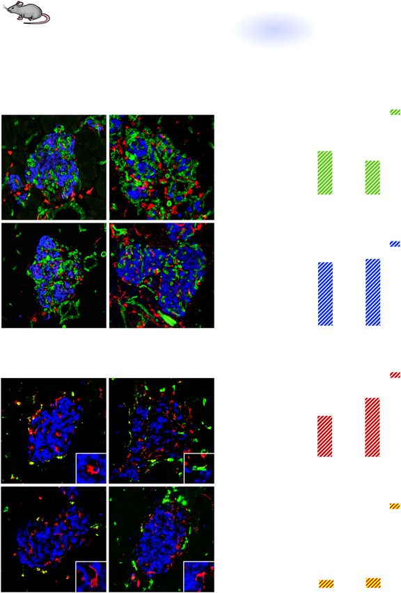
D.C. Saunders et al.
                                                                                                                                                       3

Fig. 2 Macrophages are required for β cell proliferation in βVEGF-A mice. a To deplete macrophages (MΦs) during VEGF-A induction and
normalization, βVEGF-A mice were treated with clodronate or control liposomes (150–200 μl i.v.) every other day, beginning 1 day before Dox
treatment and continuing 1 week after Dox withdrawal (1wk WD). b Representative flow cytometry plots showing circulating monocytes
(CD11b+ Ly6G–) of control and clodronate-treated βVEGF-A mice 24 h after single injection (No Dox). Approximately 10,000 white blood cells
(WBCs) were analyzed (monocyte fraction reported as mean + s.e.m.) per each animal. c Islet architecture displayed by labeling for β cells
(Insulin; blue), endothelial cells (Caveolin-1; green), and MΦs (Iba1; red) during VEGF-A induction (1wk Dox) and normalization (1wk WD). Scale
bar, 50 μm. d Quantification (mean + s.e.m.) of islet MΦ area by immunohistochemistry (5.1 ± 0.7 × 105 μm2 total islet area analyzed per
animal). e, f Quantification (mean + s.e.m.) of endothelial cell (EC) area (e) and β cell area (f) in βVEGF-A mice treated with control or clodronate
liposomes during VEGF-A induction and normalization. g Rate of β cell proliferation (mean + s.e.m.; 1138 ± 87 β cells counted per animal)
during VEGF-A normalization (1wk WD and 2wk WD) in control βVEGF-A mice was significantly reduced in clodronate-treated mice. In panels
b–g, each closed circle represents one animal; asterisks indicate unpaired two-tailed t-tests of control vs. clodronate groups; *p < 0.05; **p <
0.01; ****p < 0.0001. Dashed lines in d–g depict average values in βVEGF-A mice at baseline (No Dox).

before ultimately the impaired β cell proliferation leads to                   Proliferative ECs are required for MΦ polarization and
reduced β cell area 6 weeks after VEGF-A normalization. This                   maximal MΦ recruitment
finding suggests that MΦs play an important role in islet                       To investigate the contribution of ECs to β cell loss and recovery in
remodeling and composition in βVEGF-A mice separate from                       the βVEGF-A model, we first created a mouse line in which
their effect on β cell proliferation, most likely through their                VEGFR2, which is enriched in ECs of islet capillaries and the main
function as phagocytes.                                                        transducer of VEGF-A signal in islets, is inactivated by tamoxifen

Published in partnership with the Australian Regenerative Medicine Institute                                 npj Regenerative Medicine (2021) 22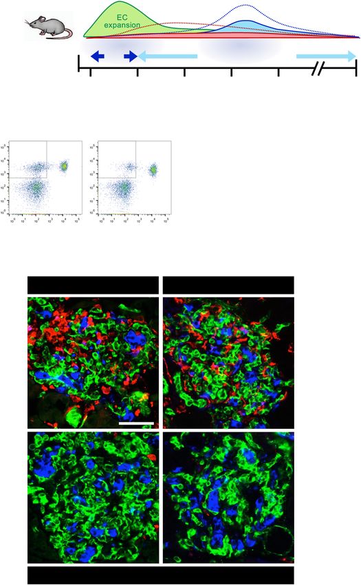
D.C. Saunders et al.
4
    (Tm)-inducible Cre-mediated excision in ECs (Cad5-CreERT2 line24).            suggesting that neither MΦ retention nor polarization at this
    Three doses of Tm to the Cad5-CreERT2; VEGFR2fl/fl (VEGFR2iΔEC)                 stage of β cell recovery are dependent on VEGFR2 signaling in
    mice achieved efficient EC-specific VEGFR2 knockdown (Supple-                   quiescent ECs.
    mentary Fig. 3a, b) without significant changes to VEGF-A                         Since VEGFR2 inactivation in quiescent ECs of βVEGF-A; R2iΔEC
    expression (Supplementary Fig. 3c), islet capillary density or size           mice leads to a relatively rapid EC decline after Tm treatment (9d
    (Supplementary Fig. 3e and d) or basal β cell proliferation                   WD), we next evaluated the islet vascular regression by visualiza-
    (Supplementary Fig. 3f), indicating that acute loss of                        tion of collagen IV, a major component of the islet ECM30,31
    VEGFR2 signaling in ECs is not detrimental to adult islet vascular            generated by intra-islet ECs. This study revealed that regressing
    homeostasis or β cell proliferation. A similar observation was made           islet capillaries leave behind vascular “casts” of ECM that are
    previously when VEGF-A was acutely inactivated in adult β cells18.            no longer associated with intact ECs (Fig. 4g, 9d WD). When
    Next, we crossed VEGFR2iΔEC mice with the existing βVEGF-A line               considered in conjunction with the degree of β cell loss, β cell
    to effectively perturb VEGF-A–VEGFR2 signaling in ECs at various              proliferation rates in βVEGF-A; R2iΔEC mice were increased at 9d
    time points during β cell loss and recovery. To control for any               WD compared to controls (Fig. 4f), suggesting that accelerated
    possible effects of Tm administration on compensatory β cell                  decline in islet ECs enhances β cell proliferation in this context.
    proliferation25, we treated all mice with Tm and designated Cre-
    negative (βVEGF-A; VEGFR2fl/fl) mice as controls to represent intact            Identifying interactions between β cells, ECs, and MΦs in the
    VEGFR2 signaling by ECs.                                                      βVEGF-A islet microenvironment
       To determine the effect of proliferative ECs on MΦ recruitment
                                                                                  Our prior studies in the βVEGF-A model localized the stimulus for
    and polarization, Tm was administered to knockdown VEGFR2 (R2)
                                                                                  β cell proliferation to the islet microenvironment, ruling out any
    in ECs prior to VEGF-A induction in β cells (Fig. 3a and
                                                                                  contribution from circulating factors that might reach the
    Supplementary Fig. 4a). VEGF-A was induced in both control
                                                                                  pancreas1. With key roles established for both MΦs and ECs in
    βVEGF-A; R2fl/fl and βVEGF-A; R2iΔEC genotypes, with efficient
                                                                                  this system, we next sought to identify potential mechanisms and
    knockdown of VEGFR2 in the latter (Supplementary Fig. 4b). As
                                                                                  signaling pathways coordinating cell–cell and cell–matrix interac-
    expected, VEGF-A induction caused a 7% EC expansion and
                                                                                  tions. To do this we isolated βVEGF-A islets at baseline (No Dox)
    associated 16% β cell loss per islet area in βVEGF-A; R2fl/fl controls,
                                                                                  and during the course of VEGF-A induction (1wk Dox) and
    whereas EC and β cell area did not change in βVEGF-A; R2iΔEC mice
                                                                                  normalization (1wk WD) and purified islet populations of β cells,
    (Fig. 3b–d). These results indicate that activation of
                                                                                  ECs, and MΦs at each of these time points for transcriptome
    VEGFR2 signaling in ECs by acute elevation of VEGF-A in the islet
                                                                                  analysis (Fig. 5a and Supplementary Fig. 6a, b).
    microenvironment is essential for islet hypervascularization and
                                                                                     All of the nine sample types analyzed (3 cell types per each of
    leads to β cell loss.
                                                                                  3 time points) demonstrate distinct transcriptional profiles,
       Inactivation of VEGFR2 signaling in ECs significantly reduced,
                                                                                  reflecting a high degree of uniqueness among three cell types
    but did not completely prevent MΦ recruitment to βVEGF-A;
                                                                                  and significant temporal changes in gene expression within each
    R2iΔEC islets compared to βVEGF-A; R2fl/fl controls (5.0 vs. 6.3% per
                                                                                  cell type (Supplementary Fig. 6c–e). By hierarchical clustering, the
    total islet area, respectively; p < 0.05) (Fig. 3e and f). This suggests
                                                                                  highest correlations were observed within one cell type across
    that MΦ infiltration can occur in response to VEGF-A alone, which
                                                                                  different time points; for example, β cells at No Dox are more
    is consistent with the fact that VEGF-A can mediate monocyte
                                                                                  similar to β cells at 1wk Dox and 1wk WD than they are to MΦs or
    recruitment through VEGFR1 activity26,27. Still, the proliferative EC
                                                                                  ECs at any time point. Of the β cell samples, regenerative β cells
    environment (intact VEGF-A–VEGFR2 signaling and β cell loss)
                                                                                  (1wk WD) are more similar to stressed β cells (1wk Dox) than
    leads to maximal MΦ recruitment. Interestingly, a subset of
                                                                                  quiescent β cells (No Dox); in contrast, regenerative MΦs (1wk WD)
    infiltrating MΦs in control islets expressed the M2-like marker
                                                                                  are more similar to resident MΦs (No Dox) than to those recruited
    CD206 (Mrc1), which is normally made only by exocrine MΦs28,29,
                                                                                  upon VEGF-A induction (1wk Dox), and quiescent ECs following
    while infiltrating MΦs in βVEGF-A; R2iΔEC islets remained CD206−
                                                                                  VEGF-A normalization (1wk WD) are more similar to quiescent
    (Fig. 3e and g). This observation suggests that VEGFR2 signaling in
                                                                                  ECs at baseline (No Dox) than to proliferative ECs (1wk Dox)
    proliferative ECs and/or β cell loss promotes a shift in MΦ
                                                                                  (Supplementary Fig. 6d).
    polarization and phenotypic signature.
                                                                                     With the induction of VEGF-A (1wk Dox), all cell populations
                                                                                  increase expression of growth factors, matrix remodeling
    VEGFR2 inactivation in quiescent ECs accelerates EC                           enzymes involved in tissue repair and matrix degradation (MMPs,
    regression, enhancing β cell recovery                                         ADAMs, ADAMTSs), as well as cell adhesion molecules involved in
    To investigate the role of VEGFR2 signaling in quiescent ECs                  cell–matrix and cell–cell interactions (ICAM1, VCAM1, selectins),
    during β cell recovery, VEGF-A-mediated EC proliferation and β                many of which remain elevated during β cell regeneration (1wk
    cell loss was first induced with 3-day Dox treatment in βVEGF-A;               WD) (Supplementary Fig. 7a). Pathway analysis shows a high
    R2iΔEC mice and controls, followed by 7 days of Dox withdrawal to             degree of cellular motility (Supplementary Fig. 7b), and extra-
    allow VEGF-A to normalize and ECs to return back to quiescence                cellular organization and cell adhesion processes are significantly
    (Fig. 4a and Supplementary Fig. 5a). Islet phenotype and β cell               enriched in all cell types during β cell recovery (Supplementary
    proliferation was assessed after 7 days of VEGF-A normalization               Fig. 8).
    (7d WD) and subsequently 2 days post-Tm treatment to inactivate                  Infiltrating MΦs show elevated expression of both pro-
    VEGFR2 (9d WD). VEGF-A-induced EC expansion, β cell loss, and                 inflammatory and pro-regenerative markers during periods of
    MΦ infiltration occurred in both genotypes, with no difference                 initial monocyte recruitment (1wk Dox) and β cell regeneration
    between the two groups at 7d WD prior to VEGFR2 inactivation                  (1wk WD). Pro-inflammatory genes like Il12b and Il1a are
    (Fig. 4b–f). As expected, VEGF-A expression continued to decline              upregulated at 1wk Dox, while M2 markers such as Ptgs1 and
    during Dox withdrawal and VEGFR2 was efficiently inactivated in                Retnla are upregulated at 1wk WD compared to No Dox
    βVEGF-A; R2iΔEC mice within 48 h of a single 4-mg Tm injection                (Supplementary Fig. 7a). Upregulation of chemokines and
    (Supplementary Fig. 5b). Surprisingly, unlike under normal                    cytokines by MΦs (e.g., Il12b, Ccl2, Ccl7) and corresponding
    homeostatic conditions (Supplementary Fig. 3), VEGFR2 inactiva-               increase in expression of receptors (e.g., Ccr2, Il12rb2) by β cells
    tion in βVEGF-A; R2iΔEC mice significantly accelerated islet EC                suggests crosstalk between the two cell types and potential
    regression compared to controls (Fig. 4c) without changes in MΦ               phenotypic effects on β cells in addition to MΦs. Similarly,
    phenotype (Supplementary Fig. 5d) and number (Fig. 4d),                       increased growth factor expression in ECs and MΦs appears in

    npj Regenerative Medicine (2021) 22                                         Published in partnership with the Australian Regenerative Medicine Institute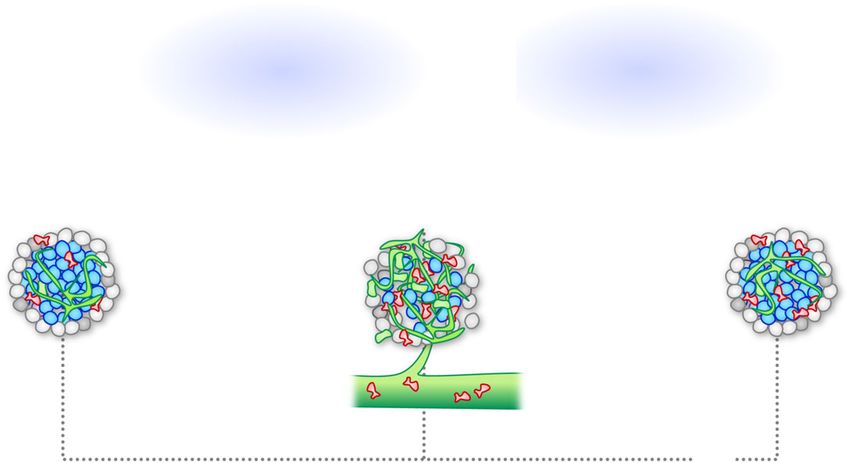
D.C. Saunders et al.
                                                                                                                                                        5

Fig. 3 Inactivation of VEGFR2 signaling in endothelial cells prevents β cell loss and M2-like macrophage polarization by acute elevation
of VEGF-A in the islet microenvironment. a To inactivate VEGFR2 in endothelial cells (ECs), control (βVEGF-A; VEGFR2fl/fl) and VEGFR2iΔEC
(βVEGF-A; VEGFR2iΔEC) mice were treated with Tamoxifen (Tm; 4 mg s.c.) prior to VEGF-A induction. b Islet architecture displayed by labeling
for macrophages (Iba1+), ECs (CD31+), and β cells (Ins+) at baseline (No Dox) and after 3d Dox. c, d Quantification (mean + s.e.m.) of islet β cell
and EC composition (10 ± 1 × 105 μm2 total islet area analyzed per animal). e Some intra-islet macrophages (MΦs) in control mice showed an
“M2-like” phenotype (CD206+) after VEGF-A induction, indicated by arrowheads. Insets show representative intra-islet MΦs in each group and
time point. f, g Quantification (mean + s.e.m.) of MΦ infiltration and M2-like intra-islet MΦs (percent CD206+ Iba1+ of Iba1+), 3 ± 2 × 105 μm2
total islet area analyzed per animal. Each closed circle in bar graphs represents one animal. Asterisks indicate unpaired two-tailed t-tests
between genotypes; *p < 0.05; ***p < 0.001. Scale bars in b and e, 50 μm; inset, 10 μm.

concert with increased expression of growth factor receptors in                in the extracellular milieu (Fig. 5b). Activation of integrin-mediated
β cells (Supplementary Fig. 7a).                                               signaling and the integrin-linked kinase pathway were accom-
   At the peak of regeneration (1wk WD), β cells highly express                panied by increases in PI3K/Akt and MAPK signaling genes (Fig. 5c
integrins and other molecules that sense and respond to changes                and Supplementary Fig. 8a), many of which are known modulators

Published in partnership with the Australian Regenerative Medicine Institute                                 npj Regenerative Medicine (2021) 22
D.C. Saunders et al.
6
Fig. 4 Phenotypic and structural changes to the islet microenvironment in response to VEGFR2 inactivation in quiescent endothelial cells
during β cell recovery. a To inactivate VEGFR2 in endothelial cells (ECs) during β cell recovery, control (βVEGF-A; VEGFR2f/fl) and VEGFR2iΔEC
(βVEGF-A; VEGFR2iΔEC) mice received Tamoxifen (Tm; 4 mg s.c.) after 7 days (d) of Dox withdrawal (WD). b Islet architecture visualized by
labeling for macrophages (Iba1+) ECs (CD31+, green), β cells (Insulin+, blue) at 7d WD (pre-Tm) and 9d WD (2d post-Tm). Scale bar, 50 μm.
c–e Area quantification (mean + s.e.m.) of islet ECs (c), MΦs (d), and β cells (e) by immunohistochemistry; 14 ± 1 × 105 μm2 total islet area
analyzed per animal. Each circle represents one animal; asterisks indicate results of unpaired two-tailed t-tests between genotypes; ***p <
0.001. f β cell proliferation rates (1,947 ± 145 β cells per animal) plotted as a function of β cell loss (% β cells of total islet area) reveal a
significant increase after VEGFR2 inactivation in quiescent ECs at 9d WD. Parentheses beside lines provide x- and y-intercepts derived from
linear regression. At 9d WD, intercepts are significantly different; **p < 0.01. g Visualization of islet extracellular matrix by immunofluorescence
(ECM; Col-IV+, red), ECs (CD31+, green), β cells (Insulin+, blue) at 7d WD (pre-Tm), and 9d WD (2d post-Tm). Scale bar, 50 μm; inset, 10 μm.
Arrowhead in bottom right panel points to ECM casts where ECs have regressed.
npj Regenerative Medicine (2021) 22 Published in partnership with the Australian Regenerative Medicine InstituteD.C. Saunders et al.
7
Fig. 5 Genes and pathways upregulated in β cells during recovery reflect activation of intracellular signaling to promote proliferation.
a Experimental schematic showing sorting of islet-derived β cells, endothelial cells (ECs), and macrophages (MΦs) from βVEGF-A mice at
baseline (No Dox), during VEGF-A induction/β cell loss (1wk Dox), and during VEGF-A normalization/β cell recovery (1wk WD). See also
Supplementary Fig. 5b–e. b Normalized expression of selected genes known to function in β cell proliferation34–37 at No Dox (n = 4 biological
replicates), 1wk Dox (n = 5), and 1wk WD (n = 3). Numbers listed in 1wk Dox and 1wk WD columns represent fold-change ≥2 orD.C. Saunders et al.
8
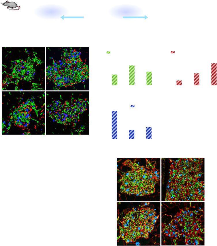
Fig. 6 Model of interactions between β cells, macrophages, endothelial cells, and the extracellular matrix in β cell regeneration. Upon
VEGF-A induction, intra-islet endothelial cells (ECs) proliferate while increasing expression of cell adhesion molecules and growth factors and
altering their expression of integrins and extracellular matrix (ECM) remodeling enzymes. These adhesion molecules help recruit macrophages
(MΦs), which upon islet infiltration also upregulate expression of cell adhesion molecules and pro- and anti-inflammatory chemokines and
cytokines, influencing further MΦ recruitment in addition to signaling through chemokine and cytokine receptors on β cells. Chemokine and
cytokines become increasingly less inflammatory as VEGF-A normalizes, as MΦs produce growth factors and matrix remodeling enzymes that
may promote β cell proliferation. Upon VEGF-A induction, β cells exhibit enrichment for several integrin pathways and other proteins involved
in ECM remodeling and cell–matrix interactions in addition to regulating expression of chemokines known to support a regenerative (M2 or
alternative) MΦ phenotype. Growth factors from all cell types act on an increased number of growth factor receptors being expressed on
β cells, activating downstream signals converging on the PI3K/Akt and MAPK pathways. Other signals from cells in the microenvironment, or
from the rapidly remodeling ECM, may also play a role in β cell proliferation.
ECs, as well as increased production of chemokines and cytokines from their effect on β cell proliferation. Together, these data
from MΦs already in the islet microenvironment39. By specifically suggest a phenotypic shift in MΦs that facilitates β cell recovery
depleting MΦs in βVEGF-A mice using clodronate liposomes, we and return to homeostasis in the islet, in line with an increasing
demonstrated that MΦs are required for β cell regeneration in the number of studies observing tissue-restorative effects of MΦs
islet microenvironment. These recruited MΦs have a unique triggered in various β cell injury models11,12,45. Further studies will
hybrid phenotype: they express markers of both classical pro- be necessary to clarify the specific signals that regulate the
inflammatory (M1) activation as well as alternative (M2) activation phenotypic shift and define the cues that govern their withdrawal.
promoting tissue repair and regeneration. Though MΦ-derived In addition, more work is required to characterize the phenotypes
cytokines and chemokines can exacerbate β cell stress40,41, the of human pancreatic islet MΦs.
comparable β cell loss in MΦ-depleted and control islets suggests Regulated EC-derived signals are necessary for pancreatic and
that MΦs do not promote β cell apoptosis in this model. Instead, islet development17,46–48, optimal adult β cell function18, and islet
MΦs downregulate key pro-inflammatory cytokines (e.g., Tnf, Il6) revascularization after transplantation49–52. In addition, ECs and
at 1wk WD, concomitant with β cell proliferation. Their phenotypic VEGFR2 signaling have been previously implicated in organ
profile at this stage is most reminiscent of phenotyping subtypes regeneration13,16. For example, during liver regeneration, sinusoi-
“M2a” (high expression of scavenger and phagocytic receptors like dal ECs appear to have a biphasic effect in mediating hepatic
Retnlb/Fizz2, Ym1, and Mrc1; secretion of profibrotic and trophic reconstitution; proliferative angiogenesis of sinusoidal ECs inhibits
factors like fibronectin, IGF, and TGFβ) and “M2c” (associated with hepatocyte self-renewal, whereas quiescent sinusoidal ECs stimu-
removal of apoptotic cells)42–44. This phagocytic phenotype is late hepatic regeneration13. To determine the role of proliferative
consistent with our finding that MΦs at this time point (1wk WD) and quiescent ECs in β cell loss and recovery, we generated a
likely have an effect on islet remodeling and composition separate compound βVEGF-A model with inducible VEGFR2 inactivation
npj Regenerative Medicine (2021) 22 Published in partnership with the Australian Regenerative Medicine InstituteD.C. Saunders et al.
9
(βVEGF-A; VEGFR2iΔEC) that allowed us to modulate VEGF- MΦs and ECs are quite attuned to their rapidly changing
A–VEGFR2 signaling in ECs. environment, with integrins and cell adhesion molecules being
Our studies show that intact VEGFR2 signaling is required for some of the most dynamically regulated genes in both cell types.
the proliferative angiogenesis of islet endothelium, which Changes in these molecules are likely regulating MΦ recruitment
ultimately results in β cell loss. Although transcriptome analysis and/or polarization70–73, which may explain the phenotypic shift
did not reveal specific signals produced by proliferating ECs that that happens between MΦ recruitment (occurring rapidly in
would lead to β cell apoptosis during VEGF-A induction, there are response to VEGF-A) and the appearance of β cell proliferation
significant changes in expression of matrix remodeling enzymes (during VEGF-A normalization).
and cell adhesion molecules. Based on the importance of It is also possible that β cells undergo intrinsic changes
cell–matrix interactions in β cell survival in vitro20,53, we heightening their sensitivity to extracellular signals, supported
hypothesize that ECM changes associated with rapidly expanding by the observation that ECs and MΦs increase expression of
endothelium likely contribute to β cell loss in the βVEGF-A system. several growth factors known to promote β cell proliferation
In addition, we show that intact VEGFR2 signaling is necessary for (IGF-1, PDGF, and CTGF)35,74–76 while β cells simultaneously
accumulation of intra-islet MΦs expressing CD206, a marker often upregulate expression of corresponding receptors (Igf1r, Pdgfr).
associated with an M2-like phenotype. This observation is Signaling cascades activated by integrins and growth factors
consistent with MΦ activation by EC-derived cues in the context exhibit extensive downstream crosstalk and protein activity that
of acute injury, a phenomenon that has been noted in numerous makes it difficult to determine pathway activation status based
tissues54–56. Furthermore, an M2 phenotype (high IL-10, CD206) solely on gene expression. Nonetheless, we did observe
was shown to be required for β cell regeneration after DT- transcriptional changes to components of the PI3K/Akt, PLC,
mediated ablation12, and elsewhere CD206 has been linked to and MAPK pathways, as well as upregulation of transcription
increased expression of Tgfb1 and Egf57 and promotion of β cell factors regulated by the MAPK pathway32–35 suggesting that
proliferation via a Smad7-cyclin pathway11. Further work is the cell–cell and cell–ECM interactions may converge on the
needed to clarify whether islet resident MΦs indeed shift towards activation of these pathways leading to β cell proliferation. We
an M2 phenotype or whether acinar stromal MΦ populations28 therefore propose a model in which coordination of growth
contribute to the increase of M2-like MΦs expressing CD206 factors—whose bioavailability is likely modulated by ECs and
observed in islets in the absence of VEGFR2. MΦs—together with increased integrin signaling promotes
In contrast to lung and liver regeneration13,16, VEGFR2 inactiva- activation of pro-proliferative pathways in surviving β cells
tion in quiescent ECs (1wk WD) resulted in accelerated β cell during VEGF-A normalization to ultimately restore β cell mass
recovery, which is associated with rapid islet capillary regression, (Fig. 6).
leaving behind vascular “casts” of ECM components. We postulate Recognizing the key role of cellular and extracellular compo-
that β cell proliferation is promoted by release of growth factors nents of the islet microenvironment on β cell development,
from degraded ECM. Though ECs are no longer expanding at the function, and homeostasis is critical to further our understanding
1wk WD time point, islets still contain quite extensive capillary of signals regulating adult β cell proliferation. Islet microenvir-
networks that might be sustained by the residual extracellular onmental signaling in the βVEGF-A system promotes human
VEGF-A we observed, as well as possibly other growth factors. β cell proliferation1, which prompted us to develop new
Interestingly, VEGFR2 inactivation during β cell homeostasis does strategies to disentangle the roles of various microenvironmental
not impact β cell proliferation and islet capillary morphology. components in this regenerative process. However, moving
It will be important to assess the role of quiescent ECs and forward it will be important to further explore the mechanisms
VEGFR2 signaling in other β cell injury models in the future. In and combination of specific cytokines, growth factors, and other
addition, these findings highlight that ECs have different roles in microenvironmental signals that activate and regulate relevant
different tissues and during various physiologic and pathologic mitogenic signaling pathways in human β cells. Overall, these
stressors. studies highlight the importance of developing innovative
By deconstructing the contributions of the cellular components approaches to examine β cells in vivo in order to more
of the islet microenvironment of βVEGF-A mice, we were able to completely understand the complex microenvironmental factors
uncover a role for ECM remodeling and signaling in β cell regulating β cell function and regeneration.
proliferation. MΦs, ECs, and β cells all show transcriptional
regulation of integrin receptors and ECM remodeling enzymes,
some of which have been shown previously to affect β cell METHODS
proliferation22,58–60. Importantly, there is mounting evidence that
ECM modulates human β cell proliferation and that these Mouse models
components can be manipulated to promote β cell growth All animal studies were approved by the Institutional Animal Care and Use
in vitro61–63. In addition to shaping cell–matrix signaling, ECM Committee at Vanderbilt University Medical Center, and animals were kept
in facilities monitored by the Vanderbilt University Division of Animal Care
reorganization can also lead to the release and/or activation of on a 12 h light/12 h dark schedule with unrestricted access to standard
matrix-sequestered growth factors64–67. This is particularly rele- chow and water. Mouse models and abbreviations used to describe them
vant given the temporal increase of growth factor receptor are summarized in Supplementary Table 2. All mice were between 8 and
expression in β cells during VEGF-A induction and normalization, 16 weeks of age at the time experiments were initiated.
suggesting heightened β cell responsiveness to signals from the
rapidly remodeling ECM as VEGF-A normalizes. Furthermore, our RIP-rtTA; TetO-VEGF (βVEGF-A) mice. The original bitransgenic mice with
observation of ECM casts following VEGFR2 knockdown suggests doxycycline (Dox)-inducible β cell-specific overexpression of human
that regression of quiescent ECs during the β cell recovery phase VEGF-A165 (abbreviated βVEGF-A) were generated by crossing RIP-rtTA
could stimulate release of growth factors from degraded ECM, male mice and TetO-VEGF female mice, both on a C57BL/6 back-
ground77–81. These mice were generously provided by Dr. Shimon Efrat of
thereby promoting β cell proliferation. Together, these data
Tel Aviv University and Dr. Peter Campochiaro of Johns Hopkins
provide evidence that ECM-bound growth factors released during University, respectively. In this βVEGF-A model the rat Ins2 promoter
EC regression promote β cell proliferation. drives expression of the tetracycline-responsive rtTA transactivator
Remodeling of extracellular milieu can also influence MΦ specifically in pancreatic β cells. Upon exposure to Dox, the rtTA
phenotype68,69, though more work is required to define specific transactivator binds the tetracycline operator (TetO), driving expression of
signaling pathways that regulate this process in the pancreas. human VEGF-A165 in β cells. Details of Dox preparation and administra-
However, our transcriptome data provides evidence that both tion are included below.
Published in partnership with the Australian Regenerative Medicine Institute npj Regenerative Medicine (2021) 22D.C. Saunders et al.
10
Cd5-CreER; VEGFR2fl/fl (VEGFR2iΔEC) mice. Mice with Tamoxifen (Tm)- overnight. After blotting to remove excess sucrose, tissues were mounted
inducible EC-specific knockout of VEGFR2 (abbreviated VEGFR2iΔEC) were in Tissue Tek cryomolds filled with Tissue-Plus optimal cutting temperature
generated by crossing Cd5-CreER male mice and VEGFR2fl/fl female mice (OCT) compound (VWR Scientific Products). Tissue molds were placed on
(see Supplementary Table 3, crosses A1-A2). Heterozygous VEGFR2fl/wt dry ice until the OCT was set, then stored at −80 °C. Tissues were sectioned
mice on a C57BL/6 background were obtained from Jackson Laboratories from 5 to 10-μm thick on a Leica CM1950 cryostat (Leica) and these
(stock #018977)82 and bred to create a homozygous VEGFR2fl/fl line. Frozen cryosections were attached to Superfrost Plus Gold slides (ThermoFischer
sperm from the Cd5-CreER line24 was generously provided by Dr. Yoshiaki Scientific).
Kubota of Keio University, and in vitro fertilization (IVF) was performed by
the Vanderbilt Genome Editing Resource using female C57BL/6 mice
(Jackson Laboratories, stock #000664). In the VEGFR2iΔEC model loxP sites Immunohistochemistry, imaging, and analysis
were inserted flanking VEGFR2 exon 3, and the Cdh5 (VE-cadherin) Immunohistochemical analysis was performed on serial 8–10-μm pancrea-
promoter drives expression of Tm-inducible Cre recombinase in vascular tic cryosections as described previously1,18. Briefly, tissue permeabilization
ECs. Upon exposure to Tm, Cre recombinase translocates to the nucleus, was conducted using 0.2% Triton-X in 10 nM PBS and blocking using 5%
excising VEGFR2 exon 3 through Cre-loxP recombination and subsequently normal donkey serum in 10 mM. Primary and secondary antibody
preventing VEGFR2 expression in ECs. Details of Tm preparation and incubations (listed in Supplementary Table 5) were performed in buffer
administration are described below. with 0.1% Triton-X and 1% BSA and nuclei were counterstained with DAPI.
Slides were mounted using SlowFade Gold antifade reagent (Invitrogen
βVEGF-A; VEGFR2iΔEC mice. To generate an inducible model of EC-specific Molecular Probes) and sealed with fingernail polish prior to imaging.
knockdown of VEGFR2 in βVEGF-A mice, Cd5-CreER and VEGFR2fl/fl mice Digital images were acquired with a Leica DMI6000B fluorescence
were crossed with RIP-rtTA and TetO-VEGF transgenic mice as outlined in microscope equipped with a Leica DFC360FX digital camera (Leica), a laser
Supplementary Table 3. The final cross produced both βVEGF-A; scanning confocal microscope (Zeiss LSM510 META or LSM880, Carl Zeiss),
VEGFR2iΔEC mice as well as Cre-negative sibling controls (βVEGF-A; and a ScanScope FL (Aperio). Image analysis was performed using
VEGFR2fl/fl). Due to the inefficient induction of VEGF-A in female mice MetaMorph 7.7 software (Molecular Devices), ImageScope software
(the single copy of the TetO transgene is subject to X chromosome (Aperio), or HALO software (Indica Labs).
inactivation), only male mice were used for experiments. For analysis of islet composition, images of entire pancreatic sections
were captured at ×20 magnification using a ScanScope FL system. Islet
βVEGF-A; MIP-GFP mice. To enable fluorescence-activated cell sorting area was annotated manually based on insulin staining, and HALO
(FACS) of pancreatic β cells from βVEGF-A mice, a transgene was separately algorithms were used to calculate area of β cells (Insulin+), ECs (CD31+),
introduced into the βVEGF-A mouse model by crossing MIP-GFP mice on a and MΦs (Iba1+). For β cell proliferation, cells were deemed positive for
C57BL/6 background83 (Jackson Laboratories, stock #006864) with RIP-rtTA Ki67 only when at least 75% of the nucleus was surrounded by insulin+
and Tet-O-VEGF-A mice (abbreviated βVEGF-A; MIP-GFP). In these mice, cytoplasm.
the mouse Ins1 promoter drives green fluorescent protein (GFP) expression
in β cells. Flow cytometry and cell sorting
Flow analysis and sorting was performed in collaboration with the
DNA extraction and genotyping. Mouse models used in our breeding Vanderbilt Flow Cytometry Core. Peripheral blood (50–100 μl) was
schemes were maintained by genotyping using the primers and PCR collected from the retro-orbital sinus of βVEGF-A mice using heparinized
conditions listed in Supplementary Table 4. DNA was extracted and PCR capillary tubes 24 h after beginning liposome injections to evaluate
reactions were performed with tail snips from mice as described depletion of circulating monocytes. Blood was incubated for 3–5 min at
previously1. Thermal cycler conditions listed in Supplementary Table 4 37 °C with 1 ml warmed, filter-sterilized erythrocyte lysis buffer (8.26 g
were used to amplify DNA before resolving on agarose gels with 100 ng/ml ammonium chloride, 1 g potassium bicarbonate, and 0.38 g EDTA in 1 L
ethidium bromide in 1X Tris/Borate/EDTA (TBE) buffer as indicated. Milli-Q water). Cells were pelleted by centrifuging at 1800 rpm for 2–3 min
at 4 °C and supernatant discarded. Incubation with erythrocyte lysis buffer
Compound preparation and administration. VEGF-A transgene expression was repeated, and then cells were washed with 1 ml FACS buffer (2 mM
was activated in βVEGF-A mice by Dox administration (5 mg/ml) in light- EDTA and 2% FBS in 10 mM PBS) prior to antibody incubation. Blood from
protected drinking water containing 1% Splenda® for a period of 3–7 days. WT mice was collected for antibody compensation controls.
VEGFR2 knockdown was induced in VEGFR2iΔEC mice by subcutaneous Isolated islets from βVEGF-A; MIP-GFP mice handpicked in Clonetics EGM
injection of 4 mg Tm (20 mg/ml; 200 μl). Tm (20 mg/ml) was prepared fresh MV Microvascular Endothelial Cell Growth Medium (Lonza) were washed
in filter-sterilized corn oil the day before each injection and allowed to three times with 2 mM EDTA in 10 mM PBS and then dispersed by
dissolve overnight on a shaker at room temperature, protected from light. incubating with Accutase (Innovative Cell Technologies) at 37 °C for 10 min
Vetbond tissue adhesive (3 M) was used to seal injection sites to prevent oil with constant pipetting. Accutase was quenched with EGM MV media, and
leakage. Clodronate-mediated macrophage depletion in βVEGF-A mice then islet cells were washed twice with the same media and counted using
was accomplished by injecting 150–200 μl clodronate liposomes (5 mg/ml; a hemocytometer prior to antibody incubation. Anti-rat Ig, κ CompBead
Clodrosome) retro-orbitally every other day for a 1–2 week period (4–8 Plus Compensation Particles (BD Biosciences), and EasyComp Fluorescent
total injections). Liposome injections began 1 day before Dox administra- Particles, GFP (Spherotech) were used as single-color compensation
tion and continued for 1 week after in mice harvested at later time points. controls for islet cell sorts.
Control liposomes with the same lipid composition (Clodrosome) were Peripheral blood and islet cells prepared as described above, and anti-
administered to βVEGF-A mice using the same route, volume, and rat Ig compensation particles were incubated for 15–20 min at 4 °C with
schedule. Mice receiving liposome injections were supplemented with fluorophore-conjugated antibodies in FACS buffer followed by one wash
Transgenic Dough Diet (21.2% protein, 12.4% fat, 46.5% carbohydrate; with FACS buffer. All antibodies for flow cytometry and their working
BioServ) throughout the course of the experiment. dilutions are listed in Supplementary Table 5. Prior to analysis or sorting,
either propidium iodide (0.05 μg/100,000 cells; Invitrogen Molecular
Glucose measurements. Random (non-fasted) plasma glucose levels Probes) or DAPI (0.25 μg/1,000,000 cells; Invitrogen Molecular Probes)
were measuring by obtaining whole blood from nicked tail veins using was added to samples for non-viable cell exclusion. Flow analysis was
an Accu-chek glucose meter (Roche Diagnostics) calibrated according to performed using an LSRFortessa cell analyzer (BD Biosciences) and a
the manufacturer’s instructions. FACSAria III cell sorter (BD Biosciences) was used for FACS. Analysis of flow
cytometry data was completed using FlowJo 7.6.5-10.2.1 (FlowJo LLC).
Tissue collection and fixation
Mouse pancreata were collected from anesthetized mice prior to cervical RNA isolation, sequencing, and analysis
dislocation. Organs were washed in ice-cold 10 mM phosphate-buffered Sorted islet-derived cells (8000–400,000/sample) were added to 200–400 μl
saline (PBS), then fat and other excess tissue was removed before lysis/binding solution in the RNAqueous micro-scale phenol-free total RNA
pancreata were weighed and processed. Fixation was performed in 0.1 M isolation kit (Ambion). Trace contaminating DNA was removed with TURBO
PBS containing 4% paraformaldehyde (Electron Microscopy Sciences) for DNA-free (Ambion). RNA quality control quantification was performed
2–3 h on ice with mild agitation, then organs were washed in four changes using a Qubit Fluorometer (Invitrogen, Carlsbad, CA) and an Agilent 2100
of 0.1 M PBS over 2 h and equilibrated in 30% sucrose/0.01 M PBS Bioanalyzer. All RNA samples had an RNA integrity number (RIN) ≥ 5.0. RNA
npj Regenerative Medicine (2021) 22 Published in partnership with the Australian Regenerative Medicine InstituteD.C. Saunders et al.
11
was amplified using the Ovation system (NuGen Technologies) according 10. Mussar, K. et al. Macrophage/epithelium cross-talk regulates cell cycle progres-
to standard protocol. Amplified cDNA was sheared to target 300 bp sion and migration in pancreatic progenitors. PLoS ONE 9, e89492 (2014).
fragment size and libraries were prepared using NEBNext DNA Library Prep 11. Xiao, X. et al. M2 macrophages promote beta-cell proliferation by up-regulation
(New England BioLabs). 50 bp paired-end (PE) sequencing was performed of SMAD7. Proc. Natl Acad. Sci. USA 111, E1211–E1220 (2014).
on an Illumina HiSeq 2500 using traditional methods84,85. Raw reads were 12. Criscimanna, A., Coudriet, G. M., Gittes, G. K., Piganelli, J. D. & Esni, F. Activated
mapped to the reference mouse genome mm9 using TopHat v2.086 and macrophages create lineage-specific microenvironments for pancreatic acinar-
aligned reads were then imported onto the Avadis NGS analysis platform and β-cell regeneration in mice. Gastroenterology 147, 1106–1118.e11 (2014).
(Strand Scientific). Transcript abundance was quantified using the TMM 13. Ding, B.-S. et al. Inductive angiocrine signals from sinusoidal endothelium are
(Trimmed Mean of M-values) algorithm87,88. Samples were compared by required for liver regeneration. Nature 468, 310–315 (2010).
principal component analysis (PCA) and hierarchical clustering analysis. A 14. Butler, J. M. et al. Endothelial cells are essential for the self-renewal and repo-
minimum expression cutoff (normalized expression ≥20 at one or more pulation of notch-dependent hematopoietic stem cells. Cell Stem Cell 6, 251–264
time points) was applied before determining differential expression (2010).
between samples, which was calculated on the basis of fold-change 15. Butler, J. M., Kobayashi, H. & Rafii, S. Instructive role of the vascular niche in
(cutoff ≥ 2 or ≤−2) with p-values estimated by z-score calculations (cutoff promoting tumour growth and tissue repair by angiocrine factors. Nat. Rev.
0.05) as determined by the Benjamini–Hochberg false discovery rate (FDR) Cancer 10, 138–146 (2010).
method89. Differentially expressed genes were further analyzed through 16. Ding, B.-S. et al. Endothelial-derived angiocrine signals induce and sustain
Ingenuity Pathway Analysis (IPA, Qiagen) and Gene Ontology (GO) analysis regenerative lung alveolarization. Cell 147, 539–553 (2011).
using DAVID90. RNA quality control, amplification, sequencing, and analysis 17. Cai, Q. et al. Enhanced expression of VEGF-A in β cells increases endothelial cell
were performed in collaboration with the Genomic Services Laboratory at number but impairs islet morphogenesis and β cell proliferation. Dev. Biol. 367,
HudsonAlpha Institute for Biotechnology. 40–54 (2012).
18. Reinert, R. B. et al. Vascular endothelial growth factor-A and islet vascularization
are necessary in developing, but not adult, pancreatic islets. Diabetes 62,
Statistical analysis 4154–4164 (2013).
Prism software (GraphPad) was used to perform all statistical analyses for 19. Sand, F. W. et al. Growth-limiting role of endothelial cells in endoderm devel-
immunohistochemistry. In all experiments manipulating MΦs and ECs, opment. Dev. Biol. 352, 267–277 (2011).
control and experimental groups were compared at each time point using 20. Hammar, E. et al. Extracellular matrix protects pancreatic β-cells against apoptosis
a two-tailed unpaired t test. For analysis of proliferative ECs, two-tailed role of short- and long-term signaling pathways. Diabetes 53, 2034–2041 (2004).
unpaired t tests were also used to compare baseline (No Dox) and VEGF-A 21. Nikolova, G. et al. The vascular basement membrane: a niche for insulin gene
induction (3d Dox) time points within each group. For analysis of quiescent expression and Beta cell proliferation. Dev. Cell 10, 397–405 (2006).
ECs, a one-way analysis of variance (ANOVA) was used to analyze time 22. Diaferia, G. R. et al. β1 integrin is a crucial regulator of pancreatic β-cell expansion.
points within each group, followed by Tukey’s multiple comparison test to Development 140, 3360–3372 (2013).
compare each time point with baseline (No Dox). Unless otherwise noted, 23. Aamodt, K. I. & Powers, A. C. Signals in the pancreatic islet microenvironment
data are expressed as mean + standard error of mean (s.e.m.). Statistical influence β-cell proliferation. Diabetes Obes. Metab. 19, 124–136 (2017).
analysis of RNA-sequencing data is described above (see “RNA isolation, 24. Okabe, K. et al. Neurons limit angiogenesis by titrating VEGF in retina. Cell 159,
sequencing, and analysis”). 584–596 (2014).
25. Carboneau, B. A., Le, T. D. V., Dunn, J. C. & Gannon, M. Unexpected effects of the
MIP-CreER transgene and tamoxifen on β-cell growth in C57Bl6/J male mice.
Reporting summary Physiological Rep. 4, e12863 (2016).
Further information on research design is available in the Nature Research 26. Luttun, A. et al. Revascularization of ischemic tissues by PlGF treatment, and
Reporting Summary linked to this article. inhibition of tumor angiogenesis, arthritis and atherosclerosis by anti-Flt1. Nat.
Med. 8, 831–840 (2002).
27. Takahashi, H. & Shibuya, M. The vascular endothelial growth factor (VEGF)/VEGF
DATA AVAILABILITY receptor system and its role under physiological and pathological conditions.
RNA-sequencing data is available in the Gene Expression Omnibus (GEO) database of Clin. Sci. 109, 227–241 (2005).
the National Center for Biotechnology Information (NCBI) under accession numbers 28. Calderon, B. et al. The pancreas anatomy conditions the origin and properties of
GSE72546 and GSE163825. Additional datasets and materials generated during the resident macrophages. J. Exp. Med. 212, 1497–1512 (2015).
current study are available from the corresponding author on reasonable request. 29. Weitz, J. R. et al. Mouse pancreatic islet macrophages use locally released ATP to
monitor beta cell activity. Diabetologia 61, 182–192 (2018).
30. Kragl, M. & Lammert, E. Basement membrane in pancreatic islet function. Adv.
Received: 22 September 2020; Accepted: 24 February 2021; Exp. Med. Biol. 654, 217–234 (2010).
31. Bogdani, M. Thinking outside the cell: a key role for hyaluronan in the patho-
genesis of human type 1. Diabetes Diabetes 65, 2105–2114 (2016).
32. Schlaepfer, D. D., Hanks, S. K., Hunter, T. & Geer, P. van der. Integrin-mediated
signal transduction linked to Ras pathway by GRB2 binding to focal adhesion
REFERENCES kinase. Nature 372, 786–791 (1994).
1. Brissova, M. et al. Islet microenvironment, modulated by vascular endothelial 33. Foulds, C. E., Nelson, M. L., Blaszczak, A. G. & Graves, B. J. Ras/mitogen-activated
growth factor-A signaling, promotes β cell regeneration. Cell Metab. 19, 498–511 protein kinase signaling activates Ets-1 and Ets-2 by CBP/p300 recruitment. Mol.
(2014). Cell Biol. 24, 10954–10964 (2004).
2. Tsunawaki, S., Sporn, M., Ding, A. & Nathan, C. Deactivation of macrophages by 34. Carlson, S. M. et al. Large-scale discovery of ERK2 substrates identifies ERK-
transforming growth factor-β. Nature 334, 260–262 (1988). mediated transcriptional regulation by ETV3. Sci. Signal 4, rs11–rs11 (2011).
3. Ricardo, S. D., van Goor, H. & Eddy, A. A. Macrophage diversity in renal injury and 35. Chen, H. et al. PDGF signalling controls age-dependent proliferation in pancreatic
repair. J. Clin. Invest. 118, 3522–3530 (2008). β-cells. Nature 478, 349–355 (2011).
4. He, H. et al. Endothelial cells provide an instructive niche for the differentiation 36. Fiaschi-Taesch, N. et al. Survey of the human pancreatic β-cell G1/S proteome
and functional polarization of M2-like macrophages. Blood 120, 3152–3162 (2012). reveals a potential therapeutic role for Cdk-6 and Cyclin D1 in enhancing human
5. Sica, A. & Mantovani, A. Macrophage plasticity and polarization: in vivo veritas. J. β-cell replication and function in vivo. Diabetes 58, 882–893 (2009).
Clin. Invest. 122, 787–795 (2012). 37. Zhang, H. et al. Gestational diabetes mellitus resulting from impaired β-cell
6. Meier, J. J., Bhushan, A., Butler, A. E., Rizza, R. A. & Butler, P. C. Sustained beta cell compensation in the absence of FoxM1, a novel downstream effector of pla-
apoptosis in patients with long-standing type 1 diabetes: indirect evidence for cental lactogen. Diabetes 59, 143–152 (2009).
islet regeneration? Diabetologia 48, 2221–2228 (2005). 38. Karnik, S. K. et al. Menin regulates pancreatic islet growth by promoting histone
7. Campbell-Thompson, M. L. et al. The diagnosis of insulitis in human type 1 methylation and expression of genes encoding p27Kip1 and p18INK4c. Proc. Natl
diabetes. Diabetologia 56, 2541–2543 (2013). Acad. Sci. USA 102, 14659–14664 (2005).
8. Halban, P. A. et al. β-cell failure in type 2 diabetes: postulated mechanisms and 39. Shi, C. & Pamer, E. G. Monocyte recruitment during infection and inflammation.
prospects for prevention and treatment. Diabetes Care 37, 1751–1758 (2014). Nat. Rev. Immunol. 11, 762–774 (2011).
9. Banaei-Bouchareb, L. et al. Insulin cell mass is altered in Csf1op/Csf1op 40. Grunnet, L. G. et al. Proinflammatory cytokines activate the intrinsic apoptotic
macrophage-deficient mice. J. Leukoc. Biol. 76, 359–367 (2004). pathway in β-cells. Diabetes 58, 1807–1815 (2009).
Published in partnership with the Australian Regenerative Medicine Institute npj Regenerative Medicine (2021) 22You can also read