Molecular analysis of the ribosome recycling factor ABCE1 bound to the 30S post-splitting complex
←
→
Page content transcription
If your browser does not render page correctly, please read the page content below
Article

Molecular analysis of the ribosome recycling factor
ABCE1 bound to the 30S post-splitting complex
Elina Nürenberg-Goloub1,†, Hanna Kratzat2,†, Holger Heinemann1,†, André Heuer2, Peter Kötter3                                                           ,
Otto Berninghausen2, Thomas Becker2 , Robert Tampé1,* & Roland Beckmann2,**

Abstract                                                                               diseases including ribosomopathies and cancer (Tahmasebi et al,
                                                                                       2018; Aspesi & Ellis, 2019). Accordingly, each of the four phases of
Ribosome recycling by the twin-ATPase ABCE1 is a key regulatory                        translation—initiation, elongation, termination, and ribosome recy-
process in mRNA translation and surveillance and in ribosome-                          cling—as well as the transitions between them must be under rigor-
associated protein quality control in Eukarya and Archaea. Here,                       ous control. While the first three phases are directly involved in
we captured the archaeal 30S ribosome post-splitting complex at                        protein biosynthesis and have therefore been extensively studied,
2.8 Å resolution by cryo-electron microscopy. The structure reveals                    ribosome recycling has only recently been structurally and function-
the dynamic behavior of structural motifs unique to ABCE1, which                       ally characterized (Hellen, 2018). Herein, the conserved and essen-
ultimately leads to ribosome splitting. More specifically, we                          tial ATP-binding cassette (ABC)-type twin-ATPase ABCE1 plays the
provide molecular details on how conformational rearrangements                         key role for Archaea and Eukarya (Pisarev et al, 2010; Barthelme
of the iron–sulfur cluster domain and hinge regions of ABCE1 are                       et al, 2011; Shoemaker & Green, 2011). ABCE1 recycles canonical
linked to closure of its nucleotide-binding sites. The combination                     70S/80S post-termination complexes (post-TCs) after stop codon-
of mutational and functional analyses uncovers an intricate allos-                     dependent termination and non-canonical post-TCs during mRNA
teric network between the ribosome, regulatory domains of ABCE1,                       surveillance and resumption of translation after cellular stress. In
and its two structurally and functionally asymmetric ATP-binding                       both cases, a decoding A-site factor (archaeal/eukaryotic release
sites. Based on these data, we propose a refined model of how                          factor 1 (a/eRF1) or its homologue a/ePelota, respectively) is deliv-
signals from the ribosome are integrated into the ATPase cycle of                      ered to the ribosomal A-site by a translational GTPase (aEF1/eRF3
ABCE1 to orchestrate ribosome recycling.                                               or aEF1/Hbs1, respectively) and forms an interaction platform for
                                                                                       ABCE1 to establish the 70S/80S pre-splitting complex (pre-SC;
Keywords ABC proteins, ribosome recycling; molecular machines; mRNA                    Becker et al, 2012; Preis et al, 2014; Brown et al, 2015; Shao et al,
surveillance; ribosome-associated quality control                                      2016). In concert with the A-site factor, ABCE1 splits the pre-SC into
Subject Categories Structural Biology; Translation & Protein Quality                   the small (SSU) and large (LSU) ribosomal subunit. In Eukarya,
DOI 10.15252/embj.2019103788 | Received 20 October 2019 | Revised 17                   other components of the post-TC stay associated with the ribosomal
January 2020 | Accepted 21 January 2020                                                subunits and are subsequently recycled by additional factors (Pis-
The EMBO Journal (2020) e103788                                                        arev et al, 2010; Skabkin et al, 2010). Canonical termination, which
                                                                                       includes peptide release by eRF1, yields 40S-mRNA-deacylated
                                                                                       tRNA complexes and free 60S subunits whereas ribosome recycling
Introduction                                                                           of non-canonical post-TCs in the presence of Pelota results in 40S-
                                                                                       mRNA and 60S-peptidyl-tRNA complexes due to Pelota’s incapacity
Protein biosynthesis via mRNA translation is a fundamental process                     to release peptides. Moreover, Pelota/Hbs1/ABCE1 not only acts in
in living cells. Strikingly, translation is interlaced in a complex                    the splitting of stalled (Shoemaker & Green, 2011), but also vacant
network of cellular pathways including mRNA surveillance, ribo-                        (van den Elzen et al, 2014), and newly synthesized ribosomes
some-associated quality control, and ribosome biogenesis (Bassler &                    (Strunk et al, 2012). Immediately after splitting, an ABCE1-bound
Hurt, 2019; Joazeiro, 2019; Nürenberg-Goloub & Tampé, 2019).                         30S/40S post-splitting complex is formed (Kiosze-Becker et al, 2016;
These crucial pathways maintain protein, mRNA, and ribosome                            Heuer et al, 2017), in which ABCE1 may remain for a defined time
homeostasis (Young et al, 2015; Mills et al, 2016), induce organelle                   span (Nürenberg-Goloub et al, 2018; Gouridis et al, 2019) to
turnover (Wu et al, 2018), assist embryonic development (Coelho                        prevent re-association of the LSU (Heuer et al, 2017). Additionally,
et al, 2005; Chen et al, 2006), and are also linked to various                         ABCE1 has been shown to interact with initiation factors and is

1 Institute of Biochemistry, Biocenter, Goethe University Frankfurt, Frankfurt a.M., Germany
2 Department of Biochemistry, Gene Center, Ludwig-Maximilians University Munich, München, Germany
3 Institute for Molecular Biosciences, Biocenter, Goethe University Frankfurt, Frankfurt a.M., Germany
  *Corresponding author. Tel: +49 069 798 29475; E-mail: tampe@em.uni-frankfurt.de
  **Corresponding author. Tel: +49 089 218 076900; E-mail: beckmann@genzentrum.lmu.de
  †
   These authors contributed equally to this work

ª 2020 The Authors. Published under the terms of the CC BY 4.0 license                                              The EMBO Journal   e103788 | 2020   1 of 13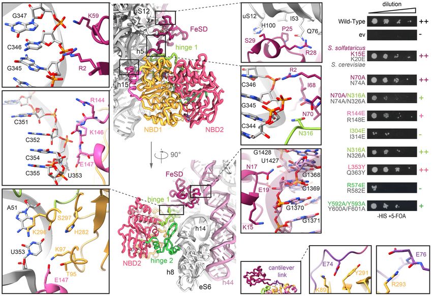
The EMBO Journal                                                                                                   Elina Nürenberg-Goloub et al

assumed to promote their recruitment to the SSU (Dong et al, 2004;       2017). To stabilize the post-SC, a well-characterized, hydrolysis-
Chen et al, 2006), thus linking ribosome recycling to translation        deficient ABCE1 mutant was used. This mutant, with both catalytic
initiation.                                                              glutamates being substituted by alanine (E238A/E485A, short IIEA),
    A key question is which molecular mechanism is employed by           efficiently split 70S ribosomes and remained quantitatively bound to
ABCE1 as an ABC-type ATPase. All members of the ABC super-               30S subunits (Nürenberg-Goloub et al, 2018) (Fig 1A). Notably, 70S
family utilize the energy of ATP binding and hydrolysis generated        from S. solfataricus are intrinsically instable (Barthelme et al, 2011)
in two conserved nucleotide-binding sites (NBS) and are ubiqui-          and thus unsuitable for our in vitro splitting approach.
tously found in numerous cellular processes. These include trans-            The purified 30S-ABCE1IIEA post-SC was subjected to single-
port of a limitless range of substrates across membranes,                particle cryo-EM analysis. 3D classification revealed that the vast
chromatin remodeling, DNA repair, or modulation of ribosomal             majority (97%) of 30S particles were associated with ABCE1IIEA.
complexes. The NBSs are formed at the interface of two nucleo-           This class was refined to an average resolution of 2.8 Å (Fig 1B).
tide-binding domains (NBDs), which are arranged reciprocally             Local resolution assessment showed that the body of the 30S formed
(Hopfner, 2016). ABCE1 additionally possesses an essential N-            a very rigid structure whereas the 30S head and ABCE1 showed
terminal iron–sulfur cluster domain (FeSD) (Barthelme et al, 2007)       flexibility and lower resolution (4–6 Å) (Fig EV1). However, using
and a composite hinge region, which comprises a hinge 1 stretch          focused refinement, the local resolution was improved to 3.0 Å for
between the NBDs and a hinge 2 stretch at the C terminus, and            ABCE1 and to 2.8 Å for the 30S head. This allowed to build a
connects the two NBDs. A unique helix-loop-helix (HLH) insertion         complete molecular model for the T. celer SSU associated with
in NBD1 distinguishes it from the otherwise superimposable NBD2          ABCE1 (Figs 1C and EV1).
(Karcher et al, 2008). The two functionally asymmetric NBSs have
distinct roles during ribosome recycling (Nürenberg-Goloub et al,       Molecular model of the Thermococcus celer small
2018) and can adopt multiple isoenergetic conformational states          ribosomal subunit
(Gouridis et al, 2019). We speculated that the state of the ribo-
some and the dynamic transitions during ribosome recycling (from         The T. celer 30S ribosome structure comprises 1,485 nucleic acid
pre-splitting to post-splitting states) can be precisely sensed by       residues of 16S ribosomal RNA (rRNA) (Appendix Fig S1) and 28
ABCE1 and are coupled to rearrangements in the NBSs                      ribosomal proteins (Fig EV2A). As an initial template, we used the
(Nürenberg-Goloub et al, 2018).                                         structure of the closely related Pyrococcus furiosus (P.fu.) ribosome
    To gain molecular information about the post-splitting complex,      modeled at 6.6 Å resolution (Armache et al, 2013), to which T. celer
we solved the structure of the archaeal post-SC by cryogenic elec-       rRNA shows 96% and ribosomal proteins 78–95% sequence iden-
tron microscopy (cryo-EM) to an overall resolution of 2.8 Å. Our        tity, respectively. All residues were manually exchanged to the
structure of ABCE1 bound to the 30S small ribosomal subunit              correct T. celer sequence and fitted into the electron density map.
allowed a thorough analysis of this asymmetric ABC protein in the        Several protein N and C termini as well as loop regions were built
nucleotide-occluded conformation at the level of individual resi-        de novo. This was possible for the entire 30S subunit except for
dues. The NBSs of ABCE1 adopt the closed, nucleotide-occluded            rRNA and proteins forming the beak (eL8, eS31, and parts of h33),
state with two ATP-mimicking Mg2+-AMP-PNP molecules bound in             which is known to be the most flexible moiety of the SSU (Fig EV1).
both NBSs. In general, both catalytic sites superimpose well with            Interestingly, we discovered a previously unobserved density for
marginal deviations. Comparison with the best-resolved structure of      a ribosomal protein on the 30S platform, which was identified as a
the pre-SC (Brown et al, 2015) reveals that the functionally impor-      so far uncharacterized protein and its structure was built de novo
tant hinge region opens up in the post-SC, allowing ABCE1 to adopt       (Figs 1B and EV2). The 59 amino acid (aa) long protein (6.6 kDa) is
the nucleotide-occluded state. Our high-resolution cryo-EM structure     located in a cleft between uS2, uS5, and uS8, close to helix (h) 36
explains how this conformational change can induce an allosteric         and h26/h26a of 16S rRNA. There, it occupies the same position as
crosstalk from the SSU into the two functionally distinct NBSs,          eS21 in the Saccharomyces cerevisiae (S.c.) 40S ribosome, whereas
giving new insights into how the different stages of ribosome recy-      in the 30S ribosome from Escherichia coli (E.c.), the equivalent posi-
cling are linked to ABCE1’s ATPase cycle.                                tion is not covered (Fig EV2B). The sequence matches UniProtKB:
                                                                         A0A218P055 (A0A218P055_THECE) and contains a zinc-binding
                                                                         zinc ribbon domain, for which we could assign density for two
Results                                                                  bound zinc ions. It is conserved in other archaeal species, yet
                                                                         sequence identity with eS21 is rather low (Fig EV2C) with 7% for
Assembly of the post-splitting complex                                   the full-length protein, but 27% for residues 10–24 representing the
                                                                         zinc ribbon. In accordance with the universal nomenclature for ribo-
To obtain archaeal post-SCs, we actively split isolated native Ther-     somal proteins (Ban et al, 2014), we will refer to the identified
mococcus celer (T. celer) 70S ribosomes using recombinant ABCE1,         protein as eS21.
aRF1, and aPelota from the related archaeon Saccharolobus solfatari-
cus (S.s.), thus ensuring to resemble the cellular recycling route for   The architecture of the post-splitting complex is conserved
all ribosomes present in the native mixture: ribosomes with the A-       among Eukarya and Archaea
site occupied by a stop codon (aRF1), a sense codon (e.g., in stalled
ribosomes) or vacant ribosomes (aPelota). Thereby, we circum-            Binding to 70S/80S ribosomes in pre-splitting and to 30S/40S ribo-
vented a low-Mg2+ and high K+ treatment necessary for facilitated        somes in post-splitting complexes is already known to be mainly
ribosome splitting as previously performed in yeast (Heuer et al,        mediated by the ABCE1-specific HLH motif and hinge region

2 of 13     The EMBO Journal e103788 | 2020                                                                                    ª 2020 The Authors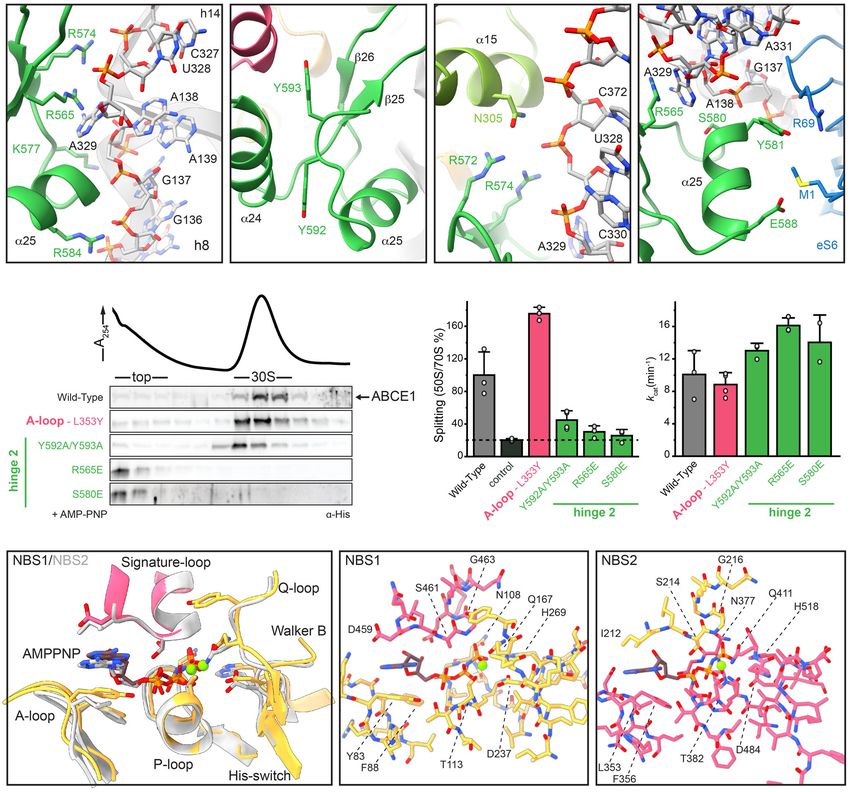
Elina Nürenberg-Goloub et al                                                                                                                       The EMBO Journal

          A                                                                                                     B

          C

  Figure 1. In vitro assembly and cryo-EM structure of the archaeal post-splitting complex.
  A ABCE1IIEA efficiently splits 70S ribosomes in the presence of AMP-PNP and aRF1/aPelota. The 30S population contains a stoichiometric ratio of ABCE1 and ribosomal
    proteins, forming the post-splitting complex. rps: small subunit ribosomal proteins.
  B Cryo-EM density of the post-SC highlights the archaeal ribosomal protein eS21 and ABCE1. Domain architecture of ABCE1 including the mutation sites is shown
    below.
  C Molecular model of the archaeal post-SC, domain colors as in (B).
  Data information: In (A), the gradient profiles are representative for the respective nucleotide condition.

contacting the body of the SSU. Upon transition from the pre- to the                        The FeSD domain establishes inter- and intramolecular
post-splitting state, the NBSs move from a semi-open to a fully                             interactions specific for the post-SC
closed, nucleotide-occluded state. Concomitantly, the FeSD rotates
around a cantilever toward the decoding site of the SSU close to                            Based on the high-resolution data, we can delineate crucial interac-
rRNA helix h44 (Heuer et al, 2017).                                                         tions between the FeSD domain, NBD1, hinge 1, and the 30S riboso-
   The overall architecture of the archaeal post-SC is similar to the                       mal subunit. The FeSD is embedded in a pocket between rRNA h44,
yeast 40S-ABCE1 complex (Heuer et al, 2017) showing the same                                the h5-h15 junction, and the universally conserved ribosomal
hallmarks. The FeSD occupies a position close to rRNA h44, hinge                            protein uS12 (Fig 2A). The majority of FeSD interactions with the
region and HLH motif anchor the NBDs to the 30S body, and the                               ribosome are conserved, while the loop regions of the FeSD opposite
two NBSs are in a closed conformation. Yet, the resolution of the                           of the ribosome (e.g., L36-K43) are variable in sequence and struc-
archaeal post-SC (2.8 Å overall) is significantly higher than the one                      ture, underlining the significance of the interaction of the FeSD with
of the yeast post-SC (3.9 Å overall), especially in NBSII and the                          the ribosome (Fig EV3A, Appendix Fig S2). The majority of interac-
hinge region, thus allowing to describe interactions between ABCE1                          tions are formed by salt bridges and hydrogen bonds established
and the SSU as well as interactions between the two NBSs on a                               between conserved residues in ABCE1 (R2, K15, N17, E19, K59) and
molecular level. These molecular insights allowed us to draw                                the phosphate backbone as well as 2’OH groups of rRNA (Fig 2A).
conclusions and make predictions about the allosteric crosstalk                             Similarly, also the interaction sites between ABCE1 and uS12 are
between the two NBSs of ABCE1 as well as ABCE1 and the ribo-                                conserved (P25, R28, and S29 of ABCE1 to Q76 and H100 of uS12)
some. Moreover, these insights guided the corresponding functional                          (Fig 2A). Interestingly, we observed a few cases where the ribosome
studies (see below).                                                                        and ABCE1 co-evolved to maintain the interaction pattern. For

ª 2020 The Authors                                                                                                        The EMBO Journal e103788 | 2020       3 of 13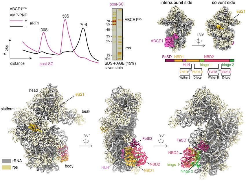
The EMBO Journal                                                                                                                          Elina Nürenberg-Goloub et al

A                                                                                                                                 B

                                                                                                C

  Figure 2. The conserved ABCE1-30S interface is formed by essential interactions.
  A Zoom-ins into ABCE1-30S connections. Most interactions are salt bridges or H-bonds between ABCE1 residues and the rRNA phosphate backbone. The FeSD cluster
    domain contacts rRNA h5 via R2 and K59, interacts with uS12 via S29 and R28, and contacts h44 by N17 and K15. The helix-loop-helix motif connects to rRNA h15 via
    R144 and E147. The positioning of the cantilever is stabilized by an interaction network of R2, I68, and N70 with N316 of hinge 1 and rRNA h5.
  B Yeast survival of ABCE1 variants (S. solfataricus colored, S. cerevisiae in gray). Most residues connecting to 30S in the post-SC show a growth defect when exchanged
    for a small one (alanine) or a negative charge (glutamate). ++ no effect, + growth defect, lethal.
  C The cantilever link forms salt bridges of E74 and E76 with NBD1 residues K89 and R293, respectively.
  Data Information: In (B), data are representative for a set of two independent experiments.

example, the interaction between S29 of ABCE1 and H100 of uS12 is                         our high-resolution structure, we identified additional stabilizing
substituted by the contact of K36 (ABCE1) with N99 (uS12) in yeast                        contacts for the cantilever loop. E74 also interacts with the side
(Fig EV3B), underlining the importance of an interaction at this                          chain of K89 (NBD1) and the carbonyl group of E76 binds the guani-
position for re-orientation of the FeSD after ribosome splitting.                         dino group of R293 (NBD1) (Fig 2C). Moreover, an interaction
   The FeSD is linked to the main twin-ATPase body via a flexible                         network is formed between R2 (R7 in S.c.) at the N terminus, I68
linker connecting the cantilever b-sheet b4 with NBD1 (Fig 2C,                            and N70 (N74 in S.c.) of the cantilever b-sheet b4, and N316 (N326
Appendix Fig S2). This linker (D73-V79 in S. solfataricus) forms an                       in S.c.) in hinge 1, as well as the phosphate groups of G345 and
a-helix in free ABCE1 and the pre-SC (Karcher et al, 2008; Brown                          G346 in rRNA h5 (Fig 2A). In yeast, the mutations Y301A and R7A
et al, 2015), but unfolds into a loop in the post-SC. As in the yeast                     impair the anti-association activity of ABCE1 in vitro and are
post-SC (Heuer et al, 2017), this cantilever helix is also unfolded in                    synthetically lethal in vivo (Heuer et al, 2017). Additionally, we con-
S. solfataricus. At high resolution, we deciphered a chain of inter-                      firm synthetic lethality of N74A with N326A (Figs 2B and EV3C).
and intramolecular interactions that are a consequence of FeSD                                Taken together, closure of the NBSs displaces the FeSD, which
repositioning after splitting. We observed a similar stabilization of                     leads to new interactions of the cantilever b-sheet and the cantilever
the cantilever loop by an interaction of Y291 in NBD1 (Y301 in S.c.)                      loop with the ribosome, NBD1 and hinge 1. This allows for an allos-
with the backbone of E74 (N78 in S.c.) (Fig 2C, Appendix Fig S2). In                      teric communication of post-SC formation to the NBSs.

4 of 13       The EMBO Journal e103788 | 2020                                                                                                           ª 2020 The Authors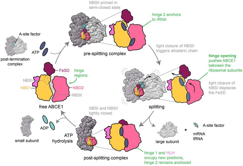
Elina Nürenberg-Goloub et al                                                                                                    The EMBO Journal

Hinge 2 serves as a linchpin during ribosome splitting                     for the post-SC. Thus, a15 of hinge 1 binds U328 and the conserved
                                                                           N316 binds to A314 as well as the phosphates of G343 and G345
The NBDs of ABCE1 are located at the body of the 30S subunit with          close to the h5-h15 junction (Fig 2A). As mentioned above, U328
main anchor points contributed by the HLH motif (to h15) and the           also contacts R574 in hinge 2 (Fig 3C) while N316 is connected to
dipartite hinge region (to junction of h8 and h14) (Fig 2A). In stark      the rearranged cantilever loop of the FeSD. Consequently, the FeSD,
contrast to the pre-splitting complex, the HLH is displaced from its       hinge 1, and hinge 2 form a post-SC state-specific intricate interac-
contact point at h5 by 16 Å toward h15. In the post-SC, h15 is in         tion network.
contact with the loop containing two basic residues (R144-G145-                Functional analyses and lethality screens confirm the essential
K146-E147) between helices a6 and a7 (Fig 2A). A charge reversion          role of the hinge 2 region for ABCE1 function. As mentioned before,
of the respective arginine in yeast (R148E) leads to a substantial         ABCE1S580E (Appendix Fig S3) exhibits wild-type ATPase activity
growth defect, confirming this important position (Figs 2B and             (Fig 3G) but neither binds to 30S ribosomes (Figs 3E and EV4A) nor
EV3C). The other residues in the HLH loop rather stabilize an inter-       splits 70S ribosomes (Figs 3F and EV4B). Additionally, the corre-
action formed by NBD1 with U353, which flips out of h15 and forms          sponding mutant is lethal in yeast (S588E) (Karcher et al, 2008).
a Watson-Crick base pair with A51 in h5, establishing the h5-h15           Interestingly, S580 is the N-terminal residue of helix a25 and does
junction. Multiple residues (T95, K97, E147, H282, K296, and S297)         not directly interact with the ribosome but points toward a25
are facing this base pair, suggesting that this specific tertiary struc-   (Fig 3D). Thus, the mutation to glutamate at this position inhibits
ture is precisely monitored by NBD1 and the HLH motif of ABCE1             ribosome binding via destabilization of helix a25 rather than by
(Fig 2A). In contrast to yeast, no contacts are observed between           direct repulsion. The importance of R574 for ribosome recognition
ABCE1 and eS24, which is also present but significantly shorter at         is confirmed by our plasmid-rescue analysis in yeast, demonstrating
its C terminus in T. celer.                                                that the respective R582E mutation is lethal (Figs 2B and EV3C).
    The ABCE1-specific hinge region is subdivided into hinge 1
(S. solfataricus 298–325) and hinge 2 (S. solfataricus 547–594;            Structural asymmetry of the nucleotide-binding sites
Appendix Fig S2). Interactions with the ribosome are mainly estab-
lished by hinge 2. Hinge 1 connects NBD1 and NBD2 via a flexible           Apparently, ABCE1 can act as timer for ribosome recycling (Heuer
linker (S. solfataricus 326–338), which is—as in other structures—         et al, 2017; Nürenberg-Goloub et al, 2018). During this process, the
only partially visible. Similar to the HLH/NBD1 region, hinge 2 also       NBSs receive and integrate signals about the state of the ribosome,
recognizes a special tertiary structure of the rRNA. It binds at the       e.g., discriminate between pre-splitting and post-splitting
junction between rRNA helices h8 and h14, where A329 flips out of          complexes. In the post-SC, both NBSs have mainly been observed in
h14 and stacks upon the ribose of A138 in h8. The geometry is read         the closed state (Gouridis et al, 2019), coinciding with a movement
out by the conserved R565 forming a cation-p-stack with A138               of the FeSD (Kiosze-Becker et al, 2016; Heuer et al, 2017) as initially
(Fig 3A and D, Appendix Fig S2). Notably, this interaction is main-        suggested (Becker et al, 2012). Yet, in all obtained cryo-EM struc-
tained during ribosome splitting (Fig 4), and exchange of the corre-       tures of pre- and post-SCs, the identity of the bound nucleotides,
sponding residue (R573E) leads to loss of function in yeast (Karcher       especially in NBSII, remained unclear. Based on our high-resolution
et al, 2008). Hence, the S. solfataricus ABCE1R565E mutant                 data, we can resolve both catalytic pockets and unambiguously
(Appendix Fig S3) was unable to bind 30S ribosomes (Figs 3E and            identify the non-hydrolysable ATP-analogue AMP-PNP complexed
EV4A) and failed to split 70S ribosomes (Figs 3F and EV4B), whereas        with a Mg2+ ion in each NBS (Figs 3H–J and EV5). In agreement
the ATPase activity was similar to wild-type ABCE1 (Fig 3G).               with the yeast post-SC and the structures of symmetric ABC-type
    The second main contact to the h8-h14 junction is formed by a          NBD dimers (Lammens et al, 2011; Korkhov et al, 2012), AMP-PNP
salt bridge between R574 and the phosphate of U328 (Fig 3A and             is sandwiched between the typical conserved motifs of ABC-type
C). Moreover, R572 and N305 in hinge 1 stabilize the interaction           ATPases. In NBSI, the A-loop residue Y83 stacks on the purine base,
network around this junction on the side of h14 (Fig 3C), while            which is contacted by the aliphatic part of D459 adjacent to the
K577, S580, and R584 are in close contact to h8 (to G137 and A139)         signature motif of the opposite NBD2. In addition, the ribose is
(Fig 3A and D). Further, hinge 2 forms an additional interaction site      stabilized by stacking with F88 (Fig 3I). The c-phosphate is directly
with eS6 by stacking Y581 against R69 (eS6) (Fig 3D). This interac-        contacted by N108 (Walker A), H269 (His-switch), S461-G463 (sig-
tion also occurs in yeast between Q589 and K58 (eS6), indicating a         nature motif), and Q167 (Q-loop), while T113 (Walker A) and D237
co-evolution of ABCE1 ribosome interactions as previously                  (Walker B) coordinate the Mg2+ ion. Analogous residues are super-
described for FeSD and uS12 (Fig EV3D).                                    imposable in NBSII, i.e., we find that N377 (Walker A), S214, G216
    While the hinge 2 region serves as a constant linchpin to the          (signature motif), and H518 (His-switch) coordinate the c-phosphate
ribosome, the interaction pattern of hinge 1 is substantially altered      while Q411 (Q-loop), T382 (Walker A), and D484 (Walker B)
compared to the pre-SC. In hinge 2, only R574 switches from U329           contact the Mg2+ ion (Fig 3J). Notably, the characteristic A-loop is
in the pre-SC to the adjacent U328 in the post-SC, while all other         degenerated in NBSII of most (but not all) organisms, featuring
residues remain with their respective interaction partners (Fig 4A).       aliphatic or even polar residues (Gerovac & Tampé, 2019). Despite
In contrast, the entire hinge 1 region opens up relative to hinge 2,       the degenerated A-loop (L353 instead of the aromatic residue), the
which results in a 5 Å shift of the hinge 2 b-sheets b25 and b26          accommodation of the purine base is similar to the one observed in
(Fig 4A, Appendix Fig S2) and a 10 Å movement of hinge 1 helix            NBSI (Fig 3H). The base is sandwiched between L353 and I212 adja-
a15. Together with the movement of the HLH (Fig 4B) and the                cent to the signature motif of NBD1. Yet, we hypothesized that
FeSD, this conformational rearrangement, which we term “hinge              higher flexibility of the nucleotide in NBSII due to the degenerated
opening”, leads to the formation of new ribosomal contacts specific        A-loop might explain (i) the reduced intrinsic ATPase activity in

ª 2020 The Authors                                                                                       The EMBO Journal e103788 | 2020   5 of 13
The EMBO Journal Elina Nürenberg-Goloub et al
A B C D
E F G
H I J
Figure 3. Structural and functional analysis of the hinge regions and NBSs.
A–D Hinge 2 (emerald) residues interacting with the ribosome. R565E forms a conserved cation-p-stacking with A329 of h8; R574 forms a salt bridge with the phosphate
backbone of U328 in h14. Aromatic C-terminal residues Y592 and Y593 adopt a parallel coordination. R572 of hinge 2 and N305 of hinge 1 (light green) form an
interaction that might be important for sensing. Essential S580 does not contact the ribosome, whereas Y581 and E588 form H-bonds to R69 and M1 of eS6 (blue),
respectively.
E Mutations in the a-helices of hinge 2 prevent 30S binding while the Y592A/Y593A (C terminus) and L353Y (A-loop in NBSII) exchanges do not influence ribosome
binding.
F 70S splitting efficiency normalized to wild type. Hinge 2 mutations Y592A/Y593A, R565E, and S580E display strongly impaired splitting activity. Unspecific ribosome
dissociation level as determined in control experiments in the absence of ABCE1 is marked by the dotted line.
G ATP turnover per ABCE1 is not affected in all tested mutants.
H–J Overview of ATP coordination in both NBSs and overlay of the two NBSs reveals only slight differences, which cannot elucidate the functional asymmetry. Residues
of NBD1 and NBD2 involved in coordination are shown in gold and punch, respectively.
Data Information: In (F) and (G), the mean SD of assay triplicates and duplicates are plotted.
Source data are available online for this figure.
6 of 13 The EMBO Journal e103788 | 2020 ª 2020 The AuthorsElina Nürenberg-Goloub et al The EMBO Journal
A D
B E
C
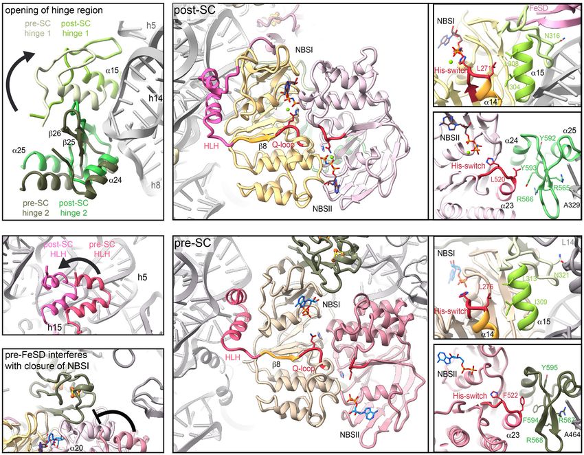
Figure 4. Hinge regions and HLH sense the ribosome splitting event and allosterically communicate with the NBSs.
A Hinge 1 moves away from hinge 2 during transition from pre-SC (cotton) to post-SC (lime), thereby forming new interactions with the ribosome. In contrast, hinge 2
movement from pre- (moss) to post-SC (emerald) does not change the interaction with the ribosome.
B The HLH motif is displaced from h5 in the pre- (watermelon) to h15 in the post-SC (pink).
C Positioning of the FeSD (sage) interferes with the closure of NBD2 (blush) in the pre-SC (rose).
D Possible communication pathways from ribosome binding sites to the NBSs in the post-SC. HLH is connected to the Q-loop of NBSI via b8. I304 of hinge 1 connects
to a14 which is adjacent to the His-switch in NBSI. Analogously, hinge 2 binding to the SSU might be communicated via Y593 and R566 to a23 next to the His-switch
of NBSII.
E Interaction pattern of the communication pathways between HLH and hinge 1 to NBSI as well as hinge 2 to NBSII is different in the pre-SC compared to the post-SC.
NBSII (Nürenberg-Goloub et al, 2018) and (ii) the lower resolution Ribosome binding is allosterically communicated to conserved
of this site in cryo-EM studies (Heuer et al, 2017). To test this motifs in the NBSs
hypothesis, we substituted L353 by a tyrosine, thereby generating a
consensus A-loop in NBSII. However, 30S binding, 70S splitting effi- Ribosome splitting completely alters the interaction pattern of
ciency, and ATPase activity of ABCE1L353Y (Appendix Fig S3) were ABCE1 with the ribosome at all contact points excluding the hinge
comparable to wild type (Figs 3E–G and EV4). Consequently, the 2 region. Based on the high-resolution structure, we elaborated
respective yeast mutation Q363Y had no effect on growth and allosteric communication pathways between the ribosome-ABCE1
survival (Figs 2B and EV3C). Thus, the functional asymmetry of interface and the NBSs. In the pre-splitting complex, the FeSD does
ABCE1 may originate from the connection of each NBS to an allos- not interfere with the NBSI semi-open state (Brown et al, 2015).
teric regulatory element on the ABCE1 surface, i.e., the FeSD, HLH However, upon closure, the loop K12-P13-D14 of the FeSD would
motif, and hinge regions, rather than from single residues within the clash into NBDII, in particular into residues preceding the NBSI
ATP-binding pockets. signature motif and a20, involving the L453-E454-S455 stretch
ª 2020 The Authors The EMBO Journal e103788 | 2020 7 of 13The EMBO Journal Elina Nürenberg-Goloub et al
(Fig 4C). The movement of NBSI is thus coupled to rearrange- “hinge opening” modulates the His-switches in both NBSs by alter-
ments of the FeSD and vice versa. Moreover, the flexible HLH ing the contact interface to adjacent a-helices. We observed that
motif via b8 is linked to the Q-loop of NBSI (Fig 4D and E). Muta- NBSI is in an active conformation with all residues needed for cata-
tions in the Q-loops strongly affect the ATPase activity of ABCE1 lytic activity in place, i.e., activation of a water molecule for nucle-
and compromise its function in yeast (Karcher et al, 2008; ophilic attack on the c-phosphate (Chen et al, 2003; Lammens et al,
Barthelme et al, 2011). As stated above, we observed clear density 2011; Hofmann et al, 2019). The functional and dynamic asymme-
for Q167 sensing the presence of the c-phosphate. Additionally, we try of the two NBSs (Barthelme et al, 2011; Nürenberg-Goloub et al,
envision that hinge opening is directly transmitted to the H-loops 2018; Gouridis et al, 2019) does not arise from incomplete ATP
in both NBSs, which are key motifs in controlling ATPase activity alignment due to a non-canonical A-loop in NBSII, as we confirmed
of ABCE1 and other ABC proteins (Zaitseva et al, 2005; Barthelme by biochemical and yeast viability studies. In the ABC transporter
et al, 2011; Hurlimann et al, 2017). In the post-SC, hinge 1 forms TAP and its homolog TmrAB, the position of the non-canonical site
a specific contact to the h5-h15 junction where N316 interacts with cannot be switched without compromising the transport function,
G345. Compared to the pre-SC, hinge 1 a15 moves closer toward indicating that additional signals from outside the binding pocket
NBSI and forms a contact with a14, directly adjacent to the H-loop are integrated into the ATPase cycle (Chen et al, 2003; Procko et al,
of NBSI (Fig 4D and E). The conserved I304 in a15 points toward 2006; Zutz et al, 2011). Consistently, we envision an allosteric regu-
a14, allowing a communication between hinge 1 and NBSI. Consis- latory network that extends from the ABCE1-ribosome interface into
tent with this essential function, the corresponding mutation I314E the NBSs. The spatial separation of hinge 1 from hinge 2 is linked to
is lethal in yeast (Figs 2B and 4D and E, and EV3C). Similarly, a both NBSs and in addition might be a prerequisite for closure of
conserved series of residues communicates ribosome binding from NBSII (Fig 4 and Movie EV1). In agreement, the introduction of
hinge 2 to the H-loop of NBSII. Herein, R565 in hinge 2 senses the mutations disrupting ribosome binding in hinge 1 (R311A in S.c.;
h8–h14 junction while R566 and Y593 contact helix a23. Analo- R301 in S. solfataricus) or hinge 2 (R573E, R582E, and S588E in S.c.;
gously to a14 in NBD1, helix a23 occupies the position adjacent to R565, R574, and S580, in S. solfataricus, respectively) compromise
the H-loop in NBSII (Fig 4D and E). We substituted the conserved ABCE1 function (Karcher et al, 2008) (Figs 2B and 3B–D, and EV3C,
Y592 and Y593 by alanine and probed for ABCE1 function. Consis- and EV4). The exchange of G303 in hinge 1 (Appendix Fig S2),
tent with the role of Y593 in ribosome sensing without direct located at the contact interface to NBD1, leads to a reduced wing
contact to rRNA or ribosomal proteins, the 70S splitting ability of size in Drosophila melanogaster (G316D in the pixie gene), further
ABCE1Y592A/Y593A (Appendix Fig S3) is substantially inhibited highlighting the role of the hinge region for ABCE1 function (Coelho
(Figs 3F and EV4B) while the 30S binding efficiency and ATPase et al, 2005). Notably, hinge 1 and hinge 2 occupy a position analo-
activity are similar to wild type (Figs 3D and E, and EV4A). Addi- gous to the regulatory elements of bacterial ABC importers (New-
tionally, the respective double-mutant Y600A/F601A exhibits a stead et al, 2009; Johnson et al, 2012; Chen et al, 2013)
growth defect in yeast (Figs 2B and EV3C). The five-stranded b- (Appendix Fig S4), showing that a regulation from this site can be
sheet harboring the degenerated A-loop in NBSII is in close prox- exploited by ABC-type proteins.
imity of hinge 2. Comparing the pre-SC with the post-SC, we Closure of NBSII allosterically activates NBSI, which is consis-
observed a conformational change in this region which contributes tent with the increased ATPase activity of ABCE1 in the presence
to ATP occlusion by allowing the hydrophobic stacking of L353 of 70S/80S ribosomes and release factors (Pisarev et al, 2010;
and the adenine base (Fig 3J). Shoemaker & Green, 2011; Nürenberg-Goloub et al, 2018). On a
We finally inspected the Walker B/D-loops, which are known to structural level, we assume that NBSII can close prior to NBSI to
assure transport directionality in the ABC transporter associated prime ribosome splitting at the pre-SC (Fig 5). In more detail, the
with antigen processing (TAP) (Grossmann et al, 2014). Notably, movement of the signature motif toward NBSII is possible when
the D-loops are, together with the H-loops, already part of the still bound to the 70S/80S ribosomes, since ABCE1 anchors via
contact interface between the NBDs in the pre-splitting state. This the hinge 2 region and HLH motif, and none of the mobile parts
interface drastically alters upon closure of the NBSs, ribosome split- participate in ribosome binding. Furthermore, 70S/80S are split as
ting, and post-SC formation, allowing a multilayered communica- soon as both sites occlude Mg2+-ATP and switch to the closed
tion network between both sites in addition to the allosteric conformation (Fig 5), as found within the post-SC (Heuer et al,
regulation by the ribosome (Fig 4D and E). 2017; Nürenberg-Goloub et al, 2018; Gouridis et al, 2019). During
the closing movement, the FeSD is pushed away by NBD2 and,
concomitantly, interactions between NBD1, the HLH motif, and
Discussion the ribosome must be temporarily broken, allowing hinge 1 to
move away from hinge 2 (Fig 5). Structurally, separation of the
By using an ATPase-deficient mutant of ABCE1 in an in vitro ribo- two hinge regions occurs concomitantly with FeSD movement and
some recycling assay, we were able to capture the archaeal adoption of the fully closed state of the ABCE1 NBDs. These struc-
post-splitting complex comprising the 30S subunit and ABCE1. Our tural rearrangements may well determine the ribosome splitting
structure reveals this essential, asymmetric ABC-type protein in a rate. Consistently, in the presence of Mg2+-AMP-PNP, ABCE1 tran-
fully nucleotide-occluded state at atomic resolution. Furthermore, siently associates with 30S ribosomes within 5 s, while closure of
the cryo-EM structure allows a prediction of the communication NBSII takes app. 7 min and stabilizes the post-SC (Gouridis et al,
pathways within the post-splitting complex, which we functionally 2019).
and genetically assessed. Ribosome binding is sensed by the HLH Remarkably, translation termination is a slow event. Several
motif and hinge region that opens up during ribosome splitting. This ribosome profiling studies showed a high enrichment of reads
8 of 13 The EMBO Journal e103788 | 2020 ª 2020 The AuthorsElina Nürenberg-Goloub et al The EMBO Journal Figure 5. Model for ribosome splitting by ABCE1. ABCE1 binds to 70S/80S ribosomes containing mRNA, tRNA in the P-site (not shown), and an A-site factor (a/eRF1 after canonical termination; a/e Pelota during stalled ribosome recognition) to form pre-splitting complexes. Here, NBSII is primed in a semi-closed state and anchored to ribosomal RNA via hinge 2. ATP occlusion and tight closure of NBSII triggers an allosteric chain within ABCE1 leading to a tight closure of NBSI. Consequently, the FeSD is displaced and the parallel hinge opening rearranges ABCE1 in the ribosomal subunit cleft. Thereby, the subunits are split apart and the FeSD is repositioned at h44. During and/or after the splitting process, the A-site factor dissociates and mRNA and tRNA are recycled (not shown). At the post-SC, ABCE1 occludes two ATP molecules in the NBSs. ATP hydrolysis is a prerequisite for NBS opening and dissociation of ABCE1 from the SSU. Black arrows indicate domain movements within ABCE1. indicating a high occupancy of ribosomes on stop codons future, the precise role of ABCE1 in initiation will need to be (Andreev et al, 2017). Moreover, a significant population of elucidated to complete the translation cycle for Eukarya and ABCE1-containing termination complexes was found in native Archaea. polysomes, along with translating ribosomes (Behrmann et al, 2015). Similarly, the half-life of ribosomes stalled during transla- tion and rescued by the Pelota/Hbs1/ABCE1 system is supposedly Material and Methods long. In light of this, it makes sense that ribosome splitting is regulated and coordinated by the action of the intrinsically slow Protein purification NBSII. Slow closure of NBSII could ensure correct engagement within the pre-splitting complex, and slow ATP hydrolysis could Construction of the pSA4 plasmids for recombinant expression of determine the dwell time of ABCE1 after splitting to prevent ABCE1, aRF1, aPelota, and aIF6 from S. solfataricus in E. coli was premature re-association with large ribosomal subunits, or coordi- described previously (Barthelme et al, 2007, 2011). All proteins nate downstream events such as translation initiation and/or were expressed, purified, and stored as previously described tRNA/mRNA recycling. In this context, the question remains open (Nürenberg-Goloub et al, 2018). Protein quality was assured by as to how ATPase activity and thus the 30S/40S dissociation is SDS–PAGE and size exclusion chromatography (Superdex 200 modulated (Fig 5). Here, external factors, e.g., components of the Increase 3.2/300, GE Healthcare) in SEC buffer (20 mM Tris–HCl pH initiation machinery, might play a direct or indirect role in 7.5, 150 mM NaCl, 2 mM b-mercaptoethanol) at 4°C recording communicating conformational rearrangements during pre-initia- absorption at 280 and 410 nm to monitor FeSD cluster integrity. tion complex formation into the NBSs of ABCE1 to trigger its release. In particular, and possibly by modulating its ATPase Ribosome purification activity, the non-essential eukaryotic eIF3j subunit (Hcr1 in S.c.) assists ABCE1 in ribosome recycling, and thereby may also Frozen cell pellets from T. celer were purchased from the Centre promote post-SC disassembly (Young & Guydosh, 2019). In the of Microbiology and Archaea, University of Regensburg, Germany. ª 2020 The Authors The EMBO Journal e103788 | 2020 9 of 13
The EMBO Journal                                                                                                    Elina Nürenberg-Goloub et al

Cell pellets were resuspended in 2.5× volume S30 buffer (10 mM            area to 70S peak area of the A254 gradient profile using OriginPro
Tris–HCl pH 7.5, 60 mM KOAc, 14 mM MgCl2, 1 mM dithiothreitol             2018 (OriginLab) and normalized to the mean value of wild-type
(DTT)) and lysed using a Branson Sonifier. Cell debris was                ABCE1. Splitting experiments were performed at least three times
removed by centrifugation 2 × 30 min at 34,000 g and 4°C. The             per ABCE1 variant; bars show mean  SD value.
supernatant was loaded on a high-salt sucrose cushion (10 mM
Hepes-KOH pH 7.5, 1.1 M sucrose, 1 M NH4Cl, 10.5 mM Mg                    30S binding assay
(OAc)2, 0.1 mM EDTA, 4 mM b-mercaptoethanol), and ribosomes
were pelleted at 200,000 g for 15 h at 4°C. For 70S preparation,          17.5 pmol T. celer 30S were incubated with 8.5 pmol ABCE1 in the
pelleted ribosomes were resuspended in S30 buffer and gradient            presence of AMP-PNP, or ADP (8.5 nmol each), or in the absence of
purified (10–40% (w/v) sucrose, S30 buffer) for 14 h at 68,000 g.         any nucleotide in S30 buffer for 10 min at 65°C. Higher molecular
Fractions were collected using a Piston Gradient Fractionator (Bio-       weight aggregates were removed for 10 min at 16,100 g and 4°C.
comp) recording the A254 profile. The buffer of 70S containing            Samples were loaded onto a 10–40% (w/v) sucrose density gradient
fractions was exchanged to TrB25 (56 mM Tris–HCl pH 8.0,                  in S30 buffer, as described. 0.5-ml fractions were collected, precipi-
250 mM KOAc, 80 mM NH4OAc, 25 mM MgCl2, 1 mM DTT) via                     tated overnight at –20°C in 2× volume acetone, and pelleted for 1 h
Econo-Pac 10DG Desalting Columns (Bio-Rad), and 70S were                  at 16,100 g and 4 °C. The pellet was resuspended in SDS loading
concentrated using a 100K Amicon Ultra (Merck). For 30S purifica-         dye and analyzed by SDS–PAGE and immunoblotting. All ABCE1
tion (for 30S binding assays), high-salt sucrose cushion pelleted         variants contained a C-terminal His6 tag and were detected using
ribosomes were resuspended in buffer A30 (10 mM Hepes-KOH                 rabbit anti-His (ab1187, Abcam) and goat anti-rabbit (AP307P,
pH 7.5, 100 mM NH4Cl, 10.5 mM Mg(OAc)2, 0.1 mM EDTA,                      Merck) antibodies. Binding assays were performed once per
4 mM b-mercaptoethanol) and loaded onto a HiPrep 16/60 Sepha-             ABCE1variant. The gradient profiles shown are representative for the
cryl S-400 HR size exclusion chromatography column (GE Health-            respective nucleotide condition.
care). Ribosome fractions were collected and again pelleted
through a low magnesium sucrose cushion in buffer A30 (2.5 mM             ATPase assay
Mg(OAc)2) for subunit dissociation. Ribosomes were resuspended
in S30 buffer (with 2.5 mM Mg(OAc)2 instead of MgCl2) and gradi-          ATPase activity was measured using a Malachite Green-based
ent purified. 30S fractions were pooled, the buffer exchanged to          assay (adapted from (Baykov et al, 1988). Samples were measured
S30 and concentrated as before.                                           at least in duplicates. 1–2 lM ABCE1 was incubated with 2 mM
                                                                          ATP in ATPase buffer (10 mM Hepes pH 7.5, 150 mM NaCl, 2.5
Assembly of the post-splitting complex for cryo-EM                        mM MgCl2) for 8 min at 80°C in a total volume of 25 ll. The reac-
                                                                          tion was stopped by addition of 175 ll ice-cold 20 mM H2SO4.
To mimic the physiological translation cycle, post-splitting              50 ll Malachite Green working solution (2 ml conc. Malachite
complexes were generated by splitting of 1 nmol purified 70S ribo-        Green solution (60 ml H2SO4 in 300 ml H2O with 0.44 g Malachite
somes from T. celer by ABCE1IIEA (8 lM), aPelota, and aRF1 (5 lM          Green), 40 ll Tween-20 (10% v/v) and 550 ll Na2MoO4) was
each) in the presence of 0.5 mM AMP-PNP in 50 mM HEPES-KOH                added per sample and incubated for 2–5 min at room temperature.
pH 7.5, 30 mM KCl, 10 mM MgCl2, and 2 mM DTT at 65 °C for 15              A620 was recorded in a CLARIOstar plate reader (BMG Labtech).
min. Samples were chilled on ice and cross-linked with 1% (v/v)           Bar diagrams represent mean  SD of two (ABCE1S580E), four
formaldehyde for 30 min on ice. Higher molecular weight aggre-            (ABCE1L353Y) or three (all other ABCE1 variants) independent
gates were removed for 15 min at 16,100 g and 4 °C. Samples were          experiments.
loaded onto 10–30% (w/v) sucrose density gradient in 50 mM
HEPES-KOH pH 7.5, 30 mM KCl, 0.5 mM MgCl2, and 2 mM DTT,                  Yeast plasmid shuffling assay
and ribosomal particles were separated by centrifugation for 13.5 h
at 78,000 g and 4°C in a SW40 rotor (Beckman Coulter Life                 In vivo function of ABCE1 mutants was checked as previously
Sciences). Gradients were fractionated into 0.3 ml using Piston           described (Heuer et al, 2017). The haploid yeast strain CEN.MG1-9B
Gradient Fractionator (Biocomp Instruments) while recording A254.         (MATa his3D1 leu2-3,112 trp1-289 MAL2-8C SUC2 ura3-52 rli1::
Fractions containing 30S were pooled, and the sucrose was removed         KanMX4 + pRS426-ABCE1) was generated in which the essential
by Sephadex G-25 gravity flow size exclusion columns (GE Health-          ABCE1 gene (RLI1) was deleted by KanMX4 and substituted by
care). Ribosomes were diluted to concentrations of 50–70 nM               pRS426-ABCE1 [URA3] expressing wild-type ABCE1 under the
(based on OD260) for quality control by negative stain EM. Samples        control of the endogenous promoter. CEN.MG1-9B strain was trans-
were vitrified immediately.                                               formed with pRS423-ABCE1 [HIS3] plasmid coding for wt and
                                                                          mutated ABCE1 and with empty vector pRS423 as negative control
70S splitting assay                                                       and selected on -HIS. If such a strain harboring both plasmids was
                                                                          grown on medium containing 5-FOA, the pRS426-ABCE1 [URA3]
7.5 pmol T. celer 70S were split using ABCE1, aRF1, aPelota, and          plasmid is lost by counter-selection as the URA3 gene product
aIF6 (75 pmol each) in the presence of 22.5 nmol AMP-PNP in S30           converts 5-FOA to a toxic compound. Consequently, the strain was
buffer at 65°C for 15 min. Higher molecular weight aggregates were        prone to survive only in the presence of pRS423-ABCE1. Growth
removed for 10 min at 16,100 g and 4°C. Samples were analyzed             and survival were checked by growth studies in a serial dilution
via 10–40% (w/v) sucrose density gradient in S30 buffer as                assay over 2–3 days. Data in Figs 2B and EV4C are representative
described. Splitting efficiency was calculated as the ratio of 50S peak   for a set of two independent experiments.

10 of 13     The EMBO Journal e103788 | 2020                                                                                   ª 2020 The Authors
Elina Nürenberg-Goloub et al The EMBO Journal
Cryo-EM analysis Table 1. Data collection, refinement, and validation statistics.
30S-ABCE1 (EMD-10519,
For the archaeal post-SC, the sample was applied to 2-nm pre-coated PDB ID 6TMF)
Quantifoil R3/3 holey carbon-supported grids and vitrified using a Data collection
Vitrobot mark IV (FEI). Data were collected on a TITAN KRIOSTM Voltage (kV) 300
cryo-TEM (Thermo Fisher) equipped with a Falcon III chip
Electron exposure (e–/Å2) 25
enhanced Falcon II direct detector at 300 keV under low-dose condi-
tions of approximately 25 e /Å2 for 10 frames in total, and a defo- Defocus range (lm) 1.1 to 2.3
cus range of 1.1 to 2.3 lm. Magnification settings resulted in a Pixel size (Å) 1.084
pixel size of 1.084 Å per pixel. Original image stacks were summed Symmetry imposed C1
and corrected for drift and beam-induced motion at the micrograph
Refinement
level by using MotionCor2 (Zheng et al, 2017). The contrast transfer
Particle images (no.) 293 010
function (CTF) estimation of each micrograph was performed with
Gctf (Zhang, 2016). Map resolution (Å) 2.8/2.8/3.0
(overall/30S head/ABCE1)
Data processing FSC threshold 0.143
Map sharpening B factor (Å2) 117.8/ 128.8/ 151.9
The ABCE1-30S data set was processed, unless otherwise stated, (overall/30S head/ABCE1)
following the standard workflow using RELION 2 and 3 (Kimanius Model composition
et al, 2016; Zivanov et al, 2018). After particle picking with GAUTO-
Correlation coefficient (%; Phenix) 0.85
MATCH (http://www.mrc-lmb.cam.ac.uk/kzhang/) and 2D classifi-
Initial model used (PDB codes) 5JBH, ABCE1: 5LL6 (chain h)
cation, particles were subjected to a thorough 3D classification
regimen. About 97% of all particles contained ABCE1 stably bound Non-hydrogen atoms 65 449
to the small ribosomal subunit. Different conformational states of Protein residues 4 172
the ribosome 30S head were separated and a homogeneous class RNA bases 1 485
with 293.010 particles was selected for further refinement. First, the
R.m.s. deviations
particles of this class were CTF-corrected and refined to an overall
resolution of 2.8 Å after post-processing. A focused refinement on Bond lengths (Å) 0.017 (30)
the head and ABCE1 could improve the local resolution of the struc- Bond angles (°) 1.208 (44)
ture. Validation
MolProbity score 1.79
Model building
Clash score 4.66
The molecular model of the small ribosomal subunit was built using Rotamer outliers (%) 0.73
the 70S model of P.fu. [4V6U (Armache et al, 2013), 5JBH (Coureux Ramachandran plot
et al, 2016)]. After rigid-body fitting of the 30S into the density, the Favored (%) 89.98
sequence was manually changed to T. celer and modeled into the
Allowed (%) 9.51
cryo-EM density using Coot (version 0.8.9.1) (Emsley & Cowtan,
2004). The sequences were taken from the T. celer Vu 13 = JCM Disallowed (%) 0.51
8558A genome, available at NCBI. A previously unidentified protein Validation RNA
could be modeled by building the sequence de novo into the density. Correct sugar pucker (%) 98
The T. celer genome was searched for characteristic sequence motifs
Good backbone conf. (%) 80
of the protein taking the approximate size of the protein into consider-
ation. An initial model of ABCE1 was generated using Phyre2 (Kelley
et al, 2015). The model of S.c. ABCE1 (Heuer et al, 2017) was used as atomic models have been deposited in the Protein Data Bank under
a template, and the resulting model was manually refined in Coot. accession number PDB ID 6TMF (https://doi.org/10.2210/pdb
After Phenix refinement (Adams et al, 2010), the models and maps of 6TMF/pdb). Correspondence and requests for materials should be
30S head, body, and ABCE1 were combined and refined again. Cryo- addressed to R.T. (tampe@em.uni-frankfurt.de) or R.B. (beck-
EM structures and models were displayed using UCSF Chimera (Pet- mann@genzentrum.lmu.de).
tersen et al, 2004) and ChimeraX (version 0.8; Goddard et al, 2018).
Expanded View for this article is available online.
Data availability Acknowledgements
The authors thank Simon Trowitzsch, Lukas Susac, Jingdong Cheng, and
The cryo-EM density maps of the archaeal 30S ribosome and ABCE1 Michael Ameismeier for discussions; Charlotte Ungewickell and Susanne Rieder
have been deposited in the Electron Microscopy Data Bank under for technical assistance; Lukas Kater for support with processing and the pre-
accession number EMD-10519 (https://www.ebi.ac.uk/pdbe/ processing pipeline of the cryo-EM data; and Petr Tesina for help with final
entry/emdb/EMD-10519) (see Table 1). Atomic coordinates for the model refinements in Phenix. E.N.G. was supported by the Christiane Nüsslein-
ª 2020 The Authors The EMBO Journal e103788 | 2020 11 of 13The EMBO Journal Elina Nürenberg-Goloub et al
Volhard Foundation, L’Oréal, and the United Nations Educational, Scientific Behrmann E, Loerke J, Budkevich TV, Yamamoto K, Schmidt A, Penczek PA,
and Cultural Organization (UNESCO). H.K. is supported by a DFG fellowship Vos MR, Burger J, Mielke T, Scheerer P et al (2015) Structural snapshots of
through the Graduate School of Quantitative Bioscience Munich (QBM). The actively translating human ribosomes. Cell 161: 845 – 857
German Research Foundation (DFG) SFB 902 “Molecular mechanisms of RNA- Brown A, Shao S, Murray J, Hegde RS, Ramakrishnan V (2015) Structural basis
based regulation” (to R.T.), TRR174 “Spatiotemporal dynamics of bacterial cells” for stop codon recognition in eukaryotes. Nature 524: 493 – 496
(to R.B.) and FOR 1805 (to R.B.) funded this work. Chen J, Lu G, Lin J, Davidson AL, Quiocho FA (2003) A tweezers-like motion of
the ATP-binding cassette dimer in an ABC transport cycle. Mol Cell 12:
Author contributions 651 – 661
EN-G, HK, HH, TB, RB, and RT designed the study. EN-G and HH developed Chen ZQ, Dong J, Ishimura A, Daar I, Hinnebusch AG, Dean M (2006) The
the preparation of the post-splitting complex. EN-G, HK, HH, and AH opti- essential vertebrate ABCE1 protein interacts with eukaryotic initiation
mized the sample preparation for cryo-EM. EN-G, HK, HH, and AH prepared factors. J Biol Chem 281: 7452 – 7457
the EM samples. HK and OB collected and HK processed the cryo-EM data. Chen S, Oldham ML, Davidson AL, Chen J (2013) Carbon catabolite repression
HK built and refined the model. HK, TB, EN-G, HH, RT and RB analyzed and of the maltose transporter revealed by X-ray crystallography. Nature 499:
interpreted the structures. HH and EN-G performed all functional assays. 364 – 368
EN-G and PK conducted the genetic analysis in yeast. EN-G, HK, TB, HH, RB, Coelho CM, Kolevski B, Bunn C, Walker C, Dahanukar A, Leevers SJ (2005)
and RT wrote the manuscript with contributions from all authors. RT initi- Growth and cell survival are unevenly impaired in pixie mutant wing
ated the project. discs. Development 132: 5411 – 5424
Coureux PD, Lazennec-Schurdevin C, Monestier A, Larquet E, Cladiere L,
Conflict of Interest Klaholz BP, Schmitt E, Mechulam Y (2016) Cryo-EM study of start
The authors declare that they have no conflict of interest. codon selection during archaeal translation initiation. Nat Commun 7:
13366
Dong J, Lai R, Nielsen K, Fekete CA, Qiu H, Hinnebusch AG (2004) The
References essential ATP-binding cassette protein RLI1 functions in translation by
promoting preinitiation complex assembly. J Biol Chem 279: 42157 – 42168
Adams PD, Afonine PV, Bunkoczi G, Chen VB, Davis IW, Echols N, Headd JJ, van den Elzen AM, Schuller A, Green R, Seraphin B (2014) Dom34-Hbs1
Hung LW, Kapral GJ, Grosse-Kunstleve RW et al (2010) PHENIX: a mediated dissociation of inactive 80S ribosomes promotes restart of
comprehensive Python-based system for macromolecular structure translation after stress. EMBO J 33: 265 – 276
solution. Acta Crystallogr D Biol Crystallogr 66: 213 – 221 Emsley P, Cowtan K (2004) Coot: model-building tools for molecular graphics.
Andreev DE, O’Connor PB, Loughran G, Dmitriev SE, Baranov PV, Shatsky IN Acta Crystallogr D Biol Crystallogr 60: 2126 – 2132
(2017) Insights into the mechanisms of eukaryotic translation gained with Gerovac M, Tampé R (2019) Control of mRNA translation by versatile ATP-
ribosome profiling. Nucleic Acids Res 45: 513 – 526 driven machines. Trends Biochem Sci 44: 167 – 180
Armache JP, Anger AM, Marquez V, Franckenberg S, Frohlich T, Villa E, Goddard TD, Huang CC, Meng EC, Pettersen EF, Couch GS, Morris JH, Ferrin
Berninghausen O, Thomm M, Arnold GJ, Beckmann R et al (2013) TE (2018) UCSF ChimeraX: meeting modern challenges in visualization
Promiscuous behaviour of archaeal ribosomal proteins: implications for and analysis. Protein Sci 27: 14 – 25
eukaryotic ribosome evolution. Nucleic Acids Res 41: 1284 – 1293 Gouridis G, Hetzert B, Kiosze-Becker K, de Boer M, Heinemann H, Nürenberg-
Aspesi A, Ellis SR (2019) Rare ribosomopathies: insights into mechanisms of Goloub E, Cordes T, Tampé R (2019) ABCE1 controls ribosome recycling by
cancer. Nat Rev Cancer 19: 228 – 238 an asymmetric dynamic conformational equilibrium. Cell Rep 28:
Ban N, Beckmann R, Cate JH, Dinman JD, Dragon F, Ellis SR, Lafontaine DL, 723 – 734.e6
Lindahl L, Liljas A, Lipton JM et al (2014) A new system for naming Grossmann N, Vakkasoglu AS, Hulpke S, Abele R, Gaudet R, Tampé R (2014)
ribosomal proteins. Curr Opin Struct Biol 24: 165 – 169 Mechanistic determinants of the directionality and energetics of active
Barthelme D, Scheele U, Dinkelaker S, Janoschka A, Macmillan F, Albers SV, export by a heterodimeric ABC transporter. Nat Commun 5: 5419
Driessen AJ, Stagni MS, Bill E, Meyer-Klaucke W et al (2007) Structural Hellen CUT (2018) Translation termination and ribosome recycling in
organization of essential iron-sulfur clusters in the evolutionarily highly eukaryotes. Cold Spring Harb Perspect Biol 10: a032656
conserved ATP-binding cassette protein ABCE1. J Biol Chem 282: Heuer A, Gerovac M, Schmidt C, Trowitzsch S, Preis A, Kötter P,
14598 – 14607 Berninghausen O, Becker T, Beckmann R, Tampé R (2017) Structure of the
Barthelme D, Dinkelaker S, Albers SV, Londei P, Ermler U, Tampé R (2011) 40S-ABCE1 post-splitting complex in ribosome recycling and translation
Ribosome recycling depends on a mechanistic link between the FeS initiation. Nat Struct Mol Biol 24: 453 – 460
cluster domain and a conformational switch of the twin-ATPase ABCE1. Hofmann S, Januliene D, Mehdipour AR, Thomas C, Stefan E, Bruchert S, Kuhn
Proc Natl Acad Sci USA 108: 3228 – 3233 BT, Geertsma ER, Hummer G, Tampé R et al (2019) Conformation space of
Bassler J, Hurt E (2019) Eukaryotic ribosome assembly. Annu Rev Biochem 88: a heterodimeric ABC exporter under turnover conditions. Nature 571:
281 – 306 580 – 583
Baykov AA, Evtushenko OA, Avaeva SM (1988) A malachite green procedure Hopfner KP (2016) Invited review: architectures and mechanisms of ATP
for orthophosphate determination and its use in alkaline phosphatase- binding cassette proteins. Biopolymers 105: 492 – 504
based enzyme immunoassay. Anal Biochem 171: 266 – 270 Hurlimann LM, Hohl M, Seeger MA (2017) Split tasks of asymmetric
Becker T, Franckenberg S, Wickles S, Shoemaker CJ, Anger AM, Armache JP, nucleotide-binding sites in the heterodimeric ABC exporter EfrCD. FEBS J
Sieber H, Ungewickell C, Berninghausen O, Daberkow I et al (2012) 284: 1672 – 1687
Structural basis of highly conserved ribosome recycling in eukaryotes and Joazeiro CAP (2019) Mechanisms and functions of ribosome-associated
archaea. Nature 482: 501 – 506 protein quality control. Nat Rev Mol Cell Biol 20: 368 – 383
12 of 13 The EMBO Journal e103788 | 2020 ª 2020 The AuthorsYou can also read