Structure-function analyses of alkylhydroperoxidase D from Streptococcus pneumoniae reveal an unusual three-cysteine active site architecture
←
→
Page content transcription
If your browser does not render page correctly, please read the page content below
JBC Papers in Press. Published on January 23, 2020 as Manuscript RA119.012226
          The latest version is at https://www.jbc.org/cgi/doi/10.1074/jbc.RA119.012226

    Structure-function analyses of alkylhydroperoxidase D from Streptococcus pneumoniae reveal an
                              unusual three-cysteine active site architecture

    Yanxiang Meng1, Campbell R. Sheen2, Nicholas J. Magon3, Mark B. Hampton3*, and Renwick C.J.
                                             Dobson1*

1
  Biomolecular Interaction Centre and School of Biological Sciences, University of Canterbury, Christchurch,
    New Zealand
2
  Callaghan Innovation, University of Canterbury, Christchurch, New Zealand
3
  Centre for Free Radical Research, Department of Pathology and Biomedical Science, University of Otago,
    Christchurch, New Zealand

                 Running title: Alkylhydroperoxidase D (AhpD) from Streptococcus pneumoniae

*To whom correspondence should be addressed: Prof. Renwick Dobson, Biomolecular Interaction Centre and
School of Biological Sciences, University of Canterbury, Christchurch, New Zealand. Telephone (+64) 3 369
5145. E-mail: renwick.dobson@canterbury.ac.nz or

                                                                                                                     Downloaded from http://www.jbc.org/ by guest on November 7, 2020
Prof. Mark Hampton, Department of Pathology and Biomedical Science, University of Otago, Christchurch,
New Zealand. Telephone (+64) 3 378 6225. E-mail: mark.hampton@otago.ac.nz

Keywords: Alkylhydroperoxidase D, hydrogen peroxide, oxidative stress, Streptococcus pneumoniae, X-ray
crystallography, analytical ultracentrifugation, small-angle X-ray scattering (SAXS), liquid chromatography-
mass spectrometry (LC-MS/MS), redox switch, electron transfer

ABSTRACT                                                    conserved Asn-76 of the AhpD core motif is
         During aerobic growth, the Gram-positive           important for SpAhpD folding. In summary, SpAhpD
facultative anaerobe and opportunistic human                is a weak peroxidase and does not transfer electrons
pathogen Streptococcus pneumoniae generates large           to AhpC, and therefore does not fit existing models
amounts of hydrogen peroxide that can accumulate            of bacterial AhpD antioxidant defense mechanisms.
to millimolar concentrations. The mechanism by              We propose that it is unlikely that SpAhpD removes
which this catalase-negative bacterium can withstand        peroxides either directly or via AhpC, and that
endogenous hydrogen peroxide is incompletely                SpAhpD cysteine oxidation may act as a redox
understood. The enzyme alkylhydroperoxidase D               switch or mediate electron transfer with other thiol
(AhpD) has been shown to contribute to                      proteins.
pneumococcal virulence and oxidative stress
responses in vivo. We demonstrate here that SpAhpD                   Streptococcus pneumoniae is a Gram-
exhibits weak thiol-dependent peroxidase activity           positive, catalase-negative, facultative anaerobe
and, unlike the previously reported Mycobacterium           associated with a variety of infections in humans (1).
tuberculosis AhpC/D system, SpAhpD does not                 During aerobic growth, S. pneumoniae produces
mediate electron transfer to SpAhpC. A 2.3-Å                millimolar concentrations of hydrogen peroxide
resolution crystal structure revealed several unusual       (H2O2) due to the activities of pyruvate and lactate
structural features, including a three-cysteine active      oxidases, and the absence of significant disposal
site architecture that is buried in a deep pocket, in       mechanisms. These levels are sufficient to kill other
contrast to the two-cysteine active site found in other     bacteria and even host cells (2–5). However, it is
AhpD enzymes. All single-cysteine SpAhpD variants           unclear how S. pneumoniae protects itself from high
remained partially active, and LC–MS/MS analyses            levels of endogenous H2O2. Elucidation of protective
revealed that the third cysteine, Cys-163, formed           mechanisms could lead to the development of novel
disulfide bonds with either of two cysteines in the         therapeutics to disrupt this process, thereby making
canonical Cys-78–X–X–Cys-81 motif. We observed              the pathogen more susceptible to oxidative stress.
that SpAhpD formed a dimeric quaternary structure                    Several aspects of the S. pneumoniae
both in the crystal and in solution, and that the highly    response to oxidative stress have been described (6),
                                                                                                                1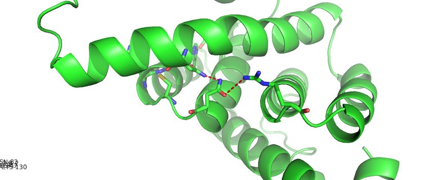
Alkylhydroperoxidase D (AhpD) from Streptococcus pneumoniae
including enzymes such as superoxide dismutase (7), SpAhpD is encoded in a bicistronic operon,
NADH oxidase (8), and thiol peroxidase (9). downstream of the gene spr0371, which is predicted
Additionally, spr0370 from S. pneumoniae strain R6 to be an integral membrane protein of unknown
encodes a putative alkylhydroperoxidase D function (10). Given the structural and functional
(SpAhpD) 1 . Knocking out spr0370 reveals that diversity of AhpD proteins, along with the
AhpD is a pneumococcal virulence determinant differences in genome organization, it is difficult to
involved in the response to oxidative stress (10). infer how AhpD contributes to the oxidative stress
Alkylhydroperoxidase (Ahp) family response in S. pneumoniae. As such, we report the
enzymes are found in both Gram-positive and Gram- first biochemical characterization and crystal
negative bacteria and convert peroxides to alcohol structure of SpAhpD to better understand this aspect
and water (6). AhpC/F is the most common bacterial of the pneumococcal antioxidant defence
Ahp family peroxidase system (Fig. 1A). AhpC is a mechanism. We demonstrate that SpAhpD exhibits
highly active two-Cys peroxiredoxin that reacts only weak peroxidase activity towards both H2O2 and
directly with peroxides, while the NADH-dependent alkyl hydroperoxides, showing rates similar to those
flavoprotein AhpF reduces AhpC via a thiol-disulfide of AhpD proteins from other organisms. However,
exchange reaction, regenerating the enzyme for unlike the AhpC/D system in M. tuberculosis,
another cycle (11). SpAhpD is unable to reduce SpAhpC. The crystal
The best-described AhpD is that of structure of SpAhpD, determined at 2.3-Å resolution,
Downloaded from http://www.jbc.org/ by guest on November 7, 2020
Mycobacterium tuberculosis (12) (Fig. 1B). Here, the reveals several unique structural features.
ahpD gene is immediately downstream of ahpC and
replaces the need for ahpF. MtAhpD contains a Results
thioredoxin-like CXXC motif. Initially, it was shown S. pneumoniae AhpD exhibits weak DTT-
to exhibit weak peroxidase activity in parallel with dependent peroxidase activity
MtAhpC in vitro by using AhpF from Salmonella The ability of SpAhpD to reduce H2O2, ter-
typhimurium as the reductase (12). It was later butyl hydroperoxide (tBuOOH), and cumene
discovered that MtAhpD has a more important hydroperoxide (CuOOH) was tested using the
function of mediating electron transfer from ferrous-oxidation xylenol orange (FOX) assay, with
dihydrolipoamide dehydrogenase (Lpd) and DTT as the reducing agent. Under assay conditions,
dihydrolipoamide succinyltransferase (SucB) to SpAhpD exhibits net peroxidase activity (rate in
oxidized MtAhpC via thiol-disulfide exchange presence of SpAhpD minus rate of peroxide with
reactions (13), linking the M. tuberculosis Lpd-SucB DTT alone) of 0.124 µM/min towards H2O2, 0.127
metabolic pathway to antioxidant defence. µM/min towards tBuOOH, and 0.575 µM/min
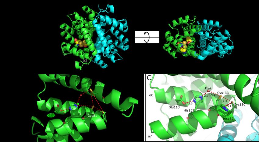
Over the last few years, the core active site towards CuOOH (Fig. 2A). While these values are
sequence motif (E(X)11CXXC(X)3H) from MtAhpD greater than the rate of peroxide decomposition in the
has been used to identify putative AhpD proteins in absence of protein, they correspond to turn-over
other bacterial species (10, 14–16) (Fig. S1). While numbers of less than 0.1 s−1. Compared to SpAhpC,
the CXXC motif is responsible for redox activity, which shows greater initial velocities by nearly two
Glu118 and His137 of MtAhpD form a proton shuttle orders of magnitude under the same assay conditions,
that activates a peroxidatic cysteine by the peroxidase activity of SpAhpD is weak.
deprotonation. These residues are also conserved in The role of each cysteine residue of SpAhpD
AhpD from S. pneumoniae (SpAhpD) (Fig. 1C). was investigated by testing the activity of single
AhpD enzymes are associated with oxidative mutants against H2O2 under the same assay
stress resistance in four other species: conditions. All three mutants, C78S, C81S, and
Corynebacterium glutamicum (15), Anabaena sp. C163S, resulted in decreased peroxidase activity
PCC7120 (16), Pseudomonas aeruginosa (14), and (Fig. 2B). Interestingly, both C78S and C81S
S. pneumoniae (10). However, the biochemical retained about 50% of the activity, while C163S
function of the AhpD enzymes in these bacteria is retained 80% of its activity.
poorly understood.
1
Abbreviations: AhpD: alkylhydroperoxidase D, AhpC: acetamido-4′maleimidylstilbene-2,2′-disulfonic acid,
alkylhydroperoxidase C, AhpF, alkylhydroperoxidase F, tBuOOH: ter-butyl hydroperoxide, CuOOH: cumene
Trx: thioredoxin, LB: Luria-Bertani medium, IPTG: hydroperoxide, HRV3C: Human Rhinovirus 3C Protease,
Isopropyl β-D-1-thiogalactopyranoside, AMS: 4- NEM: N-ethylmaleimide.
2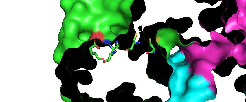
Alkylhydroperoxidase D (AhpD) from Streptococcus pneumoniae

        We confirmed that the ability of DTT to              generated H2O2 at the same rate regardless of
reduce the disulfide bond in SpAhpD was not rate-            whether or not SpAhpD was present, and H2O2
limiting, and therefore the reason for the low activity      production and subsequent reduction by the
of SpAhpD. The free thiol content of SpAhpD and              SpAhpC/F system is also unaffected by SpAhpD
SpAhpC was measured with Ellman’s reagent. DTT               (Fig. 3D).
rapidly reduced both SpAhpC and SpAhpD (Fig.                          Taken    together, these   experiments
2C). In contrast, when H2O2 was added to pre-                demonstrate that SpAhpD does not mediate electron
reduced SpAhpD, the thiol content decreased at a             transfer to SpAhpC.
much slower rate than that of SpAhpC (Fig. 2D).
                                                             Crystal structure of AhpD from S. pneumoniae
AhpD from S. pneumoniae does not mediate                              Given that SpAhpD is functionally different
electron transfer to AhpC                                    from MtAhpD, we solved the crystal structure of
         To test whether SpAhpD can reduce SpAhpC            recombinant SpAhpD to a resolution of 2.3-Å (Fig.
via a thiol-disulfide exchange reaction, as for              4A). The X-ray crystallography data collection and
MtAhpD (13), we used a gel-shift assay based on 4-           refinement statistics are shown in Table 1 (PDB:
acetamido-4′-maleimidylstilbene-2,2′-disulfonic              6E8L).
acid (AMS). In this assay, AMS irreversibly                           In contrast to the MtAhpD trimer and other
alkylates thiols, adding 0.5 kDa for each free thiol,        hexameric AhpD structures in the Protein Data Bank,

                                                                                                                      Downloaded from http://www.jbc.org/ by guest on November 7, 2020
which is observed as a shift on non-reducing SDS-            the SpAhpD structure assembled as a face-to-face
PAGE gels (17). Although oxidized AhpC forms                 dimer (Fig. 4A). The dimeric assembly of SpAhpD
intermolecular disulfide bonds and can easily be             was confirmed by subsequent small-angle X-ray
distinguished from reduced AhpC, AMS labelling is            scattering and sedimentation velocity centrifugation
required for visualizing the redox state of SpAhpD.          experiments (discussed in a separate section below).
Pre-reduced SpAhpD in five-fold excess was mixed             The structures of the three dimers in an asymmetric
with oxidized SpAhpC. No change in the redox state           unit are almost identical (RMSD = 0.297 and 0.367,
of oxidized SpAhpC was observed over the course of           respectively) (Fig. S3). Formation of the dimer
30 min (Fig. 3A). Reduced thioredoxin from S.                results in a buried surface area of ~4,000 Å2, with an
pneumoniae (SpTrx) was mixed with either pre-                average ΔGint of −31.4 kcal/mol.
oxidized SpAhpC or SpAhpD for use as positive                         Each SpAhpD monomer consists of nine α-
controls, and almost all of the oxidized SpAhpC was          helices, each ranging in size from 6–20 residues (Fig.
reduced by SpTrx upon mixing (Fig. 3B). Although             4A). SpAhpD has very low sequence homology
it was difficult to discern the change in intensity of       (~20% identity), when compared with the
the band corresponding to oxidized SpAhpD, bands             functionally well-characterized MtAhpD. However,
corresponding to reduced SpAhpD and oxidized Trx             like all AhpD structures in the Protein Data Bank,
indicated that thiol-disulfide exchange occurs               both proteins belong to a class of globin-like α-
between SpAhpD and SpTrx, albeit slowly (Fig. 3C).           proteins and comprise nine α-helices.
These findings confirm that the disulfide bond in                     The      putative     active    site   motif
both proteins can be reduced by thiol proteins under         E(X)11CXXC(X)3H is located on helices α4 and α5
the assay conditions, and that SpAhpD does not               (Fig. 4A and B), which are connected by a 180°
reduce SpAhpC.                                               (hairpin) turn motif (14). In SpAhpD, this section
         We then tested whether SpAhpD mediates              contains the redox-active CXXC motif, the highly
electron transfer in the SpAhpCF system. By                  conserved His85 and Asn76 residues, and a semi-
measuring both the consumption of NADH (at 340               conserved Glu66 residue (Fig. 1C and Fig. 4B).
nm) and H2O2 concentration (using the FOX assay),            His85, Glu66, and a structural water molecule are
we demonstrate that SpAhpF generates H2O2 in an              thought to form a proton shuttle towards the CXXC
NADH-dependent manner (Fig. 3D and Fig. S2). In              motif to deprotonate the free thiols in AhpD from
comparison, the mixture of SpAhpC and SpAhpF has             both M. tuberculosis (Fig. 4C) and P. aeruginosa
significantly reduced H2O2 production, and the H2O2          (14, 18), and these residues are conserved across
concentration decreases rapidly after initial                most species.
generation (Fig. 3D, red line). We then added                         The positioning of these conserved residues
SpAhpD in five-fold excess to test whether it                in SpAhpD (Fig. 4B) is similar to that in MtAhpD
influences activity of the SpAhpCF peroxidase                (Fig. 4C), as is the apparent interaction between
system (Fig. 3D, blue line). However, SpAhpF                 His85 and Glu66 at a distance of 2.6 Å. The water

                                                                                                                3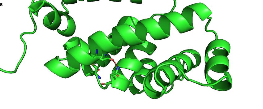
Alkylhydroperoxidase D (AhpD) from Streptococcus pneumoniae

molecules in the SpAhpD active site are                                  In MtAhpD His132 has been shown to play
inconsistently positioned among the six protomers in            a functional role by deprotonating the resolving
an asymmetric unit and have a high temperature                  cysteine (equivalent to Cys78 in SpAhpD) (18). In
factor (average of 56 Å2 comparing to global average            SpAhpD the equivalent residue is replaced by Phe80,
of 46 Å2), suggesting that the water molecule                   which is unable to deprotonate the resolving cysteine
involved in the SpAhpD proton shuttle may have                  (Cys78), and there is no basic residue near Cys78 in
high mobility while still interacting with His85 to             the crystal structure. Therefore, Cys78 of SpAhpD is
mediate the deprotonation of Cys81.                             likely to be less reactive than the equivalent resolving
         Overall, the crystal structure of SpAhpD               cysteine in MtAhpD.
shows that it has the similar all-α-helical topology as
AhpD from all other organisms and structurally                  The third cysteine residue, Cys163, forms novel
conserved active site residues (E(X)11CXXC(X)3H),               disulfide bonds with both cysteine residues in the
but the biological assembly is a distinct dimer.                CXXC motif
                                                                         We used liquid chromatography–mass
S. pneumoniae AhpD forms an unusual three-                      spectrometry (LC-MS/MS) to investigate the
cysteine active site buried in a deep pocket                    reactivity of Cys163 towards the other two cysteine
         Despite the conserved features described               residues. This was performed on chymotryptic
above, the active site of SpAhpD has several key                digests of SpAhpD treated with various

                                                                                                                           Downloaded from http://www.jbc.org/ by guest on November 7, 2020
differences that could make SpAhpD mechanistically              concentrations of H2O2 followed by N-
distinct and explain the observed functional                    ethylmaleimide (NEM), which specifically alkylates
difference.                                                     free thiols.
         The most obvious feature is that the active                     First, we identified the three free cysteine-
site consists of three cysteines, rather than the usual         containing peptides alkylated with NEM: NG-
two (Fig. 4B). In addition to the conserved Cys81 and           Cys78-AF, Cys81-VAGHTAF, and Cys163-NY, and
Cys78 residues, Cys163 from helix α9 is also present.           confirmed their fragmentation patterns in the reduced
Although AhpD proteins from P. aeruginosa                       sample (Fig. S5). Their respective relative abundance
(PaAhpD; PDB 2O4D) and Ralstonia eutropha                       decreased as they were treated with increasing
(ReAhpD; PDB 2PRR) also contain three cysteines,                concentrations of H2O2 (Fig. 6A-C). The relative
the third cysteine sits distant from the active site (Fig.      abundance of Cys163 peptide decreased more slowly
S4). Cys163 in SpAhpD is uniquely positioned in                 (Fig. 6C), suggesting that it may be less reactive than
close proximity to both Cys81 (5.1 Å) and the                   the other two cysteines. However, when treated with
structural water (3.4 Å) of the proton shuttle,                 0.5 mM H2O2, all three free cysteine containing
indicating that it may also react with Cys81 to form            peptides had near undetectable abundance, meaning
a disulfide bond, which is a previously undescribed             all of them are either involved in disulfide bonds or
mechanism in AhpD proteins. It is somewhat further              hyperoxidized.
away (8.1 Å) from the second cysteine, Cys78.                            All three possible combinations of disulfide
         The active site residues of SpAhpD are                 bonds were detected when SpAhpD was treated with
located in a deep pocket formed between helices α1,             H2O2 (Fig. 6D-F and S5). Interestingly, their relative
α3, α4, and α5, with helix α9 on the bottom. Cys81              abundance peaked at 0.1–0.5 mM and decreased at
and Cys163 are located in the bottom of the pocket,             higher H2O2 concentrations (Fig. 6D-F). This is
while Glu66 and His85 are more solvent-accessible               likely due to the formation of irreversible oxidation
and positioned closer to the entrance of the pocket             products, such as sulfinic (-SOOH) and sulfonic
(Fig. 5A). Cys78 is also solvent-accessible via the             acids (-SO3H) (Fig. S6). The relative abundance of
opposite face of the monomer. The active sites of               the disulfide bond formed within the CXXC motif
most other AhpD proteins are more exposed or in a               and between Cys78 and Cys163 peaked at 0.1 mM
shallower groove, as seen in MtAhpD (Fig. 5B). In               H2O2 treatment, while the disulfide bond between
contrast, the pocket on SpAhpD appeared to be fully             Cys81 and Cys163 peaked at 0.5 mM, suggesting
enclosed and planar in geometry, with a depth of                that the latter may be slightly less favourable. These
approximately 15 Å. It was only accessible via an               concentrations of H2O2 are generated by S.
opening ~9 Å in width. Generating vacuum                        pneumoniae grown in culture media.
electrostatic using Pymol predicted the interior of                      These experiments demonstrate that Cys163
this pocket to be mildly hydrophobic to slightly                is redox active and able to form a disulfide bond with
positive electrostatic.

                                                                                                                      4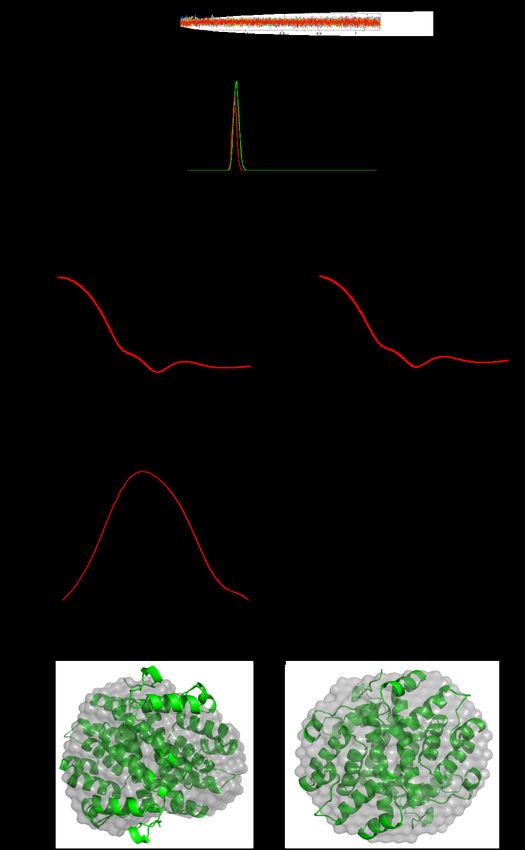
Alkylhydroperoxidase D (AhpD) from Streptococcus pneumoniae

either Cys78     or   Cys81    under    physiological       across a concentration range of 0.66–4.0 mg/ml. At
conditions.                                                 all concentrations tested, the enzyme is
                                                            monodisperse and not involved in a self-association
The highly conserved Asn76 residue within the               with higher order species (Table S1, Fig. S8). The
AhpD core motif is required for the folded structure        buoyant molecular weight of the peak is 39.5 kDa,
of the active site                                          which is very close to the mass of the dimer (39.7
         Multiple sequence alignment of the AhpD            kDa) calculated from the sequence (Fig. 9A).
core motif reveals that Asn76 of SpAhpD is also                      We also collected small-angle X-ray
highly conserved (Fig. 1C). We propose that Asn76           scattering data for both the reduced and oxidized
may serve a structural purpose, stabilizing the folded      SpAhpD enzyme to test whether the shape of the
active site structure in all AhpD proteins. In our          protein in the crystal is representative of that in
structure of SpAhpD, Asn76 forms a hydrogen bond            solution and examine whether the oxidation state of
with the sidechain of Asn164 on helix α9 (Fig. 7A).         the protein affects its structure. The redox states of
Asn164 is conserved in all AhpD sequences, except           the protein were confirmed using Ellman’s assay and
for MtAhpD (Fig. S1), and the hydrogen bonding              the scattering profiles are shown in Fig. 9 (B and C).
interaction is structurally conserved (Fig. 7B). In         The Guinier plots for both data sets are linear (R2 =
MtAhpD, Asn128 (structurally homologous to                  0.999), indicating minimal aggregation and no inter-
SpAhpD Asn76) hydrogen bonds with the side chain            particle interference.

                                                                                                                      Downloaded from http://www.jbc.org/ by guest on November 7, 2020
of Asn82, and possibly Arg47 (Fig. 7C). In all known                 The scattering profile for both the oxidized
AhpD structures, the asparagine next to the CXXC            and reduced samples are in good agreement with the
motif (Asn76 in SpAhpD) always hydrogen bonds               theoretical scattering profile calculated using the
with the sidechain of another asparagine on the             dimer from the crystal structure (χ2 values of 0.486
longest α-helix in the same subunit (Asn164 in              and 0.694, respectively) (Fig. 9B and C, red line). By
SpAhpD). This highly conserved interaction                  calculating the pairwise distance distribution (P(r)),
indicates that Asn76 is important for the function of       both the radius of gyration and the molecular weight
AhpD, probably by stabilizing the folded structure of       are estimated to be very similar for both the oxidized
the active site.                                            and reduced forms of SpAhpD, so is the maximum
         To test this theory, we constructed two            distance (Table 2). The (P(r)) plot reveals a more
mutants, N76A and N76L, by site-directed                    prominent difference, where oxidized SpAhpD had
mutagenesis and attempted to express and purify             clearly lower P(r) at distance of 35-50 Å. (Fig. 9D).
them. However, in a small-scale expression trial both       Comparison of the scattering profiles also revealed
asparagine mutants were insoluble (Fig. S7),                small differences at 0.1–0.2 Å−1 (Fig. 9E).
probably due to inability to fold properly and                       Taken together, these findings confirm that
consistent with the hypothesis that it stabilizes the       the dimer observed in the crystal structure represents
structure of the active site.                               the in-solution structure of SpAhpD and suggests that
                                                            the oxidation state of the protein may have a small
AhpD from S. pneumoniae forms an unusual                    effect on the global structure.
dimeric quaternary structure
         Our crystal structure demonstrates that            Discussion
SpAhpD forms a dimer (Fig. 4A). This was                             In this study, we reveal the unusual
surprising, since MtAhpD forms a trimer in solution         biochemical and structural properties of AhpD from
(18), and all other AhpD crystal structures were            S. pneumoniae. The activity of SpAhpD does not
predicted to form hexamers (trimer of dimers) (19)          appear to fit into the existing models of bacterial
(Fig. 8). All the hexameric and trimeric AhpD               AhpD antioxidant defence mechanisms: while it is
proteins have 3-fold rotational symmetry with a             oxidized by hydroperoxides it is a weak peroxidase,
central cavity as the axis of symmetry, whereas             and it does not mediate electron transfer to AhpC.
SpAhpD lacks both the 3-fold symmetry and a                 Results of the DTT-dependent FOX assay and
central cavity. Analytical ultracentrifugation and          Ellman’s assay clearly demonstrate that SpAhpD
small-angle X-ray scattering experiments confirm            reacts very slowly with H2O2. Because of this low
that SpAhpD is also a dimer in solution, consistent         activity, we could not induce a Michaelis-Menten-
with the crystal structure.                                 type response in the DTT-dependent kinetic assay.
         Sedimentation velocity type analytical             However, the resultant turnover (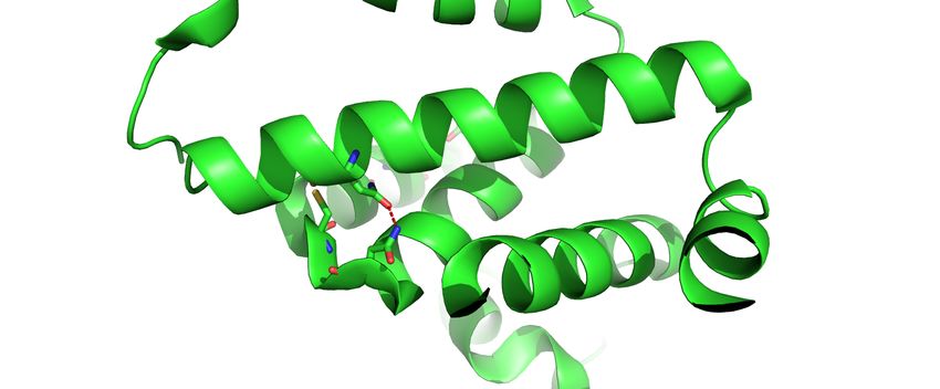
Alkylhydroperoxidase D (AhpD) from Streptococcus pneumoniae

lower than that of the characterized thiol peroxidase,      an artefact of the redox active motif. We propose that
TpxD, from S. pneumoniae (9). Given the H2O2                this may also be the case for the AhpD proteins from
concentrations to which S. pneumoniae is routinely          S. pneumoniae, P. aeruginosa, and Anabaena sp.
exposed, the direct removal of H2O2 by SpAhpD is            PCC7120 because they all share the same core
unlikely to be important for protection against             sequence motif.
oxidative damage.                                                    We sought to use structural information to
         We also tested the hypothesis that SpAhpD          unravel the biological function of SpAhpD. The
mediates electron transfer to SpAhpC, similar to its        crystal structure obtained in this study confirmed that
homologue in M. tuberculosis (13, 20). However, the         SpAhpD contains the structurally-conserved core
AMS gel-shift assay showed that SpAhpD does not             functional motif (E(X)11CXXC(X)3H). The structure
reduce SpAhpC, while the NADH-dependent FOX                 of the putative active site, with favourable
assay confirmed that SpAhpD does not affect the             conformation for the formation of disulfide bonds
putative AhpC/F system in S. pneumoniae.                    between cysteine residues, is consistent with
         SpAhpF alone appears to generate H2O2, in          SpAhpD being a redox active protein. While
an NADH-dependent manner, consistent with                   SpAhpD is ineffective at reducing AhpC it could
bifunctional AhpF (NADH oxidase and AhpC                    reduce other thiol proteins, conferring protection
reductase) and AhpC/F peroxidase systems in certain         from oxidative stress. This would explain why
other species, including Amphibacillus xylanus,             various AhpD proteins appear to contribute to the

                                                                                                                       Downloaded from http://www.jbc.org/ by guest on November 7, 2020
Sporolactobacillus inulinus, and S. mutans (21, 22).        oxidative stress response in vivo (10, 14–16), despite
Given the high degree of sequence similarity                only exhibiting weak peroxidase activity. This
between S. pneumoniae and S. mutans AhpF this dual          hypothesis is supported by studies on C. glutamicum
activity was expected, and may contribute to H2O2           AhpD (15), which is strongly linked to the cellular
generation in S. pneumoniae, in addition to pyruvate        NAD+/NADH ratio in vivo. In M. tuberculosis,
oxidase and lactate oxidase. While the ahpC                 AhpD acts in between Lpd-SucB and AhpC, but the
(GenBank:        CMAP01000085.1,         locus    tag:      reactivities between such bacterial peroxidase-
ERS022390_02365), and ahpF (NZ_CHHM010000                   related thiol proteins have been shown to be
86.1) genes are present in certain strains of S.            promiscuous and species-dependent in many cases
pneumoniae, their expression levels have not been           (11, 12, 24, 25), further complicating the question of
reported.                                                   the biological activity of AhpD. Alternatively, thiol
         The finding that SpAhpC does not                   exchange may not occur with other proteins, but
functionally interact with SpAhpD was also                  SpAhpD may interact with different bacterial
supported by the observed genome organization. In           proteins depending on whether it is reduced or
M. tuberculosis, ahpD is adjacent to ahpC in the            oxidized. Mammalian thioredoxin dissociates from
same operon, whereas spr0370 (encoding AhpD)                ASK1 upon oxidation, leading to activation of this
from S. pneumoniae is expressed in a bicistronic            kinase (26). Further investigation of the interacting
operon with downstream gene spr0371, which                  partners of SpAhpD by pull-down assays or genetic
encodes a putative voltage-dependent anion channel          complementation studies are required to understand
family membrane protein of unknown function (10,            its biological activity.
23). In this respect, SpAhpD is similar to AhpD from                 The combination of several conserved
C. glutamicum, whose corresponding gene is                  structural features with large differences in the
preceded by a four-component gene cluster encoding          folding and sequence of AhpD proteins from
an ABC-type nickel/peptide transporter (15).                different bacterial species has been attributed to
         The weak peroxidase activity of SpAhpD is          convergent evolution (14). While SpAhpD shares the
consistent with several previously-characterized            common features of the “hairpin” motif
AhpD proteins. Weak activity was initially                  (E(X)11CXXC(X)3H) (14) and an all-α-helical
demonstrated for AhpD from M. tuberculosis (12),            topology with AhpDs from other organisms, analysis
with later studies reporting similar findings in P.         of our crystal structure revealed a number of
aeruginosa (14) and Anabaena sp. PCC7120 (16).              structural features not previously observed in AhpD
Interestingly, the primary function of MtAhpD was           family proteins. We also demonstrated that a
later shown to be the linking of reducing potential         structurally well conserved asparagine residue
from lipoamide-containing metabolic enzymes to the          (Asn76 of SpAhpD) within this core motif is
peroxiredoxin AhpC via thiol-disulfide exchange             important for the folding of this core structure, and it
reactions (20), suggesting the peroxidase activity is

                                                                                                                  6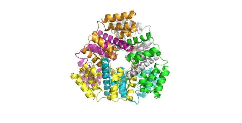
Alkylhydroperoxidase D (AhpD) from Streptococcus pneumoniae

should be considered part of the AhpD family core               peroxidases (28). In such circumstance, Cys163 may
sequence motif (E(X)9NXCXXC(X)3H).                              react with cysteine-SOH before H2O2 to prevent
         First, the redox-active CXXC motif is                  hyperoxidation, or act as a replacement when one of
located in a deep pocket in SpAhpD, while it tends to           the canonical cysteines has been hyperoxidized,
be more exposed in AhpD proteins from other                     thereby retain AhpD activity at high H2O2
species. This suggests that the reactive cysteines are          concentrations.
less accessible to larger molecules and explains why,                    Our subsequent LC-MS/MS results
in contrast to the M. tuberculosis orthologue,                  demonstrated that Cys163 is able to form disulfide
SpAhpD did not react with SpAhpC. This also                     bonds with either cysteine residues in the CXXC
indicates that the active site of SpAhpD may be more            motif. As expected, the disulfide bond between the
selective towards its substrate. The planar geometry            canonical CXXC motif cysteines is the most
and mildly hydrophobic nature of this pocket is                 common, while Cys163 appears to form disulfides at
indicative of the type of substrate it may bind. This           higher H2O2 concentrations. While Cys81 was the
explains the observation during our kinetic                     most reactive free thiol (Fig. 6B) as expected from its
experiments that reactivity of SpAhpD towards                   activation from the proton shuttle, Cys81 was the
CuOOH is five times greater than that of H2O2 and               least susceptible to form sulfonic acid (Fig. S6). This
tBuOOH. The peroxidase activity of SpAhpD may be                could be explained by the fact that a significant
selective towards aromatic alkyl hydroperoxide                  proportion of it is protected by disulfide bonding

                                                                                                                           Downloaded from http://www.jbc.org/ by guest on November 7, 2020
substrates as a result of the structure of the active site      with Cys163 after treating with high concentrations
pocket. Importantly, this is also a favourable feature          (1-3 mM) of H2O2, as shown in Fig. 6F.
for a putative drug target because it enables rational                   Thirdly, Phe80 in SpAhpD replaces the
design of a highly selective competitive inhibitor by           equivalent His132 in MtAhpD, which has been
exploiting the geometry of the active site pocket.              shown to play a functional role of deprotonating the
         Secondly, a third cysteine (Cys163) from               resolving cysteine (equivalent to Cys78 in SpAhpD)
helix α9 is also present in the deep pocket, near the           (18). Obviously, this interaction cannot occur in
CXXC motif. The position of the proton shuttle,                 SpAhpD, nor is there an adjacent basic residue that
consisting of Glu66, His85, and the conserved water             could perform a similar function. As a result, Cys78
molecule (14), suggests that it may interact with               in SpAhpD may be less reactive than the equivalent
Cys163, resulting in deprotonation. Although                    resolving cysteine in MtAhpD. His132 is unique at
SpAhpD is not the only AhpD containing three                    this position to M. tuberculosis. Substitution with
cysteines (Fig. S1), the third cysteines in AhpD from           tyrosine is more common, followed by phenylalanine
P. aeruginosa and R. eutropha are both distant from             (Fig. 1C), but neither substitution is likely to replace
the active site. In fact, the third cysteines in both           the function of His132 in MtAhpD. Therefore, the
PaAhpD and ReAhpD are symmetrically located on                  resolving cysteine in other organisms may also be
the dimer interface, positioned towards the                     less reactive than their equivalent in MtAhpD.
equivalent cysteine from another chain, suggesting                       Finally, we demonstrated that SpAhpD
intermolecular disulfide bond formation (Fig. S4).              forms a dimer in solution using AUC and SAXS
This arrangement is obviously divergent from the                analyses. The SAXS profile indicates that the
intramolecular disulfide bond in SpAhpD.                        structure of the dimer in the crystal is consistent with
         Testing the activity of cysteine mutants               that in solution. Interestingly, there is a small
demonstrated that they all had attenuated peroxidase            difference between the oxidized and reduced solution
activity, but were still reactive with peroxides. Both          structures, which may be a result of the alternative
single mutants of the canonical CXXC motif retained             disulfide bonding in the active site. This is also
50% of the wild-type activity, while C163S retained             supported by our LC-MS/MS results that show the
80% activity. This is in clear contrast to the C130S            Cys78-Cys163 disulfide bond is more readily formed
and C133S mutants of MtAhpD that were almost                    than Cys81-Cys163, despite the fact that the distance
completely inactive (27). This suggests that the                between them in the crystal structure (8.1 Å) is
additional Cys163 is involved in the activity of                clearly greater than that between the latter (5.1 Å).
SpAhpD, and it offers a degree of redundancy than               Such distance would require protein motion upon
MtAhpD. This may be important for retaining its                 oxidation, which is consistent with the subtle
function in S. pneumoniae under increased                       difference observed in our SAXS data. The
endogenous levels of H2O2, where oxidative                      biological assembly of SpAhpD is a twisted oblate-
inactivation becomes significant for some                       shaped homodimer. This is in clear contrast to the

                                                                                                                      7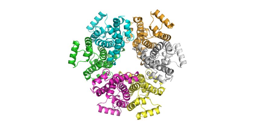
Alkylhydroperoxidase D (AhpD) from Streptococcus pneumoniae

trimeric MtAhpD enzyme and all other hexameric                        Unless otherwise indicated, chemicals were
AhpD structures, which have 3-fold rotational                 purchased from AppliChem, Roche, or Sigma-
symmetry with a central cavity as the axis of                 Aldrich.
symmetry. Although the function of trimeric and
hexameric quaternary structure in other AhpD                  Cloning, protein expression, and purification
proteins remain unclear, the unique in-solution                        The sequences of the genes encoding
structure also indicates that SpAhpD may differ               SpAhpD        (spr0370),      SpAhpC        (GenBank:
functionally from the better-characterized MtAhpD             CMAP01000085.1, locus tag: ERS022390_02365),
protein.                                                      SpAhpF (NZ_CHHM01000086.1), and SpTrx
         In conclusion, our biochemical and                   (GenBank: CJK72264.1) were obtained from the
structural studies of SpAhpD reveal a number of               NCBI GenBank database and codon-optimized for
unique features that indicate the biological role of          expression in Escherichia coli K12 (high) using
this protein is more complex than originally                  EMBOSS Backtranseq (32). Linear DNA was then
anticipated. The cysteine residues of SpAhpD active           synthesized by Thermo Fisher Scientific and cloned
site are redox active, but the structure makes them           into the expression vector pOPINF (a gift from Ray
ineffective at influencing H2O2 levels either directly        Owens; Addgene plasmid #26042), linearized with
or through provision of reducing equivalents to               KpnI/HindIII, using an In-Fusion HD cloning kit
AhpC enzymes. The structure of the unique active              (Clontech) as per the manufacturer’s instructions

                                                                                                                       Downloaded from http://www.jbc.org/ by guest on November 7, 2020
site pocket and its higher reactivity towards CuOOH           (33). Plasmids were then purified from an overnight
indicate that it may be selective towards certain             culture of a single E. coli Stellar transformant using
planar substrates. The additional Cys163 in the active        a miniprep plasmid purification kit (iNtRON
site provides SpAhpD with a degree of redundancy,             Biotechnology) and the DNA sequence was
which may make it less prone to oxidative                     confirmed (Macrogen). Purified plasmid was then
inactivation, a feature beneficial for its function           transformed into E. coli Tuner pLacI cells (Novagen)
within S. pneumoniae. Investigation of the                    for over-expression and purification.
interaction partners of SpAhpD, and assessment of its                  Recombinant protein for subsequent
redox status in vivo will help to illuminate the role of      experiments was expressed by culturing E. coli
this protein in S. pneumoniae. Moreover, the                  Tuner pLacI transformant cells in LB broth to an
difference in activities between MtAhpD and                   OD600 of 0.4–0.6 then inducing with 1 mM IPTG for
SpAhpD highlights that the functions of AhpD                  18 hours at 25°C. The cells were then collected by
enzymes can be species-dependent and need to be               centrifugation and lysed by sonication in HisTrap
examined individually in each species.                        loading buffer (20 mM Tris, 150 mM NaCl, 30 mM
                                                              imidazole, pH 8.0). The crude lysate was clarified by
Experimental procedures                                       centrifuging at 33,000 × g (Thermo Sorvall RC-6-
Bioinformatics                                                plus centrifuge) for 45 min, followed by filtration
         Protein sequences were obtained from the             through a Minisart NML Syringe Filter (0.2-µm pore
NCBI Protein database (accession numbers:                     size).
NP_357964.1, S. pneumoniae AhpD; NP 216945, M.                         The clarified lysate was then loaded onto a
tuberculosis AhpD; CAF21097, C. glutamicum                    5-ml HisTrap High Performance column (GE
AhpD; 2O4D, P. aeruginosa AhpD; BAB77070.1,                   Healthcare) pre-equilibrated with HisTrap loading
Anabaena PCC7120 AhpD; 2GMY, A. tumefaciens                   buffer using an ÄKTA pure protein purification
AhpD; 2OYO, Deinococcus geothermalis AhpD;                    system (GE Healthcare Life Sciences). Bound
2PRR, R. eutropha AhpD; 2OUW, Rhodospirillum                  protein was eluted in a gradient of high imidazole
rubrum AhpD). Multiple sequence alignments were               buffer (20 mM Tris, 150 mM NaCl, 300 mM
carried out using ClustalOmega (29) and converted             imidazole, pH 8.0) over a 50-ml retention volume.
into       graphics       using    ESPript     3.0            Fractions containing protein of interest were
(http://espript.ibcp.fr) (30). Homologues of genes            identified using SDS-PAGE and pooled for
encoding AhpC, AhpF, and thioredoxin in the S.                subsequent purification.
pneumoniae genome were identified using BLAST                          The His-tag was cleaved from recombinant
against non-redundant protein sequences database              SpAhpD using Human Rhinovirus 3C Protease
(31).                                                         (HRV3C; Novagen). The His-tagged HRV3C
                                                              protease and cleaved His-tag were removed by
Materials                                                     HisTrap chromatography (Fig. S9). The pooled

                                                                                                                  8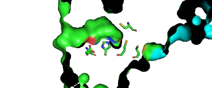
Alkylhydroperoxidase D (AhpD) from Streptococcus pneumoniae

protein was then concentrated to a final volume of 2        reaction mix were mixed with 190 µl of FOX reagent
ml before being purified via size-exclusion                 (250 μM ammonium ferrous sulfate, 125 μM xylenol
chromatography (HiLoad 16/60 Superdex 200; GE               orange, 100 mM sorbitol, and 25 mM sulfuric acid)
LifeSciences) using SEC buffer (20 mM Tris, 150             in a clear-bottom 96-well microplate and incubated
mM NaCl, 3 mM DTT, pH 8.0). Fractions containing            for 20 min at room temperature. The absorbance at
protein of interest were identified using SDS-PAGE,         560 nm was monitored using a microplate reader.
pooled, frozen in liquid nitrogen, and stored at −80°C      Peroxide concentration was calculated using a
until use.                                                  standard curve for each type of peroxide (H2O2,
         Electrospray-ionization      quadrupole-TOF        tBuOOH, and CuOOH).
mass spectrometry was used to confirm the mass of                   To test the peroxidase activity of SpAhpD,
the purified protein. The sequences of all                  10 µM SpAhpD was mixed with 300 µM DTT in
recombinant proteins used in the present study are          TBS at room temperature. The reaction was initiated
shown in Fig. S10.                                          by adding peroxide to a final concentration of 60 µM.
                                                            The concentration of peroxide was determined at
Site-directed mutagenesis                                   various time points up to 2 h, as described above.
        To construct mutant proteins of SpAhpD,
pOPINF construct encoding wild-type SpAhpD was              Ellman’s assay
mutated using the In-Fusion HD cloning kit                           Ellman’s      reagent    (5,5ʹ-dithio-bis-(2-

                                                                                                                      Downloaded from http://www.jbc.org/ by guest on November 7, 2020
(Clontech) following manufacturer’s instructions            nitrobenzoic acid) reacts readily with free thiol to
(TaKaRa) (34). The In-Fusion cloning products after         produce chromophoric 2-nitro-5-thiobenzoate,
mutagenesis were transformed into E. coli Stellar           which absorbs strongly at 412 nm, enabling
competent cells. Plasmids were then purified from an        colourimetric determination of free thiol
overnight culture of a single colony using a miniprep       concentrations. The concentration of free thiol in
plasmid purification kit (iNtRON Biotechnology)             either SpAhpD or SpAhpC was determined using
and the DNA sequence was confirmed (Macrogen).              Ellman’s reagent as per the manufacturer’s
Plasmid containing correct mutation was then                instructions (ThermoFisher Scientific). Briefly,
transformed into E. coli Tuner pLacI cells (Novagen)        Ellman’s reagent (0.08 mg/ml final concentration)
for over-expression and purification, following the         was first dissolved in buffer (0.1 M sodium
same procedures as the wild-type SpAhpD.                    phosphate, 1 mM EDTA, pH 8.0). A 200-µl volume
                                                            of the reagent solution was then mixed with 20 µl of
Preparation of protein redox state                          sample in a flat-bottom microplate and incubated at
         Unless otherwise indicated, all proteins used      room temperature for at least 15 min. The absorbance
in the following activity assays were prepared using        was then measured at 412 nm. Thiol concentration
the following methods to ensure consistent redox            was determined using a standard curve generated
state at the onset of the activity assay. To prepare        using cysteine standards.
reduced protein, freshly-prepared DTT solution was                   SpAhpD and SpAhpC were used at a final
mixed with protein at a final concentration of 3 mM         concentration of ~80 µM. To test their ability to react
and incubated for 1 h at 4°C. To prepare oxidized           with DTT, pre-oxidized protein was mixed with DTT
protein, H2O2 was mixed with protein at a final             at a final concentration of 240 µM. At various time
concentration of 1 mM and incubated at 4°C                  points, aliquots of reaction mixture were removed
overnight. DTT and H2O2 were removed prior to use           and immediately desalted into TBS using Micro Bio-
by desalting the protein into Tris-buffered saline          Spin 6 Columns (Bio-Rad) as per the manufacturer’s
solution (TBS, 20 mM Tris, 150 mM NaCl, pH 8.0              instructions. The free thiol concentration was
at 4°C) using a HiTrap Desalting column with                determined using Ellman’s assay immediately after
Sephadex G-25 resin (GE Healthcare Life Sciences)           desalting. The protein concentration was determined
following manufacturer’s instructions.                      using Bio-Rad Protein Assay Dye Reagent and
                                                            calculated using a standard curve generated using
DTT-dependent ferrous-oxidation xylenol orange              bovine serum albumin. The number of free thiols per
(FOX) assay                                                 molecule was calculated by dividing free thiol
        To test the ability of SpAhpD to reduce H2O2        concentration in µM by protein concentration in µM.
and alkyl hydroperoxides, peroxide concentrations in        To test the reactivity of SpAhpD and SpAhpC with
the reaction mixtures were measured using the FOX           H2O2, pre-reduced proteins were reacted with H2O2
assay, as previously described (14). Briefly, 10 µl of      at a final concentration of 500 µM. Free thiol

                                                                                                                 9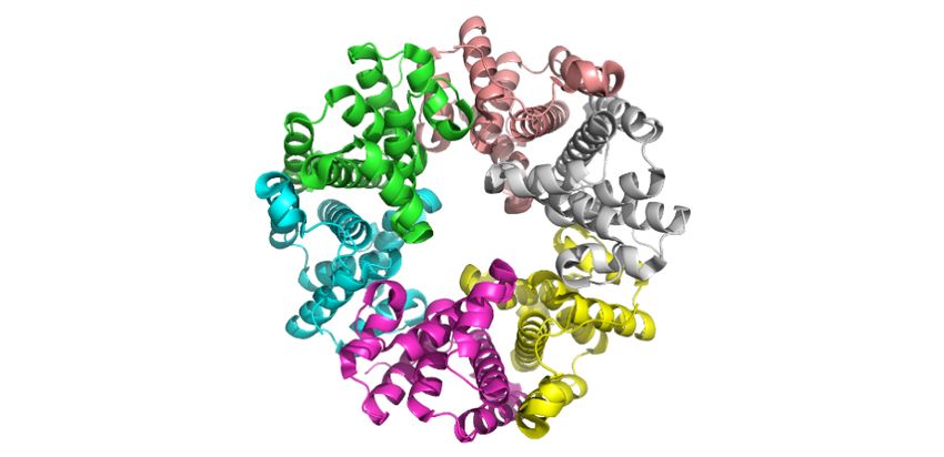
Alkylhydroperoxidase D (AhpD) from Streptococcus pneumoniae

concentration and protein concentration were                initiated using seed stock prepared from an initial
determined at various time points using the same            crystallization hit using Seed Beads (Hampton
method.                                                     Research), as per the manufacturer’s instructions.
                                                            The crystals appeared as large, transparent, triangular
Gel-shift assay using 4-acetamido-                          or quadrilateral plates.
4′maleimidylstilbene-2,2′-disulfonic acid (AMS)
          A gel-shift assay using AMS was used to           X-ray diffraction data collection, phasing, and
determine whether thiol-disulfide exchange occurs           refinement
between SpAhpD and SpAhpC. Labelling of free                         X-ray diffraction data was collected at the
thiol with AMS was carried out as described                 Australian Synchrotron on the micro-crystallography
previously (35). Briefly, 20 mM AMS was first               (MX2) beamline equipped with an EIGAR 1M
dissolved in buffer (50 mM sodium phosphate, pH             detector. The dataset was reduced using AIMLESS
7.0). Derivatization was performed by mixing equal          (36) via CCP4i2 (37). The space group was P212121,
volumes of protein sample (≤ 1 mg/ml) and AMS               with unit cell dimensions of a = 65.296 Å, b = 84.233
solution, followed by incubation at room temperature        Å, and c = 183.846 Å. Although the Matthew’s
for at least 10 min. Three standards were prepared for      coefficient (38) suggests that both five and six
each protein: reduced, oxidized, and native. The            molecules in an asymmetric unit are equally probable
reduced and oxidized standards were prepared by             (0.42 and 0.40, respectively), there were six

                                                                                                                      Downloaded from http://www.jbc.org/ by guest on November 7, 2020
derivatizing either reduced or oxidized protein with        molecules present. The solvent content was 42.12%,
AMS, while the native standard was prepared by              and the VM was 2.12 Å3 Da−1.
derivatizing reduced protein with NEM. The reaction                  The phases were solved by molecular
was initiated by mixing 20 µM oxidized SpAhpC               replacement using PHASER. AhpD from S. mutans
with 80 µM reduced SpAhpD. At various time points,          (3LVY) was used as the search model, and it was
aliquots were removed and mixed with an equal               truncated using CHAINSAW (39) prior to molecular
volume of AMS solution. Samples were then diluted           replacement. REFMAC5 (40) was initially used for
five-fold and analysed by non-reducing SDS-PAGE             rigid body refinement of the molecular replacement
on 4–12% Bis-Tris gels. Positive controls were              model, and was subsequently used in alternating
included for both SpAhpC and SpAhpD to confirm              cycles with Coot (CCP4 program suite) (41), which
that the thiol-disulfide exchange reaction did occur        was used to improve the structure model by
under the assay conditions. The control reactions           restrained refinement.
consisted of 20 µM oxidized SpAhpC or SpAhpD                         Recombinant SpAhpD consists of residues
mixed with 80 µM reduced SpTrx and were analysed            2–182 from the wild-type AhpD sequence, plus
as described above.                                         additional N-terminal Gly-Pro residues from the
                                                            expression vector. Each chain of the refined
NADH-dependent kinetic assay                                structural model contained residues 2–183, 3–183, or
        To test the reductase activity of SpAhpF            4–183 of the recombinant SpAhpD sequence. The
towards SpAhpD or SpAhpC, 2 µM AhpF was mixed               first one to three residues of the N-terminal residues,
with 2 µM SpAhpC and/or 10 µM SpAhpD. The                   which form a random coil, have high temperature
reaction was initiated by adding NADH to a final            factors and low electron density on the 2Fo-Fc map,
concentration of 300 µM. The H2O2 concentration             and were therefore not included in the structural
was determined by FOX assay. The rate of NADH               model.
reduction was monitored by measuring the                             Quaternary structure was predicted using
absorbance at 340 nm.                                       PDBePISA (19). Structure figures were generated
                                                            using PyMOL (The PyMOL Molecular Graphics
Crystallization                                             System, Schrödinger, LLC.).
         Purified AhpD in SEC buffer was
concentrated to 19 mg/ml. Crystals were grown in            Liquid chromatography–mass spectrometry (LC-
sitting drops at 8 °C by mixing 400 nl of protein           MS/MS)
solution with 400 nl of reservoir solution in a TTP                 Initially, the SpAhpD for LC-MS/MS was
LabTech Mosquito Crystal Unit. The optimal                  pre-reduced with 3 mM DTT. The DTT was removed
reservoir contained 0.1 M Bis-Tris propane, 0.2 M           using a HiTrap Desalting column. The desalted
MgCl2, 25% (w/v) PEG 3350, and 6% (v/v) 1,2-                protein solution was divided into five equal aliquots
propanediol, pH 5.5. Optimum crystal growth was             mixed with equal volume of diluted H2O2 at various

                                                                                                                10
Alkylhydroperoxidase D (AhpD) from Streptococcus pneumoniae
concentrations to make final concentrations of 0 – 3 Synchrotron. The protein sample (10 mg/ml) was
mM. The SpAhpD samples were left to incubate with loaded onto an inline Superdex 200 5/150 GL size-
H2O2 at 4°C for 18 hours, before adding NEM to a exclusion column (GE Healthcare), pre-equilibrated
final concentration of 50 mM in order to alkylate and with running buffer (20 mM Tris, 150 mM sodium
stabilize the remaining free thiols. chloride, 0.1% sodium azide, 5% glycerol, pH 8.0) to
Chymotrypsin was added to all samples at a remove any aggregate prior to data collection. The
20:1 substrate:chymotrypsin weight ratio and fractionated sample was pumped through a capillary
incubated at 25°C overnight. Digested samples were where it was exposed to the X-ray beam. The X-ray
analyzed using a Thermo Scientific Velos Pro ion wavelength was 1.0332 Å. A 1-M Pilatus detector
trap mass spectrometer coupled to a Dionex UltiMate was positioned 1600 mm from the sample. A total of
3000 HPLC system with a 50 μl injection loop 500 detector images were collected using a 1-s
(Thermo Scientific, Waltham, MA, USA). Samples exposure time and a flow rate of 0.45 ml/min.
were stored on the autosampler tray at 5°C. A Jupiter SAXS data was analysed using the ATSAS
4 μm Proteo 90Å column (150 x 2 mm, Phenomenex, programme suite (44). Data points from the peak of
Torrance, CA) was used for chromatographic the SEC chromatogram were selected using
separation using 100% water (0.1% formic acid) as CHROMIXS (45). Two-dimensional intensity plots
Solvent A and 100% acetonitrile (0.1% formic acid) were radially-averaged, normalized to sample
as Solvent B. The column temperature was set to transmission, and background-subtracted using the
Downloaded from http://www.jbc.org/ by guest on November 7, 2020
40°C. The column was equilibrated with 95% SCATTERBRAIN software package (Australian
Solvent A and 5% Solvent B for 5 min and then a Synchrotron). PRIMUS QT was used to generate the
linear gradient was run for 45 min to 5% Solvent A Guinier plots and pair-wise distance distribution P(r)
and 95% Solvent B to achieve separation. The plots, as well as for calculating the Porod volumes
column was then flushed with 95% Solvent A and and molecular weight estimates. The theoretical
5% Solvent B for 5 min and re-equilibrated at initial scattering curves based on the crystallographic
conditions for 5 min. A flow rate of 0.2 ml/min was structure were generated and compared with
used and approximately 30 μg of digested protein experimental scattering curves using CRYSOL (46).
was injected for each sample. Nitrogen was used as Scattering profiles of the reduced and oxidized
sheath gas. The temperature of the heated capillary samples were compared using ScÅtter (47).
was 275°C. Data were analysed using Thermo
Xcalibur Qual Browser 2.2 SP1.48 (Thermo Fisher Analytical ultracentrifugation (AUC)
Scientific Inc., Waltham, MA, USA). Analytical ultracentrifugation (AUC) was
The m/z values of peptides of interest were performed using a Beckman Coulter XL-I analytical
predicted and collision-induced dissociation (CID)- ultracentrifuge. The proteins were prepared in buffer
MS/MS spectra in positive-ion mode were acquired containing 20 mM Tris, 150 mM NaCl, 1 mM Tris
for each of them. The collision energy was set at 40. (2-carboxyethyl) phosphine, pH 7.5. The samples
Peptide fragments were manually assigned based on were loaded into Epon two-channel centrepieces
Roepstorff-Fohlman nomenclature (43). A with quartz windows in a four-hole An-60 Ti rotor.
representative fragmentation pattern for each peptide Sedimentation velocity experiments were performed
species is shown in Figure S5. Each peptide species at 55,000 rpm at 20°C using protein at various
was quantified by post-acquisition filtering the concentrations. Sedimentation profiles were
MS/MS spectra obtained for a chosen abundant and measured at 280 nm using absorbance mode with a
characteristic fragment ion (listed in Table S2), and step size of 0.003 cm over 200 scans.
then measuring the area under the curve of the All AUC data were analysed using SEDFIT
resulting peak (peak algorithm: Genesis, peak (48). Buffer density and viscosity, and partial
smoothing: Gaussian 7 points). specific volume of the protein, were calculated using
Small-angle X-ray scattering SEDNTERP (49). The sedimentation data were fitted
Small-angle X-ray scattering was performed to a continuous size-distribution model.
on the SAXS/WAXS beamline at the Australian
11Alkylhydroperoxidase D (AhpD) from Streptococcus pneumoniae
Acknowledgements: The authors would like to thank Prof. Christine Winterbourn and Dr Tamsin Sheen for
critical comments on the manuscript. We acknowledge technical assistance from Michael Currie, Dr Christopher
Horne, James Davies, Jenna Gilkes, Dr Rachel North, Dr Jennifer Crowther, David Coombes, and Michael Love
from Dobson Lab, University of Canterbury, Christchurch, and Dr Heather Parker and Dr Paul Pace from Centre
for Free Radical Research, University of Otago, Christchurch.
Conflict of interest: The authors declare that they have no conflicts of interest.
Data availability: Raw mass spectrometry files available at Figshare
(https://figshare.com/articles/AhpD/11634663).
Downloaded from http://www.jbc.org/ by guest on November 7, 2020
12Alkylhydroperoxidase D (AhpD) from Streptococcus pneumoniae
References
1. Henriques-Normark, B., and Tuomanen, E. I. (2013) The pneumococcus: epidemiology, microbiology,
and pathogenesis. Cold Spring Harb Perspect Med. 3, 1–16
2. Taniai, H., Iida, K. I., Seki, M., Saito, M., Shiota, S., Nakayama, H., and Yoshida, S. I. (2008) Concerted
action of lactate oxidase and pyruvate oxidase in aerobic growth of Streptococcus pneumoniae: Role of
lactate as an energy source. J. Bacteriol. 190, 3572–3579
3. Pericone, C. D., Park, S., Imlay, J. A., and Weiser, J. N. (2003) Factors contributing to hydrogen peroxide
resistance in Streptococcus pneumoniae include pyruvate oxidase (SpxB) and avoidance of the toxic
effects of the Fenton reaction. J. Bacteriol. 185, 6815–6825
4. Rai, P., Parrish, M., Tay, I. J. J., Li, N., Ackerman, S., He, F., Kwang, J., Chow, V. T., and Engelward, B.
P. (2015) Streptococcus pneumoniae secretes hydrogen peroxide leading to DNA damage and apoptosis
in lung cells. Proc. Natl. Acad. Sci. 112, E3421–E3430
5. Perry, F. E., Elson, C. J., Greenham, L. W., and Caterall, J. R. (1993) Interference with the oxidative
response of neutrophils by Streptococcus pneumoniae. Thorax. 48, 364–369
6. Yesilkaya, H., Andisi, V. F., Andrew, P. W., and Bijlsma, J. J. E. E. (2013) Streptococcus pneumoniae
and reactive oxygen species: An unusual approach to living with radicals. Trends Microbiol. 21, 187–195
7. Yesilkaya, H., Kadioglu, A., Gingles, N., Janet, E., Mitchell, T. J., Andrew, P. W., Alexander, J. E., and
Mitchell, T. I. M. J. (2000) Role of manganese-containing superoxide dismutase in oxidative stress and
Downloaded from http://www.jbc.org/ by guest on November 7, 2020
virulence of Streptococcus pneumoniae. Infect. Immun. 68, 2819–2826
8. Le Bras, G., Trombe, M.-C., Le Thomas, I., Dos Santos, D., Chapuy-Regaud, S., Garel, J.-R., Auzat, I.,
Ogunniyi, A. D., and Paton, J. C. (2003) The NADH oxidase of Streptococcus pneumoniae : its
involvement in competence and virulence. Mol. Microbiol. 34, 1018–1028
9. Hajaj, B., Yesilkaya, H., Benisty, R., David, M., Andrew, P. W., and Porat, N. (2012) Thiol peroxidase is
an important component of Streptococcus pneumoniae in oxygenated environments. Infect. Immun. 80,
4333–4343
10. Paterson, G. K., Blue, C. E., and Mitchell, T. J. (2006) An operon in Streptococcus pneumoniae
containing a putative alkylhydroperoxidase D homologue contributes to virulence and the response to
oxidative stress. Microb. Pathog. 40, 152–160
11. Poole, L. B., and Ellis, H. R. (1996) Flavin-dependent alkyl hydroperoxide reductase from Salmonella
typhimurium. 1. Purification and enzymatic activities of overexpressed AhpF and AhpC proteins.
Biochemistry. 35, 56–64
12. Hillas, P. J., Soto Del Alba, F., Oyarzabal, J., Wilks, A., and Ortiz De Montellano, P. R. (2000) The
AhpC and AhpD antioxidant defense system of Mycobacterium tuberculosis. J. Biol. Chem. 275, 18801–
18809
13. Koshkin, A., Knudsen, G. M., and Ortiz De Montellano, P. R. (2004) Intermolecular interactions in the
AhpC/AhpD antioxidant defense system of Mycobacterium tuberculosis. Arch. Biochem. Biophys. 427,
41–47
14. Clarke, T. E., Romanov, V., Chirgadze, Y. N., Klomsiri, C., Kisselman, G., Wu-Brown, J., Poole, L. B.,
Pai, E. F., and Chirgadze, N. Y. (2011) Crystal structure of alkyl hydroperoxidase D like protein PA0269
from Pseudomonas aeruginosa: Homology of the AhpD-like structural family. BMC Struct. Biol. 11, 27
15. Hong, E.-J., Jeong, H., Lee, D.-S., Kim, Y., and Lee, H.-S. (2018) The ahpD gene of Corynebacterium
glutamicum plays an important role in hydrogen peroxide-induced oxidative stress response. J. Biochem.
0, 1–8
16. Shrivastava, A. K., Singh, S., Singh, P. K., Pandey, S., and Rai, L. C. (2015) A novel alkyl
hydroperoxidase (AhpD) of Anabaena PCC7120 confers abiotic stress tolerance in Escherichia coli.
Funct. Integr. Genomics. 15, 77–92
17. Winther, J. R., and Thorpe, C. (2014) Quantification of thiols and disulfides. Biochim Biophys Acta.
1840, 838–846
18. Nunn, C. M., Djordjevic, S., Hillas, P. J., Nishida, C. R., and Ortiz De Montellano, P. R. (2002) The
crystal structure of Mycobacterium tuberculosis alkylhydroperoxidase AhpD, a potential target for
antitubercular drug design. J. Biol. Chem. 277, 20033–20040
19. Krissinel, E., and Henrick, K. (2007) Inference of macromolecular assemblies from crystalline state. J.
13You can also read