Novel anti-repression mechanism of H-NS proteins by a phage protein
←
→
Page content transcription
If your browser does not render page correctly, please read the page content below
10770–10784 Nucleic Acids Research, 2021, Vol. 49, No. 18                                                            Published online 14 September 2021
https://doi.org/10.1093/nar/gkab793

Novel anti-repression mechanism of H-NS proteins by
a phage protein
Fredj Ben Bdira 1,2,* , Amanda M. Erkelens 1,2,† , Liang Qin1,2,† , Alexander N. Volkov                                                          3,4
                                                                                                                                                       ,
Andrew M. Lippa 5 , Nicholas Bowring1,2 , Aimee L. Boyle 1 , Marcellus Ubbink 1 ,
Simon L. Dove 5 and Remus T. Dame 1,2,*
1
 Department of Macromolecular Biochemistry, Leiden Institute of Chemistry, Einsteinweg 55, 2333 CC Leiden, The
Netherlands, 2 Centre for Microbial Cell Biology, Leiden University, Einsteinweg 55, 2333 CC Leiden, The

                                                                                                                                                              Downloaded from https://academic.oup.com/nar/article/49/18/10770/6370256 by guest on 22 October 2021
Netherlands, 3 VIB-VUB Structural Biology Research Center, Pleinlaan 2, 1050 Brussels, Belgium, 4 Jean Jeener
NMR Centre, VUB, Pleinlaan 2, 1050 Brussels, Belgium and 5 Boston Children’s Hospital, Division of Infectious
Diseases, Harvard Medical School, Boston, MA 02115, USA

Received April 07, 2021; Revised August 16, 2021; Editorial Decision August 27, 2021; Accepted September 01, 2021

ABSTRACT                                                                              INTRODUCTION
H-NS family proteins, bacterial xenogeneic silencers,                                 Bacteriophages––viruses that infect bacteria - are among
play central roles in genome organization and in the                                  the most abundant and diverse organisms on earth (1,2)
regulation of foreign genes. It is thought that gene                                  and are found wherever bacteria exist. Bacteria and their
repression is directly dependent on the DNA binding                                   associated bacteriophages co-evolve in a continuous bat-
modes of H-NS family proteins. These proteins form                                    tle, each developing defensive and offensive strategies (3–
                                                                                      5). To protect themselves against bacteriophages, bacte-
lateral protofilaments along DNA. Under specific en-                                  ria have evolved various defense mechanisms, encoded
vironmental conditions they switch to bridging two                                    in their genomes, including restriction-modification (6),
DNA duplexes. This switching is a direct effect of                                    CRISPR-Cas (7), and xenogeneic silencing systems (8).
environmental conditions on electrostatic interac-                                    Bacterial xenogeneic silencers play essential roles in bac-
tions between the oppositely charged DNA binding                                      terial evolution by recognizing and silencing foreign genes
and N-terminal domains of H-NS proteins. The Pseu-                                    acquired through horizontal gene transfer (9,10), result-
domonas lytic phage LUZ24 encodes the protein gp4,                                    ing from transformation, conjugation, or transduction. The
which modulates the DNA binding and function of the                                   silencing of these foreign genes, up to the moment that
H-NS family protein MvaT of Pseudomonas aerugi-                                       their expression is induced following an environmental cue,
nosa. However, the mechanism by which gp4 affects                                     can provide bacteria with a competitive advantage under
MvaT activity remains elusive. In this study, we show                                 specific conditions without compromising global genome
                                                                                      regulation (11).
that gp4 specifically interferes with the formation and                                  Four types of bacterial xenogeneic silencers have been
stability of the bridged MvaT–DNA complex. Struc-                                     identified to date. These proteins belong to the family of
tural investigations suggest that gp4 acts as an ‘elec-                               H-NS proteins, defined by their functional similarity to the
trostatic zipper’ between the oppositely charged do-                                  H-NS protein of Escherichia coli (8,12,13). Members in-
mains of MvaT protomers, and stabilizes a structure                                   clude H-NS of Proteobacteria (14), MvaT and MvaU of
resembling their ‘half-open’ conformation, resulting                                  Pseudomonas species (15), Lsr2 of Actinomycetes (16) and
in relief of gene silencing and adverse effects on P.                                 Rok of Bacillus species (17). The gene silencing mechanism
aeruginosa growth. The ability to control H-NS con-                                   of H-NS family proteins is determined by their ability to
formation and thereby its impact on global gene reg-                                  bind and spread across genes. Characteristic of H-NS fam-
ulation and growth might open new avenues to fight                                    ily proteins is the formation of nucleofilaments along the
Pseudomonas multidrug resistance.                                                     DNA that can switch to mediate DNA–DNA bridges in re-
                                                                                      sponse to environmental changes (18–23). This interchange
                                                                                      between the DNA binding modes of H-NS family proteins
                                                                                      is thought to play an important role in the global regulation

* To
   whom correspondence should be addressed. Tel: +31 71 527 5605; Email: rtdame@chem.leidenuniv.nl
Correspondence may also be addressed to Fredj Ben Bdira. Email: fredjbdira@gmail.com
†
    The authors wish it to be known that, in their opinion, the second and third authors should be regarded as Joint Second Authors.


C The Author(s) 2021. Published by Oxford University Press on behalf of Nucleic Acids Research.
This is an Open Access article distributed under the terms of the Creative Commons Attribution License (http://creativecommons.org/licenses/by/4.0/), which
permits unrestricted reuse, distribution, and reproduction in any medium, provided the original work is properly cited.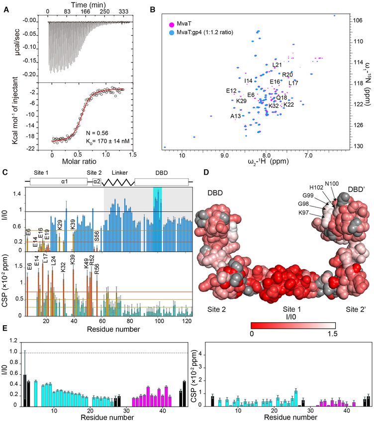
Nucleic Acids Research, 2021, Vol. 49, No. 18 10771
of gene expression and the dynamic organization of bacte- bridging steps (18,19). Therefore, gp4 may alter (i) the DNA
rial genomes (24–27). binding affinity of MvaT, which is essential for protomer
Bacteriophages have evolved resistance mechanisms by binding; (ii) the multimerization properties of MvaT, which
encoding proteins antagonizing H-NS silencing in response determine the cooperativity of nucleoprotein filament for-
to bacterial xenogeneic silencing systems. For instance, the mation or (iii) the ability of MvaT to bridge DNA. In
gp5.5 protein of E. coli phage T7 can counteract H-NS this study, we have scrutinized which of these steps in the
activity in E. coli upon phage infection (28). The gp5.5 bridged complex assembly is affected by gp4. Using bio-
binds to the central oligomerization domain of H-NS and physical and structural biology methods, we defined how
disrupts higher-order H-NS-DNA complexes, leading to gp4 interacts with MvaT and how this translates into al-
counter-silencing of genes controlled by H-NS (29,30). An- tered DNA structuring properties. We propose a molecular
other example is the Arn protein of E. coli phage T4, a mechanism by which gp4 exerts toxicity on P. aeruginosa in
DNA mimic, which has been proposed to counteract H-NS- vivo and may relieve the MvaT-mediated silencing of genes.
mediated repression by targeting its DNA binding domain
Downloaded from https://academic.oup.com/nar/article/49/18/10770/6370256 by guest on 22 October 2021
and thus interfering with DNA binding (31). E. coli phage MATERIALS AND METHODS
T4 also encodes a protein, termed MotB, that plays a role
in improving phage fitness. Its heterologous expression was Construction of plasmids
found to be toxic in E. coli B or K12 strains. It was suggested The plasmids encoding MvaT of P. aeruginosa (pRD228),
that MotB contains a DNA binding domain with which it its derivatives MvaT F36D/M44D (pRD277), MvaT 1–
can interact tightly, yet non-specifically, with DNA to al- 62-His (pRD360) and MvaT 79–124-His (pRD361)) and
ter the expression of specific host genes, including the ones gp4 of Pseudomonas phage LUZ24 (pRD232) were con-
repressed by H-NS (32). Bacteriophage Mu employs yet an- structed using pET30b as vector by Gibson assembly (40).
other mechanism. Binding of IHF to its site upstream of the The cloned sequences were verified by DNA sequencing.
early promoter (Pe) interferes with H-NS-DNA complex
formation and counteracts the H-NS-mediated repression
of this promoter (33,34). The EBPR podovirus 1 genome DNA substrates
encodes a H-NS family protein (gp43), which was pro- All tethered particle motion and bridging assay experiments
posed to repress expression of genes involved in host de- were performed using a random, AT-rich, 685 bp (32% GC)
fense mechanisms such as CRISPR-associated proteins and DNA substrate used in previous research (18,19). The DNA
a Type III restriction-modification system (35). Regrettably, substrate was generated by PCR and the products were pu-
despite their importance, detailed molecular descriptions of rified using a GenElute PCR Clean-up kit (Sigma-Aldrich).
H-NS inhibition by these phage defense mechanisms are If required, DNA was 32 P-labeled as described previously
generally lacking. (19). For the electrophoretic mobility shift assay an AT-rich
The Pseudomonas phage LUZ24 encodes the gp4 pro- 200 bp (32% GC) was generated using the same procedure.
tein that binds the H-NS family protein MvaT (36). MvaT
is thought to play an important role in the genome or-
Protein expression and purification
ganization of P. aeruginosa and in the regulation of gene
transcription (15,37). The fold architecture of the MvaT BL21 (DE3) pLysS cells transformed with pRD228,
monomer consists of an ␣-helical N-terminal oligomeriza- pRD277, pRD360, pRD361 or pRD232 were grown in
tion domain (NTD: residues 1–64) tethered to a C-terminal 2 L of LB supplemented with kanamycin (100 g/mL)
DNA binding domain (DBD) (residues 79–124) by a flexible at 37◦ C to an OD600 of ∼0.6. Protein expression was in-
linker (residues 65–78) (Figure 1A) (18). The structural unit duced with 0.5 mM isopropyl -D-1-thiogalactopyranoside
of MvaT protofilaments is a dimer (protomer) formed by (IPTG) overnight at 18◦ C. Cells were centrifuged at
‘coiled-coil’ interactions between the N-terminal ␣-helices 7000 × g for 15 min at 4◦ C. MvaT and MvaT F36D/M44D
of the MvaT monomers (␣1: residues 1–32), dimerization were purified as described previously (18). MvaT 1–62-His
site 1. The MvaT protomers form high-order oligomers and MvaT 79–124-His were purified with modifications.
through a second dimerization site, site 2 (␣2: residues 50– The harvested cells were lysed by sonication in a lysis buffer
58). Both coiled-coils display a hydrophobic core stabilized containing 20 mM Tris–HCl (pH 8.0), 1 M NaCl. The lysate
by salt bridges (18). was cleared by centrifugation at 37 000 × g for 30 min at
The open reading frame (ORF) of gp4 is located be- 4◦ C. Next, the supernatant was loaded on a HisTrap HP
tween the nucleotides 2189–2329 in the early region of the 5mL column (GE healthcare Life sciences) and the protein
LUZ24 45625bp genome (38). Proteins with ORFs within was eluted by applying an imidazole gradient from 0 to 1
the early genome region (1020–11434) have been classified M. The eluted fractions were checked by SDS-PAGE and
as ‘early proteins’ and have been presumed to be involved the fractions that contain the target protein were pooled,
early in the phage infection cycle (38,39). The heterolo- concentrated and buffer exchanged using a PD10 column
gous expression of gp4 in Pseudomonas causes adverse ef- to 20 mM tris-HCl pH 7, 100 mM KCl. Next, the MvaT 1–
fects on pathogen growth. It was proposed that gp4 inhibits 62-His and MvaT 79–124-His were loaded on a HiTrap Q
the binding of MvaT to DNA to abolish silencing of the HP 5 mL and HiTrap SP HP 5 mL column (GE healthcare
phage LUZ24 genome mediated by MvaT (36). Several pos- Life sciences) respectively and eluted by applying a NaCl
sible mechanisms for gp4 modulation of MvaT function gradient (from 0.1 to 1 M). The eluted fractions were also
can be envisioned based on the MvaT-DNA complex for- checked by SDS-PAGE and the pooled fractions were con-
mation pathway determined by the nucleation-propagation- centrated to a 500 L volume with an Amicon 3 kDa cut-off
10772 Nucleic Acids Research, 2021, Vol. 49, No. 18
Downloaded from https://academic.oup.com/nar/article/49/18/10770/6370256 by guest on 22 October 2021
Figure 1. Effect of LUZ24 gp4 protein on P. aeruginosa growth and on the DNA binding affinity of MvaT. (A) Fold topology of MvaT monomer and
protomer. The upper panel is a schematic representation of MvaT monomer fold organization. In the lower panel is the structural model of the MvaT
protomer adopted from (18). The DBD are colored in yellow, site 1 in magenta, site 2 in cyan and the linker in blue. (B) Heterologous expression of gp4 in
P. aeruginosa wild-type cells and cells lacking either mvaU or mvaT. The left panel shows serial dilutions (×d) of cells growth in the absence of inducer and
the right panel is in the presence of Rhamnose for induction of gp4 expression. (C) Electrophoretic mobility-shift assay (EMSA) of 200 bp DNA substrate
(32% GC) by 2 M of MvaT wild type and MvaT2 in the presence of different concentrations of gp4. (M) is for a 1kb DNA marker and (C) is for the
200 bp DNA substrate without proteins. (D) NMR titration of 15 N MvaT2 :gp4 complex (1:1.2 molar ratio) with a 20 bp DNA substrate. Representative
chemical shift perturbations (CSP) of the MvaT2 DBD amides are shown and the corresponded amino acid residues are depicted on the MvaT2 structure
in (A). Black arrows indicate the directions of the CSP.
filter. The concentrated protein fractions were loaded on a tube. The beads were prepared by changing the solution
GE Superdex 75 10/300 GL column and eluted with 20 mM to binding buffer (100 mM NaCl, 10 mM Tris–HCl pH
Tris-HCl pH 8, 300 mM KCl, 10% glycerol. For the purifi- 7.2, 50 mM imidazole, 20 mM MgCl2 ). The nickel beads
cation of gp4, the harvested cells were lysed by sonication in were then centrifuged at 7000 rpm for 2 min and the su-
a lysis buffer containing 20 mM Tris–HCl (pH 7.0), 100 mM pernatant was removed. 250 L of the desired combination
NaCl. Next, the supernatant was loaded on a HiTrap SP HP of MvaT 79–124-His (40 M), His-MvaT 1–62 (40 M)
5 mL column (GE healthcare Life sciences) and the protein and/or gp4 (40 M), in binding buffer, was added to the
was eluted by applying a NaCl gradient from 0.1 to 1 M. prepared nickel beads. The reaction mixture was incubated
The eluted fractions were checked by SDS-PAGE and the for 30 minutes while being shaken at 1000 rpm. After incu-
fractions that contain the target protein were pooled, con- bation the samples were centrifuged at 3000 rpm for 2 min.
centrated and loaded into a GE Superdex 75 10/300 GL The supernatant was removed, and the pellet was carefully
column and eluted with 20 mM Tris–HCl pH 8, 300 mM resuspended in 250 L of binding buffer. This step was re-
KCl, 10% Glycerol. The purity of the protein was checked ferred to as a washing step and was repeated a total of six
by SDS-PAGE and the concentration was determined using times. Following the final washing step, 250 L of elution
a Pierce BCA protein assay kit (Thermo Scientific). buffer (100 mM NaCl, 10 mM Tris–HCl pH 7.2, 1.5 M im-
idazole, 20 mM MgCl2 ) was added to the beads instead of
binding buffer to elute any protein bound to the bead. The
His-tag pull-down assay samples were centrifuged at 3000 rpm for 2 min and the su-
Typically, 120 L of the High-Density Nickel Agarose (Jena pernatant was collected. 10 L of the supernatant was run
Bioscience) suspension was pipetted into an Eppendorf on a tricine gel.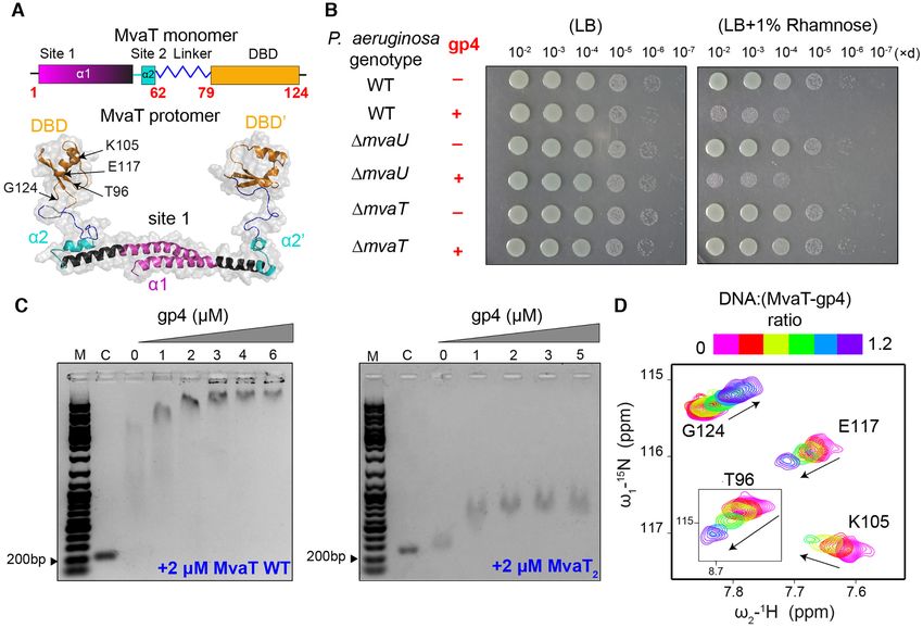
Nucleic Acids Research, 2021, Vol. 49, No. 18 10773
Tethered particle motion L of radioactive DNA was used as a reference to calculate
the bridging efficiency (DNA recovery %).
Tethered Particle Motion experiments were performed as
described previously (18,41) and the flow cells were pre-
pared with minor modifications. In short, the flow cell was Heterologous expression of LUZ24 gp4 in Pseudomonas
washed with 100 mL of wash buffer (10 mM Tris–HCl pH aeruginosa
8, 150 mM NaCl, 1 mM EDTA, 1 mM DTT, 3% glycerol,
100 g/ml BSA (ac)) and experimental buffer (10 mM Tris Wild-type P. aeurigonsa PAO1 was obtained from Arne Ri-
pH 8, 50 mM KCl, 10 mM EDTA, 5% glycerol). The flow etsch, Case Western Reserve University. The strain harbor-
cell was sealed after adding protein (MvaT alone or MvaT- ing a deletions of mvaU was previously described (42). The
gp4 mixture) in experimental buffer, followed by 10 minutes mvaT deletion strain was constructed using the pEXG2-
of incubation. The measurements were initiated 15 min, af- based plasmid pEXG2M4315 by allelic exchange (43). The
ter introducing protein. For each flow cell >200 beads were deletion retains the start codon for mvaT and replaces the
measured at a temperature of 25˚C. Two separate flow cells remaining coding sequence with DNA encoding three ala-
Downloaded from https://academic.oup.com/nar/article/49/18/10770/6370256 by guest on 22 October 2021
were measured for each concentration. nines followed by a stop codon. Rhamnose-inducible ex-
pression of gp4 was achieved by integration of the pJM220-
based plasmid pAL63 in single copy at the Tn7 attach-
Electrophoretic mobility shift assay ment site of PAO1, the mvaU strain, or the mvaT strain,
The assay was performed on an AT-rich 200 bp DNA sub- as previously described (44) followed by removal of the
strate (32% GC). The DNA substrate (25 ng) was incubated aac1 casette using pFLP2 (45). pAL63 includes the cod-
with different concentrations of the desired combination of ing sequence of gp4 inserted between the SpeI and HindIII
MvaT, MvaT F36D/M44D and gp4 in the binding buffer sites of pJM220 via isothermal assembly. Freshly streaked
(10 mM Tris–HCl, 60 mM KCl, pH 8.0). The mixture was colonies of each strain grown on lysogeny broth (LB) agar
loaded on a 1% agarose gel (containing 1:104 dilution of plates were resuspended in PBS, OD600 was determined us-
Gel red DNA stain) with loading buffer (10 mM Tris–HCl, ing a NanoDrop ND1000 spectrophotometer, normalized
pH 7.6, 0.03% bromophenol blue, 0.03% xylene cyanol FF, to OD600 = 0.01 (designated dilution 10–1 ) and serially di-
60% glycerol and 60 mM EDTA). The samples were run at luted 10-fold in PBS. 10 L of culture for each strain and
4◦ C in TAE standard buffer. The gels were visualized with dilution were plated to LB or LB supplemented with L-
a Bio-rad Gel Doc XR+ system. rhamnose (Sigma-Aldrich) to a final concentration of 1%.
Plates were incubated at 27◦ C for 18 h and photographed
under white light illumination using a Nikon D3400 cam-
Isothermal titration calorimetry
era.
Isothermal Titration Calorimetry experiments were per-
formed as described previously (18) with modifications.
Briefly, ITC experiments were performed using a MicroCal NMR titration
VP-ITC system at 20◦ C. The protein samples were dialyzed The NMR titration of the MvaT2 with gp4 was performed
to a buffer containing 20 mM Bis–Tris, pH 6, 50 mM KCl, on a 150 M (of MvaT monomer) 15 N isotopically labelled
16 mM MgCl2 . Typically, 20 M of MvaT F36D/M44D protein sample, in 20 mM Bis–Tris Buffer, 50 mM KCl,
was placed in the cell (1.4 mL) and titrated with 300 M 16 mM MgCl2 , pH 6 and 6% D2 O. A series of 1 H–15 N
(500 l) of the gp4, injected in 2 L aliquots. The delay HSQC spectra were acquired by gradually increasing the
time between the injections was 60 s with a stirring speed MvaT:gp4 molar ratio up to 1:1.2. The experiments were
of 307 rpm. The corresponding ‘protein to buffer’ controls recorded at 20 ◦ C on a Bruker Avance III (HD) 600 MHz
were performed for background correction. The ITC titra- spectrometer, equipped with TCI cryoprobe, processed by
tion data were analyzed using Origin 7.0 (OriginLab) pro- TopSpin 3.5 (Bruker Biospin) and analyzed by Sparky soft-
vided with the instrument. One set of sites model (1:1 inter- ware (46). The same experimental procedure was applied
action) was used to fit the data where N (number of bind- for the titration MvaT-gp4 (1:1.2 molar ratio) with 20 bp
ing sites), K (association constant) and H (delta enthalpy) of DNA. The DNA:MvaT-gp4 molar ratio was increased
were set free during fitting. from 0.2 to 1.2. The changes in peak positions and intensi-
ties were analyzed and the average chemical shift perturba-
Bridging assay tions (CSP) were calculated using equation (1):
The bridging assay was performed as described previously
δN 2
(18,41) with modifications. To start the bridging reaction, δ avg = δH 2 + (1)
MvaT was added, followed by 2 L of gp4 to different fi- 6.252
nal concentrations. The samples were incubated while shak-
ing at 1000 rpm at 25 ˚C. The reactions were stopped and
NMR solution structure of LUZ24 gp4
washed at certain time with buffer (10 mM Tris–HCl, pH 8,
65 mM KCl, 5% glycerol, 1 mg/ml BSA (ac), 1 mM spermi- All NMR experiments were performed at 293 K on a
dine, 20 mM CaCl2 , 0.02% Tween 20), followed by resuspen- Bruker Avance III HD 800 MHz spectrometer equipped
sion in 12 L denaturing buffer (10 mM tris pH 8, 200 mM with a TCI cryoprobe. The sample contained 1 mM U-[13 C,
15
NaCl, 1 mM EDTA, 0.2% SDS). Scintillation counting was N] labeled gp4 in 20 mM Bis–Tris pH 6.0, 150 mM
used to determine the final radioactivity of each sample. 2 KCl, 1 mM EDTA and 6% D2 O for the lock. All NMR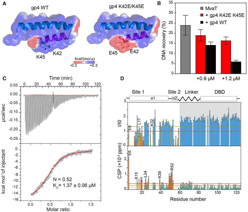
10774 Nucleic Acids Research, 2021, Vol. 49, No. 18
data were processed in TopSpin 3.6 (Bruker) or NMR- TFA:TIPS:H2O, (95:2.5:2.5) mixture. The protein was sub-
Pipe (47) and analyzed in CCPN (48). Nearly complete, sequently precipitated into ice-cold diethyl ether, collected
unambiguous 1 H, 13 C and 15 N resonance assignments of by centrifugation and freeze-dried. Purification was per-
the protein nuclei were obtained from a suite of standard formed by high-pressure liquid chromatography (HPLC)
multidimensional NMR experiments: 2D [1 H,15 N]-HSQC, using a Shimadzu system comprising two KC-20AR pumps
[1 H,13 C]-HSQC and constant-time [1 H,13 C]-HSQC and an SPD-20A UV-Vis detector, fitted with a Kinetix Evo
for the aromatic region; triple-resonance HNCACB, C18 column. The protein was eluted from the column using
HN(CO)CACB, HNCO, HN(CA)CO, HBHA(CO)NH, a linear gradient of 20–80% buffer B (A = H2 O with 0.1%
(H)CCH-TOCSY and H(C)CH-TOCSY experiments; 2D TFA, B = MeCN with 0.1% TFA) over 20 min. The purified
(HB)CB(CGCD)HD and (HB)CB(CGCDCE)HE spectra protein was freeze-dried and TFA residues were removed by
for the aromatic resonances; and 3D 15 N-edited NOESY- dialysis before the protein was utilized. TFA removal was
HSQC and 13 C-edited NOESY-HSQC for aliphatics and monitored by 19F NMR.
aromatics. The resonance assignments were deposited in
Downloaded from https://academic.oup.com/nar/article/49/18/10770/6370256 by guest on 22 October 2021
the BMRB data bank under the accession number 28112.
The 3D 15 N-edited NOESY-HSQC and 13 C-edited RESULTS
NOESY-HSQC spectra for aliphatics and aromatics, all ac- LUZ24 gp4 is selectively targeting MvaT in vivo to modulate
quired with the mixing time of 120 ms, were subsequently its DNA binding modes
used for the protein structure calculation. The NOE cross-
peaks, determined with CCPN Analysis (48), were com- Effect of gp4 on Pseudomonas growth. Previous studies
bined with the dihedral angle restraints, obtained with have suggested that the LUZ24 gp4 protein negatively af-
DANGLE (49), and used as an input for the automated fects Pseudomonas growth by targeting its H-NS family pro-
NOE assignment and structure calculations in CYANA v.3 tein, MvaT (36). To validate these findings, we first heterol-
(50), followed by the explicit solvent and torsion angle re- ogously expressed gp4 in a wild type strain of P. aeruginosa
finement in CNS (51) and Xplor-NIH (52), respectively. The (see M&M). Induction of gp4 expression by 1% rhamnose
10 lowest-energy structures were retained and deposited in caused severe inhibition of bacterial growth (Figure 1B).
the PDB bank under the accession code 6YSE. The NMR However, the expression of gp4 in cells lacking MvaT ab-
structure calculation and refinement statistics are presented rogated that toxicity. MvaU is an MvaT paralog that binds
in Supplementary Table S1. to many of the same genomic regions of P. aeruginosa and
coordinately regulates the expression of ≈350 target genes
(15,37). However, gp4 remained toxic in cells lacking MvaU.
Modelling of MvaT2 –gp4 complex These data show that gp4 is toxic to cells of P. aeruginosa in
Prediction of the MvaT2 –gp4 dimer complex was per- a manner that is dependent upon MvaT but not MvaU.
formed by HADDOCK2.2 server (53) using default setting
parameters. The data from the NMR titration of the MvaT2 Effect of gp4 on MvaT DNA binding affinity. The mech-
with gp4 was used to restrain the docking. The homology anism underlying gp4 activity was earlier proposed to be
model of MvaT dimer (18) was used. Only the amino acids inhibition of MvaT binding to DNA, based on the results
of site 1 of the MvaT2 were classified as active in the docking from an electrophoretic mobility shift assay (EMSA) (36).
settings. As most of gp4 amino acid residues appear to be To verify the reported results, we also performed an EMSA
involved in the complex formation, the full protein amino using a 200 bp DNA (32% GC) substrate. In our hands, the
acid sequence was classified as active. Amino acid residues addition of gp4 from 1 to 6 M to the DNA in the presence
indirectly involved in complex formation (passive) were au- of 2 M MvaT, does not inhibit the formation of MvaT–
tomatically identified by the server. HADDOCK clustered DNA complexes (Figure 1C, Supplementary Figure S1A).
138 structures in 16 clusters, which represents 69.0% of the The reason for the discrepancy in the EMSA results is un-
water-refined models HADDOCK generated. The statistics clear.
of the top cluster are shown in Supplementary Table S2. The To further validate our observations, we also exam-
top cluster is the most reliable according to HADDOCK. ined the effect of gp4 on the DNA binding of MvaT
The reported scores and energies are averages calculated F36D/M44D (MvaT2 ), a mutant that exists exclusively as
over the top four members of a cluster. dimer and does not oligomerize further (18). Similar to
MvaT WT, the DNA binding of MvaT2 was not inhibited
by adding increasing amounts of gp4, as concluded from
Synthesis of gp4 K42E/K45E
the EMSA results (Figure 1C, Supplementary Figure S1A).
The gp4 K42E/K45E, gp4 helix 1 (1–30) and gp4 he- Previously, the 1 H–15 N HSQC NMR spectrum of MvaT2
lix 2 (30–46) were synthesized via solid-phase peptide was assigned and used to study the protein interactions with
synthesis (SPPS) using a microwave-assisted Liberty Blue a 20 bp DNA substrate at low and high ionic strength (18).
peptide synthesizer. A Wang resin, preloaded with the Here we performed a NMR titration of a 15 N-MvaT2 sam-
C-terminal lysine residue was utilized as the solid sup- ple in the presence of gp4 (MvaT:gp4 1:1.2 ratio, see be-
port. Standard Fmoc-chemistry protocols were employed, low) with a 20 bp DNA substrate. The titration data show
with Fmoc-deprotection achieved using 20% piperidine in progressive chemical shift perturbations (CSP) of the DNA
DMF, and coupling reactions facilitated by DIC/Oxyma binding domain (DBD) resonances (Figure 1D, A), similar
as the activator/activator base. After synthesis, the pro- to the previous results obtained from the titration of MvaT2
tein was cleaved manually from the solid support using a with DNA in the absence of gp4 (18). Based on these results,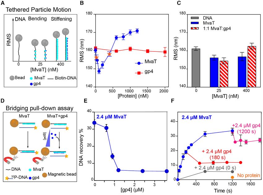
Nucleic Acids Research, 2021, Vol. 49, No. 18 10775
we conclude that gp4 does not inhibit the binding of MvaT tion at t = ∼600 s. Upon addition of gp4 at t = 180 s (pre-
to the DNA substrate. saturation level), and 1200 s (at saturation level), a reduc-
tion in DNA recovery by about 10% is observed. The loss
Effect of gp4 on MvaT DNA stiffening activity. Oligomer- in DNA recovery is maximized by adding gp4 at time zero
ization of MvaT is required for the formation of nucleopro- (∼5%). These data demonstrate that gp4 can perturb, yet
tein filaments along DNA and is essential for the function not completely disrupt, preassembled MvaT–DNA bridges.
of MvaT in gene silencing (20,54). To investigate the effect Collectively, we have shown that gp4 specifically targets
of gp4 on the formation of MvaT nucleofilaments, we used MvaT in Pseudomonas to selectively inhibit its DNA bridg-
Tethered Particle Motion (TPM) (18,19). In the absence of ing activity. This process is neither caused by inhibition of
gp4, DNA compaction (indicated by a reduction in the root the DNA binding capacity of MvaT nor by the obliteration
mean square displacement (RMS)) was observed at a low of its oligomerization in solution and along DNA.
concentration of MvaT (25 nM). This effect was attributed
to DNA bending by individually bound MvaT dimers (nu-
Downloaded from https://academic.oup.com/nar/article/49/18/10770/6370256 by guest on 22 October 2021
Structural characterization of LUZ24 gp4
cleation step) (18). Upon increasing the amount of MvaT,
stiffening of the DNA substrate is observed, and saturation The LUZ24 gp4 is a 46 amino acid peptide with no re-
is reached at 1200 nM MvaT (Figure 2A, B). ported tertiary structure. The secondary structure of the gp4
gp4 alone does not alter the RMS, attributed to lack of polypeptide chain is predicted to consist of two ␣-helices: a
DNA binding (Figure 2B), in line with the EMSA results long N-terminal (␣1: residues 4–26) and a short C-terminal
(Supplementary Figure S1A). To test whether gp4 affects (␣2: residues 31–45) helix, connected by a loop of four
the formation of the MvaT–DNA filament, TPM was per- amino acids (Figure 3A). In agreement with these predic-
formed with fixed MvaT concentrations of 25 and 400 nM, tions, gp4 exhibits a far-UV CD spectrum with two min-
in the presence of equivalent concentrations of gp4. At 25 ima at approximately 208 and 222 nm, typical of ␣-helical
nM MvaT, addition of gp4 does not significantly reduce the secondary structures (Figure 3B). The ratio between the in-
RMS of the MvaT-DNA complex (Figure 2A,C). This in- tensities of the 222 and 208 nm bands (I222 /I208 ) is equal to
dicates that gp4 exhibits negligible effect on the bending 1.13, suggesting that the peptide is assuming a coiled-coil
capacity of the MvaT protomers. At 400 nM MvaT, the structure (56). In contrast to the narrow dispersion of reso-
MvaT–DNA nucleofilament is not abolished by gp4 (Fig- nances, often observed in the 1 H–15 N HSQC spectra of ␣-
ure 2A, C). On the contrary, the increase in RMS indicates helices (centred at ∼8 ppm), gp4 has a well-dispersed HSQC
that addition of gp4 leads to a more extended MvaT–DNA spectrum (Figure 3C). This characteristic of the NMR spec-
nucleofilament. Dynamic light scattering experiment also trum reflects a non-homogeneous chemical environment of
suggests that gp4 does not inhibit the multimerization of the backbone amide nuclei, possibly due to the coiled-coil
unbound MvaT in solution (Supplementary Figure S1B). fold of the peptide.
Based on these data, we conclude that gp4 does not inter- We determined the solution structure of gp4 using NMR
fere with the oligomerization of MvaT in solution and along spectroscopy, and the bundle of the ten best structures is
DNA, and thus does not inhibit its stiffening activity. shown in Figure 3D. The structure shows that gp4 indeed
adopts an intramolecular antiparallel coiled-coil topology.
Effect of gp4 on MvaT DNA bridging activity. In conjunc- The interhelical interface is formed by a hydrophobic core
tion with DNA stiffening, MvaT can bridge two DNA du- between residues M14, L18, L21 and L25 of ␣1 and residues
plexes under specific buffer conditions (18,19,55). To de- W33, Q37 L40 and L44 of ␣2. The hydrophobic core is sta-
termine whether gp4 affects the ability of MvaT to bridge bilized by a salt bridge between residues R17 (␣1) and E36
DNA, we used a protein-DNA pull-down bridging assay (␣2) (Figure 2E) and two hydrogen bonds between the side
(see Materials and Methods, Figure 2D) (19,41). MvaT was chain atoms T22-O␥ 1 and Q36-NHε 22 and between R11-
found to bridge DNA in the presence of divalent (Mg2+ , NH1 and the carboxyl group at the C-terminus. The ␣2 is
Ca2+ ) or monovalent (K+ , Na+ ) cations, in a concentration- tilted from the ␣1 helix plane, forming an interhelical angle
dependent manner (18). Here we performed the experi- of 30◦ (Figure 3D).
ments in the presence of 16 mM MgCl2 , a condition at gp4 is a basic peptide (predicted pI = 10.5) with a
which MvaT nucleofilaments exhibit optimal DNA bridg- predominantly positive electrostatic surface. The positive
ing activity. A fixed amount of MvaT monomer (2.4 M) is charges are more clustered on the surface of ␣2, compris-
used, yielding a 35% recovery of DNA in the absence of gp4. ing six basic amino acids with the side chains exposed to
The DNA recovery decreased upon addition of gp4. Max- the solvent (Figure 3F).
imum inhibition of DNA recovery was achieved at a gp4
concentration of 1.2 M, where the MvaT:gp4 molar ratio
Structural study of MvaT–gp4 complex
is 2:1 (Figure 2E). These results demonstrate that gp4 se-
lectively inhibits the formation of MvaT-DNA bridges, and To uncover the molecular basis of gp4 inhibitory func-
does not perturb MvaT–DNA filament formation. tion on MvaT DNA bridging activity, we first investigated
Next, to examine whether gp4 affects preassembled the biophysical properties of the MvaT–gp4 complex. It
MvaT-DNA bridges, we performed a time-dependent DNA was previously suggested that gp4 interacts with the MvaT
bridging assay at a 1:1 molar ratio of MvaT:gp4 (Figure 2F). oligomerization domain and linker region (residues 1–80)
As a control, we measured the time dependence of DNA (36). To substantiate this proposal, we first studied the in-
recovery by MvaT in the absence of gp4. Under this condi- teraction of gp4 with the truncated NTD (residues 1–62)
tion, DNA recovery increases over time and reaches satura- and the DBD (residues 79–124) of MvaT using a magnetic10776 Nucleic Acids Research, 2021, Vol. 49, No. 18
Downloaded from https://academic.oup.com/nar/article/49/18/10770/6370256 by guest on 22 October 2021
Figure 2. Effect of LUZ24 gp4 protein on the DNA binding modes of MvaT. (A) Schematic representations of the Tethered Particle Motion (TPM) assay
in the absence and presence of gp4. (B) The blue curve represents the RMS of DNA bound by MvaT at concentrations from 0 to 1200 nM measured by
the TPM in the absence of gp4.The data points of the MvaT concentrations of 25 and 400 nM are highlighted in orange stars. The red curve is for the
RMS of DNA titrated by increased concentrations of gp4 between 0 and 2000 nM, in the absence of MvaT. The error bars represent the standard deviation
propagated from two independent measurements (each including ∼100 individual DNA tethers); some error bars are hidden behind the data points. (C)
Effects of gp4 on MvaT DNA stiffening activity. The error bars are for the standard deviation from duplicate experiments. (D) Schematic representations
of the DNA bridging assay in the absence and presence of gp4. (E) Effects of gp4 on MvaT DNA bridging activity. (F) Time dependent bridging assay
of DNA–MvaT–DNA complexes. The blue curve represents the kinetics of MvaT-DNA bridge formation without adding gp4. The red and pink curve
represent the kinetics of MvaT–DNA bridge formation with gp4 introduced at 180 and 1200 s, respectively. The grey curve represents the introduction
of gp4 at the beginning of the assay (t = 0 s). Error bars represent the standard deviation from at least two independent measurements. The assays were
performed in the presence of 2.4 M MvaT and 2.4 M gp4 (1:1 molar ratio). The orange point represents a control measurement without proteins at
1200 s of incubation.
bead pull-down assay (See M&M). The assay shows that pothesized that gp4 mainly interacts with site 1, to satisfy
gp4 binds to the NTD but not to the truncated DBD (Sup- the stoichiometry of the complex.
plementary Figure S2A). These results also suggest that the To corroborate this hypothesis, we performed a NMR
linker region (65–80) may not be required for the formation titration of a 15 N labeled MvaT2 sample with unlabeled gp4,
of the MvaT-gp4 complex. under the same experimental conditions as used in the ITC
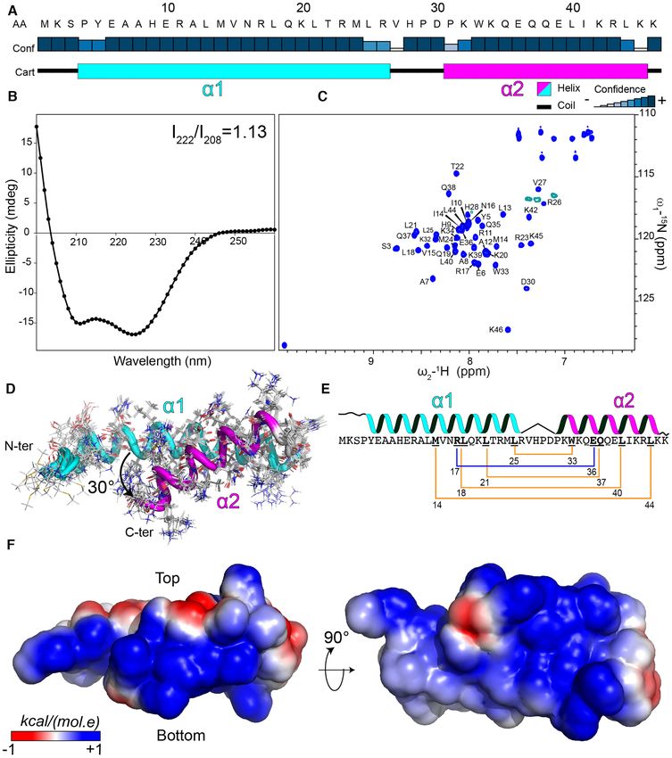
An isothermal titration calorimetry (ITC) experiment experiment. The addition of gp4 to MvaT2 induced a de-
was conducted to determine the stoichiometry and the dis- crease in the intensity of multiple resonances in the protein
sociation constant (KD ) of the MvaT2 -gp4 complex. The spectrum with minor changes in chemical shift positions of
fit of the titration data yields a KD of 170 ± 14 nM with the dimerization site1 residues (Figure 4B,C). At a molar
a number of binding sites N = 0.56 (Figure 4A, see M&M). ratio of MvaT:gp4 of 1:1.2, the ratio between the intensi-
These results indicate a tight binding between the two pro- ties of the peaks of the free MvaT2 (I0) and MvaT2 bound
teins with a stoichiometry of 2:1 (MvaT:gp4). The complex to gp4 (I) shows that the amide resonances of the dimeriza-
appears to be formed by the interaction of one molecule of tion site 1 (residues 1–22) are the most affected upon com-
gp4 with two molecules of MvaT. As the MvaT variant used plex formation (Figure 4C,D). These observations indicate
(F36D/M44D) can only form a dimer through the interac- that gp4 binds to the coiled-coil region of the MvaT2 dimer-
tion between the two N-terminal ␣-helices (site1), we hy- ization site 1.Nucleic Acids Research, 2021, Vol. 49, No. 18 10777
Downloaded from https://academic.oup.com/nar/article/49/18/10770/6370256 by guest on 22 October 2021
Figure 3. Structural characterization of LUZ24 gp4. (A) Analysis of gp4 secondary structure by JPred 4 server (57). The blue rectangles indicate the
confidence of the prediction. The predicted N-terminal helix (␣1) is shown in a cyan rectangle and the C-terminal helix (␣2) is in magenta. (B) Far UV-CD
spectrum of gp4 recorded at 5 M protein concentration in 10 mM phosphate buffer, pH 7. The ratio between the 222 nm and 208 nm bands is indicated.
(C) Assignment of the 1 H–15 N HSQC spectrum of gp4. (D) Bundle of the best 10 structures of gp4 is shown in cartoon and the amino acids side chains
in lines. The ␣−helices are colored as in (A). The angle between gp4 helices is indicated. (E) Schematic representation of the intramolecular interaction
within the gp4 coiled-coil fold. The hydrophobic interactions between the side chains of the amino acid residues (bold underlined) are shown with a yellow
line and the salt bridge in blue. (F) The electrostatic surface of gp4 is shown, generated by CHARMM-GUI (58).10778 Nucleic Acids Research, 2021, Vol. 49, No. 18
Downloaded from https://academic.oup.com/nar/article/49/18/10770/6370256 by guest on 22 October 2021
Figure 4. gp4 interacts with the N-terminal and DBD domains of MvaT. (A) ITC titration of gp4 (230 M) into MvaT2 (monomer concentration = 16
M) using 2 l of injection volume. The red line represents the best fit using a one set of sites model (see M&M). The fitting yields a KD = 170 ± 14
nM and N = 0.56. (B) The overlay between 15 N–1 H TROSY spectra of the MvaT2 in the free state (magenta) and at a molar ratio of MvaT:gp4 of 1:1.2
(blue). Amide resonances which experience severe line broadening upon titration are labelled. (C) The upper panel depicts the 15 N–1 H TROSY spectra
peak intensities ratio of MvaT in the presence of 1.2 molar ratio of gp4 (I) and the free state (I0) versus the protein residue number. Lower panel shows
weighted average CSP of the MvaT resonances between the same points of titration. Resonances with CSP more than two (orange line) or one (yellow line)
standard deviation (SD) from the 10% trimmed mean (green dashed line) are labelled and shown in orange, yellow and green bars, respectively. The blue
and green bars are for resonances with a reduction in peak intensities and CSP higher and less than the 10% trimmed mean, respectively. The grey shaded
region highlights the changes in the peak intensities of the DBD-linker resonances of MvaT. Amide resonances of the DBD which exhibit increase in peak
intensities are highlighted with a cyan rectangle and indicated on the structural model of MvaT2 in (D). (D) Mapping of the residues with a reduction in
their peak intensities (red/white gradient), upon addition of gp4, on the surface of the MvaT2 structural model (18). Residues with no data are colored
in grey spheres. (E) Analysis of the NMR titration of 15 N-gp4 with unlabeled MvaT2 . The left panel represents the change in the peak intensities of the
amide resonances (I/I0) at a 15 N-gp4:MvaT 1.2:1 molar ratio. The right panel shows weighted average CSP of the gp4 resonances between the free and
bound states. Cyan bars are for amides of gp4 ␣1, magenta bars are for ␣2 amides and black bars are for residues of the loop regions.Nucleic Acids Research, 2021, Vol. 49, No. 18 10779
A line broadening is also observed for several amide res- of gp4 in multiple orientations, similar to an encounter com-
onances of the DBD (Figure 4C). This suggests a simul- plex stabilized by electrostatic interactions. The general line
taneous interaction of gp4 with the DBD and the NTD broadening of the resonances of gp4 upon complex forma-
within MvaT2 . These interactions are possibly too weak tion is partly due to the increase in the rotational correlation
to be probed by the pull-down assay when the truncated time and may also be caused by the same effect as observed
DBD is used. Indeed, NMR titration of the 15 N gp4 with for MvaT2 . It is clear that the NMR data provide evidence
the DBD does not show significant changes in the peptide for complex formation and also help to indicate which re-
HSQC spectrum even at a molar ratio of gp4:DBD of 1:3 gion on MvaT2 is involved in the interactions with gp4.
(Supplementary Figure S2B).
Within MvaT2 , the DBD is tethered to the N-terminal
Modelling of gp4 inhibition mechanism of MvaT DNA bridg-
helices by the linker, which increases its local population
ing activity
near the NTD. Therefore, the tethering by the linker might
promote the occurrence of intermolecular interactions be- Diffractive crystals of gp4 in complex with the NTD of
Downloaded from https://academic.oup.com/nar/article/49/18/10770/6370256 by guest on 22 October 2021
tween the DBD and gp4 when occupying dimerization site 1 MvaT wild type (residues 1–62) could not be obtained, pos-
of MvaT2 . The DBD of MvaT includes negatively charged sibly because of the high flexibility of the system (see above).
residues on its surface, which may be required for direct- Therefore, we used the NMR titration data as experimen-
ing the formation of a specific complex with DNA through tal restraints to generate structural models of the MvaT2 -
molecular frustration (18,59). The observed decrease in gp4 complex with HADDOCK (53). In our modelling, we
peak intensities includes these negatively charged patches, only considered the binding of gp4 to the MvaT2 NTD
which suggests that they interact with the positively exposed (residues 1–22), as it appears to represent the primary inter-
surface of gp4. action site between the two proteins (see M&M). Multiple
Previously, we found that at low ionic strength, MvaT2 structures of the MvaT2 –gp4 dimer complex were obtained
adopts a ‘half-open’ state in which intramolecular elec- (Supplementary Figure S3A). The lowest energy structure
trostatic interactions between the DBD-linker and NTD of the complex is shown in Figure 5A. The complex forms
(site 1) take place. The interface between the two do- a trimeric coiled-coil topology, in which the N-terminal ␣1
mains includes the positively charged DBD loop 95- of gp4 is intercalated between the two N-terminal helices
ETKGGNHK-102 and the negatively charged fragment of MvaT2 site 1. The ␣2 helix of gp4 exhibits interactions
26-QDDKLKKELEDEE-38 of the NTD (18). These in- with only one of the MvaT2 coiled-coil helices (Supplemen-
tramolecular interactions cause a localized line broadening tary Figure S3B). The intermolecular interface includes 187
of the amide resonances of the DBD loop and for residues non-bonded contacts mainly formed by electrostatic and
of the linker region. An increase in ionic strength destabi- hydrophobic interactions between the interfaces of the two
lizes the electrostatic interactions between the two domains proteins. Four salt-bridges stabilize the trimeric coiled-coil
and induces an increase in the intensities of the peaks of located between residues R23, K20, H28 and D36 of ␣1 and
these MvaT regions (18). Similar to the effects of high ionic loop region of gp4 with E6, E35 and R20 of site 1 helices.
strength, an increase in the peak intensities of the loop at Residues D30 and R43 of gp4 ␣2 form two additional salt
95–102 and linker resonances is observed upon titration bridges with K31 and E16 of one of the site 1 helices to fur-
with gp4 (Figure 4C,D). This suggests that the binding of ther stabilize the complex (Supplementary Figure S3B).
gp4 to MvaT2 site 1 induces a change in the intramolecu- Our in vitro assays show that in the presence of gp4, the
lar electrostatic interactions and the conformational orien- MvaT oligomers are not able to interact with a second DNA
tation of the DBD-linker relative to the NTD. duplex to form a bridge (Figure 2E). This situation resem-
The resonances of the DBD exhibit no significant chem- bles the low ionic strength condition, in which the MvaT
ical shift perturbations (CSP) despite the apparent interac- oligomers can form filaments yet not bridge DNA. This
tions with gp4 (Figure 4C). This observation can possibly be condition is attributed to the sequestration of one of the
explained by the different binding modes of the DBD to the DBD’s within the MvaT protomers by electrostatic inter-
solvent-exposed surface of gp4. The DBD can also adopt actions with the NTD. An increase in ionic strength desta-
different spatial positions, relative to the NTD, within the bilizes these interdomain interactions and shifts the con-
conformational ensemble of the MvaT2 (open and closed) formational equilibrium of the MvaT protomer towards an
(18). The average effect of these different chemical environ- ‘open state’, able to bridge two DNA duplexes (Figure 5B)
ments may result in small CSP for the DBD resonances. (18). Although our bridging assay was performed at high
The titration of 15 N labeled gp4 with unlabeled MvaT2 ionic strength (50 mM KCl, 16 mM MgCl2 ), MvaT remains
also shows no significant CSP of the amide resonances of unable to bridge two DNA duplexes in the presence of gp4
gp4 in conjunction with a general line broadening (Figure (see M&M).
4E, Supplementary Figure S2C). Given the high affinity of Our structural studies suggest that the primary bind-
gp4 and MvaT2 , the strong reduction in the intensities in ing site of gp4 is on the dimerization site 1 of MvaT pro-
the resonances in the NTD of MvaT2 upon complex for- tomers (Figure 4C, D & Figure 5A). Upon complex for-
mation, in combination with very small CSP, suggests that mation, a structural rearrangement of the MvaT protomer
binding is in the slow-intermediate regime on the NMR seems to occur, in which one of the DBD’s interacts with
timescale. In the very slow regime new resonances would be the surface of gp4 that is not involved in the interface with
expected to appear for the bound state. However, these are MvaT2 site 1 (Figure 5C). These interactions appear to
not observed, which would be in line with line-broadening be mediated through electrostatic forces. As a result, gp4
caused by slow-intermediate exchange and perhaps binding will act as an ‘electostatic zipper’ between the NTD and10780 Nucleic Acids Research, 2021, Vol. 49, No. 18
Downloaded from https://academic.oup.com/nar/article/49/18/10770/6370256 by guest on 22 October 2021
Figure 5. gp4-MvaT complex and inhibition mechanism of gp4. (A) The best model of the complex between gp4 and MvaT2 NTD generated by HAD-
DOCK, using the NMR titration data as restraints, is shown. The electrostatic surfaces of gp4 (left) and MvaT2 (right) were generated by CHARMM-GUI
(58). (B) The switching mechanism of a MvaT nucleofilament between DNA stiffening and bridging under the influence of salt (18). (C) Schematic rep-
resentation of the gp4 anti-bridging mechanism. Note that upon interaction of the gp4 with site 1, one of the DBD’s of MvaT2 becomes restrained by
electrostatic interactions with gp4. The DBD might bind to gp4 on different sites. The electrostatic surfaces of MvaT2 and gp4 are shown schematically in
a red (negative)/white (neutral)/blue (positive) color gradient. Green and red dots are for ions (Mg2+ ) and counter ions (Cl– ), respectively.
DBD of the MvaT protomers and stabilize a structure re- their DNA bridging capacity. The anti-bridging activity of
sembling the ‘half-open’ conformational state within the gp4 K42E/K45E was monitored by the pull-down bridg-
protein oligomers (Figure 5C). Consequently, the MvaT– ing using gp4 WT as a reference. Indeed, a 50–30% loss
gp4 nucleofilaments will exhibit weak DNA bridging ac- of gp4 inhibitory function is observed at 0.9 and 1.2 M
tivity, despite forming high-order oligomers along double- gp4 respectively, added to 2.4 M MvaT (Figure 6B). How-
stranded DNA. ever, measurement at higher concentrations of gp4 were im-
possible because of proteins aggregation on the magnetic
Validation of the molecular mechanism of MvaT interference beads. Titration of MvaT with different concentrations of
by gp4 the gp4 C-terminal ␣2 (residues 30–46) did not affect the
DNA bridging activity of the protein (Supplementary Fig-
To validate the proposed molecular model for the mecha- ure S1C). The stoichiometry of the complex between gp4
nism of MvaT inhibition by gp4, we generated a gp4 vari- K42/K45E-MvaT2 appears equal to 0.52, similar to that
ant in which the solvent-exposed residues K42 and K45 obtained for the MvaT2 titration with gp4 WT, as deter-
of the ␣2 helix are substituted with glutamic acids (Fig- mined by ITC (Figure 6C). However, a tenfold increase in
ure 6A). These charge inversion mutations were aimed in- the KD value is observed (1.36 ± 0.06 M), possibly due
troducing repulsive electrostatic forces between gp4 and to loss of some intermolecular interactions within the com-
the DBD to shift the conformational equilibrium of the plex. To verify this hypothesis, we performed a NMR titra-
MvaT protomers toward an ‘open state’, thus restoring tion of 15 N labelled MvaT2 with the mutated gp4. The gp4Nucleic Acids Research, 2021, Vol. 49, No. 18 10781
Downloaded from https://academic.oup.com/nar/article/49/18/10770/6370256 by guest on 22 October 2021
Figure 6. Validation of gp4 anti-bridging mechanism by point mutagenesis. (A) electrostatic surface of gp4 WT and gp4 K42E/K45 mutant. (B) Inhibitory
effects of gp4 mutant (red bars) and gp4 WT (black bars) on MvaT DNA bridging activity at 0.9 and 1.2 M of gp4 using a 2.4 M concentration of
MvaT (grey bars). (C) ITC of MvaT2 with gp4 K42E/K45E. The red line shows the best fit of the data. (D) Analysis of the NMR titration data of 15 N
MvaT2 with gp4 K42E/K45E. The upper panel depicts the 15 N–1 H TROSY spectra peak intensities ratio of MvaT2 in the presence of 1.2 molar ratio of
gp4 (I) and the free state (I0) versus the protein residue number. Note the two-fold increase in the peak intensities of the DBD-linker region (shaded in
grey) due to the loss of the intramolecular electrostatic interactions between the DBD-linker -NTD within MvaT2 subunits (18) and the intermolecular
interactions with gp4. Lower panel shows weighted average CSP of the MvaT resonances between the same points of titration. Resonances with CSP more
than two (orange line) or one (yellow line) standard deviation (SD) from the 10% trimmed mean (green dashed line) are labelled and shown in orange,
yellow and green bars, respectively. The blue and green bars are for resonances with a reduction in peaks intensities and CSP higher or less than the 10%
trimmed mean, respectively.
mutant binds to the dimerization site 1 as concluded from DISCUSSION
the observed CSP and the reduction in the ratio of peak in-
Bacterial species and their associated bacteriophages are in
tensities (Figure 6D). In contrast to the titration with WT
a relentless evolutionary arms race in which they develop
gp4, no line broadening is observed for the DBD-linker res-
defense and counter-defense mechanisms to assure survival.
onances. These results suggest a loss of the intermolecular
One of the mechanisms that phages deploy is the production
interaction between gp4 K42E/K45E and the DBD-linker
of an infantry of proteins to shut down host defense mecha-
of the MvaT2 . Collectively, these data support our model in
nisms and co-opt host-cell machinery for efficient infection
which gp4 is acting as an electrostatic stabilizer of a struc-
(60,61). Despite representing a rich source of antimicrobial
ture resembling the ‘half-open’ state of the MvaT protomers
molecules, the structure, target, and mode of action of these
for the selective inhibition of their DNA bridging activity.
proteins have remained vastly unexplored.10782 Nucleic Acids Research, 2021, Vol. 49, No. 18
One of the first line of Enteric bacteria resistance sys- NS complex is to permit the phage growth on lambda lyso-
tems is the silencing of foreign (viral) DNA by H-NS fam- gens, although it was suggested that the mechanism might
ily proteins. However, bacteriophages encode proteins that be more complex (29). Indeed, another study has shown
counteract this host-defense system. In this work, we have that upon T7 infection, gp5.5 uses tRNA to sequester H-
investigated the molecular mechanism by which the phage NS from inhibiting DNA replication of E. coli (30).
LUZ24 protein gp4 functionally modulates the P. aerugi- Unlike the T7 phage, the LUZ24 phage has an AT-rich
nosa H-NS family protein MvaT to markedly inhibit the (52% GC) genome, prone to repression by the xenogeneic si-
growth of this important pathogen. Using (single-molecule) lencer of Pseudomonas, MvaT (38,39). Based on our molec-
biophysical methods, we found that gp4 specifically targets ular studies, we propose that the primary role of gp4 is to
the formation and maintenance of bridged DNA complexes relieve the repression of phage genes by MvaT through dis-
by MvaT without interfering with MvaT oligomerization rupting DNA bridges. Nevertheless, it remains unknown
along DNA. This situation resembles the low ionic strength whether gp4 is essential for LUZ24 development and how
conditions under which the MvaT protomers adopt a ‘half- its molecular mechanism is influenced by the environmental
Downloaded from https://academic.oup.com/nar/article/49/18/10770/6370256 by guest on 22 October 2021
open’ conformational state, incompetent to bridge two and physiological conditions of the host.
DNA duplexes while forming lateral filaments along DNA. DNA bridging by H-NS proteins is thought to be crucial
This structural configuration results from intramolecular for gene silencing and for the global organization of Enteric
electrostatic interactions between the oppositely charged bacteria genomes (67–69). Additionally, molecular studies
DBD and dimerization site 1 of the MvaT protomers (18). have suggested that a switch between linear and bridging nu-
Structural studies revealed that gp4 simultaneously inter- cleofilaments of H-NS family proteins drives the bacterial
acts with dimerization site 1 and the DBD of the MvaT pro- adaptation to environmental factors (18,19). The selective
tomers to stabilize a mimic of the ‘half-open’ state through inhibition of MvaT DNA bridging activity by gp4, which
electrostatic interactions. Introduction of repulsive electro- translates into an adverse impact on growth of P. aeruginosa
static forces at the interface of gp4 and the DBD of the provides additional evidence of the importance of DNA
MvaT dimer results in diminished anti-bridging activity of bridging by H-NS family proteins in maintaining bacterial
gp4. homeostasis. P. aeruginosa also encodes MvaU, a paralog of
Bacteria encode several proteins that modulate the struc- MvaT that binds to many of the same chromosomal regions
ture and function of H-NS–DNA complex. For instance, and also bridges DNA duplexes (15,70). Nevertheless, de-
truncated H-NS (H-NST) derivatives, which lack the C- spite the structural and functional similarity between these
terminal DNA binding domain, were found in large ge- two proteins, gp4 appears to target MvaT selectively and
nomic islands of pathogenic E. coli strains (62). It was sug- requires its presence for cytotoxicity. Future studies on the
gested that H-NST family members act as antagonists of impact of gp4 on nucleoid organization and regulation of
H-NS function by preventing its correct oligomerization gene expression in P. aeruginosa might provide more insight
through heterodimerization with the N-terminal dimeriza- into the antimicrobial mechanism of this phage protein.
tion domain of H-NS. The expression of YdgT stimulates In conclusion, a better understanding of how viral pro-
DNA bridging by enhancing the cooperativity of the lat- teins counteract H-NS proteins is essential for designing
eral nucleofilament formation of H-NS (19). Binding of molecules that modulate phage infections. Potential appli-
Hha/YdgT family proteins to the H-NS dimerization site cations of such molecules can be envisioned in biotechnol-
(residues 1–45) stimulates H-NS DNA bridging activity ogy, where phage infections are a threat to large scale cultur-
(19), and thus enhances transcriptional silencing of foreign ing, or in phage therapy where they may enhance the ability
genes in bacteria (63). Hha forms electrostatic complexes to infect and kill their host. Moreover, due to their global
with the ‘hand-shake’ fold of H-NS dimerization site 1 with role in gene regulation in bacteria, H-NS family proteins
a 1:1 stoichiometry (Hha: H-NS monomer) and exposes a are potential drug targets to mitigate recalcitrant bacterial
positively charged surface (64,65). It was proposed that the infections. A mechanistic understanding at the molecular
exposed surface of Hha enhances the binding affinity of H- level of how H-NS family protein activity is modulated will
NS filament to DNA and stabilizes the bridging complex to prove key to such developments.
promote pausing of RNA polymerase (66). The LUZ24 gp4
seems to operate analogously to Hha regarding the electro-
static interactions with dimerization site 1 of H-NS family
DATA AVAILABILITY
proteins and the exposure of a positively charged surface.
Nevertheless, the stoichiometry, topology and orientation The datasets generated during the current study are avail-
of the complex appear to differ because of the deviations in able from the 4TU repository (https://data.4tu.nl) under
the fold architectures of MvaT and H-NS site 1 (coiled-coil DOI: https://doi.org/10.4121/15097611. The resonance as-
vs. hand-shake) and of gp4 and Hha, which result in oppo- signments of gp4 were deposited in the BMRB data bank
site effects on the bridging activity of the two H-NS family under the accession number 28112. The NMR structures of
members. gp4 were deposited in the PDB bank under the accession
Previous studies have demonstrated that the gp5.5 of code 6YSE.
phage T7 interacts with oligomerization site 2 of H-NS from
E. coli to alter its high order oligomeric state without in-
terfering with DNA binding affinity (28,29). The T7 phage
SUPPLEMENTARY DATA
has a low AT-rich genome, a requirement to be silenced by
H-NS. Thus, it was proposed that the role of the gp5.5-H- Supplementary Data are available at NAR Online.You can also read