THE GLUTAMINE TRANSPORTER SLC38A1 REGULATES GABAERGIC NEUROTRANSMISSION AND SYNAPTIC PLASTICITY - MEDUNI WIEN EPUB
←
→
Page content transcription
If your browser does not render page correctly, please read the page content below
Cerebral Cortex, December 2019; 5166–5179

                                                                             doi: 10.1093/cercor/bhz 055
                                                                             Advance Access Publication Date: 3 May 2019
                                                                             Original Article

ORIGINAL ARTICLE

The Glutamine Transporter Slc38a1 Regulates

                                                                                                                                                         Downloaded from https://academic.oup.com/cercor/article/29/12/5166/5485254 by guest on 29 January 2021
GABAergic Neurotransmission and Synaptic Plasticity
Tayyaba Qureshi1, Christina Sørensen2,3, Paul Berghuis4, Vidar Jensen1,
Marton B. Dobszay4, Tamás Farkas4, Knut Tomas Dalen5, Caiying Guo6,
Bjørnar Hassel7, Tor Paaske Utheim8,9, Øivind Hvalby1, Torkel Hafting1,
Tibor Harkany4,10, Marianne Fyhn2 and Farrukh Abbas Chaudhry1,9
1
 Department of Molecular Medicine, University of Oslo (UiO), Oslo, Norway, 2Department of Biosciences, UiO,
Oslo, Norway, 3Department of Neuroscience, University of Copenhagen, Copenhagen, Denmark, 4Department
of Neuroscience, Karolinska Institutet, Stockholm, Sweden, 5Department of Nutrition, UiO, Oslo, Norway,
6
 Janelia Research Campus, Ashburn, Virginia 20147, USA, 7Department of Neurohabilitation, Oslo University
Hospital (OUH) and UiO, Norway, 8Department of Medical Biochemistry, OUH, Norway, 9Department of Plastic
and Reconstructive Surgery, OUS and UiO, Norway and 10Department of Molecular Neurosciences, Center for
Brain Research, Medical University of Vienna, Austria
Address correspondence to email: f.a.chaudhry@medisin.uio.no

Øivind Hvalby passed away 23rd May 2015

Abstract
GABA signaling sustains fundamental brain functions, from nervous system development to the synchronization of
population activity and synaptic plasticity. Despite these pivotal features, molecular determinants underscoring the rapid
and cell-autonomous replenishment of the vesicular neurotransmitter GABA and its impact on synaptic plasticity remain
elusive. Here, we show that genetic disruption of the glutamine transporter Slc38a1 in mice hampers GABA synthesis,
modifies synaptic vesicle morphology in GABAergic presynapses and impairs critical period plasticity. We demonstrate that
Slc38a1-mediated glutamine transport regulates vesicular GABA content, induces high-frequency membrane oscillations
and shapes cortical processing and plasticity. Taken together, this work shows that Slc38a1 is not merely a transporter
accumulating glutamine for metabolic purposes, but a key component regulating several neuronal functions.

Key words: GABA, neurotransmitter replenishment, SAT1, Slc38, SNAT1

Introduction                                                                 excitation by dendritic inhibition (Klausberger and Somogyi
GABA is the principal inhibitory neurotransmitter in the central             2008; Huang 2009; Hu et al. 2014). GABA signaling can be either
nervous system (CNS) with manifold functions: it regulates                   short-lived and phasic when GABA is released in quanta to act at
action potential (AP) firing and network synchrony through                    synaptic GABAA receptors or long-lasting and tonic upon ambient
perisomatic inhibition and the efficacy and plasticity of                     extracellular GABA stimulating extra-synaptic receptors (Isaacson

© The Author(s) 2019. Published by Oxford University Press.
This is an Open Access article distributed under the terms of the Creative Commons Attribution Non-Commercial License (http://creativecommons.org/
licenses/by-nc/4.0/), which permits non-commercial re-use, distribution, and reproduction in any medium, provided the original work is properly cited.
For commercial re-use, please contact journals.permissions@oup.com
Slc38a1 in GABAergic Transmission and Plasticity Qureshi et al. | 5167
and Scanziani 2011). In addition, GABA affects developmentally (Slc38) (Nissen-Meyer and Chaudhry 2013): system N transporters
coded cortical critical period plasticity by modulating interneuron Slc38a3 (SN1/SNAT3) and Slc38a5 (SN2/SNAT5) reside on astroglial
migration, placement and synaptic wiring (Ben-Ari et al. 2007). membranes and work bi-directionally to supply neurons with glu-
Indeed, at the systems level, GABA signaling underpins learning, tamine (Chaudhry et al. 1999; Hamdani et al. 2012). Heterologous
memory, cognition and sensory perception (Buzsaki et al. 2007). expression of the homologous system A transporter (SAT) Slc38a1
The life-long competence of GABA signaling relies on efficient (SAT1/SNAT1/SA2) in cultured mammalian cells shows transport
local means for neurotransmitter reuptake, replenishment and of amino acids with a preference for glutamine (Varoqui et al.
release. Considering the prominence of dysfunctional GABA sig- 2000; Chaudhry et al. 2002). We have shown that Slc38a1 is
naling in brain disorders, such as epilepsy, autism, schizophrenia enriched in GABAergic neurons and based on this localization pro-
and anxiety (Soghomonian and Martin 1998; Lewis et al. 2012), it posed that Slc38a1 could be involved in the replenishment of the
is surprising that molecular determinants rate-limiting precursor neurotransmitter GABA (Solbu et al. 2010). However, this has been
availability for metabolic replenishment and vesicular filling and contested by a number of papers reporting that Slc38a1 occurs
their impact on inhibitory synaptic plasticity remain elusive. indiscriminately in glutamatergic, GABAergic, cholinergic and
To describe precursor replenishment, the “glutamate/GABA- dopaminergic neurons and targeted primarily to their somatoden-
Downloaded from https://academic.oup.com/cercor/article/29/12/5166/5485254 by guest on 29 January 2021
glutamine (GGG) cycle” was proposed decades ago, which suggests dritic compartments implicating a broader role in general cellular
that GABA (and glutamate) upon transport into perisynaptic astro- metabolism (Mackenzie et al. 2003; Conti and Melone 2006). In
glia is first converted into glutamine, which is then transported addition, the functional significance of glutamine in GABA replen-
back into neurons to regenerate GABA as neurotransmitter (Reubi ishment and the existence of a GGG cycle remain ambiguous since
et al. 1978; Nissen-Meyer and Chaudhry 2013). This is supported by some studies have shown unchanged neurotransmission upon
elucidation of the unconventional kinetics combined with the cell- pharmacological inhibition of system A transporters, inactivation
specific localization of a family of amino acid (AA) transporters of phosphate-activated glutaminase (PAG) and/or removal of
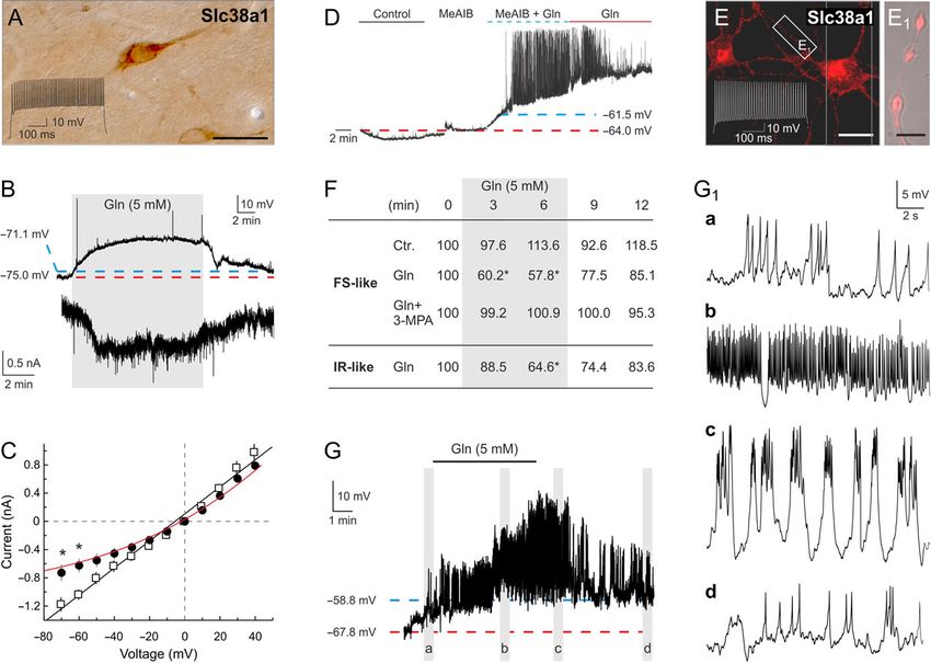
Figure 1. Genetic inactivation of Slc38a1 using the binary Cre/LoxP system and its validation. (A) Schematic drawing of the targeting vector, wild-type allele (Slc38a1wt
(Slc38a1+)), and targeted alleles (Slc38a1flox-Neo, Slc38a1flox, and Slc38a1null (Slc38a1-)). The Slc38a1 targeting vector contained a LoxP site (inserted into intron 4; white
triangles), and a selection cassette (Neomycin) flanked by FRT-sites (black triangles) and a LoxP site (inserted into intron 8). After homologous recombination in ES
cells (Slc38a1flox-Neo locus), ES cells were transected with Flp to excise the selection cassette (Slc38a1flox locus) prior to blastocyst injection. Upon expression of Cre
recombinase, exons 5–8 were excised (Slc38a1 null), generating out of reading frame splicing transcript. The binding site for the 5-end Southern screening probe and
the expected fragment sizes after KpnI digest are indicated. (B) Genotyping of ear biopsies of Slc38a1+/+, Slc38a1+/- (heterozygous) and Slc38a1−/− mice by 3 pairs of
probes gives the expected amplified fragments (see Supplementary Experimental Procedures). (C) Southern blot analysis of DNA isolated from liver of Slc38a1+/+,
Slc38a1+/−, and Slc38a1−/− mice. DNA was digested with KpnI followed by hybridization with the 5-probe to give the expected fragments for Slc38a1WT (20.2 kb) and
Slc38a1Null (15.7 kb). (D) Expression of Slc38a1 protein in brain lysates of Slc38a1+/+, Slc38a1+/−, and Slc38a1−/− mice was investigated. No protein expression is detected
in lysates from Slc38a1−/− mice, while Slc38a1+/- mice show reduced staining for Slc38a1 protein compared to Slc38a1+/+ mice. β-actin was used as loading control. E)
Expression of Slc38a1 protein was investigated by immunostaining of free-floating brain sections for Slc38a1. In the hippocampus, Slc38a1 immunoreactivity accumu-
lates in scattered interneuron-like cells in the CA1 and dentate area of Slc38a1+/+ mice. Such staining is abolished in sections from Slc38a1−/− mice. G, granulare; LM,
lacunosum-moleculare; M, moleculare; P, pyramidale; R, Radiatum. Insets: P, pyramidal cells; I, interneuron-like cell. Scale: 100 μm.5168 | Cerebral Cortex, 2019, Vol. 29, No. 12
external glutamine (Masson et al. 2006; Kam and Nicoll 2007). A new set of primers were designed for PCR genotyping. The
Thus, conclusive experimental evidence for the function of Slc38a1 template DNA was obtained by digesting an ear piece using the
in vivo and its impact on inhibitory synaptic plasticity, and the REDExtract-N-Amp™ Tissue PCR Kit (Sigma) followed by PCR-
molecular determinants of GABA replenishment and GABAergic amplification of 1 μl of the template in a 10 μl PCR reaction. The
vesicular load are lacking, and more broadly, for the existence of a reaction was carried out for 35 cycles (94 °C 30 s, 60 °C 30 s and
GGG cycle. 72 °C 30 s) followed by one cycle of 72 °C for 10 min.
Here, we have genetically inactivated Slc38a1 in mice and
characterized their phenotype at successive levels of cellular
and network complexity in vitro and in vivo. We demonstrate Primers Used for Generation of a Floxed Slc38a1 Mice
that Slc38a1 sustains replenishment of GABA, impacts vesicle and for Investigating Their Genotypes
morphology and neurotransmitter content, triggers AP genera-
The homologous arms were 2945 bp (5′) and 5440 bp (3′). Primer
tion and regulates inhibitory synaptic plasticity in vivo.
sequences for ES screening were: 5′ arm forward primers: Slc38a1
Scr F1 (5′- cgtgttctccgtcagctatt-3′) and Slc38a1 Scr F2 (5′- atgtccgg-
Downloaded from https://academic.oup.com/cercor/article/29/12/5166/5485254 by guest on 29 January 2021
Materials and Methods cagagtctttga-3′). Reverse primers: LoxP Scr R1 (5′-gagggacctaa-
taacttcgt-3′) and LoxP Scr R2 (5′-ggaattgggctgcaggaatt-3′). 3′ arm
Animal Handling forward primers: FRT Scr F1 (5′-ttctgaggcggaaagaacca-3′) and FRT
Experiments were approved and conducted in accordance with Scr F2 (5′-cgaagttattaggtggatcc-3′); Reverse primers: Slc38a1 Scr R1
the Norwegian Animal Welfare Act and the European (5′-agtgatggtaaccgtcctgt-3′) and Slc38a1 Scr R2 (5′-ctactggccaggaa-
Convention for the Protection of Vertebrate Animals used for caacgat-3′).
Experimental and Other Scientific Purposes (ETS 123). The mice A new set of primers were designed for PCR genotyping. The
were kept and handled under veterinary supervision at the primer combination Scl38a1-Scr1: (5′-tgtaggtctgttcccatgttgtcct-3′)
UiO. Mice were housed in a temperature controlled facility and Scl38a1-Scr2: (5′-gatgaaatgttccccggagtctaac-3′) generates a PCR
(22–26 °C) with 50 ± 10% humidity and a 12-hours light/dark product of 426 bp for the wild type allele, and 514 bp for the floxed
cycle. Animals were served municipal water and fed ad libitum allele. The primer set Scl38a1-scr3 (5′-tctcccactaagttgcgttgtcttc-3′)
with RM3 (E) from Special Diets Services (UK). and Scl38a1-scr4(5′-ccaaatggatgacttggagattgtc-3′) generates a PCR
Discharge/frequency experiments on animals were per- product of 372 bp for the wild type allele and 471 bp for the floxed
formed at Karolinska Institutet and conformed to the European allele. Detection of the null allele after Cre excision was detected
Communities Council Directive (86/609/EEC) and were approved using the primer combination Scl38a1-Scr1 and Scl38a1-Scr4 giving
by the Stockholms Norra Djurförsöksetiska Nämnd (N26/2005 a PCR product of 616 bp for the null allele and a fragment too large
and N38/2005). to be amplified for the wild type allele.
Generation of a floxed Slc38a1 Mouse Southern Blotting
The floxed Slc38a1 targeting construct was produced (Fig. 1A) The probe sequence was radiolabelled with [a-32P]dCTP (Perkin
using recombineering techniques (Liu et al. 2003). A 12 904 bp Elmer, Wellesley, MA) using Megaprime DNA labelling System
genomic DNA fragment containing exons 5–12 of the Slc38a1 (Amersham Biosciences) prior to hybridization of the mem-
gene was retrieved from BAC clone RP23-85D13. A LoxP brane to verify homologous recombination of the loci in the
sequence was inserted in intron 4 and a FRT-PGKNeo-FRT-LoxP animals.
cassette was inserted in intron 8. Thus, a fragment of 4504 bp
genomic DNA containing exons 5 through 8 was floxed. Cre
excision would generate an out-of-frame deletion. The target- Synaptosomes, Amino Acid Analysis, and Quantitative
ing vector was electroporated into D1 embryonic stem (ES) cells Western Blotting
which were derived from F1 hybrid blastocyst of 129S6 × C57BL/
Synaptosomes were made by homogenization of the brains in ice
6 J. G418 resistant ES colonies were isolated and screened by
cold 0.32 M sucrose 5% (w/v) followed by several steps of ultracen-
nested polymerase chain reaction (PCR) using primers outside
trifugation, as described (Bogen et al. 2006). Synaptosomal amino
the construct paired with primers inside the neo cassette.
acids were separated and quantified by HPLC and fluorescence
Fourteen clones PCR positive for both arms were identified
detection after pre-column derivatization with o-phthaldialdehyde
from 48 clones screened. Chimeric mice were generated by
(Hassel and Brathe 2000). Western blot analyses were performed
aggregating the ES cells with 8-cell embryos of CD-1 strain. The
as described (Nissen-Meyer et al. 2011). For details, see SI.
neo cassette was removed by mating the chimeras with 129S4/
SvJaeSor-Gt(ROSA)26Sortm1(FLP1)Dym/J (Stock number: 003946)
homozygous females. The F1 pups were genotyped by PCR
Light- and Electron Microscopic Cytochemistry
using primers flanking LoxP or FRT-LoxP sites. The mice were
then crossed with Deleter mice to create a systemic deletion of Ten pairs of age- and sex-matched Slc38a1+/+ and Slc38a1−/−
Slc38a1 (Schwenk et al. 1995). As Slc38a1 expression is mice were deeply anesthetized with intra-peritoneal injection
restricted to some few organs (Chaudhry et al. 2002), secondary of Zoletil-mix 125 mg/kg. Five of the pairs were fixed by trans-
effects on the CNS phenotype is not very likely. Null mutant cardiac perfusion of 4% paraformaldehyde (PFA) for immune-
(Slc38a1−/−) mice did not carry any obvious phenotypic abnor- peroxidase staining and 5 pairs were perfusion fixed with 4%
malities. We had several breedings (het x het, ko x ko, wt x wt) PFA and 2.5% glutaraldehyde (GA), hippocampal CA1 regions
and all were backcrossed between 5 and 10 times. In addition, dissected out, embedded in Durcupan (FLUKA), sectioned
the mice were crossed with wt mice regularly in order to avoid (~100 nm), immunogold labeled and analyzed by Electron
inbreeding of the mice in order to reduce the risk of differences microscopy, as described (SI; Jenstad et al. 2009; Solbu et al.
in the genetic background. 2010).Slc38a1 in GABAergic Transmission and Plasticity Qureshi et al. | 5169
Antibodies monitor centered 25 cm in front of the animal. Four days of MD
was started between postnatal day 25 (P25) and P27. Eyelids
Antibodies were generated against the most divergent and
were sutured shut under isoflurane anesthesia.
antigenic regions of the members of the Slc38 family of amino
acid/glutamine transporters, Slc17 family of vesicular gluta-
mate transporters and Slc32 family of vesicular GABA trans- Statistical Analysis
porter by subcloning these sequences C-terminal to the All values are presented as mean ± SEM. All data were analyzed
sequence for glutathione-S-transferase (GST), inducing the pro- by linear mixed models followed by comparisons of least
tein in Escherichia coli and immunizing 2–6 rabbits for each square means. Statistical tests were performed in R.
protein. The antiserum obtained has been vigorously affinity
purified by absorbing against immobilized GST followed by iso-
Results
lating on a column with immobilized GST-fusion protein con-
taining the antigenic peptide used to immunize rabbits. These Genetic Inactivation of Slc38a1 Abolishes Interneuron-
antibodies have then been properly characterized in our previ- Like Localization
Downloaded from https://academic.oup.com/cercor/article/29/12/5166/5485254 by guest on 29 January 2021
ous publications (e.g., Chaudhry et al. 1998; Boulland et al. 2003;
In order to examine the potential involvement of Slc38a1 in
Boulland et al. 2004; Jenstad et al. 2009; Solbu et al. 2010;
GABA replenishment and synaptic plasticity in vivo, we used
Hamdani et al. 2012). Antibodies against AAs were generated by
the binary Cre/LoxP system to genetically inactivate Slc38a1 in
conjugating Bovine Serum Albumin (BSA) to the AAs by glutar-
mice by deleting exons 5–8 (Fig. 1A). Genetic identity was con-
aldehyde. The BSA-conjugates were separated by dialysis and
firmed by PCR using 3 pairs of primers and by Southern blotting
mixed with adjuvant and injected intracutaneously in rabbits.
(Fig. 1B–C). The genotypes were further corroborated by analy-
Please, see detailed information on the procedure (Ottersen
ses of RNA transcripts (not shown) and Slc38a1 protein expres-
and Storm-Mathisen 1984) and the characterization of some of
sion by Western blotting showing no expression of the protein
the antibodies in our previous publications (Jenstad et al. 2009;
in Slc38a1−/− mice (Fig. 1D). In brain, overall cell numbers and
Solbu et al. 2010). Commercial antibodies against other proteins
cortical cytoarchitecture were identical in both genotypes
were used according to recommendations from the companies.
(Fig. S1A–F). Immunoperoxidase staining for Slc38a1 accumu-
Goat anti-rabbit and anti-mouse IgG Horseradish Peroxidase for
lates in scattered interneuron-like cells in Slc38a1+/+ mice
western blotting (Thermo Fischer Scientific) and the biotin-
while it is entirely abolished in brain sections from Slc38a1−/−
streptavidin-peroxidase system and DAB were used as
mice (Fig. 1E, S1G–J). Thus, DNA, RNA and protein analyses con-
described earlier (Jenstad et al. 2009; Solbu et al. 2010).
firm successful inactivation of Slc38a1 in mice, and exclude
Commercial antibodies against other proteins were only used if
major phenotypic abnormalities for brain anatomy. Moreover,
they showed right band on western blots and had been charac-
these data points to Slc38a1 as specific for neurons with
terized and published elsewhere. They were used according to
interneuron-like layer distribution.
recommendations from the companies.
Slc38a1 Inactivation Alters Levels of Key AAs and
Extracellular Recordings
Enzymes Involved in GABA Synthesis
Extracellular recordings were performed on 400 μm thick hippo-
campal slices prepared from adult (>P60) from Slc38a1+/+ and We next investigated the functional significance of Slc38a1-
Slc38a1−/− mice. Data are presented as mean ± S.E.M. Statistical mediated glutamine transport for the metabolic and/or neuro-
significance was evaluated by using a linear mixed model anal- transmitter pools of several AAs. Analyses of forebrain extracts
ysis (SAS 9.1), with P < 0.05 being designated as statistically sig- obtained from Slc38a1+/+ and Slc38a1−/− mice show signifi-
nificant. See SI for details. cantly reduced levels of AAs associated with the GABA meta-
bolism and the GGG cycle, i.e., glutamine, glutamate, GABA and
aspartate (Fig. 2A). In contrast, other AAs, such as alanine, ser-
Interneuron studies ine, glycine, and taurine, remained unchanged upon Slc38a1
Isolation of interneurons and their analyses were performed inactivation (Fig. 2A). These data implicate a role for Slc38a1 in
according to (Berghuis et al. 2004). For details, see SI. the replenishment of AAs potentially involved in GABAergic
neurotransmission in vivo.
Next, we assessed whether the reduced pools of AAs repre-
Monocular Deprivation and In vivo Electrophysiology
sented metabolic intermediates or neurotransmitters by analyz-
Extracellular recordings of single unit activity and local field ing synaptosomal fractions from Slc38a1+/+ and Slc38a1−/− mice
potentials were made using linear silicone probes with 16 for AA content. Synaptosomes are suitable for first assessment of
recording sites spaced at 50 μm intervals (NeuroNexus probes, specialized neurotransmitter pools (Biesemann et al. 2014; Hassel
A1x16-3 mm-50-177). Craniotomies to expose the primary et al. 2015) although they are contaminated with gliosomes. We
visual cortex (2 mm in diameter, 1 mm anterior and 3 mm lat- detected a significant reduction in synaptosomal glutamine and
eral to lambda) were made above one (contralateral to the aspartate levels (Fig. 2B), the latter is formed downstream of
deprived eye in MD animals or both hemispheres (control ani- GABA. In contrast, the GABA-homolog taurine—an osmolyte that
mals). The electrode was lowered into the brain to a depth of does not participate in GABA metabolism or the GGG cycle—
1000 μm in the V1B, and was allowed to settle for 20 min before remains unchanged upon Slc38a1 inactivation (Fig. 2B).
recording. Electrophysiological recordings were performed As synaptosomal GABA levels are sub-significantly reduced,
under light isoflurane anesthesia (0.5–1%) supplemented with we hypothesized that Slc38a1 disruption activates compensa-
intramuscular administration of chlorprothixene (0.2 mg). tory mechanisms to maintain GABAergic neurotransmission.
Visual stimuli was generated using Psychophysics Toolbox We therefore quantified key proteins involved in glutamate and
extension (Brainard et al. 1997) for MATLAB (Mathworks) and GABA metabolism and signaling. Relative to wild-type (wt) lit-
sinusiodal drifting gratings were displayed on a 21” computer termates, Slc38a1−/− mice showed significantly higher levels of5170 | Cerebral Cortex, 2019, Vol. 29, No. 12
A HPLC whole brain GAD67, the enzyme catalyzing glutamate to GABA conversion
(Fig. 2C). Likewise, the upstream enzyme, PAG, catalyzing gluta-
120 * mine to glutamate conversion, is up-regulated in Slc38a1−/−
nmol aa/mg protein
mice (Fig. 2C). This suggests that GABA formation is distorted
and that 2 key enzymes in the GABA synthesis pathway are up-
80
regulated to overcome metabolic restrictions to maintain GABA
(and its precursor glutamate) in nerve terminals. Expression of
40 * *
proteins contributing to the vesicular sequestration of GABA or
*
* glutamate, their receptors or plasma membrane transporters,
remained unchanged. Altogether, our data suggest a selective
role for Slc38a1 in GABA replenishment in harmony with its
Gln Glu GABA Asp Ala Ser Gly Tau
localization in interneurons (Fig. 1E, S1G–J).
B HPLC synaptosomes
Downloaded from https://academic.oup.com/cercor/article/29/12/5166/5485254 by guest on 29 January 2021
Slc38a1 Deletion Alters Vesicle Morphology and
120 Reduces Vesicular GABA Content
nmol aa/mg protein
SLC38a1–/–
SLC38a1+/+controls In order to reveal the impact of Slc38a1 on vesicular neuro-
80
transmitter content and synaptic transmission, we next stud-
ied synapse morphology and concentrations of the 2 main fast
40 neurotransmitters and their putative precursor by electron
* * microscopy. In the hippocampus, the number and morphologi-
cal appearance of synapses, as well as subcellular structures
Gln Glu GABA Asp Tau were unchanged between Slc38a1+/+ and Slc38a1−/− mice
(Fig. 3A and B, S2A). Immunogold labeling of inhibitory nerve
C Quantitative immunoblot
terminals (which represents the total vesicular and extra-
vesicular concentration of the amino acids) of the hippocampal
* CA1 demonstrated significant reduction of glutamine and glu-
*
tamate while GABA levels were sustained (Fig. 3A–C). This is
40 congruous with a shift in the equilibrium to form GABA at the
Change from WT (%)
expense of its 2 precursors, glutamine and glutamate, and is
consistent with our synaptosomal HPLC data and upregulated
20 PAG and GAD67 expressions (Fig. 2). The selective role of
Slc38a1 in GABAergic neurotransmitter replenishment is cor-
VGLUT1
roborated by a lack of any influence in adjacent glutamatergic
NR2B
nerve terminals since both glutamine and glutamate concen-
trations are preserved in Slc38a1−/− mice (Fig. 3D).
GAD67
KPAG
Slc38a2
GS
Syntaxin
GABA-T
GAD65
Rab3a
GABA AR
EAAT3
EAAT1
Synaptophysin
SNAP25
GAT1
EAAT2
Slc38a3
VGLUT2
VGAT
Synapsin
VAMP
We then specifically focused on synaptic vesicles: the relative
distribution of synaptic vesicles from the release site remains
intact in Slc38a1−/− mice (Fig. S2B). However, the density of syn-
aptic vesicles in Slc38a1−/− GABAergic terminals is increased
(Fig. 3E; ~21% augmentment). In addition, there is a small, but
Figure 2. Slc38a1 inactivation reduces selectively levels of amino acids (AA) and significant reduction (in the circumference of GABAergic synap-
increases levels of proteins sustaining GABA transmission. (A) The concentration
tic vesicles (Fig. 3F; ~8% reduction in volume). This is corrobo-
of AAs in forebrains of Slc38a1−/− (red) and Slc38a1+/+ (black) mice (n = 5 in each
group) were investigated by HPLC. Significant reduction in the levels of glutamine
rated by immunogold labeling for GABA showing reduced
(Gln), glutamate (Glu), GABA and aspartate (Asp) was detected upon Slc38a1 inacti- vesicular content (Fig. 3G; ~16% GABA reduction). However,
vation. The levels of some other AAs, such as alanine (Ala), serine (Ser), glycine Slc38a1 inactivation was not associated with any significant
(Gly) or taurine (Tau), were not altered. (B) Synaptosomes were made from brains change in the length of the post-synaptic density at GABAergic
of Slc38a1−/− (red) and Slc38a1+/+ (black) mice and their content of AAs was mea- synapses (Fig. 3H) suggesting that Slc38a1−/− synapses retain
sured by HPLC: Glutamine and aspartate are significantly reduced. Glutamate and
GABA signaling at near-physiological levels. Interestingly,
GABA are sub-significantly reduced, while there is no difference in the concentra-
tions of taurine in Slc38a1−/− compared to Slc38a1+/+. (C) Quantitative immunoblot-
Slc38a1 deletion has no impact on vesicle density, vesicle cir-
ting of total brain extracts from Slc38a1−/− and Slc38a1+/+ mice shows significant cumference or vesicular neurotransmitter content in glutama-
up-regulation of GAD67 and PAG in Slc38a1−/− mice compared to Slc38a1+/+ mice. tergic nerve terminals (Fig. 3E–G) corroborating a selective
Proteins associated with glutamatergic neurotransmission, such as NR2B and impact on GABAergic neurotransmission. Thus, Slc38a1 disrup-
VGLUT1, are not increased. GAD67, Glutamic acid decarboxylate (GAD) 67 kDa; tion significantly reduces the specific vesicular GABA content.
PAG, (kidney) Phosphate-activated glutaminase; Slc38a2, Slc38 family member 2:
The System A transporter 2 (SAT2/SNAT2); GS, Glutamine synthetase; GABA-T,
GABA aminotransferase; GAD65, GAD 65 kDa; Rab3a, The RAS-related protein Slc38a1−/− Mice Retain Normal Excitatory Synaptic
3 A; GABAAR, GABAA receptor; EAAT3, Excitatory amino acid transporter (EAAT) 3
Transmission and Synaptic Plasticity
(Slc1a1); EAAT1 (Slc1a3); SNAP25, Synaptosomal-associated protein 25; GAT1,
GABA transporter 1 (Slc6a1); EAAT2 (Slc1a2); Slc38a3, Slc38 family member 3: The To investigate any functional role for Slc38a1 in excitatory syn-
System N transporter 1 (SN1/SNAT3); VGLUT2, Vesicular glutamate transporter aptic transmission and synaptic excitability, simultaneous
(VGLUT) 2 (Slc17a6); VGAT, Vesicular GABA transporter (Slc32); VAMP, Vesicle-
electrophysiological recordings were conducted in the apical
associated membrane protein; NR2B, NR2B subunit of NMDA receptor; VGLUT1
(Slc17a7). Red and black bars in A and B: Slx38a1−/− and Slx38a1+/+, respectively.
dendritic and soma layers in the CA1 region of hippocampal
Data are mean ± SD values. Asterisk: different from control; P < 0.007 (A,C: slices from Slc38a1−/− and Slc38a1+/+ mice. We found no sub-
unpaired Student’s t-test, B: Mann–Whitney test). stantial differences in fiber density, number of afferent fibers,Slc38a1 in GABAergic Transmission and Plasticity Qureshi et al. | 5171
A B
+/+ Slc38a1–/–
Slc38a1
T T m
m
C D
120 120
Particle Density (#/µm2)
Particle Density (#/µm2)
80 80
Downloaded from https://academic.oup.com/cercor/article/29/12/5166/5485254 by guest on 29 January 2021
192 193
40 40
* *
191 185 180 120 204 214 236 249
Gln Glu GABA Gln Glu
E F G H
*
Vesicle Circumference (%)
*
Vesicle Density (%)
Particles/Vesicle (%)
100 100 100
100
PSD Length (%)
*
193/ 65/
388/ 151/ 91/ 96/
60 220 68 161/
60 528 167 60 60 102
153 121
20 20 20 20
Slc38a1–/– gabaergic Slc38a1–/– glutamatergic Slc38a1+/+ controls
Figure 3. Slc38a1 inactivation alters synaptic vesicle morphology and GABAergic vesicular load. Pieces of hippocampal CA1 from Slc38a1+/+ and Slc38a1−/− mice were
dissected out, embedded in resins and examined by immunogold electron microscopy. (A–B) Two electron micrographs (from Slc38a1+/+ and Slc38a1−/− mice, respec-
tively) showing putative GABAergic nerve terminals making symmetric synapses onto pyramidal cell dendrites and stained for glutamine. Arrow heads demark syn-
apses. (C) In GABAergic nerve terminals of the stratum radiatum, the immunoreactivities for glutamine and glutamate are significantly reduced while GABA levels
are sustained in Slc38a1−/− mice compared to Slc38a1+/+ mice. (D) Immunoreactivities for glutamine or glutamate in neighboring glutamatergic nerve terminals are
not reduced upon deletion of Slc38a1. (E) The density of synaptic vesicles in putative GABAergic nerve terminals is increased in Slc38a1−/− mice compared to
Slc38a1+/+ mice. Such change in synaptic vesicle density is not seen in adjacent glutamatergic nerve terminals. (F) The circumference of synaptic vesicles in
GABAergic and glutamatergic nerve terminals was measured. GABA+ synaptic vesicles had a small but significant reduction in the circumference in Slc38a1−/− mice.
The circumference of synaptic vesicles in glutamatergic nerve terminals withstood any changes upon genetic inactivation of Slc38a1. (G) The vesicular concentration
of GABA and glutamate was assessed by measuring immunogold labeling of synaptic vesicles. Vesicular GABA is reduced in Slc38a1 knock-out mice compared to the
wild type mice. No significant changes were detected for the vesicular glutamate concentration. (H) There is no significant change in the size of the postsynaptic den-
sity (PSD) at GABAergic synapses in Slc38a1−/− mice compared to Slc38a1+/+ mice. Structures or staining related to GABAergic or glutamatergic synapses are shown in
red and blue, respectively, while the black bars represent the corresponding structures or staining in wt mice. The numbers written on the bars indicate the number
of structures analyzed. In E-H the percentage ratio between data obtained in Slc38a1−/− and Slc38a1+/+ are shown. m, miotchondria; T, terminal. Asterisk: different
from control; P < 0.007, unpaired Student’s t-test.
amount of excitatory synaptic transmission or excitability showed no differences between the 2 genotypes (Fig. 4E–G).
(Fig. 4A–C). Thus, these results do not support any major Altogether, our data do not show any major impact of Slc38a1
changes in either excitatory synaptic transmission or in post- inactivation on glutamatergic synaptic transmission (See SI for
synaptic excitability when tested by synaptic activation, and detailed results).
reconcile well with our electron microscopy data on glutama-
tergic synapses. A comparison of paired-pulse facilitation, a
short-lasting form of synaptic plasticity primarily attributed to
Glutamine Discharges Perisomatic Interneurons
changes in presynaptic Ca2+ homeostasis (Zucker and Regehr As Slc38a1 is preferentially expressed by hippocampal and cor-
2002), reveals no significant difference between the 2 genotypes tical parvalbumin (PV)+ interneurons (also termed fast-spiking
(Fig. 4D). Finally, prolonged activation (20 Hz 50 s) of afferent (FS) cells; Fig. 5A), we hypothesized that particularly efficacious
fibers in the CA3-to-CA1 pathway or examination of LTP at hip- means of GABA replenishment might have evolved to drive AP
pocampal CA3-to-CA1 synapses in slices taken from adult mice firing at high frequencies. Here, we pharmacologically probe5172 | Cerebral Cortex, 2019, Vol. 29, No. 12
WT Slc38a1–/–
A B C D
Stim. strength (nC)
10 6 6 1.6
8
EPSP (mV)
EPSP (mV)
PPF ratio
4 4 1.4
6
4
2 2 1.2
2
0 0 0 1
0.5 1.0 1.5 0.5 1.0 1.5 (1) (2)
Prevolley (mV) Prevolley (mV) Populationspike
E
4mV
Downloaded from https://academic.oup.com/cercor/article/29/12/5166/5485254 by guest on 29 January 2021
5ms
2mV
10ms
F
2.5
a
Normalized fEPSP slope
2
b b
1.5
a c
1 0 5
Time (s)
0.5 c
0
–0.5
0 5 10 15 20 25 30
Time (s)
G
2.5
Normalized fEPSP slope
2
1.5
1
0.5
0
-10 0 10 20 30 40 50
Time (min)
Figure 4. Normal hippocampal excitatory transmission and synaptic plasticity prevail in Slc38a1−/− mice. Excitatory transmission and short- and long-term synaptic plastic-
ity were investigated at hippocampal CA3-to-CA1 synapses. (A) There are no changes in stimulation strengths necessary to elicit a fiber volley of given amplitudes (0.5, 1.0
and 1.5 mV) in Slc38a1−/− mice compared to wild type mice suggesting no difference in fiber density/number of afferents. (B) Field excitatory post-synaptic potential (fEPSP)
amplitudes as a function of the same 3 fiber volley amplitudes are equal in the 2 genotypes suggesting normal excitatory synaptic transmission. (C) The fEPSP amplitudes
necessary to elicit a just detectable population spike (1) and a population spike of 2 mV (2) are not altered suggesting no impact on pyramidal cell excitability. (D) No signifi-
cant changes were detected in the paired-pulse facilitation (PPF) ratio in the 2 genotypes at an interstimulus interval of 50 ms. (E) Top row; each trace is the mean of 5 conse-
cutive synaptic responses in stratum radiatum elicited by different stimulation strengths in slices from Slc38a1+/+ (left) and Slc38a1−/− (right) mice. The prevolleys preceding
the fEPSPs are indicated by circles. Bottom row; recordings from stratum pyramidale elicited by paired-pulse stimulation (50 ms interstimulus interval). Arrowheads indicate
the population spike thresholds. (F) Normalized and pooled fEPSP slope measurements during the initial 30 seconds of 20 Hz stimulation in stratum radiatum in slices from
Slc38a1+/+ and Slc38a1−/− mice. Vertical bars indicate S.E.M. Subtractions of the values obtained in Slc38a1+/+ mice from those obtained in Slc38a1−/− mice are represented by
the black, open symbols. The inset graph shows a the time point of the maximum magnitude of the initial frequency facilitation (as indicated by an arrow in the figure), b
time point to the transition point between the initial frequency facilitation and the delayed response enhancement (DRE), c time needed to reach the peak of the DRE. (G)
Normalized, pooled and superimposed extracellular fEPSP slopes evoked at CA3–to-CA1 synapses in slices from Slc38a1+/+ and Slc38a1−/− mice. Tetanized pathways are
shown with circles and untetanized control pathways are shown with squares. Arrowhead indicates time point of tetanic stimulation. D, F and G suggest no major differ-
ences between the genotypes in some forms of short-term and long-term synaptic plasticity. Data are shown as mean ± standard error of mean (S.E.M). Experiments are
shown with open symbols for Slc38a1+/+ mice and red, filled symbols for Slc38a1−/−. None of the comparisons between the genotypes were statistically significant (P < 0.05).Slc38a1 in GABAergic Transmission and Plasticity Qureshi et al. | 5173 Slc38a1-dependent glutamine uptake for GABA production and subsequent rapid repolarization step (Fig. 5G1, panel c). Upon glu- vesicular filling and correlate this with neuronal AP patterns. tamine wash-out, both the frequency and amplitude of mem- Besides ex vivo slice recordings, we developed a culture protocol brane depolarization gradually subsided. Pre-treatment with to specifically enrich and maintain FS cells (Berghuis et al. 100 μM 3-Mercaptopropionic acid (3-MPA), a GAD inhibitor, abol- 2004). We also used irregular-spiking (IR-like) interneurons ished glutamine-induced depolarization, suggesting end-point with discharge frequencies
5174     |   Cerebral Cortex, 2019, Vol. 29, No. 12

                                                                                                                                                                                Downloaded from https://academic.oup.com/cercor/article/29/12/5166/5485254 by guest on 29 January 2021
Figure 5. Glutamine induces high-frequency membrane oscillations in neocortical and hippocampal fast-spiking (FS) cells. (A) FS cells in CA1 stratum radiatum express
the system A transporter Slc38a1. (B) Glutamine induces membrane depolarization of FS interneurons (upper panel) and generates an inward current (bottom panel;
clamped at −70 mV), which are reversed upon wash-out. Red and blue dashed lines indicate resting membrane potentials at the start and end of current clamp record-
ings, respectively. (C) Current – voltage relationship of SAT-mediated currents. Open squares refer to superfusion in control, while filled circles in the presence of gluta-
mine (6 min). Note the glutamine-induced outward rectification. *p < 0.05 vs. control (Student’s t-test, n = 6). The reversal potential, conferring to the cross at the
abscissa, did not change. (D) Representative current clamp record shows membrane potential oscillations induced by MeAIB and glutamine. Note that glutamine but not
MeAIB alone depolarizes the recorded cell. E) Cultured FS-like cells were Slc38a1 immunoreactive at 12 days in vitro. Note Slc38a1 localization to varicose axon segments
(E1). Scale bars = 30 μm (E), 4 μm (E1). (F) Glutamine-induced membrane potential depolarization is rapid in FS-like but less so in IR-like interneurons. 3-Mercaptopropionic
acid (3-MPA) occludes this response. *P < 0.05 vs. baseline at 0 min (Student’s t-test). gray area denotes the 6-min period of glutamine superfusion. 3-MPA was applied for
16 h at 100 μM concentration. Data were expressed as means from n = 12–15 cells/condition. (G) Glutamine elicited a progressive increase in the frequency of membrane
oscillations, which can transiently exceed 0 mV after glutamine application for 6 min with gradual repolarization upon wash-out. Vertical gray bars indicate the posi-
tions of high-resolution panels (a-d) in G1. Note the highly conserved time constant and amplitude of unitary Slc38a1-mediated conductance changes.

deletion in neither excitatory nor inhibitory units (Fig. S4B–C),                        dominance plasticity during the critical period is affected in
suggesting that the Slc38a1 deletion does not influence basic                             Slc38a1−/− mice. Activity-dependent plasticity was experimen-
responses to sensory stimulation.                                                        tally induced by MD for 4 days during the critical period. MD
                                                                                         caused a shift in eye preference for excitatory units from the
                                                                                         contralateral (deprived) towards the ipsilateral (non-deprived) eye,
Slc38a1 Deletion Impairs Ocular Dominance Plasticity
                                                                                         measured as a reduction in contralateral bias, but the shift was
Altered inhibitory neuron activity of Slc38a1−/− resembles that                          only significant for Slc38a1+/+ animals (Fig. 7A–C). Conversely,
of an immature neural network and could contribute to                                    inhibitory neurons from Slc38a1+/+ displayed a shift towards pref-
impaired cortical plasticity. To test this, we used the classical                        erence for the contralateral (deprived) eye after MD, something
model of ocular dominance plasticity in the primary visual cor-                          which was not seen in Slc38a1−/− animals (Fig. 7D–F).
tex (Levelt and Hubener 2012). The majority of neurons in the                                MD significantly reduced the spontaneous firing rates of
binocular part of the primary visual cortex respond stronger to                          both Slc38a1−/− and Slc38a1+/+ inhibitory neurons (Fig. S5).
stimulation of the contralateral eye than the ipsilateral eye, a                         Despite their reduced activity, Slc38a1−/− inhibitory units still
phenomenon called ocular dominance. If the dominant eye is                               showed higher spontaneous activity than Slc38a1+/+ after MD
deprived of adequate sensory input during the critical period,                           (S5C). Absence of a normal MD response in Slc38a1−/− animals
the population of binocular neurons will shift their preference                          and the apparent reduction in stimulus-evoked rate of excit-
towards the non-deprived eye (Hubel et al. 1977). As deletion of                         atory units after MD (Fig. S5B), resemble pre-critical period con-
Slc38a1 impairs GABAergic function, we posited that ocular                               ditions (Smith and Trachtenberg 2007). This suggests that
Slc38a1 in GABAergic Transmission and Plasticity Qureshi et al. | 5175
A B C
Downloaded from https://academic.oup.com/cercor/article/29/12/5166/5485254 by guest on 29 January 2021
D E F
Figure 6. Slc38a1 inactivation alters population activity in vivo. (A) Examples of power spectra of LFP in Slc38a1−/− (red) compared to Slc38a1+/+ control (black). The
peak in the power spectrum of the local field potential was identified in the gamma range (30–90 Hz). (B) Gamma peak frequency was significantly lower in slc38a1−/−
mice (n = 10) compared to controls (n = 11) (Slc38a1−/−: 37 ± 1 Hz, wt: 44 ± 1 Hz,. Linear mixed model, *pgenotype = 0.03). (C) Example waveforms of a broad-spiking puta-
tive excitatory unit (green) and narrow-spiking putative inhibitory unit (blue) (D) Spike waveform width plotted against peak-to-trough time for all units (n = 273)
showing separate clustering of inhibitory units and excitatory units (filled circles: excitatory units; open circles: inhibitory units). Units were classified based on princi-
pal component analysis of waveform parameters. (E) Peristimulus histogram showing average firing rate of putative excitatory neurons in response to visual stimuli
of both eyes. Stimulus onset at time 0 (gray line). (F) No difference in spontaneous firing rates of excitatory neurons between the genotypes (Slc38a1−/−: 1.7 ± 0.4
spikes/s versus Slc38a1+/+: 2.1 ± 0.4 spikes/s). The spontaneous activity of inhibitory units recorded from Slc38a1−/− mice is significantly higher than that of Slc38a1+/+
(Slc38a1−/−: 3.1 ± 0.9 spikes/s versus Slc38a1+/+: 1.5 ± 0.4 spikes/s, generalized linear model, pgenotype:unit type = 0.04, comparisons of least square means, inhibitory
units: Slc38a1−/− vs. Slc38a1+/+: P = 0.009).
deletion of Slc38a1 may contribute to render the cortical net- Slc38a1 Aids Interneurons to Replenish
work in an immature state; unable to enter the critical period Neurotransmitter Pools of GABA
due to impaired GABA signaling. In this respect, Slc38a1
Slc38a1−/− mice helped us to show that Slc38a1 is intrinsic to
appears to be of fundamental importance for normal cortical
glutamine transport into GABAergic neurons to sustain GABA
development and GABA-dependent plasticity.
neurotransmitter pools: glutamine and aspartate, upstream
and downstream metabolites of GABA, respectively, are selec-
Discussion tively and significantly reduced in forebrain extracts and syn-
We establish that Slc38a1 is enriched in PV+ interneurons and that aptosomal fractions. The significant reduction in aspartate in
Slc38a1 disruption has impact on synaptic vesicle size, vesicular brain extracts and synaptosomes is indicative of a reduced
load and GABAergic signaling (for summary of the results see influx of glutamine into the tricarboxylic acid (TCA) cycle of
Fig. S6). Slc38a1 drives FS cell activity, triggers AP generation and GABAergic neurons, leading to a reduced formation of TCA
regulates network excitability. Genetic inactivation of Slc38a1 cycle end product, which equilibrates with aspartate. Our data
altered ocular dominance plasticity and γ oscillations suggesting agree with the notion that system A activity being responsible
impaired GABA signaling resembling neuronal networks in an for 87% of neuronal uptake of glutamine (Kanamori and Ross
immature state. Collectively, our data implicate that Slc38a1 contri- 2006). Furthermore, they corroborate glutamine as a major pre-
butes to normal cortical development and plasticity. Thus, dys- cursor for transmitter GABA synthesis, consistent with a large
functional glutamine supply and Slc38a1 activity may contribute to number of classical studies (e.g., Paulsen et al. 1988; Sonnewald
neurologic dysfunction such as in epilepsy, autism, schizophrenia et al. 2006). Indeed, oral glutamine supplementation increases
and anxiety (Soghomonian and Martin 1998; Lewis et al. 2012). CNS levels of GABA (Wang et al. 2007).5176 | Cerebral Cortex, 2019, Vol. 29, No. 12
A B C
Downloaded from https://academic.oup.com/cercor/article/29/12/5166/5485254 by guest on 29 January 2021
D E F
Figure 7. Altered ocular dominance plasticity in Slc38a1−/− mice. Stimulus evoked firing rates of excitatory (A,B) and inhibitory (D,E) units in response to stimulation
of contralateral (deprived) and ipsilateral (non-deprived) eyes with drifting gratings for slc38a1−/− (red) and Slc38a1+/+ (black) animals exposed to 4 days monocular
deprivation (MD) compared to untreated controls. For inhibitory units, Slc38a1−/− had significantly higher contralateral eye evoked rates compared to Slc38a1+/+ (lin-
ear mixed model, pgenotype:unit type = 0.002; Comparison of least square means, pIN: ko vs. wt = 0.008). Contralateral bias was calculated based on responses to contralat-
eral vs. ipsilateral eye (C/(C + I)), with higher score indicating greater relative response to contralateral eye. (C) For excitatory units, monocular deprivation led to a
shift to lower contralateral bias, however, on group level, this shift was only significant for Slc38a1+/+ animals (Slc38a1−/−: 0.495 ± 0.02, MD: 0.48 ± 0.02; Slc38a1+/+: 0.500 ±
0.012; MD: 0.43 ± 0.02, Linear mixed model, ptreatment = 0.04; pgenotype:treatment:unit type = 0.01; Comparison of least square means: pEX: ko vs. wt = 0.01; pEX,wt:control vs. MD = 0.004). F)
For inhibitory units, monocular deprivation led to a shift towards higher contralateral bias in Slc38a1+/+ animals (0.45 ± 0.03, pIN: wt: Control vs. wt = 0.02), while no such shift
took place in Slc38a1−/− animals (control: Slc38a1−/−: 0.50 ± 0.02; Slc38a1+/+:; MD: Slc38a1−/−: 0.52 ± 0.04; Slc38a1+/+: 0.59 ± 0.07, pwt: IN: control vs. MD = 0.02).
As the reduction of glutamine and GABA in Slc38a1−/− mice couples to VGAT on synaptic vesicle membranes to facilitate the
is significant yet they survive and process information properly, generation and accumulation of GABA inside synaptic vesicles
we suggest the existence of alternative pathways to maintain a for phasic synaptic release (Jin et al. 2003). In contrast, GAD67 is
certain level of vesicular GABA. In Slc38a1−/− mice, GAT1 shown to synthesize most GABA and is involved in generating
expression is preserved. This suggests that GABA re-uptake GABA also for non-vesicular release and tonic firing (Buddhala
into nerve terminals maintains basic GABAergic activity and et al. 2009). Interestingly, GAD67−/− mice have significantly
prevents mal-development and/or mal-function of the brain as reduced brain GABA concentrations while GABA levels appear
described for GAT1−/− mice (Jensen et al. 2003). Key enzymes normal in GAD65−/− (Soghomonian and Martin 1998). Our data
involved in GABA synthesis from glutamine (GAD67 and PAG) are thus consistent with GAD67 activity being selectively regu-
are significantly increased, and the preservation of GABA lated by GABA levels (Soghomonian and Martin 1998).
occurs at the expense of glutamine and glutamate. Tonic non-
vesicular release of GABA from GABAergic neurons or astroglial
cells, e.g., by the calcium-activated anion channel Bestrophin-1
Slc38a1 Defines GABAergic Vesicular Dynamics and
(Lee and Schwab 2011; Oh and Lee 2017; Soghomonian and Content
Martin 1998) may also contribute to maintain GABAergic activ- Slc38a1 inactivation impacts synaptic vesicle biology in GABAergic
ity. Thus, Slc38a1 is intrinsic for GABA formation, with addi- neurons in 3 ways: (1) vesicle size is reduced, (2) vesicular GABA
tional mechanisms existing to partly offset the metabolic drain content is reduced, while (3) vesicle density is increased.
that occurs in Slc38a1 ko animals. Interestingly, many pre-synaptic factors have been identified to be
Interestingly, Slc38a1 inactivation upregulates GAD67 but not involved in defining vesicular content by regulating vesicular filling
GAD65. GAD65 is enriched in nerve terminals, functionally and/or vesicular recycling (Edwards 2007). The vesicular loading ofSlc38a1 in GABAergic Transmission and Plasticity Qureshi et al. | 5177
e.g., dopamine depends on extracellular concentrations of dopa- Supplementary Material
mine and on activation of tyrosine hydroxylase (Pereira and Sulzer
Supplementary material is available at Cerebral Cortex online.
2012). We see reduced cytosolic concentrations of glutamate and
glutamine in GABAergic neurons. Our data corroborate that vesicu-
lar GABA is in steady state with extra-vesicular glutamate concen-
Notes
tration and that Slc38a1-mediated glutamine transport regulates
GABAergic quantal size (Mathews and Diamond 2003; Kanamori The Authors thank Dr. Y. Zilberter for his initial support with
and Ross 2006). The reduction in vesicle size may be due to patch-clamp electrophysiology and discussions and Dr. M.
reduced transmitter amount and shrunken vesicles, as a linear Zilberter for his involvement in preliminary experiments.
relationship has been demonstrated between quantal size and Thanks to Inger Lise Bogen for help with synaptosomes. This
vesicular volume (Karunanithi et al. 2002). Finally, the number of work was funded by the Norwegian Research Council (RCN)
synaptic vesicles may have been increased to overcome reduced (FAC; MF), the Norwegian Health Association (id 1513; FAC), the
vesicular content and to sustain GABAergic neurotransmission. Polish-Norwegian Research Program (No Pol-Nor/196190/23/
Alternatively, a reduction in the vesicular GABA content and 2013) the European Research Council (ERC-AdG-2015-695136,
Downloaded from https://academic.oup.com/cercor/article/29/12/5166/5485254 by guest on 29 January 2021
dependence relatively more on re-uptake through GAT1 may have TH), Swedish Medical Research Council (TH) and Hjärnfonden
increased the pool of releasing vesicles that may be smaller in size (TH). Conflict of Interest: None declared.
(Steinert et al. 2006). Altogether, our data demonstrate that Slc38a1
activity has impact on GABAergic vesicular content.
REFERENCES
Bartho P, Hirase H, Monconduit L, Zugaro M, Harris KD, Buzsaki
G. 2004. Characterization of neocortical principal cells and
Deletion of Slc38a1 Alters Cortical Processing and
interneurons by network interactions and extracellular fea-
Plasticity In vivo
tures. J Neurophysiol. 92(1):600–608.
Activity of PV+ interneurons is sufficient to drive γ oscillations Ben-Ari Y, Gaiarsa JL, Tyzio R, Khazipov R. 2007. GABA: a pio-
(Cardin et al. 2009). Models of immature and mature PV+ inhibi- neer transmitter that excites immature neurons and gener-
tory networks show a development of γ oscillations from low ates primitive oscillations. Physiol Rev. 87(4):1215–1284.
coherence and slower frequency in immature networks towards Berghuis P, Dobszay MB, Sousa KM, Schulte G, Mager PP, Hartig
higher γ frequency oscillations of greater coherence in mature W, Gorcs TJ, Zilberter Y, Ernfors P, Harkany T. 2004. Brain-
networks, reaching adult levels by the fourth week of life derived neurotrophic factor controls functional differentiation
(Doischer et al. 2008). Hence, the lower frequency γ oscillations and microcircuit formation of selectively isolated fast-spiking
in Slc38a1−/− mice may reflect immaturity of PV+ inhibitory neu- GABAergic interneurons. Eur J Neurosci. 20(5):1290–1306.
rons. The higher firing activity observed in Slc38a1−/− inhibitory Biesemann C, Gronborg M, Luquet E, Wichert SP, Bernard V,
neurons is in harmony with a compensation for lower GABA Bungers SR, Cooper B, Varoqueaux F, Li L, Byrne JA, et al.
quantal size as inhibitory neurons in general and PV+ neurons in 2014. Proteomic screening of glutamatergic mouse brain
particular have strong self-inhibition (Pfeffer et al. 2013). Such synaptosomes isolated by fluorescence activated sorting.
compensation may be insufficient as Slc38a1−/− mice still show EMBO J. 33(2):157–170.
slower γ oscillations and altered activity dependent plasticity. Bogen IL, Boulland JL, Mariussen E, Wright MS, Fonnum F, Kao
The PV+ inhibitory neurons are established as key players HT, Walaas SI. 2006. Absence of synapsin I and II is accom-
for critical period plasticity. In the visual cortex, this sub-class panied by decreases in vesicular transport of specific neuro-
of inhibitory neurons matures at the time of the critical period transmitters. J Neurochem. 96(5):1458–1466.
and is assumed responsible for opening the period of plasticity. Boulland JL, Qureshi T, Seal RP, Rafiki A, Gundersen V,
Impaired GABAergic synaptic transmission is therefore likely to Bergersen LH, Fremeau RT Jr., Edwards RH, Storm-Mathisen
affect refinement of cortical circuits. Slc38a1−/− neurons have J, Chaudhry FA. 2004. Expression of the vesicular glutamate
impaired ocular dominance plasticity in response to MD during transporters during development indicates the widespread
the critical period compared to Slc38a1+/+ neurons. Our results corelease of multiple neurotransmitters. J Comp Neurol. 480(3):
are similar to those from GAD65−/− mice, where impaired GABA 264–280.
release delays opening of the critical period for ocular domi- Boulland JL, Rafiki A, Levy LM, Storm-Mathisen J, Chaudhry FA.
nance plasticity indefinitely (Fagiolini and Hensch 2000), and 2003. Highly differential expression of SN1, a bidirectional
MD has no discernable effect on ocular dominance of the visual glutamine transporter, in astroglia and endothelium in the
cortex cells. This supports a role for Slc38a1 in GABA- developing rat brain. Glia. 41(3):260–275.
dependent plasticity of the developing cortex. Brainard GC, Rollag MD, Hanifin JP. 1997. Photic regulation of
melatonin in humans: ocular and neural signal transduc-
tion. J Biol Rhythms. 12(6):537–546.
Buddhala C, Hsu CC, Wu JY. 2009. A novel mechanism for GABA
Conclusions
synthesis and packaging into synaptic vesicles. Neurochem
Slc38a1 is intrinsic for the defining of the GABAergic vesicular Int. 55(1–3):9–12.
load and a key regulator of normal cortical development and Buzsaki G, Kaila K, Raichle M. 2007. Inhibition and brain work.
presynaptic inhibitory synaptic plasticity. As dysfunctional Neuron. 56(5):771–783.
GABA metabolism and signaling has been associated with sev- Buzsaki G, Wang XJ. 2012. Mechanisms of gamma oscillations.
eral neurological diseases such as in epilepsy, autism, schizo- Annu Rev Neurosci. 35:203–225.
phrenia and anxiety (Soghomonian and Martin 1998; Lewis Cardin JA, Carlen M, Meletis K, Knoblich U, Zhang F, Deisseroth
et al. 2012), dysfunctional Slc38a1 activity may play a role in K, Tsai LH, Moore CI. 2009. Driving fast-spiking cells induces
their pathogenesis. Thus, further investigations are required to gamma rhythm and controls sensory responses. Nature. 459
reveal potential contribution of Slc38a1 to pathophysiology. (7247):663–667.5178 | Cerebral Cortex, 2019, Vol. 29, No. 12
Chaudhry FA, Reimer RJ, Bellocchio EE, Danbolt NC, Osen KK, Kam K, Nicoll R. 2007. Excitatory synaptic transmission persists
Edwards RH, Storm-Mathisen J. 1998. The vesicular GABA trans- independently of the glutamate-glutamine cycle. J Neurosci.
porter, VGAT, localizes to synaptic vesicles in sets of glycinger- 27(34):9192–9200.
gic as well as GABAergic neurons. J Neurosci. 18(23):9733–9750. Kanamori K, Ross BD. 2006. Kinetics of glial glutamine efflux
Chaudhry FA, Reimer RJ, Krizaj D, Barber R, Storm-Mathisen and the mechanism of neuronal uptake studied in vivo in
J, Copenhagen DR, Edwards RH. 1999. Molecular analysis mildly hyperammonemic rat brain. J Neurochem. 99(4):
of System N suggests novel physiological roles in nitro- 1103–1113.
gen metabolism and synaptic transmission. Cell. 99(7): Karunanithi S, Marin L, Wong K, Atwood HL. 2002. Quantal size
769–780. and variation determined by vesicle size in normal and
Chaudhry FA, Schmitz D, Reimer RJ, Larsson P, Gray AT, Nicoll mutant Drosophila glutamatergic synapses. J Neurosci. 22
R, Kavanaugh M, Edwards RH. 2002. Glutamine uptake by (23):10267–10276.
neurons: interaction of protons with system a transporters. Klausberger T, Somogyi P. 2008. Neuronal diversity and tempo-
J Neurosci. 22(1):62–72. ral dynamics: the unity of hippocampal circuit operations.
Conti F, Melone M. 2006. The glutamine commute: lost in the Science. 321(5885):53–57.
Downloaded from https://academic.oup.com/cercor/article/29/12/5166/5485254 by guest on 29 January 2021
tube? Neurochem Int. 48(6–7):459–464. Lee M, Schwab C, McGeer PL. Astrocytes are GABAergic cells
Doischer D, Hosp JA, Yanagawa Y, Obata K, Jonas P, Vida I, that modulate microglial activity. Glia 2011;59:152–165.
Bartos M. 2008. Postnatal differentiation of basket cells from Levelt CN, Hubener M. 2012. Critical-period plasticity in the
slow to fast signaling devices. J Neurosci. 28(48):12956–12968. visual cortex. Annu Rev Neurosci. 35:309–330.
Edwards RH. 2007. The neurotransmitter cycle and quantal Lewis DA, Curley AA, Glausier JR, Volk DW. 2012. Cortical par-
size. Neuron. 55(6):835–858. valbumin interneurons and cognitive dysfunction in schizo-
Fagiolini M, Hensch TK. 2000. Inhibitory threshold for critical-period phrenia. Trends Neurosci. 35(1):57–67.
activation in primary visual cortex. Nature. 404(6774):183–186. Liu P, Jenkins NA, Copeland NG. 2003. A highly efficient
Galarreta M, Hestrin S. 1999. A network of fast-spiking cells in recombineering-based method for generating conditional
the neocortex connected by electrical synapses. Nature. 402 knockout mutations. Genome Res. 13(3):476–484.
(6757):72–75. Mackenzie B, Schafer MK, Erickson JD, Hediger MA, Weihe E,
Hamdani eH, Gudbrandsen M, Bjorkmo M, Chaudhry FA. 2012. Varoqui H. 2003. Functional properties and cellular distribu-
The system N transporter SN2 doubles as a transmitter pre- tion of the system A glutamine transporter SNAT1 support
cursor furnisher and a potential regulator of NMDA recep- specialized roles in central neurons. J Biol Chem. 278(26):
tors. Glia. 60(11):1671–1683. 23720–23730.
Hassel B, Brathe A. 2000. Neuronal pyruvate carboxylation supports Masson J, Darmon M, Conjard A, Chuhma N, Ropert N, Thoby-
formation of transmitter glutamate. J Neurosci. 20(4):1342–1347. Brisson M, Foutz AS, Parrot S, Miller GM, Jorisch R, et al.
Hassel B, Elsais A, Froland AS, Tauboll E, Gjerstad L, Quan Y, 2006. Mice lacking brain/kidney phosphate-activated gluta-
Dingledine R, Rise F. 2015. Uptake and metabolism of fruc- minase have impaired glutamatergic synaptic transmission,
tose by rat neocortical cells in vivo and by isolated nerve altered breathing, disorganized goal-directed behavior and
terminals in vitro. J Neurochem. 133(4):572–581. die shortly after birth. J Neurosci. 26(17):4660–4671.
Hensch TK. 2005. Critical period mechanisms in developing Mathews GC, Diamond JS. 2003. Neuronal glutamate uptake
visual cortex. Curr Top Dev Biol. 69:215–237. contributes to GABA synthesis and inhibitory synaptic
Hu H, Gan J, Jonas P. 2014. Interneurons. Fast-spiking, parvalbu- strength. J Neurosci. 23(6):2040–2048.
min(+) GABAergic interneurons: from cellular design to Nissen-Meyer LS, Chaudhry FA. 2013. Protein kinase C phos-
microcircuit function. Science. 345(6196):1255263. phorylates the system N glutamine transporter SN1
Huang ZJ. 2009. Activity-dependent development of inhibitory (Slc38a3) and regulates its membrane trafficking and degra-
synapses and innervation pattern: role of GABA signalling dation. Front Endocrinol (Lausanne). 4:138.
and beyond. J Physiol. 587(Pt 9):1881–1888. Nissen-Meyer LS, Popescu MC, Hamdani eH, Chaudhry FA. 2011.
Hubel DH, Wiesel TN, LeVay S. 1977. Plasticity of ocular domi- Protein kinase C-mediated phosphorylation of a single serine
nance columns in monkey striate cortex. Philos Trans R Soc residue on the rat glial glutamine transporter SN1 governs its
Lond B Biol Sci. 278(961):377–409. membrane trafficking. J Neurosci. 31(17):6565–6575.
Isaacson JS, Scanziani M. 2011. How inhibition shapes cortical Oh SJ, Lee CJ. Distribution and Function of the Bestrophin-1
activity. Neuron. 72(2):231–243. (Best1) Channel in the Brain. Exp Neurobiol. 2017;26:
Jensen K, Chiu CS, Sokolova I, Lester HA, Mody I. 2003. GABA 113–121.
transporter-1 (GAT1)-deficient mice: differential tonic acti- Ottersen OP, Storm-Mathisen J. 1984. Glutamate-and GABA-
vation of GABAA versus GABAB receptors in the hippocam- containing neurons in the mouse and rat brain as demon-
pus. J Neurophysiol. 90(4):2690–2701. strated with a new immunocytochemical technique. J Comp
Jenstad M, Quazi AZ, Zilberter M, Haglerod C, Berghuis P, Neurol. 229:374–392.
Saddique N, Goiny M, Buntup D, Davanger S, S Haug FM, Paulsen RE, Odden E, Fonnum F. 1988. Importance of glutamine
et al. 2009. System A transporter SAT2 mediates replenish- for gamma-aminobutyric acid synthesis in rat neostriatum
ment of dendritic glutamate pools controlling retrograde in vivo. J Neurochem. 13:637–641.
signaling by glutamate. Cereb Cortex. 19(5):1092–1106. Pereira DB, Sulzer D. 2012. Mechanisms of dopamine quantal
Jin H, Wu H, Osterhaus G, Wei J, Davis K, Sha D, Floor E, Hsu CC, size regulation. Front Biosci (Landmark Ed). 17:2740–2767.
Kopke RD, Wu JY. 2003. Demonstration of functional cou- Pfeffer CK, Xue M, He M, Huang ZJ, Scanziani M. 2013. Inhibition
pling between gamma -aminobutyric acid (GABA) synthesis of inhibition in visual cortex: the logic of connections
and vesicular GABA transport into synaptic vesicles. Proc between molecularly distinct interneurons. Nat Neurosci.
Natl Acad Sci USA. 100(7):4293–4298. 16(8):1068–1076.You can also read