Photo-protection/photo-damage in natural systems: general discussion
←
→
Page content transcription
If your browser does not render page correctly, please read the page content below
Faraday Discussions
Cite this: Faraday Discuss., 2019, 216, 538
View Article Online
Published on 05 July 2019. Downloaded by Lawrence Berkeley National Laboratory on 2/3/2021 12:38:40 AM.
DISCUSSIONS View Journal | View Issue
Photo-protection/photo-damage in
natural systems: general discussion
Michael Ashfold, Shuming Bai, Stephen Bradforth,
Pavel Chabera, Jeffrey Cina, Carlos E. Crespo-Hernández,
Natércia das Neves Rodrigues, Marta Duchi, Graham Fleming,
Christopher Grieco, Scott Habershon, Michael Haggmark,
Sharon Hammes-Schiffer, Shou-Ting Hsieh, Bern Kohler,
Heiko Lokstein, Andrew Marcus, Todd Martinez,
Spiridoula Matsika, Thomas A. A. Oliver, Luis Ortiz-Rodrı́guez,
Tomas Polivka, Minjung Son, Vasilios Stavros, Collin Steen,
Matthew Turner, Peter Jomo Walla and Jack Woolley
DOI: 10.1039/C9FD90031D
Collin Steen opened discussion of Tomas Polivka’s paper: It would be very
interesting to study the excited state dynamics of this E-FCP protein in the
context of the whole organism. Dynamics and behavior are frequently altered in
an in vivo sample compared to an isolated one. As an example, I’d like to present
some recent results from Graham Fleming’s group at UC Berkeley in which we
apply in vivo spectroscopy to study the photoprotective quenching mechanisms
in intact cells of the heterokont algae Nannochloropsis oceanica.1 N. oceanica is
a small marine algae, 2–3 mM in size, which results in minimized light scattering
compared to larger organisms such as E. huxleyi. It also possesses a relatively
simple membrane structure and pigment composition including the higher
plant xanthophyll cycle carotenoids (violaxanthin-antheraxanthin-zeaxanthin),
one type of chlorophyll, and Chl a-binding LHCX proteins, which are closely
related to the FCP proteins discussed in the present contribution (DOI: 10.1039/
c8fd00193f). We have also made use of two knock-out mutants of N. oceanica:
vde, which is unable to synthesize Zea in high light, and lhcx1, which lacks the
LHCX1 protein.
Fluorescence lifetime snapshots revealed that wild type cells undergo
a signicant decrease in the average chlorophyll uorescence lifetime (tavg)
during 10 min of high-light exposure, which is indicative of the onset of
quenching. However, both mutants lack any substantial decrease in tavg (Fig. 1B
in Park et al.1), indicating that VDE and LHCX1 are both required for photo-
protection in this species of algae.
In order to gain insight into the mechanism behind the dissipation of excess
excitation energy, we employed in vivo Transient Absorption (TA) spectroscopy on
N. oceanica cells that had been dark acclimated (unquenched) and aer exposure
538 | Faraday Discuss., 2019, 216, 538–563 This journal is © The Royal Society of Chemistry 2019
View Article Online
Discussions Faraday Discussions
to high light for 30 min (quenched). The decay of the signal at 980 nm following
excitation of Chl a was clearly different in the unquenched and quenched states
(Fig. 2C in Park et al.1). Given that this spectral region corresponds to Car+ ESA,
Published on 05 July 2019. Downloaded by Lawrence Berkeley National Laboratory on 2/3/2021 12:38:40 AM.
this observed difference provides evidence for charge transfer (CT) quenching in
which energy is transferred from the Qy state of Chl to a heterodimer CT state
consisting of Car+ and Chl. Probing at 540 nm also revealed differences
between the unquenched and quenched conditions (Fig. 2A in Park et al.1),
thereby conrming the presence of excitation energy transfer (EET) quenching
from the Chl Qy to the Car S1 state. Interestingly, the vde and lhcx1 mutants
exhibited no difference between unquenched and quenched samples probed at
either 980 nm or 540 nm (Fig. 3 in Park et al.1). This strongly suggests that VDE
(and thus Zea) as well as LHCX1 are required for the CT and EET photoprotective
quenching mechanisms in intact N. oceanica.
So in summary: (1) We observe Chl-Car energy transfer processes in living
cells of the algae Nannochloropsis oceanica; (2) these energy transfer processes
involve the Car+ (CT) and Car S1 (EET) states; (3) this energy transfer specically
requires VDE (Zea) as well as the LHCX1 protein, which is closely related to FCP.
I understand that it is not possible to directly compare energy transfer dynamics
in intact N. oceanica and isolated E-FCP proteins, but what do you make of the
differences between your results and ours? Additionally, do you have plans to
study energy transfer dynamics of the FCP protein in vivo? What differences
would you anticipate between FCP and LHCX1 in the context of a fully intact
system?
1 S. Park, C. J. Steen, D. Lyska, A. L. Fischer, B. Endelman, M. Iwai, K. K. Niyogi and G. R.
Fleming, Proc. Natl. Acad. Sci. U. S. A., 2019, 116, 3385–3390.
Tomas Polivka responded: We do not fully understand the energy transfer
pathways in isolated E-FCP, especially their response to high- or low-light
conditions. We know that the carotenoid composition (Fx to hFx ratio) varies
with light conditions, but we do not know how this may affect the energy transfer
pathways. So we rst plan to explore the isolated protein in detail, later we may
focus on an in vivo system containing E-FCP. Concerning the differences between
E-FCP and an intact system from N. oceanica, the key difference is the carotenoid
composition. The direct Chl-Car quenching observed in N. oceanica requires
carotenoids with low S1 energy, such as zeaxanthin. Since such carotenoids are
not present in E-FCP, the absence of quenching is rather expected. Fucoxanthin is
obviously unable to quench the singlet states of Chl-a, because its S1 energy is too
high to do it.
Tomas Polivka addressed Collin Steen: The kinetics you showed in your
presentation certainly suffers from annihilation. Then, when normalizing to the
long-time plateau of your kinetics, if the amount of annihilation is different in the
quenched and unquenched sample, such a difference must affect your results.
How do you deal with this?
Collin Steen replied: Thanks for bringing up the topic of Chl*–Chl* annihi-
lation as it does inuence the observed kinetics. The signals measured at 540 nm
are a combination of Car S1 ESA and Chl ESA. High-light exposure activates
This journal is © The Royal Society of Chemistry 2019 Faraday Discuss., 2019, 216, 538–563 | 539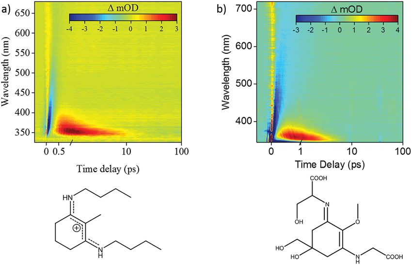
View Article Online
Faraday Discussions Discussions
a variety of de-excitation pathways, resulting in a reduced overall amplitude of Chl
ESA in the quenched sample. However, the kinetics of the ESA signals were
observed to be kinetically equivalent at time delays $40 ps where Car S1 states are
Published on 05 July 2019. Downloaded by Lawrence Berkeley National Laboratory on 2/3/2021 12:38:40 AM.
nearly fully de-populated. In order to extract the contribution of Car S1 ESA from
the overall Chl ESA, we scale the kinetic prole measured in the dark (mostly Chl
ESA) to match that in high light based on signals at long time delays and subtract
the two.
This normalization procedure of the EET kinetic traces at long time delays is
based on the assumption that the amount of annihilation is consistent during the
transition from dark to high light. If the amount of annihilation were different
under the quenched and unquenched conditions, we would expect to see
a signicant change in the Chl ESA and GSB dynamics. However, in a previous
paper, we found that the decay of the Chl ESA and GSB signals probed at 620 and
680 nm respectively were identical for dark- and high-light-exposed spinach
thylakoid membranes.1 Therefore, it is reasonable to assume that Chl*–Chl*
annihilation is consistent during the transition from dark to high light and thus
the decay of Chl ESA is kinetically equivalent. This is further supported by the N.
oceanica vde mutant that lacks Zea, which upon scaling exhibits identical decays
at 540 nm under unquenched and quenched conditions.2
1 S. Park, A. L. Fischer, C. J. Steen, M. Iwai, J. M. Morris, P. J. Walla, K. K. Niyogi and G. R.
Fleming, J. Am. Chem. Soc., 2018, 140, 11965–11973.
2 S. Park, C. J. Steen, D. Lyska, A. L. Fischer, B. Endelman, M. Iwai, K. K. Niyogi and G. R.
Fleming, Proc. Natl. Acad. Sci. U. S. A., 2019, 116, 3385–3390.
Natércia das Neves Rodrigues addressed Collin Steen: This is a question
regarding your TA experiments on living cells, which are of course rather complex
samples. I am curious to know how much this was a problem experimentally, i.e.
how difficult it was to generate signal, if there were problems with excessive
scatter or any other experimental challenges that arose from using a complex
sample like this. Moreover, I am interested in knowing how you addressed these,
both experimentally – e.g. did you have to avoid translating the sample, or were
there other things you had to adjust experimentally – and in terms of analysis, e.g.
did you have problems with over-complicated spectra, how you addressed these
and how you isolated the spectral features of interest?
Collin Steen answered: Experimentally, it is important that the sample is
continuously translocated during the experiment and bubbled regularly between
measurements to prevent sample damage. To minimize scattering of the pump
we use a polarization lter set to the probe polarization. To ensure a clean probe
signal we also add a notch lter specic to the pump wavelength prior to our
detector. In terms of analysis, we are able to identify the molecular species
involved in the quenching of excitation energy based on the features of the
difference kinetic decay prole, which is taken as the difference between the
sample in its quenched (high-light) state and its unquenched (dark) state. By
tting this difference prole, we obtain rise and decay time constants for the
species of interest, which can be compared to literature values for the lifetimes for
various pigments. Verication of the molecular species of interest is also based on
comparing the observed excited-state absorption spectrum with the literature.
Additionally, the use of mutants or chemical treatments that target specic
540 | Faraday Discuss., 2019, 216, 538–563 This journal is © The Royal Society of Chemistry 2019
View Article Online
Discussions Faraday Discussions
molecules in the organism can provide further insight into assignment of the
spectra and decay proles.
Published on 05 July 2019. Downloaded by Lawrence Berkeley National Laboratory on 2/3/2021 12:38:40 AM.
Pavel Chabera asked Tomas Polivka: It would seem that the synthetic pathway
for producing carotenoid with an extra hexanol group is rather complicated. Why
does the organism (plankton) undertake this challenge? You indicate in the paper
(DOI: 10.1039/c8fd00193f) that this is not happening for protection against excess
light – why? Is there any better plausible explanation?
Tomas Polivka replied: Our results show that the overall efficiency of
carotenoid-chlorophyll energy transfer is not affected by fucoxanthin (Fx) - hex-
anoylfucoxanthin (hFx) exchange, though the energy transfer is slower if hFx is
the donor. Similarly, there is no additional quenching of Chl singlet excited states
if hFx is present. Yet, the triplet quenching, which has not been studied in detail
in our paper, could still be affected by Fx-hFx exchange. The enhanced synthesis
of hFx under high-light conditions is clearly proven, so there should be some
advantage of having hFx in the antenna under high-light conditions. But the data
we have collected so far do not give any clear answers to what this advantage
actually is.
Pavel Chabera followed: In a “standard carotenoid model”, it is believed that
only groups attached to the conjugated backbone of a carotenoid can inuence its
excited state properties. How do you explain that the hexanol attached to the
molecule but not conjugated to carotenoid’s conjugated system can inuence its
properties? In particular, that it can switch off the ICT state?
Tomas Polivka answered: Yes, it is correct that we have always considered the
groups that are not in conjugation to be spectroscopically silent. On the other
hand, we know that spectroscopic properties of keto-carotenoids are strongly
affected by solvent polarity. Thus, there is clearly a way to modify spectroscopic
properties by something that is not a part of the conjugated system. Here, the
attached hexanoyloxy tail obviously affects the electron distribution along the
conjugated backbone, preventing stabilization of the ICT state.
Graham Fleming asked: Can you add a bit more detail on why you say charge
transfer is necessary for rapid energy transfer?
Tomas Polivka responded: The coupling of the ICT state to the S1 state
modies the transition dipole moment of the donor transition. It presumably
enhances the transition dipole moment (as evidenced by e.g. increased S1 uo-
rescence quantum yield of keto-carotenoids), making the donor–acceptor
coupling stronger, resulting in more efficient energy transfer.
Heiko Lokstein remarked: There has been a lot of discussion recently about
the so-called S* states of carotenoids. I wonder whether you have observed an S*
state in either one of your investigated carotenoids, or, more specically, whether
there was any difference in S* transient absorption spectra between them.
This journal is © The Royal Society of Chemistry 2019 Faraday Discuss., 2019, 216, 538–563 | 541View Article Online
Faraday Discussions Discussions
Tomas Polivka answered: No, we have not observed any spectral features
attributable to S* signal in these carotenoids. It is not so surprising as the S*
signal is typically reported for carotenoids with long conjugation length, let’s say
Published on 05 July 2019. Downloaded by Lawrence Berkeley National Laboratory on 2/3/2021 12:38:40 AM.
about N¼11 or longer. Fucoxanthin and its derivatives are much shorter.
Vasilios Stavros said: Tomas, you present some really interesting data. I was
wondering whether you had considered looking at the inuence of temperature
on the excited state dynamics of hFx, as conformational exibility may inuence
the role of S1/ICT on excited state energy transfer.
Tomas Polivka responded: No, we have not studied temperature dependence
so far though I agree it could provide valuable information as it indeed can reveal
some extra information about the role of the hexanoyloxy tail in hFx. In the future,
we plan to run experiments at both low temperature (down to about 200 K to keep
the solution liquid) and high (up to about 325 K to prevent degradation).
Shuming Bai opened discussion of the paper by Scott Habershon: As I
understand, you apply the machine learning diabatization to obtain an "on-the-
y" PES for the MCTDH method. My rst question: In principle, other effective
diabatization methods should be applicable in your approach, right? My second
question: The universal diabatization method is still quite challenging for the
systems with more than several atoms. For “on-the-y” applications it can be
more difficult because different congurations of state can join in along the
dynamics, as you have found. Is it possible to monitor situations like this, and
provide warning information during the calculation?
Scott Habershon replied: For the rst question, the answer is yes; in principle,
our approach is compatible with any diabatization scheme.
For the second part of the question, we agree that some sort of diabatization
monitoring is essential; we are already thinking about this somewhat. In the
projection diabatization scheme which we employ, for example, one can keep
track of the energies of the different adiabatic states throughout the sampled
congurations in our on-the-y scheme; if adiabatic states change in energy such
that they become relevant to the diabatization scheme (as in the example
considered in our paper), then we can of course go back and re-run the dynamics
with these additional states included. This procedure could be repeated until no
further states are required. As an alternative, we are also investigating how one
might include more than one set of reference electronic states in our projection
diabatization scheme; this is ongoing work, however.
Bern Kohler asked: Have you considered applying your on-the-y simulations
to uracil, a nucleobase with well-understood internal conversion pathways?
Scott Habershon answered: Yes - that is exactly the sort of molecular system
we’d like to be in a position to directly simulate using these on-the-y methods.
With 12 atoms, so a maximum of 30 active degrees-of-freedom (3N-6), this is at the
upper-end of the system sizes we can currently model; however, the development
of more efficient potential energy surface sampling schemes and improved so-
ware parallelisation should help us push towards this limit.
542 | Faraday Discuss., 2019, 216, 538–563 This journal is © The Royal Society of Chemistry 2019View Article Online
Discussions Faraday Discussions
Natércia das Neves Rodrigues said: In the model you describe, do you have to
restrict and select which coordinates to include or are you able to use the full 3N
dimensional space?
Published on 05 July 2019. Downloaded by Lawrence Berkeley National Laboratory on 2/3/2021 12:38:40 AM.
Scott Habershon responded: Although the methods we describe can in prin-
ciple be used to describe the full 3N-6-dimensional space of an N-atom molecule,
the expense of ab initio calculations and propagation of the MCTDH equations-of-
motion mean that one usually has to make a selection of the “active” vibrational
coordinates. Various approaches towards this have been suggested previously,
including the one in our contribution (DOI: 10.1039/c8fd00228b), but this aspect
remains something of an open problem.
Natércia das Neves Rodrigues remarked: In my gas-phase experiments, aer I
pump my system with one photon, I then ionize the system in order to probe it.
This ionization is effectively a second transition from the excited state to a given
cationic state. I have found that the Franck–Condon factor for this transition—
which changes in time as the wavepacket samples the excited state surface and is
projected onto different parts of the cationic state surface—affects my experi-
mental observable. In particular, the auto-correlation function which describes
the time-dependent wavepacket behavior alone does not successfully model my
experimental observations, as it does not account for the changing probe step
Franck–Condon factor. Is it realistic that your models may be expanded to model
this time-dependent projection of the excited state population onto the cationic
state? Alternatively, do you know of any other methods which may model this
behavior?
Scott Habershon answered: Yes, we believe that wavefunction methods like
MCTDH could be used to model processes such as that described, although we
expect it would be very challenging. For example, one could imagine introducing
a time-dependent term to the Hamiltonian to simulate excitation to the ionic
states at different stages during the MCTDH evolution. However, accounting for
the conguration-dependent Franck–Condon factors seems like it would require
further work; performing these “jumps” for trajectory-based methods might be
more straightforward using Franck–Condon factors calculated at each congu-
ration for each trajectory, but performing a similar simulation for a grid-based
method would be much more demanding.
It is already possible within the MCTDH framework, using pre-tted potential
energy surfaces, to model time-resolved photoelectron spectroscopy experiments,
so such an extension to the on-the-y method should be possible. The cation
potential energy surfaces can be tted as for neutral states, and time-dependent
elds can be applied to model laser pulses. So, assuming that the transition
dipole (and polarisability, etc., if necessary) matrix elements are also tted, then
excitations to the cation states my also be modelled. It is also necessary to account
for the energy of the ejected electron; this can be achieved by introducing an extra
discrete variable representation-type grid into which the electron wavefunction is
excited, a so-called discretised continuum.
Jeffrey Cina asked: Could you please comment on the relative computational
overhead of the MCTDH and electronic structure portions of these calculations?
This journal is © The Royal Society of Chemistry 2019 Faraday Discuss., 2019, 216, 538–563 | 543View Article Online
Faraday Discussions Discussions
Scott Habershon replied: In the initial calculation, where one is running
a MCTDH simulation while simultaneously sampling congurations at which to
perform ab initio electronic structure calculations, the computational burden lies
Published on 05 July 2019. Downloaded by Lawrence Berkeley National Laboratory on 2/3/2021 12:38:40 AM.
towards the electronic structure calculations. In follow-up calculations, where one
can perform an MCTDH simulation on the constructed potential energy surface,
the computational expense lies rmly with the propagation of the MCTDH
equations-of-motion.
It is also worth noting that the distribution of computational expense will be
highly problem-dependent. For example, problems with many strongly-coupled
degrees-of-freedom might require more ab initio electronic structure calcula-
tions in order to generate an accurate representation of the potential energy
surface, such that this stage may be extremely costly. In addition, the expo-
nential scaling of the MCTDH algorithm itself will eventually become the bottle-
neck in the simulations as the system-size increases. In the initial dynamics
calculations reported here, using the normal modes chosen using forces and
couplings, the overwhelming computational cost was in the electronic structure
calculations. For each of the molecules studied in this paper (DOI: 10.1039/
c8fd00228b), the percentage CPU times for the dynamics were: 0.35% (1a),
4.6% (2a), 0.11% (3a) and 0.5% (4a).
For the simulations of 4a using the 5 “steepest” normal modes, as well as the
“false” normal mode directed towards the conical intersection, MCTDH
accounted for about 40% of the total computational cost. This much larger
proportion was due to the much larger DVR grids being used.
In addition to the point about highly-correlated potential energy surfaces
being more difficult to t, we also note that such systems also require more single-
particle functions in the MCTDH calculation to account for the motion of the
coupled modes.
Sharon Hammes-Schiffer questioned: How do you generate the initial condi-
tions, and how sensitive are the results to them? Does MCTDH properly describe
zero-point energy and the transfer of energy between vibrational modes?
Scott Habershon answered: The initial wavepacket is a Hartree product of
ground-state harmonic oscillator eigenfunctions, centered at the minimum-
energy geometry of the electronic ground-state. As such, harmonic zero-point
energy is included in the initial wavefunction.
The MCTDH equations-of-motion are derived using the time-dependent
variational principle. As such, they correctly describe zero-point energy conser-
vation and other quantum-mechanical effects such as tunnelling. In addition,
energy transfer between different vibrational modes is also captured (as long as
this coupling is also described by the underlying potential energy surface, of
course).
We have not investigated the effects of alternative initial conditions in this
paper, although this is possible in the MCTDH method. For example, an initial
imaginary-time relaxation of the wavepacket on the ground-state would account
for any effects of anharmonicity on the shape of the initial wavepacket. This
wavepacket can then be vertically promoted to the excited electronic state or,
pushing towards higher accuracy, a laser pulse can be modelled as a time-
dependent eld which, allied with a representation of the transition-dipole
544 | Faraday Discuss., 2019, 216, 538–563 This journal is © The Royal Society of Chemistry 2019View Article Online
Discussions Faraday Discussions
moment surface, allows a more realistic representation of the excitation. To adopt
this more accurate representation of the excitation process in our on-the-y
method requires more work to model the transition dipoles, but this should be
Published on 05 July 2019. Downloaded by Lawrence Berkeley National Laboratory on 2/3/2021 12:38:40 AM.
feasible.
Spiridoula Matsika asked: In this study you discovered that there was a problem
with the chosen diabatization scheme because the DD-MCTDH predictions did not
agree with the known ultrafast dynamics. Is there a way to test/know if your
calculations predict the correct behavior if this behavior is not known a priori?
Scott Habershon responded: Yes - one can, in principle, compare the features
of the diabatic and adiabatic states as an indicator of potential problems in
diabatization. In addition, in one of the diabatization schemes used in this paper
(projection diabatization) (DOI: 10.1039/c8fd00228b), one can monitor the adia-
batic states as they change in energy across the range of sampled conguration
space, allowing one to self-consistently update the manifold of states used in the
diabatization scheme. Finally, although we have not investigated fully, we believe
that the projection and propagation diabatization schemes certainly have room
for improvement, for example by combining the two different approaches to seek
a “best-of-both-worlds” approach.
Jack Woolley commented: Given that certain vibrational modes are required
to traverse conical intersections, could this method, knowing the correct active
space, be used as a black box soware to explore how different chemical
functionalities associated with the two nitrogen atoms inuence the excited
state dynamics? Recent experimental data on a model MAA and the MAA Shi-
norine shows subtle differences (see Fig. 1 below) suggesting that careful
thought behind choice of functionality will be crucial in developing next
generation, nature inspired sunscreens, and hence theory will be very impor-
tant, to aid with functionalisation of core structures, like those discussed in the
contribution.
Fig. 1 (a) False colour heat map of transient absorption data, reported by Woolley et al.1
for a similar system to those studied by Habershon and co-workers. (b) False colour heat
map of transient absorption date of the MAA Shinorine.
This journal is © The Royal Society of Chemistry 2019 Faraday Discuss., 2019, 216, 538–563 | 545View Article Online
Faraday Discussions Discussions
1 J. M. Woolley, M. Staniforth, M. D. Horbury, G. W. Richings, M. Wills and V. G. Stavros, J.
Phys. Chem. Lett., 2018, 9, 3043–3048.
Scott Habershon replied: Yes, the ultimate (if ambitious) aim of this line of
Published on 05 July 2019. Downloaded by Lawrence Berkeley National Laboratory on 2/3/2021 12:38:40 AM.
research is to try to move towards a more “black box” method for modelling
quantum chemical dynamics as a route to exploring the impact of different
chemical functionality on molecular photochemistry. Such a method would
enable design of photo-functional molecules such as sensors or switches.
However, as noted in the question, barriers remain to this, such as the accuracy
and expense of ab initio electronic structure calculations and methodological
aspects such as the appropriate choice of active vibrational modes. Knowing
which vibrational modes are essential to capture the experimentally-observed
dynamics is very challenging, and may require a multi-method approach, such
as using trajectory-surface hopping to inform the set-up of MCTDH simulations.
Todd Martinez asked: Is the diabatization process that you are using depen-
dent on the path taken? If so, how do you deal with the resulting ambiguity?
Scott Habershon answered: In the propagation diabatization scheme, we
propagate the diabatization matrix outwards from some initial geometry (usually
the minimum-energy geometry on the lowest electronic state). In this scheme, the
diabatization matrix is propagated using line integrals along the shortest-path
between congurations (dened using simple Euclidean distance). As a result,
there is some ambiguity here; if one changes the set of sampled congurations,
then the propagation of the diabatization matrix will also change, and so the
diabatic states will also change.
To date, we have not noticed this becoming a problem in our simulations. Our
impression is that the density of congurations which must be sampled to generate an
accurate potential energy surface implicitly means that there is little variation between
the propagation of the diabatization matrix between different simulation runs. In
addition, we have recently begun using Sobol grid sampling to generate congurations
which are subsequently used to construct the diabatic potential energy surfaces and
inter-state couplings; this means that the MCTDH dynamics is less susceptible to
“jitter” in the constructed potential energy surfaces which might arise from differences
in sampled congurations. This point is discussed more in a previous publication.1
1 G. W. Richings, C. Robertson and S. Habershon, J. Chem. Theory Comput., 2019, 15, 857–870.
Vasilios Stavros said: You discuss possible factors that could inuence the
agreement between your calculations and those of Losantos and coworkers. One
is the inclusion of more diabatic states in the projection scheme, which would
lower the barrier along a relaxation coordinate. Do your calculations include
solvation effects and if not, is it possible that the disagreement may also be due to
the absence of these effects?
Scott Habershon replied: Our calculations do not include solvation effects, and
are performed in vacuum. It is possible that solvent effects can change the
character of the electronic states, and that could, of course, inuence the accuracy
of comparison with experiment.
546 | Faraday Discuss., 2019, 216, 538–563 This journal is © The Royal Society of Chemistry 2019View Article Online
Discussions Faraday Discussions
Including solvation effects directly into our on-the-y scheme is the next
frontier here. We can see some ways forward, such as using an Ehrenfest-type
scheme for mixed quantum/classical dynamics whereby the solvent molecules
Published on 05 July 2019. Downloaded by Lawrence Berkeley National Laboratory on 2/3/2021 12:38:40 AM.
are modelled using classical molecular dynamics, but the on-the-y nature of
our approach might enable alternatives; we are actively investigating these
aspects.
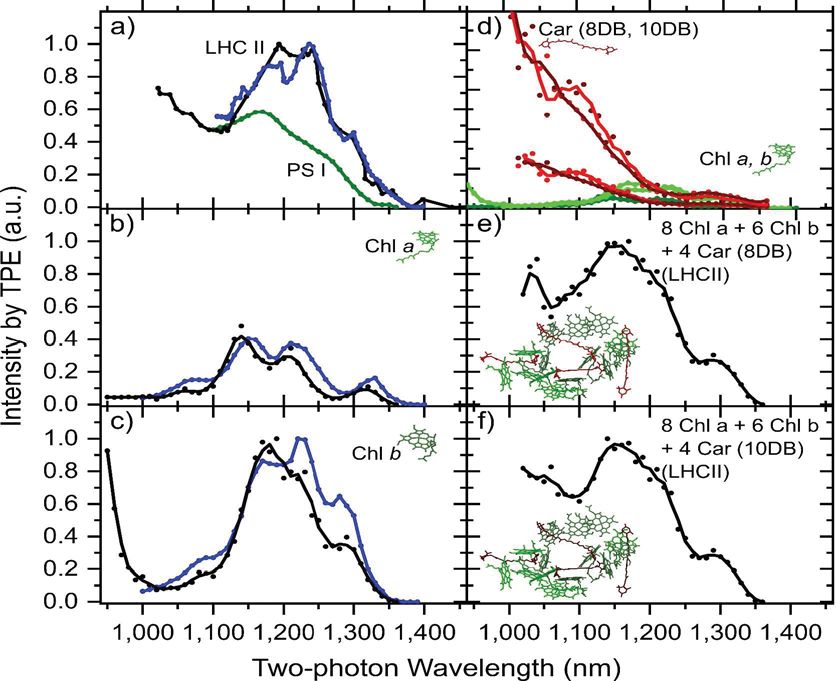
Peter Jomo Walla opened discussion of the paper by Heiko Lokstein: I would
like to make a comment about the relative contributions of chlorophyll two-
photon excitation and carotenoid two-photon excitation to the two-photon exci-
tation spectra of LHC II and other pigment-protein complexes. In 2017 we
addressed this question in a paper in which we explored relative cross-sections in
two-photon excitation spectra of chlorophylls (Chls) as well as carotenoids.1 Aer
recognizing Dr Lokstein’s contribution (DOI: 10.1039/c8fd00198g) I contacted
him because, based on his reported results, he concluded that there is almost no
contribution from carotenoid two-photon excitation to the two-photon spectra of
LHC II. However, these results did not consider the two-photon excitations
spectra of carotenoids as observed by us.1 We therefore agreed to join forces and
directly compare all two-photon results that we both have obtained in our
previous studies. I have prepared a slide that summarizes the major conclusions
from these comparisons (shown in Fig. 2).
Fig. 2 Comparison of experimental two-photon excitation spectra of LHC II, PS I and
isolated pigments. (a) Two-photon excitation spectra of LHC II by Walla et al.2 (black
curves) and Betke and Lokstein (blue) (DOI: 10.1039/c8fd00198g), as well as, PS I by
Wehling et al.3 (green). (b) and (c) Two-photon excitation spectra of isolated Chl a and
b from Gacek et al.1 and Betke and Lokstein (blue) (DOI: 10.1039/c8fd00198g). (d) Two-
photon excitation spectra of carotenoids with 8 and 10 conjugated double bonds from
Gacek et al.1 (red) in comparison to two-photon excitation spectra of Chls a and b. (green).
The relative magnitudes of the spectra in (d) reflect the relative contributions of the
individual pigments to two-photon spectra in ref. 1. (e) and (f) LHC II two-photon exci-
tation spectra reconstructed from summing experimental two-photon excitation spectra
of 8 Chls a, 6 Chls b and 4 carotenoids (xanthophylls) from the individual pigment two-
photon excitation spectra shown in (d). The protein and pigment structures were created
with VMD: Visual molecular dynamics,4 from a data set of Su et al.5
This journal is © The Royal Society of Chemistry 2019 Faraday Discuss., 2019, 216, 538–563 | 547View Article Online
Faraday Discussions Discussions
In general, the two-photon spectra observed in our work are in good agreement
with those reported by Dr Lokstein. Fig. 2a link accordingly demonstrates that the
LHC II spectrum reported in Betke and Lokstein (blue) agrees quite well with the
Published on 05 July 2019. Downloaded by Lawrence Berkeley National Laboratory on 2/3/2021 12:38:40 AM.
one we reported in 2000 in Walla et al.,2 (black in Fig. 2a) only that the spectral
range reported in our publication is larger. For comparison, Fig. 2a also shows the
two-photon excitation spectrum of PS I that we reported in 2005, for example.3
Also the agreement of the two-photon spectra of Chl a reported in Gacek et al.1 as
well as Betke Lokstein (DOI: 10.1039/c8fd00198g) in solution (Fig. 2b, black and
blue, respectively) and of Chl b in solution (Fig. 2c) is quite reasonable. Minor
differences are likely due to different solvents that were used in both studies. A
surprising result observed in Gacek et al. was that Chl b has, in general,
a signicantly larger two-photon absorption cross-section than Chl a.1
However, the larger spectral range reported for LHC II in Walla et al.1 (Fig. 2a,
black) demonstrates that there are more contributions from pigments other than
Chls since the Chls two-photon absorption cross section drastically drops in the
spectral range below 1100 nm (Fig. 2b and c). These additional signals from other
pigments to the two-photon excitation spectra of LHC II can be readily explained
by signicant contributions from carotenoid dark states since the two-photon
excitation spectra of carotenoids reported in Gacek et al.1 are quite large in this
spectral range (Fig. 2d).
Relative magnitudes of the two-photon excitation spectra of individual carot-
enoids and Chls were determined in Gacek et al.1 Thus, it appears to be reason-
able to reconstruct a LHC II two-photon excitation spectrum by using the sum of 8
Chls a, 6 Chls b and 4 carotenoids corresponding to the stoichiometric ratio of
these pigments in LHC II. In fact, the agreement of these reconstructed spectra
(Fig. 2e and f) with that of the experimental two-photon spectrum of LHC II
(Fig. 2a) is quite reasonable.
The spectrum of PS I (green curve in Fig. 2a) demonstrates that the relative
contribution of carotenoids may be even larger here. PS I contains exclusively Chl
a (besides b-carotene), hence, this is in good agreement with the observation that
Chl a has a smaller two-photon cross-section than Chl b.
The observations made by Dr Lokstein (DOI: 10.1039/c8fd00198g) that LHC II,
containing different complements of carotenoids having also different numbers
of conjugated double bonds, have very similar two-photon excitation spectra
agrees well with our observation (Gacek et al.1), that carotenoids with different
numbers of conjugated double bonds have surprisingly similar two-photon
excitation spectra (Fig. 2d).
In summary, carotenoid two-photon excitation contributes signicantly to the
two-photon excitation spectra of PS I and LHC II below a spectral range of around
1100 -1200 nm. This observation also provides evidence that in both PS I and LHC
II, excitation energy transfer occurs from carotenoid dark states to Chls; otherwise
no Chl uorescence would be observed aer carotenoid two-photon excitation. Dr
Lokstein and we agree to join forces and further explore our observations, in order
to provide more quantitative information about what the contributions of indi-
vidual pigments, Chls and carotenoids (xanthophylls) at various excitation
wavelengths in two-photon excitation and absorption spectra of photosynthetic
pigment-protein complexes are.
Dr Lokstein might also elucidate further, to what extend these insights as well
as further observations obtained by his group support current suggestions about
548 | Faraday Discuss., 2019, 216, 538–563 This journal is © The Royal Society of Chemistry 2019View Article Online
Discussions Faraday Discussions
the molecular mechanisms relevant in the regulation of photosynthetic light-
harvesting.
1 D. A. Gacek, A. L. Moore, T. A. Moore and P. J. Walla, J. Phys. Chem. B, 2017, 121, 10055.
Published on 05 July 2019. Downloaded by Lawrence Berkeley National Laboratory on 2/3/2021 12:38:40 AM.
2 P. J. Walla, J. Yom, B. P. Krueger and G. R. Fleming, J. Phys. Chem. B, 2000, 104, 4799.
3 A. Wehling and P. J. Walla, J. Phys. Chem. B, 2005, 109, 24510.
4 W. Humphrey, A. Dalke and K. Schulten, J. Mol. Graph., 1996, 14, 33.
5 X. Su, J. Ma, X. Wei, P. Cao, D. Zhu, W. Chang, Z. Liu, X. Zhang and M. Li, Science, 2017,
357, 815.
Heiko Lokstein responded: I became aware of Dr Walla’s 2017 paper1 when I
was contacted by him following the online publication of our current Faraday
Discussions paper (DOI: 10.1039/c8fd00198g). We both do agree that our exper-
imental data are rather similar, but our interpretations of these were considerably
differing, namely regarding the relative contributions of carotenoids and chlo-
rophylls (Chls) to the two-photon excitation (TPE) spectra of various photosyn-
thetic pigment-protein complexes, and, in particular those of plant major light-
harvesting complex II (LHC II). In fact, we concluded that there is (almost) no
contribution from carotenoids to two-photon excitation spectra of LHC II.
Moreover, we pointed out that direct TPE of (Bacterio)Chls in the presumed
spectral region of the forbidden carotenoid S1 (21Ag) state(s) was largely neglected
in previous publications.
However, aer discussions with Dr Walla and careful re-examination of our
own data we both came to the conclusion that our previous bold statements have
to be reconsidered. We therefore agreed to join forces and directly compare all the
two-photon excitation results that we had obtained.
Indeed, Walla and co-workers had previously reported TPE data for a more
extended spectral range, extending below 1100 (550 nm) for LHC II. These data
may indicate that there may be contributions from pigments other than Chls
(namely from dark states of carotenoids), since the Chls two-photon absorption
cross section apparently drops in the TPE range below 1100 (550 nm) nm, possibly
carotenoid contributions may even be apparent already at 1200 (600) nm.
Summarizing, we do agree that, apparently, carotenoid two-photon excitation
can signicantly contribute to the two-photon excitation spectra of photosyn-
thetic pigment protein complexes, including PS I and LHC II, in particular in the
spectral range below 1100 (550 nm), possibly even already at 1200 (600) nm,
however this would need to be further quantied.
Dr Walla and we agreed to further examine our observations, in order to
provide a more quantitative view of the contributions of individual pigments,
Chls and carotenoids (xanthophylls), to two-photon excitation at various excita-
tion wavelengths in photosynthetic pigment-protein complexes.
Moreover, we would like to stress that the mechanism of photoprotective
non-photochemical Chl-uorescence quenching (NPQ) proposed by Dr Wallas
group, based on a combination of TPE and pulse-amplitude modulated uo-
rescence (PAM) experiments2 would be consistent with our own observations:
Non-linear polarization spectroscopy in the frequency domain (NLPF) experi-
ments with LHC II in different states of aggregation (commonly serving as
a model system for NPQ) indicated altered xanthophyll-Chl interactions: Upon
pumping in the Chl a+b Qy spectral region (640–680 nm) and probing at selected
wavelengths in the energetically much higher allowed xanthophyll absorption
This journal is © The Royal Society of Chemistry 2019 Faraday Discuss., 2019, 216, 538–563 | 549View Article Online
Faraday Discussions Discussions
region (490–510 nm) it was found that a xanthophyll molecule absorbing at
510 nm strongly interacts with a Chl b absorbing at about 650 nm.3 Upon
aggregation of LHC II the picture changes completely: now the same xantho-
Published on 05 July 2019. Downloaded by Lawrence Berkeley National Laboratory on 2/3/2021 12:38:40 AM.
phyll molecule, probed at 510 nm, interacts with Chl(s) a absorbing at 682 nm,
comprising the terminal emitter in LHC II.4
1 D. A. Gacek, A. L. Moore, T. A. Moore and P. J. Walla, J. Phys. Chem. B, 2017, 121, 10055.
2 S. Bode, C. C. Quentmeier, P. N. Liao, N. Ha, T. Barros, L. Wilk, F. Bittner and P. J. Walla,
Proc. Natl. Acad. Sci. U. S. A., 2009, 106, 12311.
3 B. Voigt, M. Krikunova and H. Lokstein, Photosynth. Res., 2008, 95, 317.
4 H. Rogl, R. Schödel, H. Lokstein, W. Kühlbrand and A. Schubert, Biochemistry, 2002, 41,
2281.
Tomas Polivka remarked: Your detection of two-photon absorption relies on
carotenoid-chlorophyll energy transfer. Can the absence of any carotenoid
contribution to the two-photon signal in LHCII simply mean there is no
carotenoid-chlorophyll energy transfer via the S1 state in LHCII?
Heiko Lokstein responded: A very good question. Indeed, it seems that exci-
tation energy transfer onto chlorophyll S1 following supposed two-photon exci-
tation of carotenoid S1 states is somewhat inefficient (or even negligible) as also
suggested by previous studies with purple bacterial LH2 complexes (e.g., Kriku-
nova et al.1) – but further experimental work is clearly required.
1 M. Krikunova, A. Kummrow, B. Voigt, M. Rini, H. Lokstein, A. Moskalenko, H. Scheer, A.
Razjivin and D. Leupold, FEBS Lett., 2002, 528, 227–229.
Tomas Polivka addressed Peter Jomo Walla and Heiko Lokstein: From the data
presented by Peter Walla it seems that there is onset of carotenoid contribution to
the two-photon absorption in LHCII at signicantly higher energies than ex-
pected. Do you think this imply that the energy threshold for carotenoid-
chlorophyll S1-mediated energy transfer channel is much higher than expected?
Only hot S1 contributes to energy transfer? Or even some other state?
Peter Jomo Walla responded: We were also surprised by this observation and it
might indeed indicate energy transfer from hot carotenoid S1 states. We have already
speculated about this possibility in a paper in 2002 (Walla et al.1, see also A. Wehling
and P. J. Walla2). When the 0-0-transition of the carotenoid S1 state is signicantly
higher than that of the tetrapyrrole acceptor, the contributions of the carotenoid S1 0-
0-transtion relative to hot states might be larger in two-photon excitation spectra. On
the other hand, the reported two-photon excitation spectra (Gacek et al.3) might also
indicate the presence of further dark states between the carotenoid S1 and carotenoid
S2 state such as the putative carotenoid Sx or carotenoid S* states proposed by others.
We currently cannot disentangle the exact nature of these states but the results
provide evidence that there are dark carotenoid states, either hot S1, Sx or S* states,
that can efficiently transfer energy to tetrapyrroles and contribute to two-photon
excitation in dyads and pigment-protein complexes.
1 P. J. Walla, P. A. Linden, K. Otha and G. R. Fleming, J. Phys. Chem. B, 2002, 106, 1909.
2 A. Wehling and P. J. Walla, J. Phys. Chem. B, 2005, 109, 24510.
3 D. A. Gacek, A. L. Moore, T. A. Moore and P. J. Walla, J. Phys. Chem. B, 2017, 121, 10055.
550 | Faraday Discuss., 2019, 216, 538–563 This journal is © The Royal Society of Chemistry 2019View Article Online
Discussions Faraday Discussions
Heiko Lokstein replied: It seems, taking also the data of Dr Walla and co-
workers into account, that obviously two-photon absorption in LHC II starts at
signicantly higher energies than previously expected, (i.e., for S1 of the xantho-
Published on 05 July 2019. Downloaded by Lawrence Berkeley National Laboratory on 2/3/2021 12:38:40 AM.
phylls). In fact, we cannot exclude that the energy threshold for carotenoid-to-
chlorophyll excitation energy transfer is much higher, so that possibly hot S1
states or, even, some other, yet unassigned, state(s) contribute. Clearly, further
experimental work is required.
Minjung Son addressed Heiko Lokstein and Peter Jomo Walla: Could you
make some general comments on the sensitivity of this experiment as to whether
it can distinguish between contributions from different carotenoid molecules,
i.e., whether the carotenoid is a b-carotene or a lutein, or whether the carotenoid
has eight conjugated bonds or nine, for example?
Peter Jomo Walla replied: The two-photon excitation spectra of carotenoid-
tetrapyrrole dyads containing carotenoids with different numbers of conjugated
double bonds looked surprisingly similar (Gacek et al.1). Again, dominant
contributions from hot carotenoid S1 energy transfer might be an explanation in
this case. Then, the cut off-wavelength in two-photon excitation spectra and
energy transfer for each vibrational donor level would rather depend on the
limiting energy level of the acceptor and not so much on varying 0-0-transtion
energies of the carotenoid donors themselves. However, our studies also indicate
that the overall efficiency of energy transfer depends on the number of conjugated
double bonds or the carotenoid used, i.e. the relative contribution of the carot-
enoid donors to two-photon spectra varies compared to that of the tetrapyrrole
acceptor. Two-photon excitation spectra of pigment-protein complexes recon-
stituted with different carotenoids that include more of the spectral range
dominated by carotenoid dark states might provide further insights into that.
1 D. A. Gacek, A. L. Moore, T. A. Moore and P. J. Walla, J. Phys. Chem. B, 2017, 121, 10055.
Heiko Lokstein answered: It appears that – regarding basically our data (see
also Fig. 5 of the current paper (DOI: 10.1039/c8fd00198g)) – we cannot distin-
guish contributions from different carotenoids, or, whether the carotenoid has
eight conjugated bonds or nine, or even more. This may, at least in part, also be
related to inefficient excitation energy transfer to chlorophylls.
Stephen Bradforth asked: As your experiment uses two photons from the same
beam, this limits somewhat the polarization information available. However, in
your presentation you showed polarization ratio data comparing two circular
polarized photons to two linearly polarized photons. Can you interpret what that
ratio information is revealing?
Heiko Lokstein answered: The polarization-dependent experiment does not
aim at obtaining information about (relative) orientations of the molecules.
However, unlike in linear absorption, for two-photon absorption the polarization
of the exciting light matters, even for randomly oriented molecules.1 Thus, two-
photon excited uorescence was recorded for linear and circular excitation with
otherwise identical measuring conditions.
This journal is © The Royal Society of Chemistry 2019 Faraday Discuss., 2019, 216, 538–563 | 551View Article Online
Faraday Discussions Discussions
The two-photon polarization ratio can be an indicator for the symmetry of the
molecules investigated.1 For the S0 / S1 transition of a carotenoid (or linear
polyene), a value of 0.8 would be expected.2 In our polarization measurements
Published on 05 July 2019. Downloaded by Lawrence Berkeley National Laboratory on 2/3/2021 12:38:40 AM.
with LHCII, however, values between 1 and 1.2 were obtained for the spectral
range of 610–690 nm and close to 1 below 600 nm. This would suggest that the
excited molecules have, unlike carotenoids, a molecular symmetry different from
the C2h point group. Furthermore, the polarization ratio has been measured for
pure chlorophyll a in the range between 660 to 680 nm. It was found to be
comparable to the values for LHCII. These observations, in turn, can be taken as
further indication that the two-photon-excited molecules in this spectral region
are not carotenoids, but chlorophylls in LHCII.
1 W. M. McClain, J. Chem. Phys., 1971, 55, 2789–2796.
2 R. R. Birge and B. M. Pierce, J. Chem. Phys., 1979, 70, 168–178.
Vasilios Stavros addressed Heiko Lokstein and Peter Jomo Walla: In terms of the
(simplied) energy level scheme presented in Fig. 2 (DOI: 10.1039/c8fd00198g), I
would anticipate large spectral changes in the excited state absorption (ESA)
following excited state energy transfer from the carotenoid to the chlorophyll. From
the previous studies that you cite in your paper,1–4 is there any evidence that such
large spectral shis are observed in, say, transient absorption studies? If so, I would
expect this to be indicative of initial TPE of the carotenoid.
1 B. P. Krueger, J. Yom, P. J. Walla and G. R. Fleming, Chem. Phys. Lett., 1998, 310, 57–64.
2 P. J. Walla, P. A. Linden, C. P Hsu, G. D. Scholes and G. R. Fleming, Proc. Natl. Acad. Sci. U.
S. A., 2000, 97, 10808–10813.
3 P. J. Walla, J. Yom, B. P. Krueger and G. R. Fleming, J. Phys. Chem. B, 2000, 104, 4799–4806.
4 P. J. Walla, P. A. Linden, K. Ohta and G. R. Fleming, J. Phys. Chem. A, 2002, 106, 1909–1916.
Peter Jomo Walla responded: Actually, there is also some evidence for carot-
enoid two-photon excitation from two-photon ESA studies. When exciting pure
carotenoids at two-photon wavelengths corresponding to energies signicantly
below the allowed carotenoid S2 absorption (lTPE1200–1310 nm), carotenoid
dark state ESA decay kinetics (ldet550 nm) are observed corresponding to the
lifetime of the dark carotenoid S1 state in the absence of any energy transfer (9–
15 ps). When repeating the same experiment with the carotenoids in bacterial
light-harvesting complexes the carotenoid dark state ESA decay shis to 2 ps in
Rb. sphaeroides,1,2 conrming the time constant for carotenoid dark state to
bacteriochlorophyll energy transfer previously reported by Zhang et al.3 In
complexes with less overall carotenoid-chlorophyll energy transfer (Rps. acid-
ophila) two-photon ESA constants are observed that are closer to the carotenoid S1
lifetime in the absence of energy transfer ( 7 ps). Also, different ESA kinetics are
observed when exciting the same state energy by two- as well as one-photon
excitation in green plants or algae. For example, the fast decay kinetics ( 0.8
ps) observed in the spectral range of the carotenoid dark state ESA (ldet550 nm)
with a two-photon excitation wavelength of lTPE1150 nm is not observed in the
same complex with one-photon excitation at lOPE575 nm.4 In both cases iden-
tical state energies are excited with both types of excitation but different kinetics
are observed. This excludes that the same pigment, i.e. only Chlorophylls, is
excited in both cases as exciting the same pigment at the same state energy should
result in the same ESA kinetics. Again, the faster kinetics observed aer two-
552 | Faraday Discuss., 2019, 216, 538–563 This journal is © The Royal Society of Chemistry 2019View Article Online
Discussions Faraday Discussions
photon excitation in the spectral range of the carotenoid dark state ESA that is not
observed aer chlorophyll one-photon excitation of the same state energies
corresponds to the time scales for energy transfer from forbidden carotenoid
Published on 05 July 2019. Downloaded by Lawrence Berkeley National Laboratory on 2/3/2021 12:38:40 AM.
states to chlorophylls.
1 P. J. Walla, P. A. Linden, C. P. Hsu, G. D. Scholes and G. R. Fleming, Proc. Natl. Acad. Sci. U.
S. A., 2000, 97, 10808–10813.
2 Walla, Fleming et al., Ultrafast Phenomeny XII, 671–673, (2000).
3 J.-P. Zhang, R. Fujii, P. Qian, T. Inaba, T. Mizoguchi, Y. Koyama, K. Onaka and Y. Wata-
nabe, J. Phys. Chem. B, 2000, 104, 3683.
4 A. Wehling and P. J. Walla, J. Phys. Chem. B, 2005, 109, 24510.
Heiko Lokstein answered: A very good question, indeed. I have to admit that
we have not performed ourselves excited state absorption (ESA) studies following
two-photon excitation of carotenoids, except for b-carotene in solution. However,
as apparent from the discussion, it seems that excitation energy transfer to
(bacterio)chlorophylls following two-photon excitation of carotenoids, at least in
the spectral region above 1200 (600) nm, is inefficient (or even negligible). Thus,
the origin of the previously observed ESA signals following (supposed) two-photon
excitation of carotenoids in pigment-protein complexes may well have to be
reconsidered, as also suggested by our current paper.
Graham Fleming asked: The potential surfaces of carotenoids are very complex
and there may be multiple minima on a single electronic state surface such as S1.
In fact, in experiments we did some time ago we found that the transient infrared
spectra of b-apo carotenal obtained by either exciting S2 or two photon excitation
of S1 were not the same and the lifetime was different also.1 Thus representing the
energy levels by a single line can lead to signicant ambiguity in my view.
1 Y. Pang, M. A. Prantil, A. J. Van Tassle, G. A. Jones and G. R. Fleming, J. Phys. Chem. B, 2009,
113, 13086–13095.
Heiko Lokstein replied: Indeed, we have to admit that the (generally) used
energy level schemes for carotenoids are somewhat over-simplied. There is also
theoretical evidence that the states reached by different excitation schemes are
not the same. However, the allotted presentation time precluded introduction of
any more sophisticated representations!
Andrew Marcus opened a discussion of the paper by Spiridoula Matsika: You
found that in general the distance between adjacent pyrimidine bases is one of
the key factors that can affect the energies of charge transfer states and the rate of
cyclobutane pyrimidine dimer (CPD) formation. Is CPD formation similarly
sensitive to the local conformation (or relative orientation) of adjacent pyrimidine
bases?
Spiridoula Matsika answered: This question has been addressed before by
many studies. Studies by Schatz and co-workers examined that questions several
years ago1,2 and found that while the orientation plays some role, the most
important factor is the distance between the two thymine double bonds. More
recent studies have looked at many geometrical parameters in more detail,3 and
This journal is © The Royal Society of Chemistry 2019 Faraday Discuss., 2019, 216, 538–563 | 553View Article Online
Faraday Discussions Discussions
they nd other geometrical parameters affecting CPD formation but the distance
is still the most signicant.
1 M. McCullagh, M. Hariharan, F. D. Lewis, D. Markovitsi, T. Douki and G. C. Schatz, J. Phys.
Published on 05 July 2019. Downloaded by Lawrence Berkeley National Laboratory on 2/3/2021 12:38:40 AM.
Chem. B, 2010, 114, 5215–5221.
2 Z. Z. Pan, M. McCullagh, G. C. Schatz and F. D. Lewis, J. Phys. Chem. Lett., 2011, 2, 1432–
1438.
3 I. Conti, L. C. Martinez Fernandez, L. Esposito, S. Honger, A. Nenov, M. Garavelli and R.
Improta, Chem.–Eur. J., 2017, 23, 15177–15188.
Luis Ortiz-Rodrı́guez commented: It is known that DNA can adopt different
conformations such as A-, B-, or Z-forms. In your work, it is mentioned that
conformational restrictions exerted by the helical structure of DNA are involved in
the stabilization of the charge transfer (CT) states. One parameter highlighted in
the paper (DOI: 10.1039/c8fd00184g) is the distance between the stacking bases,
which is very sensitive to the conformation of DNA. Could you comment on the
DNA conformation selected in the investigation? Do other parameters such as the
sugar pucker, intrastrand phosphate-phosphate distance and twist angle play an
important role in the stabilization of the CT-states?
Spiridoula Matsika answered: The DNA conformation that we adopted in this
study is a regular B DNA form. Other parameters may play some role in the
stabilization of the CT states, but we did not examine them in detail. The distance
however seems to explain the trends we observe, and there is a direct correlation
between the distance and energy of the CT states, so our results indicate that
distance is the most important parameter.
Marta Duchi remarked: McCamant’s group have reported that the AMBER
ff14SB and ff14 force elds designed for double stranded DNA do not accurately
predict the conformations of single-stranded thymine due to the over-
stabilisation of 50 -thymine in a syn conformation.1 Do you observe similar
effects in your molecular dynamics simulations?
1 C. Nganou, S. D. Kennedy and D. W. McCamant, J. Phys. Chem. B, 2016, 120, 1250–1258.
Spiridoula Matsika responded: The study by McCamant focused on single
stranded thymine strands and dinucleotides, and it was concluded that the
current AMBER force eld does not accurately predict the conformation of single-
stranded thymine. Our work considers double stranded oligonucleotides which is
expected to be much better represented by AMBER force elds. That said, we do
not have a direct way to compare our results to experiments to verify the validity of
the simulations. Our results, however, indirectly agree with experiment, and we
believe this is a form of validation.
Bern Kohler said: You argue persuasively that a low-lying CT state can quench
CPD formation by depopulating a dimer precursor (DP) state that would other-
wise form the photoproduct. My question has to do with whether the probability
that an initial excited state can reach the DP state in the rst place could also be
an important factor in determining sequence-dependent CPD quantum yields. In
your earlier study,1 you argue that the energetic proximity of the bright exciton
state formed from the two pp* states of the two T bases to the DP state will
554 | Faraday Discuss., 2019, 216, 538–563 This journal is © The Royal Society of Chemistry 2019You can also read


