Untangling the multi regime molecular mechanism of verbenol chemotype Zingiber officinale essential oil against Aspergillus flavus and aflatoxin B1
←
→
Page content transcription
If your browser does not render page correctly, please read the page content below
www.nature.com/scientificreports
OPEN Untangling the multi‑regime
molecular mechanism
of verbenol‑chemotype
Zingiber officinale essential
oil against Aspergillus flavus
and aflatoxin B 1
Prem Pratap Singh1, Atul Kumar Jaiswal2, Akshay Kumar1, Vishal Gupta1 & Bhanu Prakash1*
Aflatoxin B1 (AFB1), the natural polyketide produced by Aspergillus flavus, has a potent carcinogenic
effect on humans as well as animals. In the present study, the antifungal and anti-aflatoxigenic
B1 activity of chemically characterized Zingiber officinale essential oil (ZOEO) was investigated
via in vitro analysis aided with molecular dynamics (MD) approaches. The GC–MS results revealed
verbenol (52.41%) as the major component of oil. The antifungal and anti-aflatoxigenic activity
of ZOEO was found to be 0.6 µl/ml and 0.5 µl/ml respectively. In-vitro analysis targeting the cell
membrane, mitochondria and carbohydrate catabolism elucidated the probable antifungal mode
of action. Further, docking and MD simulation results confirmed the inhibitory action of verbenol
on the structural gene products (Nor-1, Omt-1, and Vbs) of aflatoxin biosynthetic machinery.
Biochemical assays revealed the fungitoxic potential of the ZOEO while, computational results infers
the stabilizing effects on the gene products upon verbenol binding leads to the impairment in its
functionality. This is the first attempt to assess the multi-regime anti-AFB1 mechanism of verbenol
chemotype-ZOEO targeting the Nor-1, Omt-1, and Vbs via computational approaches.
Aspergillus flavus, the predominant aflatoxin B1 producing species, is frequently present in the soil, agricul-
tural crops and food products. With the ability to colonize the plant seeds, it poses the major safety problems
for humankind and livestock. Being a food-borne fungus, it produces diverse secondary metabolites (aflatoxin
B1 (AFB1) and cyclopiazonic acid) which contaminate and deteriorate feeding stuff1. Aflatoxin B 1 is a potent
mycotoxin that cause acute toxicity to the human lungs, kidneys, and colons of r odents1. According to the avail-
able literature, AFB1, among all the forms of aflatoxins, causes most severe health-related problems viz. acute
intoxication, hepatotoxicity, teratogenicity, and immunosuppressive effects along with c arcinogenicity2. The Joint
FAO/WHO Expert Committee on Food Additives (JECFA) explained the carcinogenicity of aflatoxin in terms of
‘potency factor’ for cancer development, i.e., 0.01 cases/year per 100,000 people per ng AFB1 kg−1 body weight
per day3. Because of all these traits, aflatoxins (specially A
FB1) are set forth to be a major threat for humans as
well as animals in the global context.
A range of physical and chemical management approaches have been practiced to reduce the risk factor asso-
ciated with post-harvest aflatoxin contamination. However, all the strategies end up with several limitations like
residual toxicity, microbial resistance, and loss of sensory and nutritional properties of food c ommodities4. In this
context, the use of ‘green chemicals’ especially plant-based compounds would have better prospects as a safe and
efficient control method of A FB1. Aromatic plant harbor essential oils (EOs: valuable secondary metabolites) that
possess tremendous biological activity against harmful microbes such as pathogenic bacteria, molds, viruses, and
pests5. Being natural in origin, some of the EOs and their constituent compounds viz. thymol, eugenol, estragole,
1
Centre of Advanced Study in Botany, Institute of Science, Banaras Hindu University, Varanasi 221005,
India. 2Department of Biochemical Engineering and Biotechnology, Indian Institute of Technology Delhi, New
Delhi 110016, India. *email: bprakash@bhu.ac.in
Scientific Reports | (2021) 11:6832 | https://doi.org/10.1038/s41598-021-86253-8 1
Vol.:(0123456789)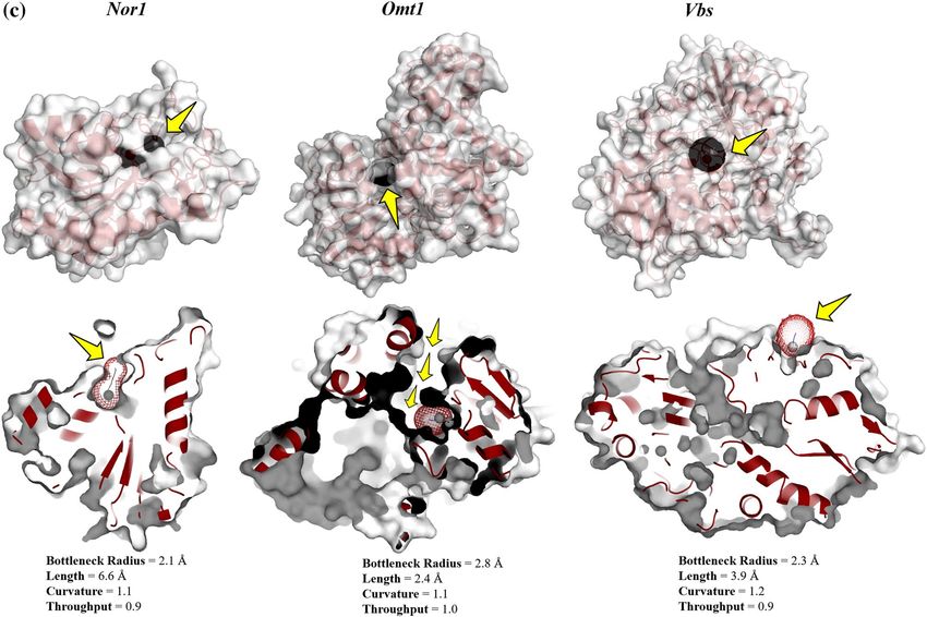
www.nature.com/scientificreports/
anethole, and verbenol are considered environmentally acceptable, and listed as “Generally Recognized as Safe”
(GRAS) by the Joint FAO/WHO Expert Committee on Food Additives (JEFCA) for m ankind6.
Zingiber officinale Roscoe is one of the oldest medicinal herbs exhibited a wide range of bioactivity such
as anti-cancerous, neuroprotective, antidiabetic, cardiovascular protective, and antiemetic7. Furthermore, the
antifungal activity of Zingiber officinale essential oil (ZOEO) has already been reported against Fusarium verticil-
lioides, F. moniliforme, F. oxysporum, Candida albicans, Penicillium chrysogenum, Aspergillus solani, A. oryzae,
A. niger, A. ochraceus, and A. flavus8.
In the past few decades, the use of in-silico techno-advancements, molecular dynamics (MD) has become
a potential tool in elucidating the behavioral patterns, strength, properties of the protein, and their interactive
sessions with ligand molecules. MD simulation studies are well suited to explain the working mode of action
(MOA) of the bioactive compounds in the context of drug d esigning9. In the molecular context of aflatoxin bio-
synthetic machinery, the products of structural genes such as aflK (AFLA_139190), aflP (AFLA_139210), and
aflD (AFLA_139390), i.e., VERB synthase (Vbs), Omt-1 and Nor-1 respectively, are the prime targets to elucidate
the molecular MOA of bioactive compounds10–12. Therefore, investigation of ligand specificity and interactive
signature receptor residues is essential to decipher the inhibitory MOA of the bioactive compounds (ligands).
The present study aimed to reveal the antifungal and anti-AFB1 potential of chemically characterized ZOEO
by deciphering the mechanism underlying its fungicidal and aflatoxin B 1 inhibitory actions. The fungal plasma
membrane, mitochondria and cell carbohydrate catabolism were taken as the prime targets for the biochemical
assays, with the intent of investigating the mechanism of antifungal activity of ZOEO. While, to understand the
multi-regime anti-AFB1 action of ZOEO at molecular level, the three major gene products Nor-1, Omt-1, and Vbs
that are critical in aflatoxin B1 biosynthetic cascade were targeted. Due to the lacking of the crystal structure of
protein–ligand complex, docking protocols were simulated to predict the complex matrix defining the favored
binding poses (energetically) of the selected ligand (compound) on the targeted receptor proteins (Nor-1, Omt-1,
and Vbs). Furthermore, MD simulations were used to investigate the role of these supramolecular complexes in
the dynamics of the targeted proteins, which may enhance or inhibit their biological function. The methodologi-
cal scheme summarizing the computational studies in this paper has been presented in Fig. 1.
Results
Chemo‑typing of essential oil and validating its antifungal and anti‑aflatoxigenic activi‑
ty. Chemical profile of ZOEO. Zingiber officinale rhizome EO was isolated with the hydro-distillation process
with yield of 12.32 ml/kg. GC–MS analysis revealed a total of 15 volatile components constituting ~ 99% of the
complete chemical profile of the isolated ZOEO. The significant peaks were identified as the verbenol (52.41%)
followed by 7-epi-Sesquithujene (6.8%), and γ-terpinene (5.18%) (Fig. 2a).
Antifungal and anti‑aflatoxigenic activity: biochemical analysis. The antifungal activity of the ZOEO was calcu-
lated in terms of minimum inhibitory concentration (MIC) against the A. flavus PN-05. The effect of ZOEO on
fungal dry weight and aflatoxin production was shown in Fig. 2b. The MIC of ZOEO was found to be 0.6 µl/ml;
however, its inhibitory activity was recorded at 0.5 µl/ml for the AFB1 content (Fig. 2b). The decrease in mycelial
dry weight and AFB1 content was in direct proportion with the doses of ZOEO.
The fungitoxic mechanism of ZOEO was elucidated through biochemical analysis of membrane integrity
(perturbance in ergosterol content and membrane cations), mitochondrial membrane potential (MMP) and
carbohydrate catabolism.
The efficacy of ZOEO in perturbing the cell membrane integrity of A. flavus was deciphered by its effect on
production of ergosterol and perturbance in membrane cations (Ca++, K+ and M g++) flow. The percent reduction
of ergosterol content was found to be 37.95%, 44.38% and 62.77% respectively, exposed to 0.125, 0.25 and 0.5 µl/
ml doses of ZOEO, compared with the control (Fig. 2c); while, the increase in the ion leakage was observed from
the mycelia of the A. flavus fumigated with the ZOEO (Fig. 2d).
The MMP plays an important role in energy-coupling phenomena of mitochondria via generation of ATP.
The reduction in the fluorescence intensity of the dye reflects the degradation of MMP. The reduction in the
fluorescence intensity of the A. flavus cells exposed to different doses of ZOEO (0.2, 0.4 and 0.6 µl/m) can be
easily visualized in Fig. 2e.
There were three categories of the carbon sources (monosaccharide, oligosaccharide and amino based carbon
sources) which were the soul carbon sources of metabolic pathway. Therefore, these carbon sources were analyzed
to visualize the alterations in the carbohydrate catabolism of the fungal cell exposed to ZOEO (0.6 µl/ml). The
result indicated that the d-Arabinose, l-Arabinose, d-Ribose, d-Trehalose, d-Xylose (monosaccharide), Malto-
triose, Palatinose, Stachyose, Sucrose, (oligosaccharide), and Alaninamide, l-Asparagine, l-Ornithine, l-Proline,
l-Pyroglutamic acid (amino based) carbon sources of control sets were highly utilized over the other moderately
or least utilized carbon sources. Whereas, the A. flavus cells treated with ZOEO showed the significant decline
in the utilization of the above-mentioned carbon sources (Fig. 3).
Molecular docking: the binding affinity of the receptor‑ligand systems. Target proteins selec-
tion. The selection of the target proteins was based on their crucial activity reported in aflatoxin-biosynthesis
in the previous studies with A. parasiticus and A. flavus10–12. Therefore, to decipher the preferred mechanism of
aflatoxin biosynthetic inhibition, Nor-1, Omt-1, and Vbs were selected as the target proteins. They serve as the
potential targets to assess the anti-aflatoxigenic activity of the selected compound (verbenol) as they perform the
essential functions in aflatoxin b iosynthesis12–16. The Nor-1 (aflD gene product), a short-chain dehydrogenase/
reductase enzyme is required to convert the norsolorinic acid (NOR) into the averantin (AVN)13. Papa13,14 dem-
onstrated the conversion of NOR to AVN as one of the critical intermediate steps for the aflatoxin production,
Scientific Reports | (2021) 11:6832 | https://doi.org/10.1038/s41598-021-86253-8 2
Vol:.(1234567890)
www.nature.com/scientificreports/
Figure 1. Methodological scheme summarizing the flow-chart of computational analysis.
while Trail et al.15 proved the involvement of the aflD (Nor-1) in aflatoxin production. Another selected target
protein in the pathway was Vbs (VERB synthase: aflK gene product). This enzyme catalyzes the synthesis of ver-
sicolorin B (VERB) from versiconal (VAL), another key step in aflatoxin biosynthesis16. Vbs was held responsible
for aflatoxin’s toxicity and carcinogenicity12. The Omt-1/Omt-A (aflP gene product), a methyltransferase enzyme
play a crucial role in the later steps of the biosynthetic pathway, catalyzes two reactions in the pathway, i.e., con-
version of sterigmatocystin (ST) to o-methylsterigmatocystin (OMST) and dihydrosterigmatocystin (DHST)
to dihydro-o-methylsterigmatocystin (DHOMST)16,17. These steps are the second last steps for the synthesis of
aflatoxins (AFB1 from OMST and A FB2 from DHOMST). Significance of Omt-1 can be seen with the fact that,
Aspergillus nidulans has ST as the end product rather than aflatoxins since it lacks an aflP orthologue12.
Scientific Reports | (2021) 11:6832 | https://doi.org/10.1038/s41598-021-86253-8 3
Vol.:(0123456789)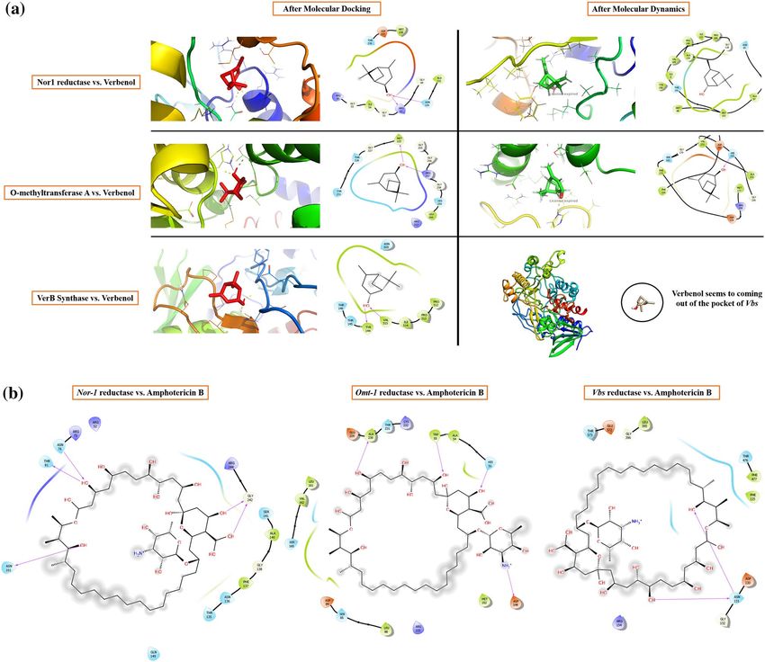
www.nature.com/scientificreports/
Figure 2. In-vitro analysis: (a) chemical profile of ZOEO; (b) anti-fungal and anti-aflatoxigenic activity of
ZOEO; (c) % inhibition of ergosterol content; (d) vital cellular ion leakage; (e) mitochondrial membrane
potential.
Homology modeling and model validation. In the present study, the target proteins Nor-1, Omt-1, and Vbs were
lacking the crystal structure, no hits were found when searched with the keywords ‘Nor-1’, ‘Omt-1’, and ‘Vbs’
converging the search on ‘Aspergillus flavus’ on the RCSB-PDB website. Therefore, homology modelling was
used to generate the atomic coordinates (3D structure) of the target proteins to perform the further compu-
tational studies18,19. For homology modeling, templates were identified subjecting the maximum query (target
protein FASTA sequence) coverage, maximum identity (PSI-BLAST), and maximum hits by pGEN THREADER
as the filters. The selected templates were 3WXB, 6IX8, and 5NCC for Nor-1, Omt-1, and Vbs, respectively. At
last, 3D models having stable and functional quality structures were filtered out based on the highest GMQE and
lowest QMEAN scores (Fig. SM1)20.
The stereochemical-stability validation of the 3D models of Nor-1, Omt-1, and Vbs gave the overall quality
score of 86.7, 88.3, and 89.07, respectively, out of 100 scoring function of SAVES v5.0. The Ramachandra plot
statistics validate the acceptance of the 3D models (Fig. ST3). For reliability of the 3D models, their validity has
been cross checked by analyzing their RMSD values in reference to their template crystal s tructures21,22. Nor-1
superimposed with protein PDB ID: 3WXB having RMSD 0.586, Omt-1 superimposed on protein PDB ID: 6IX8
with RMSD 0.713 and Vbs showed RMSD of 1.524 on superimposition with PDB ID: 5NCC (superimposed
images: Fig. SM2). The RMSD less than 2 Å indicates a close homology and ensures reliability of the model in
reference to the experimental data. P roTSAV23 assesses the quality of the protein structure at various interfaces
viz. PROCHECK , ProSA , ERRAT26, Verify3D27, and P
24 25
roQ28. It revealed the stable stereochemical properties
of all the 3D models with RMSD values in the range of good model (green-yellow zone) (Fig. SM3). Further,
protein information concerning its motifs, helices, strands, domains, tunnels, angles, positions, and errors were
assessed via P DBsum21 web-tool to decipher the additional features of the target proteins (Fig. SM4).
Active site analysis. The active site analysis is a crucial task in view of setting the docking environment and
ensuring the reliability of the docking analysis. However, due to lack of the crystal structure of receptor-ligand
complex, computational algorithms were used to identify the active site of the proteins. In the present study,
CASTp 3.0 tool, working on alpha-shape theory, was used to investigate the surface region details of a protein to
analyze its interactive function with other molecules (ligand)29,30. CASTp 3.0 tool predicts the interior voids and
surface pockets of proteins. These internal cavities of proteins are of great interest in discovery of small drug-
like molecules that are associated with their binding e vents31–33. For all the target proteins, the binding pocket
selection was made following the coordinated study of structure information obtained from the PDBsum server
Scientific Reports | (2021) 11:6832 | https://doi.org/10.1038/s41598-021-86253-8 4
Vol:.(1234567890)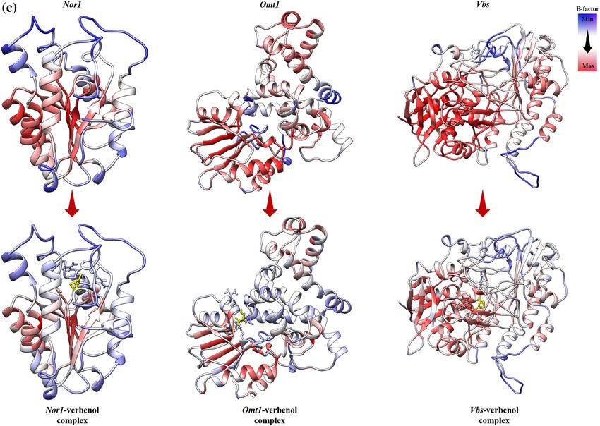
www.nature.com/scientificreports/

                                  Figure 3.  Carbon sources utilization pattern of A. flavus exposed to ZOEO (a) monosaccharide C-sources; (b)
                                  disaccharide C-sources; and (c) amino-acid based C-sources.

                                  and CASTp 3.0 webtool. The pockets selected for Nor-1, Omt-1, and Vbs contain the maximum number of the
                                  functionally critical secondary structures (Fig. SM5).

                                  Ligand selection and its toxicity estimation (ADMET properties). Verbenol being > 50% of the total chemical
                                  profile of the ZOEO and all the other compounds had < 10% in quantity individually (Fig. 2a). Therefore, it can
                                  be stated that the isolated ZOEO was of verbenol-chemotype. Hence, verbenol was selected as the ligand for in-
                                  silico assessment of the working MOA of anti-aflatoxigenic activity of ZOEO.

Scientific Reports |   (2021) 11:6832 |               https://doi.org/10.1038/s41598-021-86253-8                                                 5

                                                                                                                                          Vol.:(0123456789)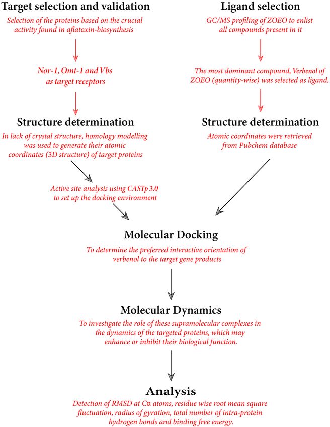
www.nature.com/scientificreports/
QikProp module, TEST, and VEGA v1.1.5 results
PubChem ID 61126 PlogS − 2.384
Reactive functional groups 0 PlogHERG (IC50) − 2.65
CNS 1 PCaco (nm/s) 4076.554
MW (g/mol) 154.252 PlogBB 0.19
Molecular formula C10H16O PMDCK (nm/s) 2259.46
SASA (Å2) 378.466 PlogKp − 2.173
FOSA 337.803 #metab 1
FISA 40.663 PlogKhsa − 0.064
Volume (Å3) 621.779 Human oral absorption 3 (high)
H-bond donor 1 PSA 20.497
H-bond acceptor 1.7 Number of nitrogen and oxygen atoms 1
Cohesive interaction index in solids 0.004492 Rule of five 0
Globularity Index 0.930885 Rule of three 0
Oral rat LD50 mg/kg (predicted) 2547.06 Tumorigenicity Negative
Mutagenicity Negative Carcinogenicity Negative
Drug-induced liver injury (Hepatotoxicity) Negative Biodegradation Positive
Persistence (sediment) nP (70 days) Persistence (water) nP (10 days)
Persistence (soil) nP (5 days) Air half-life 0.4162 h
Molinspirtation server (bioactivity) Swiss target prediction (Homo sapiens)
GPCR ligand − 0.18 Target Probability
Ion channel modulator 0.03 Androgen receptor 0.61
Kinase inhibitor − 1.42 Sodium-dependent noradrenaline transporter 0.51
Nuclear receptor ligand − 0.17 Sodium-dependent dopamine transporter 0.51
Protease inhibitor − 0.54 Muscarinic acetylcholine receptor M1 0.49
Enzyme inhibitor 0.02 Muscarinic acetylcholine receptor M2 0.43
PreADMET (ADMET) Muscarinic acetylcholine receptor M3 0.43
hERG inhibition Low risk Muscarinic acetylcholine receptor M4 0.43
Carcino mouse/rat Negative Muscarinic acetylcholine receptor M5 0.43
CYP 2C19 inhibition Inhibitor Cytochrome P450 19A1 0.43
CYP 2C9 inhibition Inhibitor Cholinesterase 0.39
CYP 2D6 inhibition Non Acetylcholinesterase 0.39
CYP 2D6 substrate Non Sodium-dependent serotonin transporter 0.39
CYP 3A4 inhibition Inhibitor Tyrosyl-DNA phosphodiesterase 1 0.38
CYP 3A4 substrate Non Estrogen receptor 0.38
Human intestinal absorption (%) 100 Estrogen receptor beta 0.38
Table 1. ADMET profiling and ecological risk assessment. MW molecular weight, SASA total solvent
accessible surface area, FOSA hydrophobic component of the SASA, FISA hydrophilic component of the SASA,
PlogS predicted aqueous solubility (log S), PlogHERG predicted IC50 value for blockage of HERG K+ channels,
PCaco predicted apparent Caco-2 cell permeability (model for the gut-blood barrier), PlogBB predicted
brain/blood partition coefficient, PMDCK predicted apparent MDCK cell permeability, PlogKp predicted
skin permeability (log Kp), PlogKhsa prediction of binding to human serum albumin, PSA Van der Waals
surface area of polar nitrogen and oxygen atoms, Rule of five number of violations of Lipinski’s rule of five,
#metab number of likely metabolic reactions, Rule of three number of violations of Jorgensen’s rule of three
(PlogS > − 5.7, PCaco > 22 nm/s, #Primary Metabolites < 7), nP non-persistant.
The toxicity assessment of the compound is used to estimate its deleterious effects over both human health
and the environment. Verbenol was assessed for determining its effects on human health, environment, and other
rediction34, Molinspirtation server, PREADMET, QikProp module (QikProp,
ecological factors. Swiss Target P
Schrodinger LLC, NY, 2017), TEST (Toxicity Estimation Software Tool) of US Environmental Protection Agency
and VEGA v1.1.5 were assessed to generate a comparative result analysis for more accurate result prediction.
Based on the results shown in Table 1, verbenol does not seem to violate the measured parameters of safety (if
provided at the acceptable range) in all the in-silico protocols used, it is also biodegradable, non-mutagenic,
non-hepatotoxic, non-carcinogenic and non-tumorigenic. All the parameters have been calculated and verified
for compliance with their standard ranges.
Molecular docking: assessing the molecular affinity of verbenol against target proteins. The lack of crystal struc-
tures of receptor-ligand complexes for target proteins, led to use the docking algorithms to identify the binding
position and the affinity persuaded between the small molecule (ligand) and the target proteins at the molecular
Scientific Reports | (2021) 11:6832 | https://doi.org/10.1038/s41598-021-86253-8 6
Vol:.(1234567890)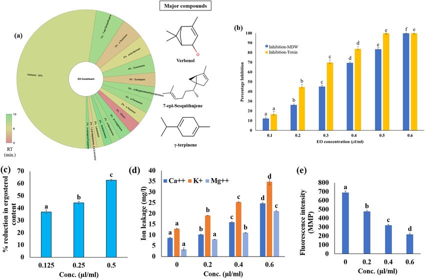
www.nature.com/scientificreports/
Receptor Ligand* Coordinates of the Grid Centre (Å) Grid dimensions (Å)
X = 49.626 X = 104
Verbenol Y = − 10.685 Y = 86
Z = − 15.266 Z = 106
Nor-1
X = 47.426 X = 80
Amphotericin B Y = − 10.76 Y = 96
Z = − 14.997 Z = 104
X = 23.525 X = 118
Verbenol Y = − 31.482 Y = 108
Z = − 18.444 Z = 126
Omt-1
X = 22.871 X = 116
Amphotericin B Y = − 31.482 Y = 108
Z = − 19.501 Z = 126
X = 34.635 X = 104
Verbenol Y = 38.764 Y = 82
Z = 48.296 Z = 126
Vbs
X = 32.894 X = 122
Amphotericin B Y = 38.896 Y = 76
Z = 48.664 Z = 124
Table 2. Details of the docking environment used for the molecular docking study.
Interactions Bond type Resides and their legends Binding energy (kcal/mol) Inhibition constant (µM) Ligand efficiency
Hydrogen bond ARG52, GLY49, GLY55, ASN130
Hydrophobic bond MET238, ALA132, ILE54
Verbenol with Nor-1 − 6.10 33.52 − 0.55
Polar bond ASN130, THR236
Charged bond ASP237, ARG56, ARG52
Hydrogen bond MET225, ARG257, HIS259
Hydrophobic bond MET225, LEU260, GLY227, GLY255
Verbenol with Omt-1 − 5.81 55.49 − 0.53
Polar bond THR228, THR231, HIS259
Charged bond ARG257, ARG313
Hydrogen bond TYR144, VAL515
THR146, PRO512, PRO513, ILE514,
Verbenol with Vbs Hydrophobic bond − 5.56 84.62 − 0.51
VAL515
Polar bond THR145, THR146, ASN505
Hydrogen bond ASN76, THR81, ASN101, GLY242
Hydrophobic bond PHE137, GLY138, ALA140
Amphotericin B with Nor-1 − 6.76 11.00 − 0.10
Polar bond THR135, ASN136, SER141, GLN149
Charged bond ARG52, ARG75, ARG244
Hydrogen bond HIS51, TRP55, ASP148, ALA230,
Hydrophobic bond ALA54, LEU88, MET152, LEU161, VAL162
Amphotericin B with Omt-1 − 7.49 3.23 − 0.12
Polar bond SER85, SER165, THR231
Charged bond ASP84, GLU229, LYS232
Hydrogen bond ASN131
PHE125, GLY132, LEU395, GLY396,
Hydrophobic bond
Amphotericin B with Vbs PHE477 − 6.72 11.95 − 0.10
Polar bond THR476, THR573
Charged bond ASP130, ARG154
Table 3. Docking details of all the three protein–ligand systems.
l evel35. Prepared structure of the verbenol molecule was docked into the pre-adjusted docking environment in
the target proteins as per the details given in Table 2. Binding predictions made by AutoDock were based on
the empirical force field and the Lamarackian Genetic Algorithm, that scores the receptor-ligand binding posi-
tions in terms of the binding energy in kcal/mol (sum of the intermolecular energy and the torsional energy)36.
The obtained docking energies and binding details were shown in Table 3, and binding poses were presented
Scientific Reports | (2021) 11:6832 | https://doi.org/10.1038/s41598-021-86253-8 7
Vol.:(0123456789)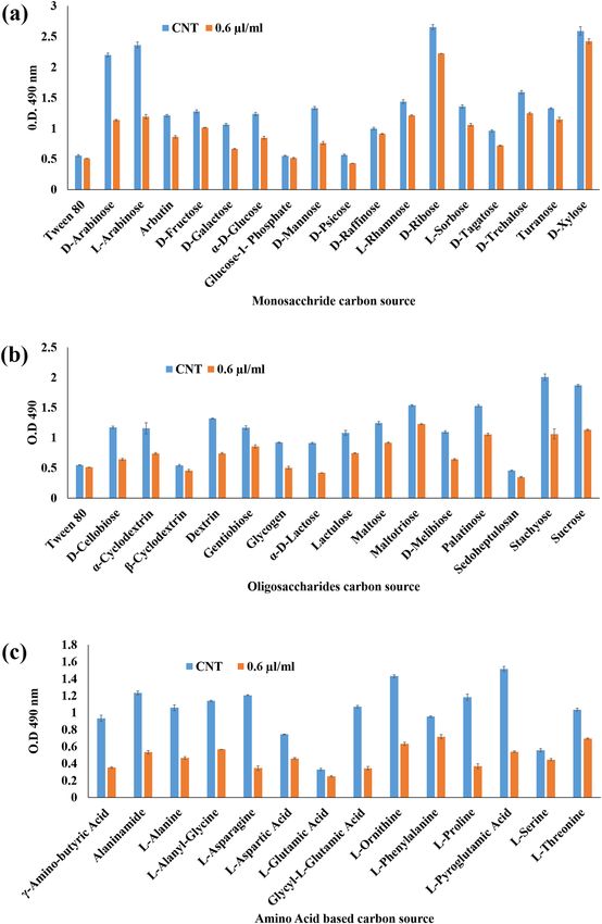
www.nature.com/scientificreports/

                                            Figure 4.  (a) Interactive behavior of target proteins with verbenol; (b) the binding details of amphotericin with
                                            target proteins; (c) B-factor profiling of proteins and protein-verbenol complexes.

          Scientific Reports |   (2021) 11:6832 |                https://doi.org/10.1038/s41598-021-86253-8                                                  8

Vol:.(1234567890)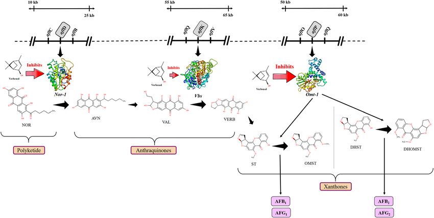
www.nature.com/scientificreports/
Figure 4. (continued)
in Fig. 4a. Furthermore, for the validation of the predicted binding poses of the receptor-ligand complexes, all-
atom MD simulation was performed37.
Verbenol showed the dynamic multi-regime inhibitory activity against the target proteins. It showed the
highest binding affinity with the Nor-1 followed by Omt-1 and Vbs. Verbenol formed the hydrogen bonds with
ARG52, GLY49, GLY55, and ASN130 in Nor-1, MET225, ARG257, and HIS259 in Omt-1 and with TYR144,
and VAL515 in Vbs. Based on the binding energy profile of verbenol, its Ki (inhibition constant) value for each
target proteins were calculated following the Eq. (1).
Ki = exp GR−1 T−1 , (1)
where ∆G = Binding energy in cal/mol, R = Gas Constant (1.9187 cal/mol/K), T = Temperature (300 K).
The Ki value is a valid parameter to measure the efficacy of the inhibitor towards the enzyme quantitively.
Low Ki denotes a high binding affinity of the inhibitor38. Verbenol showed the lowest possible inhibition constant
towards the Nor-1, followed by Omt-1 and Vbs (Table 3). Furthermore, Ki values of verbenol against all the target
proteins/enzymes only differed by a little amount, which represents the multi-regime inhibitory potential of the
verbenol against aflatoxin biosynthesis.
Comparative analysis with Amphotericin B (commercial antifungal drug). The commercial antifungal drug
amphotericin B has been selected for the comparative study of molecular docking results. MIC of amphotericin
B (1.8 µl/ml)39 was comparatively higher than the ZOEO (0.6 µl/ml) (Fig. 2a) A comparative study of binding
energy, inhibition constant and ligand efficiency has been shown in Table 3. The results revealed that the ampho-
tericin B showed the better efficacy in inhibiting the target proteins (Nor-1, Omt-1, and Vbs) that have crucial
roles in AFB1 biosynthesis than verbenol. As well as, verbenol also showed remarkable binding affinity near
to amphotericin B with target proteins which reveals its utility as a plant-based antifungal agent. The binding
details of amphotericin with target proteins were represented in Fig. 4b. Since amphotericin B is commercially
available and established antifungal drug, hence, we have not performed the molecular simulation analysis with
amphotericin B.
MD simulation approach: validatory investigation of receptor‑ligand complexes. To study the
effect of verbenol binding on the targeted gene products (i.e., Nor-1, Omt-1 and Vbs) and establish their validity,
Scientific Reports | (2021) 11:6832 | https://doi.org/10.1038/s41598-021-86253-8 9
Vol.:(0123456789)www.nature.com/scientificreports/
all-atom molecular dynamics simulation of 300 ns for receptor-ligand complexes of target proteins was in refer-
ence to the modelled structures of the target proteins employed (except Nor-1 for which 500 ns simulation length
was used). A total of 2 µs simulation was achieved for all six systems under study. On the obtained trajectory,
several calculations (structural, dynamical, and thermodynamic) were performed to understand the inhibitory
potential of verbenol against Nor-1, Omt-1, and Vbs. In the following sections, we have shown that the effect of
verbenol binding on Nor-1, Omt-1, and Vbs at structural, dynamical, and thermodynamics level.
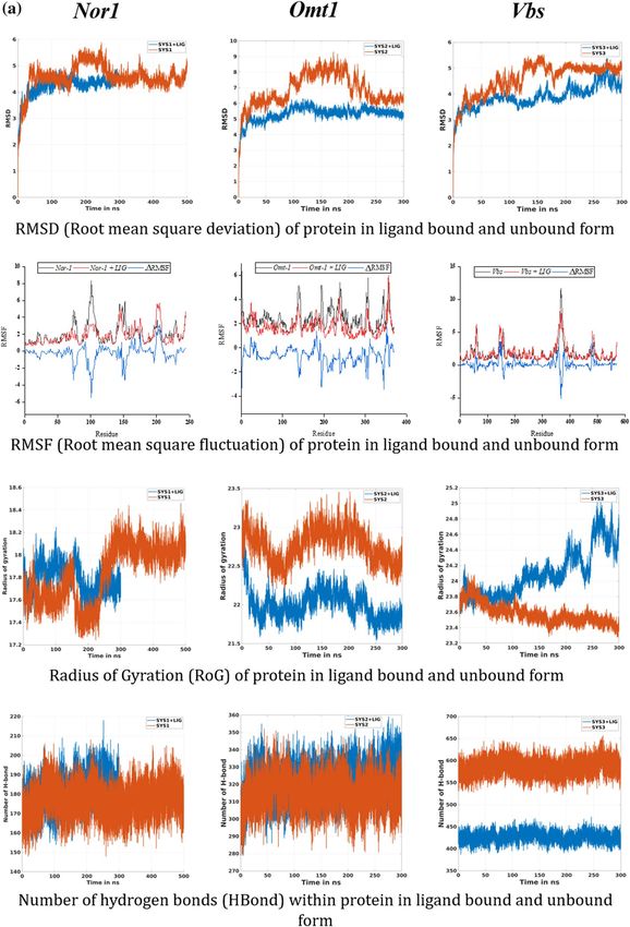
Structural and dynamic behavior analysis. The structural behavior of the target proteins in their ligand-bound
and unbound forms were assessed in the form of their backbone RMSD, Cα RMSF, B-factor, and Rg analysis.
RMSD value quantifies the flexibility difference between two structures, considering one of them as reference
structure (initial structural conformation was taken reference here). In terms of RMSD, protein structures hav-
ing less deviation are the more stable ones, and vice versa40,41. As shown in Fig. 5a, all the three receptor-ligand
systems attained the stability earlier than their reference proteins (without ligand) (~ 100 ns for both Nor-1/ver-
benol and Omt-1/verbenol, ~ 200 ns for Vbs/verbenol and ~ 250 ns for all the three target proteins Nor-1, Omt-1,
and Vbs). With the inspection of the average RMSD values, complex systems showed stable conformations than
their respective target proteins, which emphasizes the attainment of stability by target proteins on ligand bind-
ing. Nor-1/verbenol system showed lower RMSD (4.21 Å) than the Nor-1 (4.57 Å), similarly, RMSD reported for
the Omt-1/verbenol (5.31 Å), and Vbs/verbenol systems (3.89 Å) were lower than that of native target proteins
Omt-1 (6.84 Å) and Vbs (4.58 Å).
Likewise, the fluctuation of residues from their time-averaged position during the simulation was observed
as their RMSF (root mean square fluctuation). It determines the flexibility of the proteins as a function of their
residue numbers40. With the graphs presented in Fig. 5a, it could be seen that the RMSF values of the target
proteins (Nor-1 and Omt-1) were relatively higher in their ligand-unbound forms on the contrary to the ligand-
bounded systems. In the case of the Vbs, the fluctuation seems to be neutral, emphasizing no effects of the ligand
binding on the residue positioning. This was further investigated by the visual inspection of the trajectories, and
the fluctuation of target proteins from their ligand-unbound form to ligand-bound form was presented as the
ΔRMSF in the Fig. 5a.
The B-factor or Debye–Weller factor value of protein structure reflects its local motion, i.e., fluctuation from
their average positions due to the kinetic energy of a toms42. The average B-factor fluctuation was calculated using
the coordinates of the atoms (r) of the protein backbone from the Eq. (2).
B = 8π2 �r 2 /3. (2)
The results showed in Fig. 4c, it could be inferences that the B-factor of the ligand-bound proteins were
relatively lower than that of their ligand-unbound forms, i.e., the local motions of the atoms of the proteins were
stabilizing upon ligand binding.
The radius of gyration (Rg) is the measure of protein compactness that works in an inversely proportional
manner like RMSD40. The compactness of the protein differs in its bound and unbound states with the ligand.
The Rg analysis of protein in ligand unbound and bound systems also support the previous finding (RMSF). The
target proteins in their ligand unbound form portrayed the gyradius of 17.82 nm (Nor-1), 22.76 nm (Omt-1),
and 23.57 nm (Vbs), while the proteins in ligand-bound form have the gyradius of 17.78 nm (Nor-1/verbenol),
22.0 nm (Omt-1/verbenol), and 24.10 nm (Vbs/verbenol). As it also can be seen in the Fig. 5a, Gyradius of the
target proteins Nor-1 and Omt-1, in their ligand unbound systems, were relatively higher than their ligand-bound
form that characterizes the overall decrease in the compactness of protein–ligand binding. However, for the Vbs,
there were higher gyradius for the ligand-bound form instead of ligand-unbound form.
Ligand binding might lead to some conformations changes in protein structure, to study the same we per-
formed PCA analysis on obtained trajectories. With the help of PCA, we can characterize significant motions
taking place in a protein. The eigenvector with the highest eigenvalue (PC1) captures maximum variance (largest
collective motion) in the protein structure. In contrast, the eigenvector with the second largest eigenvalue (PC1)
describes the second-largest collective motion in the protein. When we plot PC1 vs. PC2 (in 2D) obtain from
MD simulation data, we observe similar conformations of protein falling into the same cluster. As can be seen
from Fig. 5b, all the target proteins with or without ligand reach a stable conformation represented by the yel-
low cluster. However, the cluster form throughout 300 ns varies significantly with verbenol bound and unbound
form. It showed that the conformational sampling of target proteins Nor-1, Omt-1, and Vbs varies significantly
in verbenol bound and unbound form, which supports the study via RMDS, RMSF, Rg, and H-bond analysis.
H‑bond analysis: the dynamic behavior of proteins. We assessed the total H-bonds of target proteins in the
ligand bound and unbound states along with the ‘unique’ H-bonds between the target proteins and verbenol
with 3 Å of donor–acceptor distance and 45° of angle cutoff which can significantly affect the protein stability40.
The average number of hydrogen bonds detected in each frame as a function of time were 176 (Nor-1), 315 (Omt-
1), 585 (Vbs) for ligand unbound states of proteins and 181 (Nor-1/verbenol), 322 (Omt-1/verbenol), and 426
(Vbs/verbenol) for their ligand-bound states. The H-bonding pattern deciphered the high number of H-bond-
ing in the ligand-bound form of the proteins Nor-1 and Omt-1. However, Vbs showed the higher H-bonds in
its ligand unbound form, which lined in the result patterns of RMSF and gyradius analysis. The number of
H-bonds formed in-between the verbenol, and target proteins were 31, 37, and 120 for the Nor-1/verbenol,
Omt-1/verbenol and Vbs/verbenol systems, respectively. The details regarding these unique H-bonds were given
in Supplementary Table ST1. There was only a single -OH group present in the structure of verbenol, which
was involved in all the H-bonding with the target proteins. Therefore, there was a smaller number of H-bonds
Scientific Reports | (2021) 11:6832 | https://doi.org/10.1038/s41598-021-86253-8 10
Vol:.(1234567890)www.nature.com/scientificreports/
Figure 5. (a) Molecular dynamics simulation analysis; (b) PCA analysis; (c) tunnel analysis of protein–ligand
complexes.
Scientific Reports | (2021) 11:6832 | https://doi.org/10.1038/s41598-021-86253-8 11
Vol.:(0123456789)www.nature.com/scientificreports/
Figure 5. (continued)
Scientific Reports | (2021) 11:6832 | https://doi.org/10.1038/s41598-021-86253-8 12
Vol:.(1234567890)www.nature.com/scientificreports/
Figure 5. (continued)
between the ligand and receptor proteins with minimal occupancy. Instead, Nor-1/verbenol and Omt-1/verbenol
system showed the occupancy of some H-bonds above 10% while Vbs/verbenol system has all the H-bonds with
occupancy < 3%.
In order to ensure the positioning of verbenol in the binding cavity of the target proteins in due course of
simulation time, the distance between the center of mass (COM) of the ligand with the COM of the binding
pocket of the target proteins as a function of time were assessed (Fig. SM6). Results assured the closeness of the
COM of verbenol to the COM of binding pockets of the Nor-1 and Omt-1 proteins with an average distance of
13.23 Å and 17.7 Å while verbenol seems to be coming out of the Vbs binding pocket with an average distance
of 34.65 Å.
Binding free energy analysis: binding selectivity prediction by MM/PBSA. In the enzyme-inhibitor systems, the
efficacy of inhibitors to inhibit the enzymes are substantially related to the stability of their binding against
enzyme. Therefore, the MD simulation study of the inhibitors should be evaluated in terms of their free energy
of binding at the active sites of the target proteins. The binding affinity profiles of verbenol against target proteins
were used for the selectivity of the best inhibitory action43. In the present study, the MD simulations were com-
bined with the MM/PBSA approach to predict the binding free energies for all three receptor-ligand complex
systems, i.e., Nor-1/verbenol, Omt-1/verbenol, and Vbs/verbenol. The combinatorial approach of MM/PBSA and
MD simulations are thought to be more accurate to identify the correct binding conformations. With the results
given in Table 4, the predicted binding free energies (ΔGbind) with Poison Boltzmann (PB) model for Nor-1/
verbenol, Omt-1/verbenol, and Vbs/verbenol systems were − 2.95 ± 2.96, − 3.79 ± 4.05, and − 0.83 ± 3.17, respec-
tively. These results impact the superiority of the Nor-1/verbenol and Omt-1/verbenol systems over the Vbs/
verbenol. Along with, ΔEEEL (change in electrostatic energy), ΔEVDW (change in van der Walls energy), ΔGPBSA
(change in solvation energy, polar and non-polar contributions), and ΔGbind (MM-PBSA binding free energy)
also support the pattern of ΔGbind.
Ligand pathway: CAVERDOCK analysis. The analysis of the pathway for the ligand (inhibitor) transport to the
binding site of the protein holds a cornerstone in unlocking the conformational cascades taking place during the
process. Furthermore, protein tunnels and channels are the key factors that assist the ligand passage to proteins’
external and internal environments. CaverDock, a fast-computational tool that facilitates the analysis of tunnel
detection and ligand transport within a single environment, has been used in this s tudy44. The ‘easiness’ in the
ligand placement has been depicted in the form of energy values: EMax (the highest binding energy in the trajec-
tory) and E
a (activation energy of association: EMax − ESurface for reactants). The lower the EMax and Ea values, the
Scientific Reports | (2021) 11:6832 | https://doi.org/10.1038/s41598-021-86253-8 13
Vol.:(0123456789)www.nature.com/scientificreports/
Nor-1 Omt-1 Vbs
ΔEVDW − 22.73 ± 1.78 − 21.10 ± 2.59 − 11.73 ± 7.20
ΔEEEL − 3.73 ± 2.83 − 5.25 ± 5.35 − 1.83 ± 3.78
ΔGPBSA 23.52 ± 3.34 22.56 ± 4.25 12.73 ± 7.67
ΔGbind − 2.95 ± 2.96 − 3.79 ± 4.05 − 0.83 ± 3.17
Table 4. The energy components predicted by MM/PBSA analysis (kcal mol−1). *Mean ± SD. ΔEEEL change in
electrostatic energy, ΔEVDW change in van der Walls energy, ΔGPBSA change in solvation energy both polar and
nonpolar, and ΔGbind MM-PBSA binding free energy.
Parameters Nor-1 Omt-1 Vbs
Bottleneck radius (Å) 2.1 2.8 2.3
Length (Å) 6.6 2.4 3.9
Curvature 1.1 1.1 1.2
Throughput 0.9 1 0.9
Ebound (kcal/mol) − 5.2 − 4.7 − 3.7
Emax (kcal/mol) 0.4 − 4.7 − 3.3
Esurface (kcal/mol) − 0.1 − 4.7 − 3.3
Ea (kcal/mol) 0.5 0 0
ΔEBS (kcal/mol) − 5.1 0 − 0.4
Table 5. CaverDock energy profile of all three target proteins.
Figure 6. Schematic representation of molecular mechanism of action of anti-aflatoxigenic activity of verbenol.
more the easiness in the ligand placement. The possible routes detected for the target proteins were shown in
Fig. 5c, along with their energy profiles (Table 5).
Discussion
Prior to detailed investigation, ZOEO was chemically characterized by GC–MS, which revealed verbenol
(52.41%) as the major compound of oil (Fig. 2a). The chemical profile of ZOEO was in accordance with the
previous reports45,46. However, the quantity of the chemical constituents varied because of the various abiotic
and biotic factors such as method of extraction, edaphic factors, age and part of plants47. Antifungal efficacy of
Scientific Reports | (2021) 11:6832 | https://doi.org/10.1038/s41598-021-86253-8 14
Vol:.(1234567890)www.nature.com/scientificreports/
ZOEO and verbenol has been previously reported by Prakash et al.48 and Al-Ja’Fari et al.49. However, detailed
investigation on the molecular details of the AFB1 inhibition and mechanism of fungitoxic potential of ZOEO is
still lacking in literature so far. Therefore, in the present study, the mechanism of fungitoxic potential along with
the molecular mechanism of aflatoxin B1 inhibition/dysfunction of ZOEO was investigated via the biochemical
and computational based approaches.
The present study showed the fungi-toxic nature of the ZOEO and its components (mainly verbenol) due to
their negative implications on the fungal cell membrane, depolarization of the mitochondrial membrane and
hampering of the carbohydrate catabolism pathway of the cell. The lipophilic nature of the EO made its entry
through cell membrane resulting the damaging effects on the membrane integrity. The reduction in the ergosterol
production in treated A. flavus cells (Fig. 2c), ensures the effect of the ZOEO on the major sterol of fungal cell
membrane. OuYang et al.50 have previously reported that the EO or its component cause the downregulation of
the significant genes involved in ergosterol biosynthesis. These phenomena might be responsible for the alteration
in the permeability of the membrane and causing the leakage of the various cations as confirmed in the present
study (Fig. 2d). The study also revealed the significant alteration in the membrane potential of mitochondria in
a dose-dependent manner (Fig. 2e), which suggested the disruption in the functioning of mitochondria. The
alterations in the MMP were observed using a cell permeable cationic dye, Rh123, which can penetrate into
the mitochondria and reflect the M MP51,52. Above all, ZOEO may shattered the various metabolic pathways
viz pentose phosphate pathway (PPP), glycolysis and tricarboxylic cycle (TCA) by declining the utilization of
the significant carbon sources (Fig. 3). Thus, ZOEO inhibits the nutrient supply and damages the secondary
metabolite production pathway of the A. flavus cells.
The anti-AFB1 efficacy of the ZOEO was reported in many reports but the detailed molecular mechanism
was still lacking in the literature48. Therefore, to assess the comprehensive molecular mechanism of ZOEO,
computational approach has been set out targeting the major gene products of the biosynthesis pathway. Since,
the bioactivities of essential oils are related to their chemotype i.e., their major compound, hence all the com-
putational studies were performed with the major compound (verbenol) for untangling the site of action of
AFB1 biosynthesis in comparison with the commercial antifungal drug, amphotericin B. This is the first attempt
to assess the multi-regime anti-AFB1 mechanism of verbenol targeting the aflD, aflK, and aflP gene products.
Also, the safety limit profile of verbenol was explored using its ADMET profile (Table 1). The ADMET results
revealed its favorable safety limit profile, biodegradable and non-toxic nature. The conclusions drawn were based
on the comparative MD simulation approach of the target proteins in their ligand-bound and unbound forms.
The docking analysis confirmed the inhibitory binding of verbenol to all the target proteins. In absence of
experimental data regarding the receptor-ligand complexes of target proteins, docking results were verified using
the all-atom MD simulation a pproach37. It provides the ultimate details concerning the conformational changes of
the target proteins upon ligand binding. These structural details of the docking-based receptor-ligand complexes
of the target proteins led us to understand the molecular attributes of the verbenol-inhibitory action. RMSD and
B-factor values confirmed the gain of stability to all the target proteins upon ligand binding (Fig. 4a). While,
RMSF and Rg analysis showed the gain of compactness and less fluctuant motion only for the Nor-1 and Omt-1
on ligand binding, which is further verified by the higher number of the H-bonding in their ligand-bound state
(Fig. 5a). In Vbs, there were fewer H-bonds in the ligand-bound state in comparison to its ligand unbound state,
which might be the possible explanation of its relaxed gyradius and fluctuant motion of the protein residues. The
H-bonds pattern between the verbenol and target proteins were also aligned with the above total H-bond results.
These phenomena strike out the importance of the H-bonding for the stability of the protein 3D conformation.
Essential dynamics analysis also provided some insight into the dynamical behavior of Nor-1, Omt-1, and Vbs
in their ligand-bound and unbound form. The mode of significant motions occurring in the target proteins were
captured via PC1 and PC2, which reveals a significant difference in major modes of motion in all three-target
protein (Nor-1, Omt-1, and Vbs) in their ligand-bound form as compared to that of their ligand unbound form
(Fig. 5b). These dynamics analysis was found to be consistent with the other analysis exploring the structural
behaviors of the same. The MM/PBSA energy profiling preferred the Nor-1/verbenol and Omt-1/verbenol systems
simultaneously as the better inhibitory action over the Vbs/verbenol system (Table 4). The ΔGbind calculated with
Poison Boltzmann (PB) model have the scheme of Omt-1/verbenol ~ Nor-1/verbenol > Vbs/verbenol. The other
factors of thermo dynamical behavior viz. ΔEMM, solvation energy (ΔGPBSA), polar and non-polar contributions
demonstrate a similar aspect. All of the above MD simulations results were converging at the inference that, in
Nor-1 and Omt-1, verbenol was bind at the interior of the protein structure while in Vbs, verbenol has the surface
binding. Therefore, verbenol has the inhibitory action over the Nor-1 and Omt-1, making them more stable than
their natural form. While in Vbs, verbenol was also making the stability actions. However, its surface binding was
causing the formation of the lower number of H-bonds that, in turn, perturbing the RMSF and gyradius data of
the protein. The binding positions of receptor-ligand complexes obtained after MD calculations were shown in
Fig. 4a. At last, the CaverDock analysis predicted the possible route of ligand placement into the active site of
target proteins. The energy data obtained from the CaverDock reveals that the inhibitory action of the verbenol
was only because of its binding at the active site rather than any tunnel blockage in the target proteins (Table 5).
The predicted molecular mechanism of action of verbenol has been presented (Fig. 6).
Conclusion
The findings of present investigation unravel the antifungal mode of action and molecular site of action of the
verbenol-chemotype ZOEO against the biosynthesis of aflatoxin B 1 using the biochemical and computational
approaches. The investigation provided the antifungal and A FB1 inhibitory mode of action of ZOEO related with
the damaging effects on the biochemical aspects (ergosterol production, MMP and carbohydrate catabolism) of
the A. flavus. Further, the binding positions of verbenol with the target gene products (Nor-1, Omt-1, and Vbs)
Scientific Reports | (2021) 11:6832 | https://doi.org/10.1038/s41598-021-86253-8 15
Vol.:(0123456789)www.nature.com/scientificreports/
involved in aflatoxin B
1 biosynthesis were investigated using docking procedures and validated with all-atom MD
simulation approach. ADMET profile of verbenol revealed its favorable safety limit profile, biodegradable and
non-toxic nature, strengthen its use as an eco-friendly antifungal preservative. In view of considerable antifungal,
AFB1 inhibition and multi-regime inhibition efficiency of ZOEO and its compound verbenol, further practical
investigations are warrant against A. flavus and A FB1 contamination in the food system.
Methods
Chemicals and equipment. All the chemicals used in the present study were procured from the Hi-Media
Laboratories Pty Ltd., Mumbai, and SRL, Mumbai, India.
Extraction and chemical profiling of ZOEO by GCMS. The rhizomes of Zingiber officinale were pro-
cured from the Botanical garden of the Banaras Hindu University, Uttar Pradesh state, India and authenticated
using the relevant taxonomic literature53. A voucher specimen (Zin./Zin-149/2018) has been deposited in the
herbarium of Department of Botany, Banaras Hindu University, Varanasi. The rhizomes (500 g) underwent
hydro-distillation at 95 °C for 4 h using the Clevenger’s apparatus (Merck Specialties Pvt. Ltd., Mumbai, India),
and the essential oil was collected in sterilized glass vial. The ZOEO was dried overnight using the anhydrous
sodium sulfate ( Na2SO4) and stored at 4 °C for chemical profiling54. The chemical profile of ZOEO was authenti-
cated using the GC/MS (PerkinElmer, Turbomass Gold, USA) with the PerkinElmer Elite-5 column. The experi-
mental procedure was similar to our previous published paper4.
Efficacy against Aspergillus flavus and AFB1 contamination. The PN-05 strain of Aspergillus flavus,
previously isolated in our laboratory, was chosen for the present investigation52. The antifungal activity of the
ZOEO was tested against the A. flavus PN-05 using the poison food technique where the potato dextrose agar
(PDA) plates were poisoned in a gradient manner from 0.1 to 0.6 μl/ml of ZOEO dissolved in 0.5 ml tween 20
(5%). Afterward, these plates were seeded with a 5 mm disc of the fungal hyphae at the center of each plate and
incubated at 27 ± 2 °C for 7 days. The lowest ZOEO concentration having no visible fungal growth, was recorded
as the minimum inhibitory concentration (MIC)4.
For investigating the anti-AFB1 efficacy, gradient concentrations of ZOEO (0.1 to 0.6 μl/ml) were separately
mixed with the 24.5 ml of SMKY liquid medium (sucrose, 20 g; magnesium sulfate, 0.5 g; potassium nitrate, 3 g;
yeast extract, 7 g; and distilled water, 1000 mL) and inoculated with a hundred microliter spore suspension of A.
flavus PN-05. After the incubation of 10 ten days, the filtered media was used for the estimation of AFB1 content
following the protocol of Prakash et al.55, based on the following formula:
D×M
AFB1 content µg/ml = × 1000
E×L
where, D, absorbance; M, the molecular weight of A
FB1 (312); E, the molar extinction coefficient of AFB1
(21,800); L, path length (1 cm cell was used).
Antifungal mode of action. Effect of ZOEO on ergosterol production in the plasma membrane of A. fla-
vus. The effect of different doses of ZOEO on the ergosterol production in the plasma membrane of the A. fla-
vus was analyzed following the protocol of Kumar et al.4. The A. flavus PN-05 mycelia were treated with different
dosses of ZOEO (0.125, 0.25 and 0.5 µl/ml) in SMKY medium and kept for five days’ incubation. Afterwards,
mycelia harvest was done followed by addition of 5 ml of freshly prepared 25% alcoholic KOH solution and
vortex for 2 min. Then after, the solutions were evaporated on water both at 85 °C for 4 h. followed by addition of
5 ml n-heptane and 2 ml sterile distilled water and vortex for 2 min. At last, optical density of the n-heptane layer
was analyzed at 230 and 300 nm and ergosterol content was calculated using the following equation:
A282 /290
%Ergosterol + %24(28) dehydroergosterol = ,
Pellet Weight
A230 /518
%24(28) dehydroergosterol = ,
Pellet Weight
A282 /290 A230 /518
%Ergosterol = −
Pellet Weight Pellet Weight
Effect of ZOEO on cellular ions. The 5-days incubated mycelia of A. flavus PN-05 was mixed with 20 ml of
0.85% saline solution followed by treatment of different doses of ZOEO (0.2, 0.4 and 0.6 µl/ml) and incubation at
room temperature for 12 h. Afterwards, solution was filtered using the Whatman filter and filtrate was analyzed
using atomic absorption spectrometry56.
Effect of ZOEO on mitochondrial membrane potential (MMP). For analyzing the alterations in the membrane
potential of mitochondria, the spore suspension (106 spores/ml in tween 20 (0.5%)) of A. flavus PN-05 was
treated with ZOEO at concentration of 0.2, 0.4 and 0.6 µl/ml for ten hours. Afterwards, samples were centrifuged
at 5000 rpm for 5 min. and pellets were suspended in phosphate buffer saline solution. Subsequently, the stain-
Scientific Reports | (2021) 11:6832 | https://doi.org/10.1038/s41598-021-86253-8 16
Vol:.(1234567890)www.nature.com/scientificreports/
ing of sample solutions was done using rhodamine 123 (1 μg/ml) for 15 min in the dark. Again, samples were
centrifuged and mixed with the PBS solution followed by optical density quantification of Rh123 dye at 488 nm
(excitation) and 525 nm (emission)51. A control set (without treatment) was also kept for the simultaneous
assessment.
Effect of ZOEO on carbon‑source utilization. The carbon-source utilization pattern of A. flavus PN-05 exposed
ublications4 with Biolog FF Microplate
to ZOEO (0.6 µl/ml) was assessed following the method of our previous p
(94545, Hayward, CA).
Molecular docking: the binding affinity of the receptor‑ligand systems. Homology modeling and
its authentication. To assess the molecular details of the inhibitory action of the ZOEO, computational analysis
has been set out. Firstly, three-dimensional (3D) structures of the target proteins were required. Therefore, in
lack of crystal structures of receptor-ligand complexes for target proteins, homology modeling technique was
used to generate the 3D models of the target proteins. Firstly, the amino acid sequences of the target proteins
were retrived from the NCBI database corresponding to the accession number EED51173 (Nor-1), EED51156
(Omt-1), and EED51154 (Vbs) in FASTA format. Further, the sequences were submitted to the PSI-BLAST
and pGEN THREADER (http://bioinf.cs.ucl.ac.uk/psipred) web services for the template selection. Afterward,
SWISS-MODEL web atmosphere (https://swissmodel.expasy.org)20 was used to generate the 3D models in refer-
ence to the selected templates. Validation and determining the acceptability of the generated 3D models were
done with the Structure Analysis and Verification Server (SAVES) v5.0 at https://servicesn.mbi.ucla.edu/SAVES
and ProTSAV function of SCFBIO23.
Binding site prediction. The active site prediction requires crystal structure of receptor–ligand complex. Hence
in the absence crystal structure, computational analysis was deployed to predict the active site of the target pro-
teins. It is the hydrophobic cavities (interior voids and surface pockets) of the proteins that are held responsible
for their specific binding e vents30. Therefore, the theoretical determination of these cavities was necessary for
predicting more accurate binding results. The Computed Atlas of Surface Topography of proteins (CASTp) 3.0
web facility was used to predict these kinds of active sites in the target proteins27. The modeled 3D structures of
the proteins were submitted to the CASTp web facility to predict the necessary amino acids.
Ligand preparation and its ADMET profiling. The native structure of ligands (verbenol and amphotericin B)
were retrieved from the PubChem database in .sdf format and processed to convert in the .pdbqt form for
further computational analysis. In addition, Swiss Target Prediction34, Molinspirtation server, PREADMET
(https://preadmet.bmdrc.kr), QikProp module (QikProp, Schrodinger LLC, NY, 2017), TEST (Toxicity Estima-
tion Software Tool) of US Environmental Protection Agency and VEGA v1.1.5 (https://www.vegahub.eu) were
assessed for the ADMET profiling and predicting the oral rat LD50, mutagenicity, drug-induced liver injury
(hepatotoxicity), tumorigenicity, carcinogenicity, biodegradation, and other ecological risk assessment factors
associated with verbenol.
Docking algorithm. The docking algorithm was performed using the AutoDock 4.2 docking protocols to deter-
mine the preferred interactive orientation of verbenol to the target gene products57. The docking parameters
used were according to the standard genetic algorithm (GA) characters i.e., 10 runs of GA, population size
150, maximum number of ratings 250,000, maximum number of generations 27,000, and crossover rate 0.8.
The docking environment were adjusted on the target proteins with the grid box having dimensions tabulated
in Table 2. The grid-box dimensions were constructed for each target protein following their CASTp structure
information. AutoDock 4.2 scoring function generates 120,000 ligand poses and rank ten superior conforma-
tions according to their binding energies. The selection of the receptor-ligand complexes was based on their
binding energies and their positioning in the predictive active sites (visual inspection).
MD simulation approach: validatory investigation of receptor–ligand complexes. Molecular
dynamics simulation. Nor-1, Omt-1, and Vbs were subjected to all-atom MD simulation with and without
ligand. So, a total of six systems were simulated. For MD simulation, all the initial structures were prepared
in tleap module of Amber16 by utilizing the ff14SB force field58. The ligand forcefield parameters were gener-
ated using antechamber and gaff while the charges were generated using AM1-bcc method. TIP3P water model
was used to solvate the simulation box with a padding distance of 12 Å and NaCl were used as counter ions
to neutralizing the total charge of the system59,60. Joung and Cheatham ion parameters were used for Na+ and
Cl− ions61. All MD simulations were performed using the Amber16 package62. For the electrostatic interaction
calculation, Particle-mesh Ewald (PME) method was used. To restrain the bonds involving hydrogen SHAKE
algorithm was used. For temperature and pressure control, Langevin dynamics and Berendson barostat were
utilized59.
First, to minimize the energy of the six systems, a restrain was kept of protein–ligand complex, and only the
solvent was minimized. After this round of minimization, the restrain potential was removed from complex, and
the entire system was minimized. Followed by minimization, the gradual heating of the systems was performed
at NVT with a 100 kcal restrain potential on complex and temperature was increased to 300 K in 300 ps. After
heating, each system was subjected to equilibration for 1.8 ns in six cycles of 300 ps in which the restrain poten-
tial was subsequently reduced from 100 kcal/mod to 0 kcal/mol (100, 50, 20, 5, 0.5, 0 kcal/mol) at NPT59. After
equilibration, all six systems were subjected to a production run of 300 ns each (except the Nor-1-ligand system
Scientific Reports | (2021) 11:6832 | https://doi.org/10.1038/s41598-021-86253-8 17
Vol.:(0123456789)You can also read