CAPN3: A muscle specific calpain with an important role in the pathogenesis of diseases (Review)
←
→
Page content transcription
If your browser does not render page correctly, please read the page content below
INTERNATIONAL JOURNAL OF MOlecular medicine 48: 203, 2021
CAPN3: A muscle‑specific calpain with an important
role in the pathogenesis of diseases (Review)
LIN CHEN1,2*, FAJUAN TANG1,2*, HU GAO1,2, XIAOYAN ZHANG1,2, XIHONG LI1,2 and DONGQIONG XIAO1,2
1
Department of Emergency Medicine, West China Second University Hospital, Sichuan University;
2
Key Laboratory of Birth Defects and Related Diseases of Women and Children (Sichuan University),
Ministry of Education, Chengdu, Sichuan 610041, P.R. China
Received May 13, 2021; Accepted September 10, 2021
DOI: 10.3892/ijmm.2021.5036
Abstract. Calpains are a family of Ca2+‑dependent cysteine release from skeletal muscle fibers, calcium uptake of sarco‑
proteases that participate in various cellular processes. Calpain 3 plasmic reticulum, muscle formation and muscle remodeling.
(CAPN3) is a classical calpain with unique N‑terminus and Studies have indicated that recessive mutations in CAPN3
insertion sequence 1 and 2 domains that confer characteristics cause limb‑girdle muscular dystrophy (MD) type 2A and other
such as rapid autolysis, Ca2+‑independent activation and Na+ types of MD; eosinophilic myositis, melanoma and epilepsy
activation of the protease. CAPN3 is the only muscle‑specific are also closely related to CAPN3. In the present review, the
calpain that has important roles in the promotion of calcium characteristics of CAPN3, its biological functions and roles in
the pathogenesis of a number of disorders are discussed.
Contents
Correspondence to: Professor Xihong Li or Professor Dongqiong
Xiao, Department of Emergency Medicine, West China Second
University Hospital, Sichuan University, 20 Renmin South Road 1. Overview of CAPN3
Section 3, Chengdu, Sichuan 610041, P.R. China 2. CAPN3 activation and inhibition
E‑mail: lixihonghxey@163.com 3. Features of CAPN3
E‑mail: m13881749494@163.com 4. Physiological function
5. CAPN3 in diseases
*
Contributed equally 6. Conclusions and future perspectives
Abbreviations: CAPN3, calpain 3; NS, N‑terminus; IS1, insertion
sequence 1; MD, muscular dystrophy; PC1, protease core domain 1; 1. Overview of CAPN3
PEF, penta‑EF‑hand (E, E‑helix; F, F‑helix); CBSW, calpain‑type
β‑sandwich; PLEIAD, platform element for inhibition of autolytic
degradation; CTBP1, C‑terminal binding protein 1; CaM,
Discovery. In 1989, Sorimachi et al (1) discovered a molecular
calmodulin; CBS‑1, calcium binding site; iMOC, intramolecular clone named p94 that encodes a novel calcium‑dependent
complementation; AldoA, aldolase A; RyR, ryanodine receptor; protease comprising 821 amino acid residues with a rela‑
NCX3, Na+‑Ca2+ exchanger isoform 3; FLNC, filamin C; MLC1, tive molecular mass of 94 kDa. In 2013, p94 was renamed
myosin light chain 1; PIAS, protein inhibitor of activated states; calpain 3 (CAPN3) (2), or calcium‑activated neutral protease,
LGMD2A, limb‑girdle muscular dystrophy type 2A; CaMKIIβ, by the American Association of Experimental Biology.
calmodulin kinase IIβ; C3KO, CAPN3 knockout mice; NF‑κ B, In humans, CAPN3 is located in the chromosomal region
nuclear factor κ B; GFAP, glial fibrillary acidic protein; SVZ, 15q15.1‑q21.1 (3). Mainly expressed specifically in skeletal
sub‑ventricular zone; Ca‑CaMKII, Ca2+/CaM‑dependent protein muscle (1), the mRNA of CAPN3 has also been detected in
kinase II; PGC1α, peroxisome proliferator‑activated receptor‑γ the early embryonic heart, but it will gradually disappear from
coactivator‑1α; CARP, cardiac myotonic repeat protein; c‑FLIP, cell
the ventricular area, and finally, only the transcript of CAPN3
FLICE inhibitor protein; DYSF, dysferlin; TMD, tibial MD; DMD,
Duchenne MD; FSHD, facioscapulohumeral MD; FRG1, FSHD
is present (4,5). In addition, CAPN3 is also expressed in the
region gene 1; CAPN3 E6‑, CAPN3 subtypes lacking exon 6; UCMD, cytoplasm and nucleus of neuron‑like PC12 cells, as well as in
Ullrich congenital MD; IEM, idiopathic eosinophilic myositis; IBM, rat astrocytes and in the brain of Microcebus (6,7).
inclusion body myositis; AD, Alzheimer's disease; FUS, fusiform
gyrus; CAD, coronary artery disease; AAV, adeno‑associated virus Structure. So far, the calpain family has 16 members, including
classic calpains and non‑classical calpains (summarized in
Key words: CAPN3, muscle formation, muscle remodeling, Fig. 1). As a classic calpain, CAPN3 exhibits a large subunit
limb‑girdle muscular dystrophy type 2A containing four domains (I‑IV). Domain I is distinctly
different from the other domains. Domain II is a conserved
cysteine protease
domain comprising protease core domain 1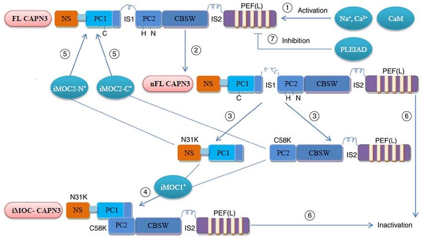
2                                     CHEN et al: CAPN3 IN THE PATHOGENESIS OF DISEASE

(PC1) and PC2 that confer protease activity. Domain III,          3. Features of CAPN3
also known as the calpain‑type β‑sandwich domain (CBSW
or C2L), is responsible for protein structural changes upon       Autolysis. Since CAPN3 contains specific sequences NS, IS1
CAPN3 activation. Domain IV, or the penta‑EF‑hand (E,             and IS2 that are not present in other calpains or proteases,
E‑helix; F, F‑helix) (PEF) domain, mainly participates in         CAPN3 exhibits unique characteristics. For instance, the
calcium ion binding and CAPN3 homodimerization. The               complete autolysis of CAPN3 is rapid and is independent of
sequences of domain II and IV in CAPN3 are homologous             Ca2+ activation (22). CAPN3 is so far the only intracellular
with those in other calpains (1).                                 enzyme that relies on Na+ activation.
    In particular, CAPN3 contains three additional insertion          The half‑life of CAPN3 in vitro is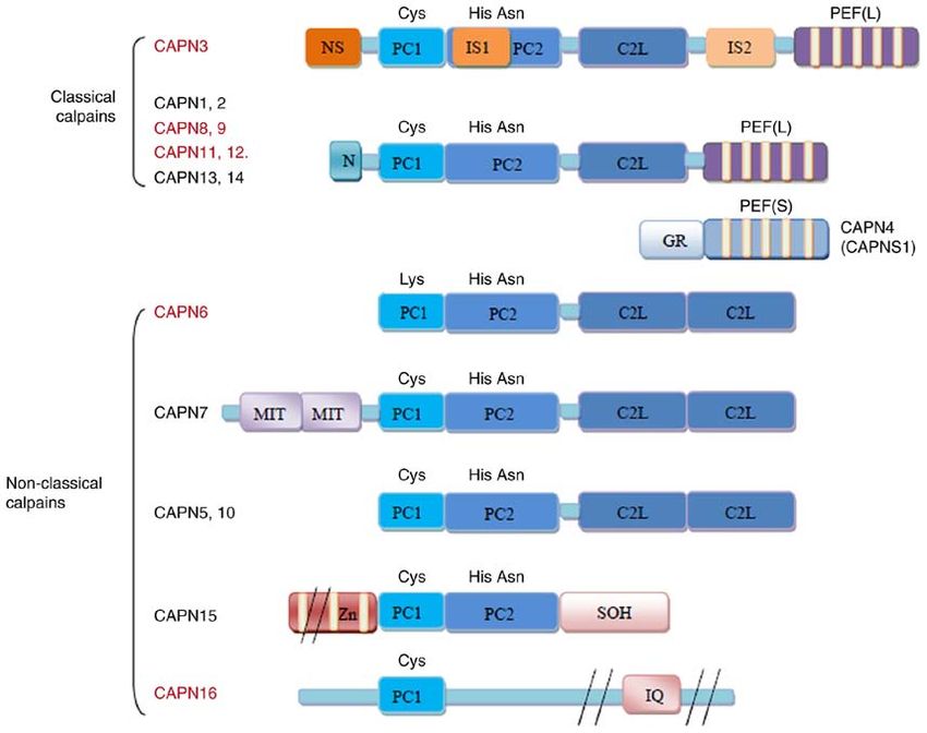
INTERNATIONAL JOURNAL OF MOlecular medicine 48: 203, 2021                                                            3

Figure 1. Structure of the human calpains. The calpains presented in red are predominantly expressed in specific tissues or organs, while those in black are
more widely expressed. The major difference in the structure of CAPN3 is that it contains three additional insertion sequences, namely NS at the N‑terminus,
IS1 of PC2 and IS2 between CBSW/C2L and PEF (L). Small subunits are not present in CAPN3. CAPN3, calpain 3; GR, Gly‑rich domain; MIT, micro‑
tubule‑interacting and transport motif; Zn, zinc‑finger motif; SOH, small optic lobes product homology domain; IQ, calmodulin‑interacting motif; NS,
N‑terminus; IS1, insertion sequence 1; PC1, protease core domain 1; PEF, penta‑EF‑hand (E, E‑helix; F, F‑helix); CBSW/C2L, calpain‑type β‑sandwich.

while CAPN3 localized in the cytoplasm may be related to the                    significantly increase cellular Ca2+ uptake through the reverse
control of cell motility or skeletal plasticity (37,38). CAPN3                  mode of NCX3 ingestion. Exercise causes excitation‑contrac‑
has both proteolytic and non‑proteolytic functions.                             tion uncoupling, NCX3 increases the uptake of Ca 2+ and
                                                                                supplements Ca2+ in the sarcoplasmic reticulum (41).
CAPN3 promotes calcium release of skeletal muscle fibers
and calcium uptake of sarcoplasmic reticulum. The triad is                      CAPN3 promotes muscle formation and muscle remodeling.
the structural component of the muscle responsible for calcium                  CAPN3 mRNA is expressed at high levels in muscles (42).
transport and excitation‑contraction coupling. Aldolase is also                 CAPN3 is distributed in the amorphous plaques of myoblasts,
present as one of the components in muscles rich in triads.                     the area close to the myotube nucleus, the adhesion plaques
Glycolytic enzyme aldolase A (AldoA) is the binding partner                     and stress fibrous structures of myotubes and the filaments of
of CAPN3. CAPN3 is able to degrade AldoA, but AldoA is                          fibroblasts (43). CAPN3 is located in the Z line and N2 line
not the body substrate of CAPN3. Aldolase and CAPN3 are                         regions of the sarcomere by binding to its chaperone protein
able to interact with ryanodine receptor (RyR) to form the                      titin (33). The most characteristic function of CAPN3 is that
main calcium release channel. Compared with the wild‑type,                      it is located in the sarcomere. CAPN3 is an important protein
the levels of AldoA and RyR associated with the triplet in                      required for muscle formation and a prerequisite for main‑
CAPN3‑deficient muscle are decreased; hence, CAPN3                              taining normal muscle function (42).
helps maintain the integrity of the triplet in skeletal muscle,
which then promotes the release of calcium from muscle                          Muscle formation. CAPN3 is mostly inactive in muscles. It
fibers (39,40).                                                                 may be activated by autolysis in the active site to destroy the
    The Na + ‑Ca 2+ exchanger isoform 3 (NCX3) is also                          cytoskeleton of actin. By lysing several endogenous proteins,
expressed in the triad of skeletal muscle. A number of studies                  the sarcomere and sarcomere components are lysed; this may
have indicated that NCX3 is associated with the increased                       enhance the adaptability of muscle cells to external and/or
Ca2+ content in the sarcoplasmic reticulum. CAPN3 is able to                    internal stimuli. Titin and filamin C, substrates of CAPN3,
increase the activity of NCX3, but only the NCX3‑AC variant,                    are co‑localized in multiple locations within the cytoskeleton
which is mainly expressed in the skeletal muscle, is sensitive to               structure in the body (38). Therefore, CAPN3 is a muscle cyto‑
calpain. Increased intracellular levels of Ca2+ ([Ca2+]i) or [Na+]i             skeleton regulator.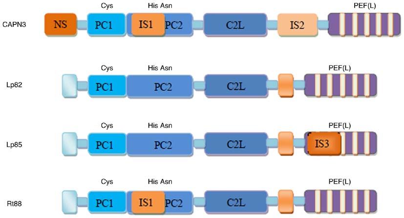
4                                             CHEN et al: CAPN3 IN THE PATHOGENESIS OF DISEASE

Figure 2. Representative CAPN3 isoforms generated by alternative splicing of CAPN3. Lp82, lack of exons 6, 15 and 16; Lp85, Lp82+IS3; Rt88, Lp82+IS1. IS3,
insertion sequence 3; CAPN3, calpain 3; PEF, penta‑EF‑hand (E, E‑helix; F, F‑helix); NS, N‑terminus; IS1, insertion sequence 1; PC1, protease core domain 1;
C2L, calpain‑type β‑sandwich.

Figure 3. Regulation of CAPN3 activity. ① Translated FL‑CAPN3 (FL‑CAPN3) is activated by physiological Na+/Ca2+ and CaM; ② FL‑CAPN3 undergoes
intramolecular dissolution and the IS1 sequence produces a nick (but not dissociated), which is nFL‑CAPN3; ③ N31K and C58K are dissociated from each
other; ④ N31K and C58K were recombined into active CAPN3 (iMOC‑CAPN3) by iMOC1, and its location and function were completely different from
FL/nFL‑CAPN3; ⑤ N31K /C58K and full‑length CAPN3 reorganization by iMOC2‑N/C to restore partial activity; ⑥ The IS2 and other sites in nFL‑CAPN3
undergo further autolysis and the recombined iMOC‑CAPN3 enters the next round of autolysis, and will eventually be inactivated; ⑦ PLEIAD is able to
bind to FL‑CAPN3 and inhibit CAPN3 activity through its C‑terminus. FL‑CAPN3, full‑length calpain 3; nFL‑CAPN3, nick full‑length CAPN3; PEF,
penta‑EF‑hand (E, E‑helix; F, F‑helix); NS, N‑terminus; IS1, insertion sequence 1; PC1, protease core domain 1; CBSW, calpain‑type β‑sandwich; iMOC,
intramolecular complementation; PLEIAD, platform element for inhibition of autolytic degradation; CaM, calmodulin.

    As a scaffold protein, CAPN3 may have a role during                         order to eliminate the interaction between FLNC and δ‑ and
the early stages of myogenesis. Muscle‑specific filamin C                       γ‑glycans, while the FLNC‑sarcoglycan interaction may have
(FLNC) is a candidate substrate of CAPN3. The C‑terminus                        a regulatory effect on CAPN‑mediated myoblast fusion (44).
of FLNC binds to the cytoplasmic domain of δ‑ and γ‑glycans.                    M‑cadherin has a role in the fusion of myoblasts. CAPN3
CAPN3 cleaves the C‑terminus of FLNC in living cells in                         is able to cut M‑cadherin and β ‑catenin. In the absence of
INTERNATIONAL JOURNAL OF MOlecular medicine 48: 203, 2021 5
CAPN3, M‑cadherin and β ‑catenin accumulate abnormally patients with residual CAPN3, the signs of abnormal regen‑
on the myometrial membrane, while myoblasts and myotubes eration of spiral fibers were highly enhanced in patients with
continue to fuse, which may inhibit muscle differentiation complete CAPN3 deficiency (52). Therefore, during sarcomere
steps, such as integrin complex body rearrangement and sarco‑ remodeling, CAPN3 is necessary for the muscle regeneration
mere assembly, inhibiting the formation of sarcomere (45). process.
Therefore, down‑regulation of CAPN3 leads to a decrease
in the number of muscle cells and in the size of myotubes Promotion of neurodegenerative processes. The Iκ Bα/nuclear
formed (43). factor‑κ B (NF‑κ B) signaling pathway is related to cell apop‑
tosis and it regulates the process of neurodegeneration by
Muscle remodeling. During the development of myoblasts into direct alteration of gene expression in neurons. Loss of CAPN3
fully differentiated myotubes in vitro, a number of ‘reserve proteolytic activity has been observed to severely interfere
cells’ are maintained. These reserve cells are closely related to with the Iκ Bα /NF‑κ B pathway (53). On the contrary, when
satellite cells responsible for muscle regeneration. The level of CAPN3 is activated, the Iκ Bα /CAPN3 complex is formed.
endogenous CAPN3 mRNA expressed in the reserve cells is Increased levels of calpain‑dependent Iκ Bα cleavage products
higher than that of proliferating myoblasts. CAPN3 is able to in the nucleus subsequently stimulate the activation of nuclear
decrease the transcriptional activity of MyoD, a key myogenic CAPN3‑like proteases in neurons and calpain‑dependent cell
regulator, through proteolysis in a manner that is independent death. The proteolysis of Iκ Bα, which activates the Iκ Bα/NF‑κ B
of the ubiquitin‑proteasome degradation pathway, participate pathway, promotes neurodegenerative processes (7).
in the establishment of the reserve cell bank and promote the
renewal of satellite cell compartments (46). The expression of A role in astrocyte plasticity and/or motility. Most astrocytes
CAPN3 mRNA after muscle injury (denervation‑devascular‑ in the brains of rats and Microcebus co‑express glial fibril‑
ization) is related to the muscle wet weight ratio. CAPN3 is lary acidic protein (GFAP), which is an ubiquitous target of
upregulated from day 7 to 14 in order to promote satellite cell calpain in vitro (54). GFAP is necessary for the movement of
renewal by inhibiting cell differentiation. CAPN3 is decreased astrocytes (55). CAPN3 is located in the cytoplasm of astro‑
significantly from day 14 to 28 in order to promote myoblast cytes, where GFAP is closely related to CAPN3. In addition,
differentiation in L6 cells and enhance the recovery function. cells expressing GFAP and CAPN3 are particularly common
Isoforms lacking exon 6 dominate the early regeneration in the sub‑ventricular zone (SVZ) and SVZ astrocytes are
process (47,48). actually the progenitor cells of stem cells that produce new
Substrates of CAPN3 are divided into two major functional neurons (56). Therefore, CAPN3 may have a role in astrocyte
categories: Metabolic substrates and myofibrils, including motility or cytoskeletal plasticity (6).
myosin light chain 1 (MLC1). CAPN3 has a proteolytic effect
on MLC1 in vitro (49). Among these substrates, there are three 5. CAPN3 in diseases
E3 SUMO ligases belonging to the protein inhibitor of activated
states (PIAS) family. CAPN3 is able to cleave PIAS protein Since calpain participates in various physiological processes
and negatively regulate the activity of PIAS3 sumoylase. In the in cells, dysregulation of CAPN3 may cause different diseases
muscle tissue of patients with limb‑girdle muscular dystrophy (Table I), such as MD, myositis or epilepsy.
(MD) type 2A (LGMD2A), SUMO2 is dysregulated (50).
Therefore, CAPN3, through fast‑acting one‑way proteolytic Muscular diseases
switch, has a significant role in muscle remodeling. CAPN3 MD. MD is a group of muscular diseases caused by genetic
also has a role upstream of the ubiquitin‑proteasome pathway factors characterized by progressive muscle weakness and
by targeting ubiquitination and proteasome degradation. degeneration of muscles that govern exercise. A common path‑
Increased expression of CAPN3 has been reported to enhance ological feature is muscle atrophy accompanied by hyperplasia
ubiquitination and promote sarcomere remodeling by cutting of fibrous tissue and adipose tissue. MD has a high degree of
and releasing myofibrillar proteins (51). genetic heterogeneity characterized by autosomal dominant or
In addition, in the triplet, CAPN3 co‑localizes with recessive inheritance of genes (57). CAPN3 is closely related
calmodulin kinase IIβ (CaMKIIβ). CAPN3 and CaMKIIβ to the occurrence of a variety of MD.
have a role in gene regulation during adaptive endurance exer‑
cise. Muscles of CAPN3 knockout mice (C3KO) have been LGMD2A. LGMD2A is the most commonly diagnosed type
reported to exhibit decreased triplet integrity and weakened of LGMD, accounting for 30‑40% of all LGMD cases (58).
CaMKIIβ signaling. After atrophy induction, it has been indi‑ LGMD2A is caused by a recessive mutation in CAPN3. So far,
cated that C3KO muscles were unable to activate CaMKIIβ >500 mutation sites have been reported. Different pathogenic
signaling and inducible heat shock protein 70, and that the CAPN3 mutations include missense mutations, frameshift
expression of cell stress‑related genes remained unchanged; mutations, nonsense mutations, deletions/insertions, splice site
hence the inflammatory response required to promote muscle mutations and single mutations. Among them, missense muta‑
recovery was absent. Meanwhile, C3KO muscles have been tions that are distributed along the entire CAPN3 gene are
indicated to exhibit decreased immune cell infiltration and most common (59‑64). These mutations have been observed
decreased expression of myogenic genes (34). In patients with to weaken CAPN3 proteolytic activity by affecting the protein
severe LGMD2A, recent muscle regeneration determined by inter‑domain interactions, decrease CAPN3 autocatalytic
the number of neonatal myosin heavy chain/vimentin‑positive activity by lowering its affinity towards Ca2+, cause complete
fibers was significantly decreased. Compared with those in or partial loss of CAPN3 protein (62,65‑67) or result in changes6 CHEN et al: CAPN3 IN THE PATHOGENESIS OF DISEASE
Table I. CAPN3 protein and genes in different diseases.
A, MD
Disease Protein expressiona Gene mutationa (Refs.)
Limb‑girdle MD type 2A Normal or low expression Mutation (59‑69)
Limb‑girdle MD type 2B Low expression Mutation (86,87)
Limb‑girdle MD type 2J Low expression ‑ (89,90)
Tibial MD Low expression ‑ (89,90)
Duchenne MD Low expression ‑ (91)
Facioscapulohumeral MD Overexpression Mutation (92‑94)
Ullrich congenital MD Low expression ‑ (95)
B, Other diseases
Disease Protein expression Gene mutation (Refs.)
Idiopathic eosinophilic myositis ‑ Mutation (96)
Inclusion body myositis Low expression ‑ (97,98)
Rhabdomyolysis syndrome Low expression ‑ (99,100)
Melanoma Overexpression ‑ (101‑106)
Epilepsy ‑ Mutation (107,108)
Alzheimer's disease Uncertain ‑ (111)
Diabetes Uncertain ‑ (112,113)
Cardiovascular diseases ‑ Mutation (114,115)
Vitiligo Overexpression ‑ (116)
Age‑related cataract Overexpression ‑ (117)
Not all diseases have clear protein expression changes and gene mutations. MD, muscular dystrophy.
a
of other characteristics of CAPN3. For instance, D705G and co‑regulator that coordinates muscle adaptive response) is
R448H mutations affect the ability and stability of CAPN3 to decreased, leading to the decreased levels of transcription
bind titin in vitro (68,69). These mutations have been reported of genes involved in muscle adaptation (40). In addition, the
to cause skeletal muscle Ca 2+ imbalance (70,71), abnormal loss of CAPN3 protease activity has been observed to impair
muscle adaptation (72,73), sarcomere disorder (74,75), oxida‑ the upregulation of muscle ankyrin repeat protein 2 and hsp,
tive damage (23,76), mitochondrial abnormality (42,77) and causing stress and muscle degeneration in skeletal muscles.
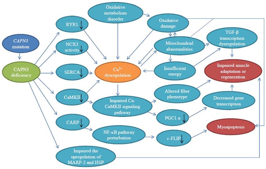
impaired muscle regeneration (77,78) (summarized in Fig. 4), Exercise has been indicated to exacerbate this change (73).
which then lead to inflammation, necrosis, fibrosis, atrophy In the end, long‑term failure of muscles to adapt and reshape
and progressive muscle degeneration. All these complications causes the occurrence of LGMD2A.
are the characteristics of LGMD2A. In addition, a multi‑protein complex comprising CAPN3
Dysregulation of Ca2+ in the skeletal muscles is a poten‑ and cardiac myotonic repeat protein (CARP) is present in the
tial event of MD, including LGMD2A (71,79,80). As one of N2A region of the sarcomeric protein titin. CAPN3 regulates
the constituents of the triplet, the absence of CAPN3, which CARP subcellular localization by cleaving the N‑terminus of
destroys the triplet integrity, decreases the release of Ca2+ in CARP. The higher the activity of CAPN3, the more impor‑
the skeletal muscles. CAPN3 deficiency also leads to degra‑ tant is the retention of CARP's sarcomere. Overexpression of
dation and dysfunction of skeletal muscle sarco‑endoplasmic CARP decreases the DNA binding activity of NF‑κ B p65, a
reticulum Ca2+‑ATPase 1 and 2 proteins, resulting in impaired factor that exhibits anti‑apoptotic effects. CAPN3 is unable
Ca2+ homeostasis in human myotubes (79) and NCX3 dysfunc‑ to decrease the inhibitory effect of CARP on NF‑κ B, which
tion, causing impaired reticular Ca2+ storage (41). may decrease the survival rate of muscle cells (84). In addi‑
CAPN3 knockout studies have indicated that Ca2+/calmod‑ tion, the activity of anti‑apoptotic factor cell FLICE inhibitor
ulin‑dependent protein kinase II (Ca‑CaMKII)‑mediated protein (c‑FLIP) depends on the NF‑κ B pathway in normal
signal transduction is impaired (81), which not only decreases muscle cells. CAPN3 is involved in regulating the expression
the slow muscle fiber phenotype and the fast muscle fiber of c‑FLIP. CAPN3‑dependent Iκ Bα is expressed after NF‑κ B
phenotype (82,83), but also lowers the level of p38 MAPK activation (75). In the muscle cells of patients with LGMD2A,
activation. Eventually, the level of peroxisome proliferator‑acti‑ NF‑κ B is activated under cytokine induction in the absence of
vated receptor‑γ coactivator‑1α (PGC1α; a transcriptional CAPN3, Iκ Bα accumulation prevents nuclear translocation ofINTERNATIONAL JOURNAL OF MOlecular medicine 48: 203, 2021 7
Figure 4. Schematic diagram of the pathogenic mechanism of CAPN3 deficiency. CAPN3 deficiency results in reduced levels of RyR1, SERCA, CaMKII and
CARP, decreased NCX3 activity and impaired upregulation of MARP‑2 and HSP, causing Ca2+ imbalance and compromise Ca‑CaMKII and NF‑κ B down‑
stream signaling pathways, which may lead to impaired gene transcription, mitochondrial abnormalities, oxidative damage and altered fiber phenotype. This
eventually leads to abnormal muscle adaptation, myocyte apoptosis and impaired regeneration. Among these mechanisms, there are multiple feedback loops.
Black arrows indicate a decline. CAPN3, calpain 3; RyR, ryanodine receptor; NCX3, Na+‑Ca2+ exchanger isoform 3; SERCA, sarco‑endoplasmic reticulum
Ca2+‑ATPase; Ca‑CaMKII, Ca2+/calmodulin‑dependent protein kinase II; CARP, cardiac myotonic repeat protein; MARP‑2, muscle ankyrin repeat protein 2;
HSP, heat shock protein; PGC1α, peroxisome proliferator‑activated receptor‑γ coactlvator‑1α; c‑FLIP, cell FLICE inhibitor protein.
NF‑κ B, the NF‑κ B pathway is dysregulated, c‑FLIP expres‑ resulting in an exceptionally benign phenotype (32). Therefore,
sion is downregulated, and finally, cell apoptosis and muscle the severity and progression of the disease are dependent on
shrinking occur (74,75). different gene mutations.
CAPN3 deficiency may also weaken the antioxidant defense
mechanism of skeletal muscles in LGMD2A patients [super‑ LGMD2B. Mutations in dysferlin (DYSF) are responsible
oxide dismutase (SOD)‑1 and nuclear factor erythroid‑2‑related for LGMD2B. DYSF is absent or minimally expressed
factor 2, but not SOD‑2], as well as increase lipid peroxidation in the muscle tissues of patients (85). DYSF is thought to
and protein ubiquitination, causing obvious oxidative damage have a role in membrane repair. CAPN3 and recombinant
and redox imbalance (76). desmoyokin (AHNAK; a protein involved in subsarcolemmal
Muscles of C3KO induced by cardiotoxin have too many cytoarchitecture and membrane repair) coexist in the DYSF
lobulated fibers belonging to the type of oxidative metabolism, protein complex. In skeletal muscle cells with normal CAPN3
increased connective tissue, insufficient muscle energy and no expression activity, the expression of AHNAK is decreased;
increase in mitochondrial content, PGC‑1α and ATP synthase conversely, in cells without CAPN3 expression, the expression
subunit δ transcripts, resulting in significant TGF‑ β tran‑ of ANHAK is increased. Furthermore, cleaved fragments of
scription level increases with microRNA dysregulation, and AHNAK by CAPN3 lose their affinity for DYSF in skeletal
the radial growth of muscle fibers is weakened, Akt/mTOR muscles. Therefore, CAPN3 is a modulator of the DYSF
complex 1 signaling is disturbed, this signal is uncoupled from protein complex in skeletal muscles (86). The expression of
protein synthesis, and in C3KO myoblast cultures, myotube CAPN3 in the skeletal muscles of patients with LGMD2B is
fusion is defective (77), so that healthy sarcomere cannot be decreased. Further analysis has indicated missense mutations
reconstructed. This is the pathogenesis of abnormal muscle in CAPN3 that change the amino acids of CAPN3 into the
regeneration in patients with LGMD2A. amino acids present in CAPN1 and CAPN2 (87). Therefore,
Although the symptoms of most patients with LGMD2A CAPN3 is associated with the onset of LGMD2B, but the
are usually uniform, studies have indicated that, compared specific mechanism exerted by CAPN3 in LGMD2B requires
with homozygous missense mutations, compound heterozygous further exploration.
mutations (such as pG222R/pR748Q) have a compensatory
effect and that the presence of ‘molecular complementation’ may LGMD2J and tibial MD (TMD). Titin C‑terminal mutations
rescue a certain amount of the proteolytic activity of CAPN3, in the M‑band of striated muscles may cause LGMD2J and8 CHEN et al: CAPN3 IN THE PATHOGENESIS OF DISEASE
TMD (88). CAPN3 binds titin at the C‑terminus and uses it as no corresponding weakness); and iii) increased peripheral
a substrate in vitro. There are several CAPN3 cleavage sites in blood eosinophils (96); its pathogenesis is related to mutation
the C‑terminus of titin. Hydrolysis of titin at the C‑terminus by of CAPN3. Further studies that focus on the physiological
CAPN3 may have an important role in normal muscles (89). functions of CAPN3 in skeletal muscles, particularly analyses
Titin C‑terminal mutations lead to the loss of binding site of CAPN3 protein and gene in patients, are required to better
for CAPN3 and also to the lack of CAPN3 in muscle (89,90). understand IEM.
Therefore, CAPN3 is involved in the pathogenesis of LGMD2J
and TMD. Inclusion body myositis (IBM). IBM is the most commonly
diagnosed primary myopathy in the elderly (97). Disorders of
Duchenne MD (DMD). DMD is a lethal X‑linked muscle calcium homeostasis in cardiomyocytes may exacerbate factors
disease caused by defective expression of cytoskeletal protein that mediate IBM muscle degeneration. Immune‑mediated
dystrophin (Dp)427. Certain retinal neurons express Dp427 membrane damage and/or abnormal accumulation of proteins
and/or the short subtype of dystrophin. Therefore, patients may be the cause of calcium disorders in patients with IBM.
with DMD may also experience specific visual defects. A Analysis of muscles in patients with IBM indicated that insuf‑
study that analyzed whether the lack of Dp427 affects the late ficient expression of CAPN3 mRNA and protein, which is
development of retina in mdx mice (the most in‑depth study conducive to proper calcium homeostasis and increased abun‑
of DMD animal models) indicated that, compared with that of dance of two CAPN3 substrates (97,98). The pathogenesis of
age‑matched wild‑type mice, the expression of genes E18‑P5 IBM remains to be clarified and the role of CAPN3 in IBM
and P5 that encode proteins related to retinal development and remains to be explored.
synaptogenesis, including CAPN3, is transiently decreased in
mdx mice (91). Rhabdomyolysis syndrome. Rhabdomyolysis syndrome is
a medical emergency caused by exposure to external trig‑
Facioscapulohumeral MD (FSHD). FSHD is a muscle disease gers and may be related to increased genetic susceptibility.
related to the loss of heterozygous D4Z4 on chromosome Previously, two patients have been diagnosed with rhabdomy‑
4q35. In previously reported atypical cases, DNA analysis olysis syndrome after consuming wild quail meat. Analysis
of patients indicated that the loss of 4q35 is associated with has indicated that the expression of CAPN3 protein in these
heterozygous CAPN3 mutations (92,93). Overexpression of two patients was decreased and that there was no pathogenic
FSHD region gene 1 (FRG1) may disrupt muscle development mutation in the CAPN3 gene (99). Another study suggested
and cause FSHD‑like phenotypes. FSHD may also be related that certain patients with underlying genetic diseases (such
to splicing. FRG1 is related to RNA‑binding fox‑1 homolog 1 as recurrent episodes, a positive family history and high or
(Rbfox1) RNA and may reduce its stability. Rbfox1 is down‑ persistently increased CK levels) have clear genetic defects and
regulated in mice with high FRG1 expression and in patients that CAPN3 may be related to increased genetic susceptibility
with FSHD; on the contrary, CAPN3 subtypes lacking exon 6 to rhabdomyolysis syndrome (100).
(CAPN3 E6) were increased. Rbfox1 decreased and CAPN3
E6 overexpression inhibited muscle differentiation. Therefore, Melanoma. Numerous genes involved in intracellular calcium
FSHD is caused by FRG1 overexpression, decreased Rbfox1 and G protein signal transduction are highly expressed in mela‑
expression and high CAPN3 protein expression through noma, with CAPN3 being one of the most highly expressed
misplaced splicing (94). genes (101).
In the process of induction of irreversible growth arrest and
Ullrich congenital MD (UCMD). UCMD is a common MD terminal differentiation, and subsequent apoptosis of mela‑
caused by abnormality of COL6A2 that leads to a defect in noma cells, the expression of CAPN3 variant 6 is inhibited,
collagen VI. UCMD is characterized by unequal muscle fiber but still maintained at a high level. After apoptotic injury, the
size and loss of muscle mass, as well as hyperplasia of connec‑ high expression of CAPN3 has no critical role in the regulation
tive tissue and adipose tissue. Studies have indicated that the of growth dynamics and/or cell viability. It has been suggested
expression of CAPN3 mRNA is decreased in patients with that the upregulation of CAPN3 may not be a pathogenic
UCMD and that the downregulation of CAPN3 is related to factor, but is related to the development of melanoma (102).
altered nuclear immunolocalization of NF‑κ B. The weakening Both hMp78 and hMp84 are established new splice variants of
of CAPN3 and NF‑κ B signal transduction may cause muscle CAPN3. They are localized in the cytoplasm and in nucleoli.
cell reduction and mass loss (95), but the exact mechanism Compared with that in benign melanoma cells, the expression
remains to be elucidated. of these variants is downregulated in malignant melanoma
cells. In A375 and HT‑144 cells, hMp78 and hMp84 are over‑
Other diseases expressed. In A375 cells, hMp84 exhibits catalytic activity that
Idiopathic eosinophilic myositis (IEM). EM is frequently induces p53 stability, regulates the expression of certain genes
related to parasitic infections, systemic diseases, drugs or related to p53 and oxidative stress and increases the produc‑
L‑tryptophan intake. When these causes can be excluded, tion of cellular reactive oxygen species, which then leads to
the pathology is IEM. A study has indicated that CAPN3 oxidative modification of phospholipids (F2‑isoprostaglandin
is a candidate gene associated with IEM, which has an formation) and DNA damage, and ultimately decreased cell
autosomal recessive inheritance pattern. Idiopathic patients proliferation ability and cell death. In HT‑144 cells, in addi‑
have the following characteristics: i) EM in the first decade; tion to p53 accumulation, the effects of hMp84 are consistent
ii) elevated serum creatine phosphokinase levels (isolated or with those observed in A375 cells. Therefore, it is thought thatINTERNATIONAL JOURNAL OF MOlecular medicine 48: 203, 2021 9
downregulation of CAPN3 may contribute to the progression FERM domain containing 5 genes are related to blood lipid
of melanoma (103,104). variables (115). Therefore, CAPN3 is associated with blood
CAPN3 is also associated with the invasion of melanoma lipids and related cardiovascular diseases.
cells (105). Compared with that of M14C2/C4 cells with a
low‑invasive phenotype, the growth rate of highly invasive Vitiligo. Vitiligo is a disease that features skin depigmenta‑
M14C2/MK18 cells is more rapid. In M14C2/MK18 cells, the tion. Its pathogenesis involves factors such as the environment,
expression of CAPN3 is downregulated. Inhibition of CAPN3 genetics and biology of melanocytes. RNA sequencing has
activity in M14C2/C4 cells significantly increases the inva‑ identified CAPN3 as one of the five differentially expressed
siveness of M14C2/C4 cells, indicating that downregulation of genes, with the RNA levels of CAPN3 being significantly
CAPN3 promotes malignant melanoma invasion (106). increased (116). Further study of these differentially expressed
CAPN3 has a vital role in the development and metastasis genes is required to understand the pathogenesis and disease
of melanoma, but the underlying mechanism and whether the progression of vitiligo.
specific role of CAPN3 is related to tumor cell types require
further clarification. Age‑related cataract. Age‑related cataracts are related to
degenerative changes that slowly occur in old individuals. In
Epilepsy. There has been a case report of CAPN3 mutation certain cases, vision is affected by lens opacity. Studies have
in a pediatric case of LGMD with hereditary generalized suggested that 129α3Cx46 ‑/‑ mice are able to form age‑related
epilepsy. It is thought that CAPN3 mutation is related to the cataracts, while in the absence of CAPN3, the formation of
occurrence of hereditary generalized epilepsy (107). Recently, cataracts is delayed and the cataract appearance becomes more
members of a family were diagnosed with generalized epilepsy diffuse and of the pulverulent type. Analysis has indicated
and LGMD phenotypes. It has been determined that patients that CAPN3 is directly involved in the γ‑crystallin cleavage
with CAPN3 homozygous mutations developed LGMD, while pathway; CAPN3 is therefore associated with the formation of
subjects with CAPN3 heterozygous mutations developed age‑related cataracts in α3Cx46 ‑/‑ mice (117). Since age‑related
epilepsy (108). Hence, CAPN3 mutations may have a role in cataract is the leading cause of blindness worldwide, it is
hereditary generalized epilepsy. important to better understand its pathogenesis.
Alzheimer's disease (AD). AD is the most common type 6. Conclusions and future perspectives
of dementia and its onset and development are related to
specific changes in DNA methylation in affected brain CAPN3, a member of the calpain family, has certain common
regions (109,110). Patients with advanced AD frequently lose features of calpains, but due to its special structure, it has
the ability to recognize family members. The fusiform gyrus complex biological functions, including fast autolysis. To
(FUS) of the brain is important in facial recognition. Studies better understand CAPN3, it is necessary to study the quater‑
have indicated that the expression levels of CAPN3 and other nary structure, activity and natural substrates of CAPN3,
four characteristic genes are abnormal in FUS and that there particularly by using full‑length CAPN3 purified protein.
are related changes in the DNA methylome profiles of these Although certain functions of CAPN3 have been under‑
genes (111). Compared with that of currently used clinical stood, due to its inherent instability, the molecular mechanisms
standards, the level of sensitivity of these related methylome of its substrate or CAPN3 activation remain elusive. In addi‑
profile changes is higher and may effectively predict the tion, iMOC may help regain the activity of the mutant CAPN3,
prognosis of AD. which in turn helps to partially alleviate LGMD2A caused by
missense mutations in the CAPN3 allele. Therefore, revealing
Diabetes. Obesity is an important factor in the development of the physiological significance of iMOC and the molecular
insulin resistance, which is the basis of type 2 diabetes. Studies mechanisms of CAPN3 is of great significance.
have indicated that the expression levels of CAPN3 in skeletal Existing CAPN3 and disease‑related studies have been
muscle are positively correlated with carbohydrate oxidation helpful in explaining the pathogenesis of associated diseases,
but negatively correlated with circulating glucose and insulin which may facilitate the prediction of disease development
concentrations, as well as body fat (112). Another study that and prognosis, but there is a relatively large number of studies
performed fasting (inducing insulin resistance) and refeeding on LGMD2A and only a small number of studies on other
(reversing insulin) in healthy patients suggested that the diseases; its role in numerous diseases remains controversial
expression of CAPN3 mRNA or protein is unaffected (113). and may be further investigated. In terms of treatment, recom‑
Further studies are required to explore the association between binant adeno‑associated virus (rAAV) that mediates CAPN3
CAPN3 and diabetes. gene transfer and autologous induced pluripotent stem cells
have been used to treat LGMD2A (118,119), both of which
Coronary artery disease (CAD). Lipid homeostasis is closely have certain curative effects, but with certain issues, including
related to cardiovascular risk. Although hundreds of loci the potential immune response caused by the introduction of
associated with blood lipids and related cardiovascular traits rAAV or CAPN3, or the persistence of genes. CAPN3‑related
have been identified, there is only a small number of known diseases, particularly MD, are genetic‑related. The combina‑
genetic links that explore long‑term changes in blood lipids. tion of genetics and biochemical research will help to further
A genotyping analysis has revealed that there is a variant site clarify the pathogenic roles of this unusual calpain molecule in
(P=1.2x10‑4) in CAPN3 that is related to CAD (114). The inte‑ order to provide a basis to facilitate the development of preci‑
gration variation, haplotype and double ploidy of CAPN3 and sion gene and cell therapy in the future.10 CHEN et al: CAPN3 IN THE PATHOGENESIS OF DISEASE
In addition, current studies on CAPN3 mainly focus on 4. Fougerousse F, Durand M, Suel L, Pourquié O, Delezoide AL,
Romero NB, Abitbol M and Beckmann JS: Expression of
skeletal muscle. Although CAPN3 is a skeletal muscle‑specific genes (CAPN3, SGCA, SGCB, and TTN) involved in progres‑
calpain, is not limited to skeletal muscle due to its diverse sive muscular dystrophies during early human development.
function and damage it causes in humans after mutation, Genomics 48: 145‑156, 1998.
5. Fougerousse F, Anderson LV, Delezoide AL, Suel L, Durand M
which requires to be further explored. and Beckmann JS: Calpain3 expression during human cardio‑
genesis. Neuromuscul Disord 10: 251‑256, 2000.
Acknowledgements 6. König N, Raynaud F, Feane H, Durand M, Mestre‑Francès N,
Rossel M, Ouali A and Benyamin Y: Calpain 3 is expressed in
astrocytes of rat and Microcebus brain. J Chem Neuroanat 25:
Not applicable. 129‑136, 2003.
7. Marcilhac A, Raynaud F, Clerc I and Benyamin Y: Detection
Funding and localization of calpain 3‑like protease in a neuronal cell line:
Possible regulation of apoptotic cell death through degradation
of nuclear IkappaBalpha. Int J Biochem Cell Biol 38: 2128‑2140,
The present study was supported by the National Key 2006.
R&D Programme of China (grant nos. 2017YFA 0104201 8. McCartney CE, Ye Q, Campbell RL and Davies PL: Insertion
sequence 1 from calpain‑3 is functional in calpain‑2 as an
and 2017YFA 0104200), the National Science Foundation of internal propeptide. J Biol Chem 293: 17716‑17730, 2018.
China (grant nos. 81330016, 82071353 and 82001593) and the 9. Ye Q, Campbell RL and Davies PL: Structures of human
Key R&D projects of Science and Technology Department of calpain‑3 protease core with and without bound inhibitor reveal
mechanisms of calpain activation. J Biol Chem 293: 4056‑4070,
Sichuan Province (grant no. 2020YFS 0104). 2018.
10. Imajoh S, Kawasaki H and Suzuki K: The COOH‑terminal E‑F
Availability of data and materials hand structure of calcium‑activated neutral protease (CANP) is
important for the association of subunits and resulting proteo‑
lytic activity. J Biochem 101: 447‑452, 1987.
Data sharing is not applicable to this article, as no datasets 11. Partha SK, Ravulapalli R, Allingham JS, Campbell RL and
were generated or analyzed during the current study. Davies PL: Crystal structure of calpain‑3 penta‑EF‑hand (PEF)
domain‑a homodimerized PEF family member with calcium
bound at the fifth EF‑hand. FEBS J 281: 3138‑3149, 2014.
Authors' contributions 12. Hata S, Doi N, Shinkai‑Ouchi F and Ono Y: A muscle‑specific
calpain, CAPN3, forms a homotrimer. Biochim Biophys Acta
Proteins Proteom 1868: 140411, 2020.
LC, FT, HG, XZ, XL and DX were involved in the conceptu‑ 13. Ma H, Fukiage C, Azuma M and Shearer TR: Cloning and
alization of the study. LC, FT, XL and DX were involved in expression of mRNA for calpain Lp82 from rat lens: Splice
software applications. LC, FT, HG, XZ, XL and DX provided variant of p94. Invest Ophthalmol Vis Sci 39: 454‑461, 1998.
the study resources. LC and FT were involved in the writing 14. Ma H, Shih M, Fukiage C, Azuma M, Duncan MK, Reed NA,
Richard I, Beckmann JS and Shearer TR: Influence of specific
and preparation of the original draft, and in the writing, regions in Lp82 calpain on protein stability, activity, and local‑
reviewing and editing of the study. HG and XZ were involved ization within lens. Invest Ophthalmol Vis Sci 41: 4232‑4239,
in the processing of the figures. XL and DX supervised the 2000.
15. Ma H, Shih M, Hata I, Fukiage C, Azuma M and Shearer TR:
study. All authors read and approved the final manuscript. Lp85 calpain is an enzymatically active rodent‑specific isozyme
Data authentication is not applicable. of lens Lp82. Curr Eye Res 20: 183‑189, 2000.
16. Herasse M, Ono Y, Fougerousse F, Kimura E, Stockholm D,
Beley C, Montarras D, Pinset C, Sorimachi H, Suzuki K, et al:
Ethics approval and consent to participate Expression and functional characteristics of calpain 3 isoforms
generated through tissue‑specific transcriptional and posttran‑
Not applicable. scriptional events. Mol Cell Biol 19: 4047‑4055, 1999.
17. Azuma M, Fukiage C, Higashine M, Nakajima T, Ma H and
Shearer TR: Identification and characterization of a retina‑specific
Patient consent for publication calpain (Rt88) from rat. Curr Eye Res 21: 710‑720, 2000.
18. Nakajima T, Fukiage C, Azuma M, Ma H and Shearer TR:
Different expression patterns for ubiquitous calpains and Capn3
Not applicable. splice variants in monkey ocular tissues. Biochim Biophys
Acta 1519: 55‑64, 2001.
Competing interests 19. Ono Y, Iemura S, Novak SM, Doi N, Kitamura F, Natsume T,
Gregorio CC and Sorimachi H: PLEIAD/SIMC1/C5orf25, a
novel autolysis regulator for a skeletal‑muscle‑specific calpain,
The authors declare that they have no competing interests. CAPN3, scaffolds a CAPN3 substrate, CTBP1. J Mol Biol 425:
2955‑2972, 2013.
20. Ermolova N, Kramerova I and Spencer MJ: Autolytic activation
References of calpain 3 proteinase is facilitated by calmodulin protein. J Biol
Chem 290: 996‑1004, 2015.
21. Ono Y and Sorimachi H: Calpains: An elaborate proteolytic
1. Sorimachi H, Imajoh‑Ohmi S, Emori Y, Kawasaki H, Ohno S, system. Biochim Biophys Acta 1824: 224‑236, 2012.
Minami Y and Suzuki K: Molecular cloning of a novel mamma‑ 22. Sorimachi H and Kawabata Y: Calpain and pathology in view of
lian calcium‑dependent protease distinct from both m‑ and structure‑function relationships. Nihon Yakurigaku Zasshi 122:
mu‑types. Specific expression of the mRNA in skeletal muscle. 21‑29, 2003 (In Japanese).
J Biol Chem 264: 20106‑20111, 1989. 23. Sorimachi H, Toyama‑Sorimachi N, Saido TC, Kawasaki H,
2. Ono Y, Ojima K, Shinkai‑Ouchi F, Hata S and Sorimachi H: Sugita H, Miyasaka M, Arahata K, Ishiura S and Suzuki K:
An eccentric calpain, CAPN3/p94/calpain‑3. Biochimie 122: Muscle‑specific calpain, p94, is degraded by autolysis immedi‑
169‑187, 2016. ately after translation, resulting in disappearance from muscle.
3. Ohno S, Minoshima S, Kudoh J, Fukuyama R, Shimizu Y, J Biol Chem 268: 10593‑10605, 1993.
Ohmi‑Imajoh S, Shimizu N and Suzuki K: Four genes for the 24. Rey MA and Davies PL: The protease core of the muscle‑specific
calpain family locate on four distinct human chromosomes. calpain, p94, undergoes Ca2+‑dependent intramolecular autol‑
Cytogenet Cell Genet 53: 225‑229, 1990. ysis. FEBS Lett 532: 401‑406, 2002.INTERNATIONAL JOURNAL OF MOlecular medicine 48: 203, 2021 11
25. Diaz BG, Moldoveanu T, Kuiper MJ, Campbell RL and 44. Guyon JR, Kudryashova E, Potts A, Dalkilic I, Brosius MA,
Davies PL: Insertion sequence 1 of muscle‑specific calpain, p94, Thompson TG, Beckmann JS, Kunkel LM and Spencer MJ:
acts as an internal propeptide. J Biol Chem 279: 27656‑27666, Calpain 3 cleaves filamin C and regulates its ability to interact with
2004. gamma‑ and delta‑sarcoglycans. Muscle Nerve 28: 472‑483, 2003.
26. Fukiage C, Nakajima E, Ma H, Azuma M and Shearer TR: 45. Kramerova I, Kudryashova E, Wu B and Spencer MJ: Regulation
Characterization and regulation of lens‑specific calpain Lp82. of the M‑cadherin‑beta‑catenin complex by calpain 3 during
J Biol Chem 277: 20678‑20685, 2002. terminal stages of myogenic differentiation. Mol Cell Biol 26:
27. Ono Y, Torii F, Ojima K, Doi N, Yoshioka K, Kawabata Y, 8437‑8447, 2006.
Labeit D, Labeit S, Suzuki K, Abe K, et al: Suppressed disas‑ 46. Stuelsatz P, Pouzoulet F, Lamarre Y, Dargelos E, Poussard S,
sembly of autolyzing p94/CAPN3 by N2A connectin/titin Leibovitch S, Cottin P and Veschambre P: Down‑regulation
in a genetic reporter system. J Biol Chem 281: 18519‑18531, 2006. of MyoD by calpain 3 promotes generation of reserve cells in
28. Ono Y, Hayashi C, Doi N, Tagami M and Sorimachi H: The C2C12 myoblasts. J Biol Chem 285: 12670‑12683, 2010.
importance of conserved amino acid residues in p94 protease 47. Stockholm D, Herasse M, Marchand S, Praud C, Roudaut C,
sub‑domain IIb and the IS2 region for constitutive autolysis. Richard I, Sebille A and Beckmann JS: Calpain 3 mRNA expres‑
FEBS Lett 582: 691‑698, 2008. sion in mice after denervation and during muscle regeneration.
29. Hata S, Doi N, Kitamura F and Sorimachi H: Stomach‑specific Am J Physiol Cell Physiol 280: C1561‑C1569, 2001.
calpain, nCL‑2/calpain 8, is active without calpain regulatory 48. Wu R, Yan Y, Yao J, Liu Y, Zhao J and Liu M: Calpain 3
subunit and oligomerizes through C2‑like domains. J Biol expression pattern during gastrocnemius muscle atrophy and
Chem 282: 27847‑27856, 2007. regeneration following sciatic nerve injury in rats. Int J Mol
30. Parr T, Sensky PL, Scothern GP, Bardsley RG, Buttery PJ, Sci 16: 26927‑26935, 2015.
Wood JD and Warkup C: Relationship between skeletal 49. Cohen N, Kudryashova E, Kramerova I, Anderson LV,
muscle‑specific calpain and tenderness of conditioned porcine Beckmann JS, Bushby K and Spencer MJ: Identification of puta‑
longissimus muscle. J Anim Sci 77: 661‑668, 1999. tive in vivo substrates of calpain 3 by comparative proteomics of
31. Ono Y, Shindo M, Doi N, Kitamura F, Gregorio CC and overexpressing transgenic and nontransgenic mice. Proteomics 6:
Sorimachi H: The N‑ and C‑terminal autolytic fragments of 6075‑6084, 2006.
CAPN3/p94/calpain‑3 restore proteolytic activity by inter‑ 50. de Morrée A, Lutje Hulsik D, Impagliazzo A, van Haagen HH,
molecular complementation. Proc Natl Acad Sci USA 111: de Galan P, van Remoortere A, ‘t Hoen PA, van Ommen GB,
E5527‑E5536, 2014. Frants RR and van der Maarel SM: Calpain 3 is a rapid‑action,
32. Sáenz A, Ono Y, Sorimachi H, Goicoechea M, Leturcq F, unidirectional proteolytic switch central to muscle remodeling.
Blázquez L, Ga rcía‑Bragado F, Ma r ina A, Poza JJ, PLoS One 5: e11940, 2010.
Azpitarte M, et al: Does the severity of the LGMD2A phenotype 51. Kramerova I, Kudryashova E, Venkatraman G and Spencer MJ:
in compound heterozygotes depend on the combination of muta‑ Calpain 3 participates in sarcomere remodeling by acting
tions? Muscle Nerve 44: 710‑714, 2011. upstream of the ubiquitin‑proteasome pathway. Hum Mol
33. Ojima K, Ono Y, Hata S, Koyama S, Doi N and Sorimachi H: Genet 14: 2125‑2134, 2005.
Possible functions of p94 in connectin‑mediated signaling 52. Hauerslev S, Sveen ML, Duno M, Angelini C, Vissing J and
pathways in skeletal muscle cells. J Muscle Res Cell Motil 26: Krag TO: Calpain 3 is important for muscle regeneration:
409‑417, 2005. Evidence from patients with limb girdle muscular dystrophies.
34. Kramerova I, Torres JA, Eskin A, Nelson SF and Spencer MJ: BMC Musculoskelet Disord 13: 43, 2012.
Calpain 3 and CaMKIIβ signaling are required to induce HSP70 53. Richard I, Roudaut C, Marchand S, Baghdiguian S, Herasse M,
necessary for adaptive muscle growth after atrophy. Hum Mol Stockholm D, Ono Y, Suel L, Bourg N, Sorimachi H, et al: Loss
Genet 27: 1642‑1653, 2018. of calpain 3 proteolytic activity leads to muscular dystrophy and
35. Murphy RM, Vissing K, Latchman H, Lamboley C, McKenna MJ, to apoptosis‑associated IkappaBalpha/nuclear factor kappaB
Overgaard K and Lamb GD: Activation of skeletal muscle pathway perturbation in mice. J Cell Biol 151: 1583‑1590, 2000.
calpain‑3 by eccentric exercise in humans does not result in its 54. DeArmond S, Fajardo M, Naughton S and Eng L: Degradation of
translocation to the nucleus or cytosol. J Appl Physiol (1985) 111: glial fibrillary acidic protein by a calcium dependent proteinase:
1448‑1458, 2011. An electroblot study. Brain Res 262: 275‑282, 1983.
36. Baghdiguian S, Martin M, Richard I, Pons F, Astier C, Bourg N, 55. Lepekhin EA, Eliasson C, Berthold CH, Berezin V, Bock E and
Hay RT, Chemaly R, Halaby G, Loiselet J, et al: Calpain 3 defi‑ Pekny M: Intermediate filaments regulate astrocyte motility.
ciency is associated with myonuclear apoptosis and profound J Neurochem 79: 617‑625, 2001.
perturbation of the IkappaB alpha/NF‑kappaB pathway in 56. Alvarez‑Buylla A, Seri B and Doetsch F: Identification of neural
limb‑girdle muscular dystrophy type 2A. Nat Med 5: 503, 1999. stem cells in the adult vertebrate brain. Brain Res Bull 57:
37. Sorimachi H, Kinbara K, Kimura S, Takahashi M, Ishiura S, 751‑758, 2002.
Sasagawa N, Sorimachi N, Shimada H, Tagawa K, Maruyama K, 57. Schmidt WM, Uddin MH, Dysek S, Moser‑Their K, Pirker C,
et al: Muscle‑specific calpain, p94, responsible for limb girdle Höger H, Ambros IM, Ambros PF, Berger W and Bittner RE: DNA
muscular dystrophy type 2A, associates with connectin through damage, somatic aneuploidy, and malignant sarcoma susceptibility
IS2, a p94‑specific sequence. J Biol Chem 270: 31158‑31162, in muscular dystrophies. PLoS Genet 7: e1002042, 2011.
1995. 58. Oliveira Santos M, Ninitas P and Conceição I: Severe limb‑girdle
38. Taveau M, Bourg N, Sillon G, Roudaut C, Bartoli M and muscular dystrophy 2A in two young siblings from Guinea‑Bissau
Richard I: Calpain 3 is activated through autolysis within the associated with a novel null homozygous mutation in CAPN3
active site and lyses sarcomeric and sarcolemmal components. gene. Neuromuscul Disord 28: 1003‑1005, 2018.
Mol Cell Biol 23: 9127‑9135, 2003. 59. Ono Y, Sorimachi H and Suzuki K: New aspect of the research
39. Kramerova I, Kudryashova E, Wu B, Ottenheijm C, Granzier H on limb‑girdle muscular dystrophy 2A: A molecular biologic and
and Spencer MJ: Novel role of calpain‑3 in the triad‑associated biochemical approach to pathology. Trends Cardiovasc Med 9:
protein complex regulating calcium release in skeletal muscle. 114‑118, 1999.
Hum Mol Genet 17: 3271‑3280, 2008. 60. Richard I, Roudaut C, Saenz A, Pogue R, Grimbergen JE,
40. Kramerova I, Ermolova N, Eskin A, Hevener A, Quehenberger O, Anderson LV, Beley C, Cobo AM, de Diego C, Eymard B, et al:
Armando AM, Haller R, Romain N, Nelson SF and Spencer MJ: Calpainopathy‑a survey of mutations and polymorphisms. Am J
Failure to up‑regulate transcription of genes necessary for Hum Genet 64: 1524‑1540, 1999.
muscle adaptation underlies limb girdle muscular dystrophy 2A 61. Chae J, Minami N, Jin Y, Nakagawa M, Murayama K,
(calpainopathy). Hum Mol Genet 25: 2194‑2207, 2016. Igarashi F and Nonaka I: Calpain 3 gene mutations: Genetic and
41. Michel LY, Hoenderop JG and Bindels RJ: Calpain‑3‑mediated clinico‑pathologic findings in limb‑girdle muscular dystrophy.
regulation of the Na+ ‑Ca² + exchanger isoform 3. Pflugers Neuromuscul Disord 11: 547‑555, 2001.
Arch 468: 243‑255, 2016. 62. Fanin M, Nascimbeni AC, Fulizio L, Trevisan CP,
42. Beckmann JS and Spencer M: Calpain 3, the ‘gatekeeper’ Meznaric‑Petrusa M and Angelini C: Loss of calpain‑3 auto‑
of proper sarcomere assembly, turnover and maintenance. catalytic activity in LGMD2A patients with normal protein
Neuromuscul Disord 18: 913‑921, 2008. expression. Am J Pathol 163: 1929‑1936, 2003.
43. de Andrade Rosa I, Corrêa S, Costa ML and Mermelstein C: 63. Peddareddygari LR, Surgan V and Grewal RP: Limb‑girdle
The scaffolding protein calpain‑3 has multiple distributions in muscular dystrophy type 2A resulting from homozygous G2338C
embryonic chick muscle cells and it is essential for the formation transversion mutation in the calpain‑3 gene. J Clin Neuromuscul
of muscle fibers. Tissue Cell 67: 101436, 2020. Dis 12: 62‑65, 2010.12 CHEN et al: CAPN3 IN THE PATHOGENESIS OF DISEASE
64. Perez F, Vital A, Martin‑Negrier ML, Ferrer X and Sole G: 83. Liu J, Campagna J, John V, Damoiseaux R, Mokhonova E,
Diagnostic procedure of limb girdle muscular dystrophies 2A Becerra D, Meng H, McNally EM, Pyle AD, Kramerova I
or calpainopathies: French cohort from a neuromuscular center and Spencer MJ: A small‑molecule approach to restore A
(Bordeaux). Rev Neurol (Paris) 166: 502‑508, 2010 (In French). slow‑oxidative phenotype and defective CaMKIIβ signaling in
65. Fanin M, Fulizio L, Nascimbeni AC, Spinazzi M, Piluso G, limb girdle muscular dystrophy. Cell Rep Med 1: 100122, 2020.
Ventriglia VM, Ruzza G, Siciliano G, Trevisan CP, Politano L, 84. Laure L, Danièle N, Suel L, Marchand S, Aubert S, Bourg N,
et al: Molecular diagnosis in LGMD2A: Mutation analysis or Roudaut C, Duguez S, Bartoli M and Richard I: A new pathway
protein testing? Hum Mutat 24: 52‑62, 2004. encompassing calpain 3 and its newly identified substrate
66. Fanin M, Nascimbeni AC and Angelini C: Screening of calpain‑3 cardiac ankyrin repeat protein is involved in the regulation of
autolytic activity in LGMD muscle: A functional map of CAPN3 the nuclear factor‑κB pathway in skeletal muscle. FEBS J 277:
gene mutations. J Med Genet 44: 38‑43, 2007. 4322‑4337, 2010.
67. Pathak P, Sharma MC, Sarkar C, Jha P, Suri V, Mohd H, Singh S, 85. Fanin M, Pegoraro E, Matsuda‑Asada C, Brown RH Jr and
Bhatia R and Gulati S: Limb girdle muscular dystrophy type 2A Angelini C: Calpain‑3 and dysferlin protein screening in patients
in India: A study based on semi‑quantitative protein analysis, with limb‑girdle dystrophy and myopathy. Neurology 56:
with clinical and histopathological correlation. Neurol India 58: 660‑665, 2001.
549‑554, 2010. 86. Huang Y, de Morrée A, van Remoortere A, Bushby K,
68. Chrobáková T, Hermanová M, Kroupová I, Vondrácek P, Frants RR, den Dunnen JT and van der Maarel SM: Calpain 3 is
Maríková T, Mazanec R, Zámecník J, Stanek J, Havlová M and a modulator of the dysferlin protein complex in skeletal muscle.
Fajkusová L: Mutations in Czech LGMD2A patients revealed Hum Mol Genet 17: 1855‑1866, 2008.
by analysis of calpain3 mRNA and their phenotypic outcome. 87. Anderson LV, Harrison RM, Pogue R, Vafiadaki E, Pollitt C,
Neuromuscul Disord 14: 659‑665, 2004. Davison K, Moss JA, Keers S, Pyle A, Shaw PJ, et al: Secondary
69. Ermolova N, Kudryashova E, DiFranco M, Vergara J, reduction in calpain 3 expression in patients with limb girdle
Kramerova I and Spencer MJ: Pathogenity of some limb muscular dystrophy type 2B and Miyoshi myopathy (primary
girdle muscular dystrophy mutations can result from reduced dysferlinopathies). Neuromuscul Disord 10: 553‑559, 2000.
anchorage to myofibrils and altered stability of calpain 3. Hum 88. Haravuori H, Vihola A, Straub V, Auranen M, Richard I,
Mol Genet 20: 3331‑3345, 2011. Marchand S, Voit T, Labeit S, Somer H, Peltonen L, et al:
70. Duguez S, Bartoli M and Richard I: Calpain 3: A key regulator of Secondary calpain3 deficiency in 2q‑linked muscular dystrophy:
the sarcomere? FEBS J 273: 3427‑3436, 2006. Titin is the candidate gene. Neurology 56: 869‑877, 2001.
71. Groen EJ, Charlton R, Barresi R, Anderson LV, Eagle M, 89. Charton K, Sarparanta J, Vihola A, Milic A, Jonson PH, Suel L,
Hudson J, Koref MS, Straub V and Bushby KM: Analysis of the Luque H, Boumela I, Richard I and Udd B: CAPN3‑mediated
UK diagnostic strategy for limb girdle muscular dystrophy 2A. processing of C‑terminal titin replaced by pathological cleavage
Brain 130: 3237‑3249, 2007. in titinopathy. Hum Mol Genet 24: 3718‑3731, 2015.
72. Chiannilkulchai N, Pasturaud P, Richard I, Auffray C and 90. Charton K, Danièle N, Vihola A, Roudaut C, Gicquel E, Monjaret F,
Beckmann JS: A primary expression map of the chromosome Tarrade A, Sarparanta J, Udd B and Richard I: Removal of the
15q15 region containing the recessive form of limb‑girdle calpain 3 protease reverses the myopathology in a mouse model for
muscular dystrophy (LGMD2A) gene. Hum Mol Genet 4: titinopathies. Hum Mol Genet 19: 4608‑4624, 2010.
717‑725, 1995. 91. Persiconi I, Cosmi F, Guadagno NA, Lupo G and De Stefano ME:
73. Ojima K, Kawabata Y, Nakao H, Nakao K, Doi N, Kitamura F, Dystrophin is required for the proper timing in retinal histo‑
Ono Y, Hata S, Suzuki H, Kawahara H, et al: Dynamic distri‑ genesis: A thorough investigation on the mdx mouse model of
bution of muscle‑specific calpain in mice has a key role in duchenne muscular dystrophy. Front Neurosci 14: 760, 2020.
physical‑stress adaptation and is impaired in muscular dystrophy. 92. Pastorello E, Cao M and Trevisan CP: Atypical onset in a series
J Clin Invest 120: 2672‑2683, 2010. of 122 cases with facioscapulohumeral muscular dystrophy. Clin
74. Baghdiguian S, Richard I, Martin M, Coopman P, Beckmann JS, Neurol Neurosurg 114: 230‑234, 2012.
Mangeat P and Lefranc G: Pathophysiology of limb girdle 93. Sacconi S, Camaño P, de Greef JC, Lemmers RJ, Salviati L,
muscular dystrophy type 2A: Hypothesis and new insights into Boileau P, Lopez de Munain Arregui A, van der Maarel SM
the IkappaBalpha/NF‑kappaB survival pathway in skeletal and Desnuelle C: Patients with a phenotype consistent with
muscle. J Mol Med (Berl) 79: 254‑261, 2001. facioscapulohumeral muscular dystrophy display genetic and
75. Benayoun B, Baghdiguian S, Lajmanovich A, Bartoli M, epigenetic heterogeneity. J Med Genet 49: 41‑46, 2012.
Daniele N, Gicquel E, Bourg N, Raynaud F, Pasquier MA, 94. Pistoni M, Shiue L, Cline MS, Bortolanza S, Neguembor MV,
Suel L, et al: NF‑kappaB‑dependent expression of the anti‑ Xynos A, Ares M Jr and Gabellini D: Rbfox1 downregulation
apoptotic factor c‑FLIP is regulated by calpain 3, the protein and altered calpain 3 splicing by FRG1 in a mouse model of
involved in limb‑girdle muscular dystrophy type 2A. FASEB Facioscapulohumeral muscular dystrophy (FSHD). PLoS
J 22: 1521‑1529, 2008. Genet 9: e1003186, 2013.
76. Nilsson MI, Macneil LG, Kitaoka Y, Alqarni F, Suri R, Akhtar M, 95. Paco S, Ferrer I, Jou C, Cusí V, Corbera J, Torner F, Gualandi F,
Haikalis ME, Dhaliwal P, Saeed M and Tarnopolsky MA: Redox Sabatelli P, Orozco A, Gómez‑Foix AM, et al: Muscle fiber
state and mitochondrial respiratory chain function in skeletal atrophy and regeneration coexist in collagen VI‑deficient human
muscle of LGMD2A patients. PLoS One 9: e102549, 2014. muscle: Role of calpain‑3 and nuclear factor‑κB signaling.
77. Yalvac ME, Amornvit J, Braganza C, Chen L, Hussain SA, J Neuropathol Exp Neurol 71: 894‑906, 2012.
Shontz KM, Montgomery CL, Flanigan KM, Lewis S and 96. Krahn M, Lopez de Munain A, Streichenberger N, Bernard R,
Sahenk Z: Impaired regeneration in calpain‑3 null muscle is Pécheux C, Testard H, Pena‑Segura JL, Yoldi E, Cabello A,
associated with perturbations in mTORC1 signaling and defec‑ Romero NB, et al: CAPN3 mutations in patients with idiopathic
tive mitochondrial biogenesis. Skelet Muscle 7: 27, 2017. eosinophilic myositis. Ann Neurol 59: 905‑911, 2006.
78. Gallardo E, Saenz A and Illa I: Limb‑girdle muscular dystrophy 97. Parker KC, Kong SW, Walsh RJ; Bch, Salajegheh M,
2A. Handb Clin Neurol 101: 97‑110, 2011. Moghadaszadeh B, Amato AA, Nazareno R, Lin YY,
79. To r a l‑ O j e d a I, A ld a n o n d o G, L a s a ‑ E lg a r r e s t a J, Krastins B, et al: Fast‑twitch sarcomeric and glycolytic enzyme
Lasa‑Fernández H, Fernández‑Torrón R, López de Munain A protein loss in inclusion body myositis. Muscle Nerve 39:
and Vallejo‑Illarramendi A: Calpain 3 deficiency affects SERCA 739‑753, 2009.
expression and function in the skeletal muscle. Expert Rev Mol 98. Amici DR, Pinal‑Fernandez I, Mázala DA, Lloyd TE,
Med 18: e7, 2016. Corse AM, Christopher‑Stine L, Mammen AL and Chin ER:
80. Lasa‑Elgarresta J, Mosqueira‑Martín L, Naldaiz‑Gastesi N, Calcium dysregulation, functional calpainopathy, and endo‑
Sáenz A, López de Munain A and Vallejo‑Illarramendi A: plasmic reticulum stress in sporadic inclusion body myositis.
Calcium mechanisms in limb‑girdle muscular dystrophy with Acta Neuropathol Commun 5: 24, 2017.
CAPN3 mutations. Int J Mol Sci 20: 4548, 2019. 99. Musumeci O, Aguennouz M, Cagliani R, Comi GP, Ciranni A,
81. DiFranco M, Kramerova I, Vergara JL and Spencer MJ: Rodolico C, Messina C, Vita G and Toscano A: Calpain 3 defi‑
Attenuated Ca(2+) release in a mouse model of limb girdle ciency in Quail Eater's disease. Ann Neurol 55: 146‑147, 2004.
muscular dystrophy 2A. Skelet Muscle 6: 11, 2016. 100. Kruijt N, van den Bersselaar LR, Kamsteeg EJ, Verbeeck W,
82. Kramerova I, Kudryashova E, Ermolova N, Saenz A, Jaka O, Snoeck MMJ, Everaerd DS, Abdo WF, Jansen DRM,
López de Munain A and Spencer MJ: Impaired calcium Erasmus CE, Jungbluth H and Voermans NC: The etiology of
calmodulin kinase signaling and muscle adaptation response in rhabdomyolysis: An interaction between genetic susceptibility
the absence of calpain 3. Hum Mol Genet 21: 3193‑3204, 2012. and external triggers. Eur J Neurol 28: 647‑659, 2021.You can also read