Role of peptidyl-prolyl cis-trans isomerases in infectious diseases and host-pathogen interactions
←
→
Page content transcription
If your browser does not render page correctly, please read the page content below
REVIEW ARTICLE

Role of peptidyl-prolyl cis–trans isomerases in
infectious diseases and host–pathogen
interactions
Gargi Biswas and Rahul Banerjee*
Crystallography and Molecular Biology Division, Saha Institute of Nuclear Physics, 1/AF-Bidhannagar, Kolkata 700 064, India;
Homi Bhabha National Institute, Anushaktinagar, Mumbai 400 094, India

                                                                         lular functions has been observed which includes cell-
Peptidyl-prolyl cis–trans isomerases (PPIases) catalyse
the cis–trans isomerization of Cα atoms about the pep-                   cycle progression3–5, gene expression6, signal transduc-
tide bond preceding a proline residue, thereby regulat-                  tion7–9, immune response10 and neuronal functions11. In
ing a conformational switch which controls a plethora                    addition, this class of proteins has been implicated in
of cellular processes. PPIases play a key role in the                    cancer12, cardiovascular diseases13, Alzheimers14 and
survival, reproduction, proliferation and virulence of                   in the regulation of microbial infections1, consequently
microbial pathogens vis-à-vis their human host. In                       identifying them as potential drug targets in numerous
addition, human PPIases either aid or retard viral                       ailments.
replication and modulate host immune response. The                          To date, PPIase activity has been observed in four dis-
article discusses the structure–function relationships                   tinct protein folds, namely cyclophilin, FK506 binding
of PPIases in the context of microbial virulence (with                   protein (FKBP), parvulin and protein Ser/Thr phospha-
an emphasis on viruses), and on targeting PPIases for
                                                                         tase 2A (PP2A) activator (PTPA)15. The cyclophilin fold
COVID-19, responsible for untold human sufferings.
                                                                         consists of an eight-stranded β-barrel (consisting of two
                                                                         anti-parallel β-sheets) capped by two helices at either end
Keywords: Host immune response, infectious diseases,                     (Figure 1 a). These helices prevent access to the highly
pathogenicity, virulence, peptidyl-prolyl cis-trans isome-               conserved hydrophobic core within the barrel and thus
rase.                                                                    the active site of the enzyme lies on one of the faces of
                                                                         the barrel16. The FKBP fold on the other hand, consists of
PEPTIDYL-PROLYL cis–trans isomerases (PPIases) are                       a short, centrally located α-helix enwrapped by five anti-
enzymes found universally in the plant, animal and                       parallel β-strands in a right-handed twist17 (Figure 1 b).
microbial kingdoms, which catalyse the cis–trans isome-                  The hydrophobic core of FKBP lies at the helix–sheet in-
rization of peptide bonds preceding a proline residue. In                terface which also accommodates the active site of the
proteins, the trans disposition of Cα atoms about the pep-               enzyme. Cyclophilins and FKBPs are jointly referred to
tide bond is almost invariably favoured energetically over               as immunophilins, as they are receptors of the immuno-
cis (by 2.6 kcal/mol)1. However, this preference for trans               suppressive drugs cyclosporine and FK506 respectively, a
to cis is substantially reduced in case of proline, due to               fact unrelated to their PPIase activity18. A few of the resi-
the cyclization of its side chain in the form of a pyrroli-              dues of both the cyclosporine and FK506 binding sites
dine ring, thereby reducing the energy difference between                overlap with the native active sites of the respective en-
the two conformers to only about 0.5 kcal/mol (refs 1, 2).               zymes, and immunosuppressive action arises as a conse-
Relative to spontaneous interconversion, PPIases increase                quence of cyclophilin–cyclosporine or FKBP–FK506
by several orders of magnitude the cis/trans transition in               forming a stable ternary complex with calcineurin (Cn/
such conformers. Initially, PPIases were found to increase               CaN), a Ca2+/calmodulin-dependent serine/threonine pro-
the rate of protein folding by facilitating the rearrange-               tein phosphatase (which dephosphorylates NF-AT, a tran-
ment of prolyl isomerization states to their native con-                 scription factor involved in T-cell activation)19. FKBPs
formation. However, with time the importance of PPIases                  are also receptors for the drug rapamycin, the binary com-
has increased enormously. This is due to the recognition                 plex interacting with mTOR to inhibit T-cell activation20.
that prolyl cis/trans isomerization states (regulated by                 Parvulins are somewhat similar to FKBPs in that they re-
PPIases) could act as a molecular or conformational                      tain the β-sheet at the centre of the fold. However, re-
switch, leading to the synergistic coordination of several               placement of two loops by helices (in parvulin) results in
proteins, to accomplish a complex cellular function.                     the latter packing on both sides of the four-stranded
Thus, the involvement of PPIases in a plethora of key cel-               twisted (half barrel) β-sheet21 (Figure 1 c), while the en-
                                                                         zyme active site lies at the concave surface of the sheet.
*For correspondence. (e-mail: rahul.banerjee@saha.ac.in)                 Despite such structural diversity, the fact that they
758                                                                             CURRENT SCIENCE, VOL. 121, NO. 6, 25 SEPTEMBER 2021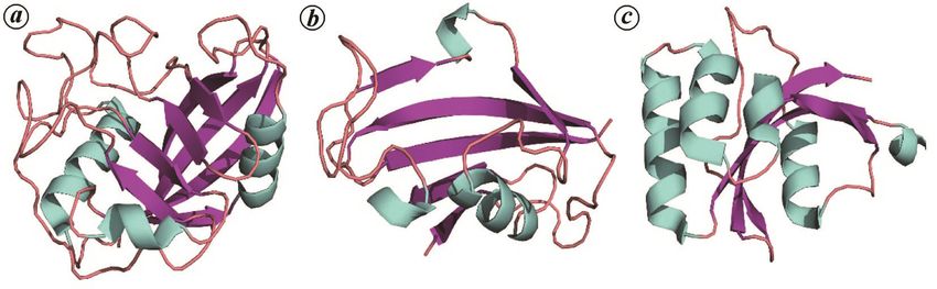
REVIEW ARTICLE

        Figure 1. Structure of three major peptidyl-prolyl cis-trans isomerase (PPIase) folds, namely cyclophilin, FKBP and parvulin.
        a, Human cyclophilin G (PDB ID:2GW2) showing a centrally located β-barrel and three surrounding α-helices. b, Human
        FKBP12 (PDB ID: 1FKJ) showing an α-helix surrounded by a five-stranded β-sheet. c, Human parvulin-14 (PDB ID: 3UI4)
        showing a four-stranded β-sheet system packed with four α-helices.

have identical enzymatic activity is an interesting problem             PPIases as virulence factors
in the structure–function paradigm, indicative of conserved
structural features in their active sites22. PPIases, however,          Pathogenic FKBP proteins
demonstrate variable recognition and enzymatic activity
depending upon the residue preceding proline (P1). For                  Virulence factors are generally proteins that promote the
example, human parvulin PIN1 preferentially recognizes                  colonization, multiplication and propagation of pathogens
phospho-threonine/serine residues at P1, whereas hFKBP12                in a host. In bacteria, two extensively studied PPIases are
is specific for either leucine or phenylalanine at the same             trigger factor (TF) and SurA, both of which probably play
residue position.                                                       an indirect role in virulence, arising as a consequence of
   In addition, individual cyclophilin, FKBP and parvulin               either their chaperone function or involvement in the
domains have been found conjoined with other non-                       secretion of virulence factors, such as adhesins. In Strep-
PPIase domains such as the WW domain, U-box, EF-hand                    tococcus pneumoniae, TF was found to be necessary for
motifs, TPR, WD40 and RRM to form larger multi-                         the adhesion of bacterial cells to epithelial tissues in the
domain proteins, probably targeting or coordinating                     human lung and has been identified as a potential drug
PPIase function in the context of specific organelles23,                target for pneumonial infections24. TF deletion mutants of
allowing their interactions with multiple partners and                  Listeria monocytogenes, the causative agent for listeri-
conferring chaperone activity.                                          osis, manifest reduced response to heat shock and ethanol
   The scope of this article will encompasses the func-                 exposure although the deletion appeared to have little
tions of three significant folds of PPIase (cyclophilins,               effect upon bacterial cell growth in vitro. However, such
FKBPs and parvulins) proteins in the regulation of                      mutants exhibit reduced intercellular survival and multi-
growth, physiological functions and pathogenicity in case               plication in vivo, probably significant for bacterial
of pathogenic infection-causing bacteria, parasites and                 virulence25. TF deletion mutants in Streptococcus mutans
fungi. PTPA proteins have not been included here since                  were observed to have reduced growth rate coupled to an
their function as virulence factors is yet to be characte-              inability to form biofilms25,26. In Streptococcus pyogenes
rized. Host PPIases which play a crucial role in the regu-              TF assisted in the maturation and secretion of specific
lation of pathogenic virulence and are considered as drug               cysteine proteases27, while its deletion decreased toler-
targets, have also been included here. The article summa-               ance to oxidative stress and reduced growth rate27.
rizes the available structural information of some essen-                  TF is a modular protein consisting of a centrally located
tial PPIase proteins and their strategic interaction sites,             FKBP domain flanked on both sides by a predominantly
which might aid in designing efficacious drugs against                  α-helical C-terminal domain and an N-terminal (α/β) ribo-
specific pathogens. One outstanding example in this                     some-binding domain, capable of chaperone activity
regard is the design of non-immunosuppressive cyclo-                    (Figure 2 a)28. The crystal structure of TF (from Escheri-
sporin (CsA) derivatives currently under advanced stage                 chia coli) in complex with the 50S ribosomal subunit
of clinical trial in the treatment of SARS-CoV-2. In short,             (Haloarcula marismortui) showed the molecule (with all
the compact presentation of structure–function relation-                the three domains in an extended conformation) at the exit
ships of virulence-associated PPIase proteins in this arti-             of the ribosome tunnel from where nascent polypeptide
cle might assist in designing new PPIase inhibitors,                    chains emerge29. The extended conformation of TF enclo-
which can be further administrated in the treatment of a                ses a predominantly hydrophobic space at the exit of the
number of pathogen-borne infectious diseases.                           ribosomal tunnel (characteristically called the ‘cradle’),
CURRENT SCIENCE, VOL. 121, NO. 6, 25 SEPTEMBER 2021                                                                                     759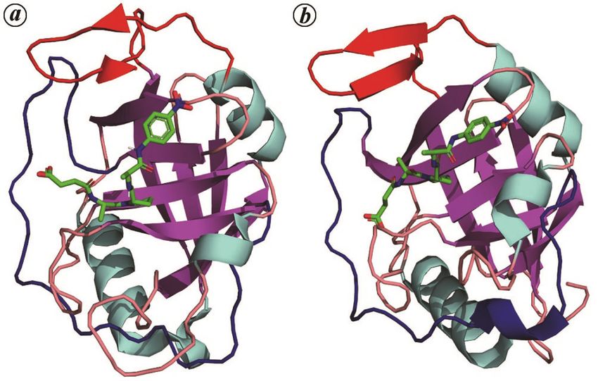
REVIEW ARTICLE

Figure 2. Structure of some important FKBP proteins. a, Escherichia coli trigger factor (PDB ID: 1T11), consisting of three individual domains,
viz. FKBP domain (yellow), N-terminal ribosome-binding domain (red) and C-terminal helical domain (green). The linker between FKBP-domain
and ribosome-binding domain is also shown (violet). b, Dimeric MIP protein from Legionella pneumophila (PDB ID: 1FD9) showing the FKBP
domain and large linker alpha helix (α3) which gives flexibility to the whole protein. c, FKBP12, FK506 and calcineurin complex (PDB ID:6TZ8
from Cryptococcus neoforms). Calcineurin A (CnA) is shown in orange, the calcineurin B (CnB) subunit in green and FKBP12 in yellow. FK506 is
shown in sticks representation (blue).

which facilitates the progress of protein folding unhin-                 tissue, thereby initiating the penetration of bacteria into
dered by proteases or aggregation.                                       the alveolar epithelial cells (across the epithelial cell
   The PPIase domain was positioned away from the                        barrier). The PPIase active site appeared to be involved in
tunnel exit and thus could access the polypeptide chain                  the host–pathogen interaction as bacterial entry could be
only at an advanced stage in folding upon its dissociation               inhibited by rapamycin36.
from the cradle. The tethering of the peripheral FKBP to                    Crystal structures of MIP (from L. pneumophila and
the other two cradle-forming domains using an extended                   T. cruzi) show the enzyme to be constituted of a centrally
double linker also provided structural rationale to the                  located FKBP-type PPIase core, flanked on both sides by
observation that PPIase activity was not necessary for                   α-helices (Figure 2 b). Two α-helices joined by a six-
either peptide binding or the chaperone function of TF.                  residue loop (α1 and α2) are present at the N-terminal of
   Another FKBP class of proteins that are confirmed                     the molecule and are responsible for the formation of bio-
virulence factors and therefore validated drug targets are               logically active MIP-dimer. The longest helix (α3) joins
the macrophage infectivity potentiator (MIP) proteins,                   the N-terminal domain (α1 and α2) with the PPIase do-
which are typically localized in the outer membranes of                  main of the molecule and probably confers structural sta-
Gram-negative bacteria. The virulence-associated activity                bility to the molecule, while the N-terminal helices are
of MIP was first identified in Legionella pneumophila,                   responsible for the flexibility in the molecule. Since the
where it was found necessary for the invasion and intra-                 centrally located PPIase has an FKBP fold, a six-stranded
cellular replication of the pathogenic bacteria within                   β-sheet wraps around the shortest α-helix (α4), with the
human alveolar macrophages30. Subsequently, MIP-like                     active site embedded in a deep hydrophobic pocket
proteins were also found in the obligate intracellular                   bounded by β-strands (3, 4, 6), helix α4 and a loop con-
pathogen Chlamydia, where its inhibition (by FK506) led                  necting strands 5, 6 (ref. 37). Generally, MIPs are secreted
to irregularities in inclusion-body formation within the                 into the environment and so similar are the proteins from
host cell31. In addition, surface-exposed NgMip contri-                  T. cruzi and L. pneumophila that recombinant addition of
buted to the persistence of Neisseria gonorrhoeae within                 the enzyme (from T. cruzi) can recover function in a dele-
the macrophages32, while NmMip was essential for the                     tion mutant (in L. pneumophila). The rms deviation in Cα
survival of Neisseria meningitidis in the blood33. Apart                 atoms between TcMIP and the corresponding LpMIP is
from bacteria, MIP PPIases in protozoan parasites Trypa-                 only about 1.00 Å, and the most significant difference in
nosoma cruzi and Leishmania infantum facilitate entry of                 their structures is with regard to the relative length of
the parasites into mammalian epithelial cells. TcMIP                     helix α3 (ref. 38).
(MIP from T. cruzi) was shown to be involved in host-                       In various pathogenic fungi, FKBP12 was identified as
cell invasion34, and the immunosuppressant FK506 was                     an important virulence factor and the target for the drugs
found to reduce parasitic burden by binding to TcMIP,                    FK506 or rapamycin, which have been widely used as anti-
possibly disrupting the signalling pathway involving Ca2+                fungal agents. In Aspergillus fumigatus, deletion muta-
(ref. 35). However, till date the maximum information                    tional analysis was performed for four FKBP-encoding
with regard to the role of MIP in pathogenic virulence is                genes, namely FKBP12-1, FKBP12-2, FKBP12-3 and
in the case of L. pneumophila, where the enzyme contri-                  FKBP12-4, in order to specifically understand the func-
buted to the proliferation of bacteria in the lungs and                  tional roles of these proteins in the fungi. The study showed
spleen36. Here, MIP was found to bind collagen IV (a                     that ΔFKBP12-1 and ΔFKBP12-4 generally resulted in
component of the extra cellular matrix: ECM) in the lung                 enhanced sensitivity towards FK506 and reduced growth
760                                                                             CURRENT SCIENCE, VOL. 121, NO. 6, 25 SEPTEMBER 2021
REVIEW ARTICLE
(coupled with growth defects) respectively39. In addition, ognition and binding, as evident from the crystal struc-
FK506 treatment modified the localization of FKBP12-1 tures of (truncated) SurA–peptide complexes46.
from the cytoplasm to the hyphal septa of the fungi. The protein homologous to SurA in Gram-positive bac-
FKBP12 deletion mutants in Beuveria bassiana, Candida teria is PrsA, of which two isoforms (PrsA1 and PrsA2)
albicans and Cryptococcus neoformans exhibited in- have been identified in Listeria monocytogenes (Lm), a
creased resistance to antifungal drugs FK506 and rapa- bacterium that resides in the soil but transforms into a
mycin39,40. The transcriptional analysis had already pathogen upon mammalian contact. Central to this transi-
confirmed that FKBP12 forms ternary complexes with tion is the secretion of a host of virulence factors such as
calcineurin (Cn) and FK506 (refs 41, 42). Crystal struc- internalin A, B to initiate host-cell invasion and listeri-
tures of the Cn–FKBP12-FK506 ternary complexes have olysin O (LLO), phospholipases for the lysis of vacuolar
been solved for A. fumigatus and C. neoformans. As has membranes, to enable bacterial entry into the host cyto-
been mentioned previously, Cn is a Ca2+/calmodulin sol47. PrsA2, a post-translocation secretion chaperone, is
(CaM)-dependent serine, threonine-specific protein phos- necessary for correct folding and activity of these
phatase principally responsible for T-cell activation along secreted proteins, in addition to being involved in flagel-
with other cellular functions42. Heterodimeric Cn consists lum-based motility48. Although PrsA1 and PrsA2 share
of two domains – a catalytic domain (CnA) and a regula- 58% sequence identity, there is little overlap between
tory (CnB) domain which interacts with Ca2+-CaM. Com- their functions and it is only fairly recently that PrsA1
parison of the individual components of the fungal and has been implicated in bacterial ethanol resistance49.
human ternary complexes showed a high degree of struc- LmPrsA is a homodimer with each monomer consisting
tural conservation. FKBP12–FK506 binds to an extended of two distinct domains – a predominantly α-helical ‘fol-
hydrophobic groove formed by both subunits of Cn (Fig- dase’ domain and a parvulin-type PPIase domain, with
ure 2 c). However, despite the structural conservation of considerable flexibility in their relative geometry. Com-
individual units, critical amino acid differences were ob- parison of the PrsA1 crystal structure and a three-
served in the loops corresponding to the human and fungal dimensional model of PrsA2 identified the ‘hotspots’ in
FKBPs, which were exploited to rationally design non- this fold which are probably responsible for transforming
immunosuppressive inhibitors of fungal FKBP12 and Cn42. PrsA2 into a virulence factor (in contrast to PrsA1). Al-
though there are several random amino acid differences
Parvulins of disease-causing bacteria and protozoa between the surfaces of both proteins, the most concerted
difference appears to be in the vicinity of the highly con-
An extensively studied multidomain protein (which inclu- served active site of the PPIase domain. Encircling the
des a PPIase) is the periplasmic (survival factor A) SurA, (PPIase) active site there are eight amino acid substitu-
which facilitates the folding and assembly of several out- tions which make the neighbourhood of the PrsA2 active
er membrane proteins (OMP) in Gram-negative bacteria. site less charged or polar compared to PrsA1. In other
SurA-deficient bacteria (as in Yersinia pseudotuberculo- words (in conformity with these substitutions), the PPIase
sis) significantly reduce the surface localization of two domain in PrsA1 appears highly electronegative in con-
key adhesins, Ail and OmpA, which hinders the attach- trast to the relatively uncharged domain in PrsA2. Inte-
ment of the parasite to its eukaryotic host cells43. In addi- restingly, the hydrophobic pocket in the foldase domain
tion, SurA-deficient strains (in Pseudomonas aeruginosa
and Salmonella enteritidis) exhibit perturbations in their
outer membrane structure, permeability and molecular
constitution leading to reduced virulence (in a Galleria
mellonella infection model) and increased sensitivity to a
number of antibiotics44,45.
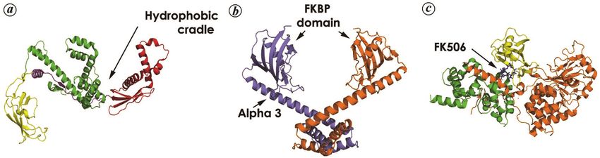
SurA consists of four domains, an N-terminal fragment
(N) of about 150 amino acids, followed by two PPIase
domains (P1, P2) of the parvulin class, and finally ending
with a predominantly α-helical C-terminal domain (C)
(Figure 3 a). The polypeptide segments N, P1 and C form
the densely packed ‘core module’, whereas P2 is tethered
to the core by a linker of approximately 30 Å in length46.
Completely abolishing all PPIase activity from the mole-
Figure 3. Structures related to the parvulin fold. a, SurA protein from
cule by mutagenesis does not appear to hinder the chape- E. coli (PDB ID:1M5Y) showing its four domains in four different
rone function carried out by the core module, as mutant colours, namely N, P1, P2 and C in green, red, yellow and blue respec-
cells exhibit wild-type phenotype and intact outer mem- tively. b, Ess1 protein from Candida albicans (CaEss1), which has a
WW domain (red) in addition to the PPIase domain (yellow). The link-
branes46. Interestingly, the PPIase activity of SurA lies er domain consists of an alpha helix (green) which is dissimilar to the
exclusively with P2 whereas P1 mediates molecular rec- human PIN1 protein.
CURRENT SCIENCE, VOL. 121, NO. 6, 25 SEPTEMBER 2021 761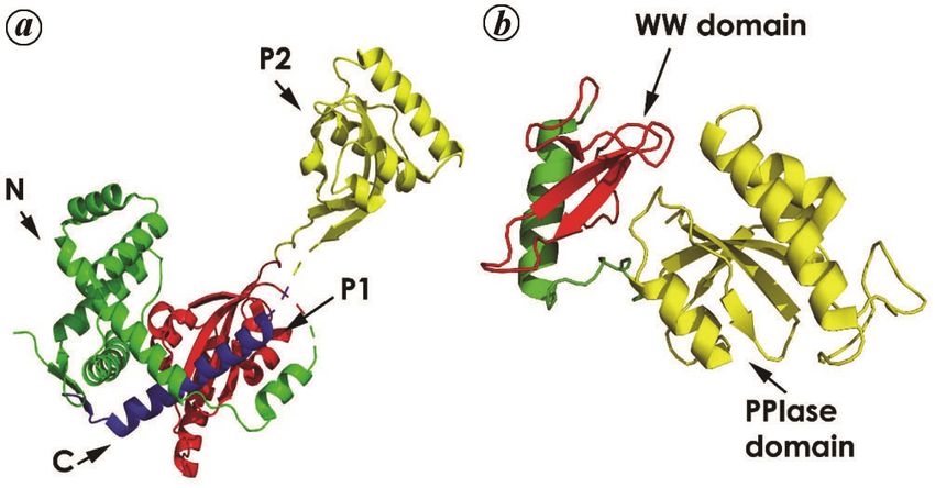
REVIEW ARTICLE
which is expected to bind to protein substrates for            Bacterial, protozoan and fungal cyclophilins as
chaperone activity was found largely conserved in both         virulence factors
proteins49.
   There is a relative dearth of information with regard to    Amongst the cyclophilins, two prominent virulence
parasitic parvulins with the exception of a few proteins       factors are the homologous enzymes PpiA and PpiB
from Trypanosoma, Toxoplasma and Theileria. Depletion          located at different cellular sites, as a consequence of
of the parvulin 42 gene from Trypanosoma brucei                specific signal peptides incorporated in their nascent
(TbPar42) by RNA interference, reduced both the viabili-       polypeptide chains. In Gram-negative bacteria PpiA is
ty and proliferation rate of parasitic cells50. Homologs of    periplasmic, while in Gram-positive bacteria it is asso-
TbPar42 are found exclusively only in protozoa and thus        ciated with (the external side of) the cytoplasmic mem-
could be favoured as a drug target. TbPar42 consists of        brane and is implicated in the transport of secreted
two domains – an N-terminal FHA domain followed by a           proteins57. In contrast, PpiB is invariably a cytoplasmic
parvulin-type PPIase domain, which has high structural         protein. Both proteins probably play a synergistic role in
similarity to human PIN1. FHA domains typically recog-         modulating cell division, resistance to extracellular stress,
nize phosphopeptides and play a significant role in DNA        chaperone activity associated with virulence factors and
damage, replication and cell-cycle progression51. No cata-     regulation of pathogenic virulence. For example, overex-
lytic activity was observed for the parvulin domain, in        pression of PpiA and PpiB from pathogenic Sinorhizo-
case of phosphorylated peptides (which included p-Thr).        bium meliloti in E. coli BL21 cells, increased by several
NMR studies did not appear to indicate any interaction         folds bacterial survival under heat and salt stress58. In
between the two domains and there is every possibility         Mycobacterium tuberculosis the protein homologous to
that the native substrate for this enzyme is yet to be iden-   PpiA was found to be secreted into host cells, due to the
tified51. It has been hypothesized that TbPar42 could be a     presence of an N-terminal signal sequence, conspicuously
scaffold protein that participates in higher-order assem-      absent in non-pathogenic strains59. Overexpression of
blies, enabling the association of weakly binding              PpiA and PpiB (from M. tuberculosis) in E. coli cells also
proteins. Other parasitic parvulins include the 22 kDa         conferred fitness to sustain oxidative, hypoxic stress con-
TgMIC5 (Toxoplasma gondii) found in secretory orga-            ditions generated by H2O2 and CoCl2 (ref. 59). In addi-
nelles called micronemes, contributing to host cell adhe-      tion, both enzymes were found to modulate host immune
sion and invasion52. TgMIC5 mimics the function of GPI         response, as treatment of THP-1 cells with recombinant
(glycosyl phosphatidyl inositol)-anchored microneme            PpiA promoted the expression of pro-inflammatory TNF-
protein TgSUB1, which processes secreted micronemal            α and IL-6 cytokines, whereas similar treatment with
(MIC) proteins on the surface of the parasite to enhance       PpiB inhibited TNF-α and induced IL-10 secretion in-
their adhesive functions53. A parvulin from Theileria          stead60. In dental caries causing bacteria, S. mutans, a
annulata (TaPIN1) similar to human PIN1, has been              PpiA-deficient strain was subject to increased phagocyto-
found to be secreted into host cells (by the parasites) so     sis by human macrophages61, thus exposing the crucial
as to hijack the host oncogenic signalling pathways.           role of the bacterial enzyme in antiphagocytic activity.
TaPIN1 also interacts with the host ubiquitin ligase           Likewise PpiA/PpiB in Enterococcus faecalis was found
FBW7 (which promotes the degradation of oncogenic              to play a significant role in stress response to high NaCl
proteins like c-JUN), thereby causing an elevation of          concentration and regulation of virulence in bacterial (E.
c-JUN levels in the host54.                                    faecalis) infection of G. mellonella larvae62.
   Parvulin proteins have also been found to be important         Several crystal structures are currently available for
for survival and virulence for a number of pathogenic          bacterial cyclophilins, of which CypA (from E. coli) is
fungi. The parvulin CaEss1 from Candida albicans has           structurally similar to human CypA, though marginally
been found to be essential for the proliferation and sur-      differing in irregular loop regions. Only eight out of the
vival of the fungal pathogen inside mammalian host             11 active site residues in bacterial CypA are conserved
cells55. In addition, the virulence of Ess1 deletion mutants   with respect to humans, though leaving the catalytic func-
tested on a murine model, demonstrated no disease symp-        tion of the bacterial enzyme unaffected63. Comparison of
toms in contrast to infection by wild type which resulted      pathogenic bacterial cyclophilins (including crystal struc-
in severe cryptococcusis56. The crystal structure of           tures of PpiA from M. tuberculosis, cyclophilin A–Azoto-
CaEss1 showed structural similarity to the human PIN1          bacter vinelandii, CypA–Schistosoma mansoni) suggests
protein, except for the linker region between PPIase and       overall conservation of the protein fold, except for minor
WW domain (Figure 3 b). In human hPIN1, this flexible          variations in the L1 and L4 loop regions64–66. An espe-
domain is devoid of any regular secondary structure. In        cially interesting study compared crystal structures of the
contrast, the linker domain of CaEss1 consists of a large      complexes (E. coli) CypA bound to Suc–Ala–cis–Pro–Ala–
helix which restricts the mobility of this region and pro-     pNA (Figure 4 a) and CypB associated with Suc–Ala–
bably suggests a different mode of protein–protein inte-       trans–Pro–pNA (Figure 4 b). Although the crystals of
ractions than found in hPIN1 (ref. 55).                        both complexes were grown under identical conditions,

762                                                                 CURRENT SCIENCE, VOL. 121, NO. 6, 25 SEPTEMBER 2021
REVIEW ARTICLE
the relative affinities (of CypA, CypB) were for the cis                 from another Schistosoma species (Schistosoma japoni-
and trans forms of the peptide respectively67,68. No confor-             cum–SjCyp18) has been shown to favour an IL-4 produc-
mational differences were observed in 13 (of the 14) con-                ing TH2 response in vivo, which appeared to promote
served binding site residues and it appeared that the                    immunopathological changes such as liver fibrosis71. In
difference in specificities was probably due to the loops in             case of visceral leishmaniasis (L. infantum), the use of re-
the neighbourhood of the binding site (extending from                    combinant leishmanial cyclophilin (LiCyp1) as an anti-
strand β4 and interconnecting β4 to β5; Figure 4 a and b).               gen to immunize BALB/c mice led to a significant
These loops condition the orientation of the residue at site             reduction in parasitic burden in liver and spleen cells.
P2 (the second residue from the N-terminal of proline in                 Further, the process also stimulated the circulation
the substrate), which in turn was postulated to determine                of specific CD4+ and CD8+ T-cells at infection sites,
the difference in specificities (vis-à-vis CypB, CypA) and               promoting the release of relevant effector cytokines and
reaction rates67,68. In E. coli, CypA and CypB correspond to             attenuating subsequent leishmanial infection72. The
PpiA and PpiB, and as has been mentioned previously are                  significant reactivity of cyclophilin from Echinococcus
localized in the periplasm and cytoplasm respectively.                   granulosus, with human IgG and IgE, proves it to be the
   Cyclophilins from several parasitic protozoa are also                 principal causative agent in allergic cystic echinococco-
known to modulate the host immune system by altering                     sis73. It is well known that interferon-gamma (IFN-γ )
T-cell responses, consequently interfering in the secre-                 produced during any microbial infection improves the
tions of their associated cytokines which leads to the                   protective immunity of the host. However, continuous
eventual acceptance of the parasite as ‘self’ by the host.               production of IFN-γ and its subsequent depletion precede
Such a role in host–pathogen interactions qualifies para-                acute-phase neosporosis infection (caused by Neospora
sitic cyclophilins as potential vaccine candidates. It may               caninum), probably due to a secretory cyclophilin
be recalled that immune response by CD4+ TH cells is                     (NcCyp) from the parasite74. Similarly, cyclophilin of the
primarily through secreted cytokines and based on the                    protozoan parasite T. gondii (TgCyp18) secreted through
repertoire of these molecules, T-helper cells can be dis-                tachyzoites, induces the production of tumour necrosis
tinguished into subclasses TH1, TH2, TH3 and TH17. For                   factor (IL-12) and nitric oxide (NO) by binding to CCR5
example, recombinant cyclophilin A of S. mansoni                         (cystine cystine chemokine receptor 5) located on surface
(SmCyp), responsible for schistosomasis, modulated the                   macrophages and spleen cells. TgCyp18 also induces IL-
immune function of bonemarrow-derived dendritic cells                    6 and IFN-γ in a CCR5-independent manner75. Possibly,
(DC) by attenuating the DC-mediated CD4+ T-cell activa-                  as a consequence of these interactions, TgCyp18 enhan-
tion and concomitant induction of the Treg cell response69.              ces the migration of parasites to host macrophages and
Vaccination with a synthetic peptide derived from                        promotes invasion and proliferation within macrophages,
SmCyp induced a reduction in parasitic burden and sig-                   thereby consolidating the survival of the parasite within
nificantly enhanced antibodies against the parasitic anti-               the host76,77.
gen in immunized mice70. Such observations appear to                        A large body of work supports the involvement of cyc-
identify SmCyp (or SmCyP-derived peptides) as promising                  lophilins in the growth, reproduction, virulence and
vaccine candidates against schistosomasis. Cyclophilin                   extracellular stress response of pathogenic fungi. The
                                                                         genome of C. neoforms, a pathogenic fungus affecting the
                                                                         human central nervous system, comprises two cyclophilin
                                                                         isoforms, namely Cpa1 and Cpa2, exhibiting diverse func-
                                                                         tions. ΔCpa1 mutants demonstrated a lower survival rate
                                                                         relative to wild type in infected murine and rabbit cells.
                                                                         Also, in mating assays the double disruption fungal mutants
                                                                         were sterile78. A cyclophilin protein from the opportunistic
                                                                         human pathogen Lemontospora prolificans (causing a wide
                                                                         range of diseases in immune-compromised humans) was
                                                                         recognized by human salivary Immunoglobin A (IgA),
                                                                         leading to the identification of this protein as an immu-
                                                                         nogenic antigen79. Similar binding to cyclophilin A. fumi-
                                                                         gatus as a conidial antigen has identified cyclophilins as a
                                                                         conserved immunogen, which could be used to develop
                                                                         vaccines against this class of pathogenic fungi79.
Figure 4. Difference in substrate binding and structural diversity in
CypA and CypB of E. coli. a, Structure of cyclophilin A in complex
with succinyl-Ala-Pro(cis)-Ala-p-nitroanilide (PDB ID:1LOP). The L1      Viral interactions of host PPIases
and L4 loops are shown in blue and red respectively, which differs
from cyclophilin B. b, Structure of cyclophilin B in complex with suc-
cinyl–Ala–Pro(trans)–Ala–p-nitroanilide (PDB ID:1V9T). The L1 and        As is well known, all viruses co-opt the replicative mole-
L4 loops have been highlighted in blue and red respectively.             cular machinery of the host to proliferate as virions,
CURRENT SCIENCE, VOL. 121, NO. 6, 25 SEPTEMBER 2021                                                                              763
REVIEW ARTICLE
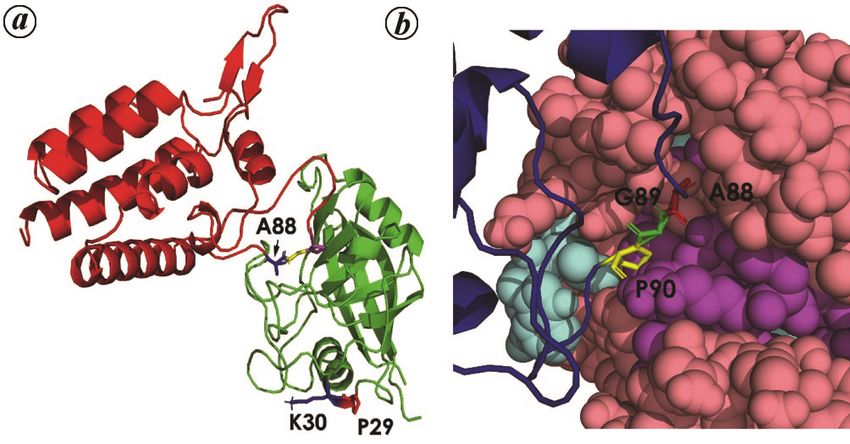
thereby completing their life cycle. This is accomplished inserted into the CypA active site (Figure 5 b)81. The pep-
by the interaction between viral and host proteins (also tidyl–prolyl bond corresponding to Pro-90 was found to
referred to as host cofactors). Several well-studied exam- be in the trans conformation, which along with the pres-
ples demonstrate the crucial role played by host PPIases ence of glycine at position 89, allowed for deep penetra-
in the viral life cycle, either as an essential factor neces- tion of the loop into the CypA active site. However, cryo
sary for its replication or as an antiviral agent80–84. Thus, EM studies of CypA complexed with (supramolecular)
cyclophilin promotes the replication of the human immu- tubular CA assemblies, found CypA bridging two CA
nodeficiency virus 1 (HIV-1), hepatitis B virus (HBV), molecules by means of an additional ‘non-canonical’
hepatitis C virus (HCV) and severe acute respiratory syn- binding site involving Pro-29 and Lys-30 (apart from the
drome coronaviruses (SARS-CoVs), while inhibiting canonical active site; Figure 5 a). This ability of CypA to
influenza and rotaviruses (RV)80. bridge two CA molecules might perhaps be structurally
HIV-1 belonging to the Retroviridae family consists of relevant in stabilizing the viral capsid86.
a positive-stranded RNA genome. Gaining entry into a Studies indicate that similar to HIV-1, CypA also
host cell, viral pol encoded reverse transcriptase (RT) promotes HCV replication by interacting with the viral
initiates the incorporation of double-stranded DNA (de- protein NS5A. This protein consists of three domains and
rived from the viral genome) into the chromosomal DNA experiments implicate Pro-319 (in the second domain)
of the host80. An essential step in the life cycle of the patho- embedded in a WARPDYN motif as interacting with
gen is the translation of the viral Gag polyprotein (an CypA83. All mutations in NS5A which confer resistance
extended polypeptide consisting of several viral factors) to cyclophilin inhibitors such as CsA are found in the
which encodes the information necessary for the assem- WARPDYN motif. Additionally, NS5B with RNA poly-
bly and release of virions. As the virions mature, Gag is merase activity and essential for viral RNA replication,
cleaved into three polypeptide fragments by a (viral) pro- forms a replication complex in the endoplasmic reticulum
tease to yield the matrix protein (MA – 132 residues – along with host proteins, of which human CypA is an im-
lining the inner surface of the viral membrane), the capsid portant constituent87. Likewise, HBV replication is also
protein (CA – 230 residues – forming the distinctive core facilitated by the human enzymes parvulin 14 and parvulin
which envelopes the NC/RNA complex at the virion cen- 17 interacting with viral small surface proteins (SHBs),
tre) and the nucleocapsid protein (NC – 54 residues – which engages the immune system of the host84. In con-
coating the genomic RNA)80. Thus, these proteins form trast to all the examples given above, incorporation of
key components of the fully infective mature virion. CypA within the virion core of influenza virus inhibits its
Human CypA interacts with the Gag polyprotein by bind- infective cycle. The most abundant protein in case of in-
ing to a proline-rich loop (specifically at Gly 89–Pro 90) fluenza is the matrix protein M1 which is essential for
corresponding to the CA region of Gag, resulting in CypA viral replication, assembly and budding. The antiviral
incorporation into the virions81. This is essential for the effect of CypA was due to its role in the degradation of
effective replication of HIV-1, as inhibiting the packag- M1 via the ubiquitin/proteosome-dependent pathway82.
ing of CypA within the virion by CsA or Gag-specific Severe acute respiratory syndrome coronavirus-2
mutations leads to a reduction in viral infectivity. CD147 (SARS-CoV-2) is the latest amongst seven previously
is the primary signalling receptor for CypA on the surface known viruses belonging to the Coronaviridae family,
of human leucocytes and CypA–CD147 interaction could
be instrumental in releasing the viral RT complex into the
cytoplasm, along with facilitating virion attachment to
host cells82. Interestingly, cell lines from Old World
primates were found to be HIV-1-resistant due to the
presence of a CyP-TRIM5α fusion protein. The cyclophilin
domain in the chimerical protein was responsible for direct-
ly targeting TRIM5α onto incoming HIV-1 capsids, leading
to the arrest of viral proliferation. In humans, TRIM5α is
only weakly resistant to HIV-1 probably due to sequence
divergence from the homologous protein in primates85.
Crystal structure of human CypA complexed with the
(151 residue) N-terminal domain of HIV-1 CA revealed
in atomic detail, their mutual interaction sites (Figure Figure 5. Interaction of human cyclophilin A with capsid protein
(CA) of HIV-1 virus. a, Human cyclophilin A (hCypA) in complex
5 a). CA151 has an all-α-fold constituted of seven helices, with N-terminal domain of HIV-1 capsid (PDB ID:1AK4). The cyclo-
with α-helices 1–4 and 7 packing along their helical axes, philin protein is shown in green and CA in red. Two residues of hCypA
while 5, 6 stack on the top of this helical bundle. Resi- involved in non-cannonical interaction with the CA protein have been
highlighted, namely K30 and P29. b, The interacting loop of the CA
dues Ala-88, Gly-89 and Pro-90 within the CypA binding protein consisting of three residues, viz. A88, G89 and P90 which get
loop of CA151 (from residues 85 to 93) were completely inserted into the active site of CypA and interact canonically.
764 CURRENT SCIENCE, VOL. 121, NO. 6, 25 SEPTEMBER 2021REVIEW ARTICLE
Table 1. A detailed list of residues of cyclophilin and FKBP proteins interacting with some important binding partners (interactions have been
calculated from crystal structures of complexes with the distance cut-off 3.8 Å), namely (a) human cyclophilin A (hCypA) and alisporivir [PDB ID:
5HSV], (b) hCypA and cyclosporin A (CsA) [PDB ID: 2RMA], (c), (d) hCypA and calcineurin A (CnA) and calcineurin B (CnB)[PDB ID: 1MF8],
(e) hCypAand HIV-1 capsid [PDB ID: 1AK4], (f) hCypA active site residues determined from the complex structures with dipeptides Ser–Pro, His–
Pro and Gly–Pro [PDB ID: 3CYH, 4CYH and 5CYH respectively], (g) human FKBP-12 (hFKBP12) and FK506 [PDB ID: 1FKJ], (h)–(j) FKBP12
of Cryptococcus neoforms and FK506, CnA and CnB [PDB ID:6TZ8], (k) hFKBP12 and rapamycin [PDB ID: 1FKB] and (l) active site residues
obtained from a previous study98 (All the structures have been obtained from RCSB PDB, https://www.rcsb.org/)
(e) Cyp- (f) Cyp (g) FKBP- (h) FKBP- (i) FKBP- (j) FKBP- (k) FKBP- (l) FKBP
(a) Cyp- (b) Cyp- (c) Cyp- (d) Cyp- HIV1 active FK506 FK506 (C. CnA (C. CnB (C. Rapamycin Active
Alisporivir CsA CnA CnB capsid site (human) neoforms) neoforms) neoforms) (human) site
R55 R55 R55 R55 Y26 Y27 Y26 Y26
D33
P58 K36
F60 F60 F60 F60 F36 F37 F37 F36 F36
D37 D38 D38 D37 D37
Q63 Q63 Q63 Q63 S39
R69 R41
N71 N71 D42
G72 G72 G72 R42 R43 R43 R43 R42
T73 T73 T73 T73 T45
E81 F46 F47 F47 46F F46
L82 V48
A101 A101 A101 A101 53Q
N102 N102 N102 N102 E54 E55 54E
A103 A103 A103 V55 V56 55V V55
Q111 Q111 Q111 I56 I57 56I I56
F113 F113 F113 F113 W59 W60 59W
W121 W121 W121 W121 A81
L122 L122 L122 L122 Y82 Y83 82Y Y82
H126 H126 H126 H126 R86
S147 H87 87H H87
F88
P90
V91
R148 R148 I92
F100 F99
which infects humans. As this article is being written the its association with the viral N protein. A high-through-
COVID-19 pandemic (caused by SARS-CoV-2) is ravag- put yeast two-hybrid screen (HTY2H) identified the bind-
ing the world in terms of human loss and suffering, with ing of CyPA to the nonstructural Nsp1 in SARS-CoV and
over two lakh deaths in India alone. Four previous corona abrogation of CyPA through SiRNA hindered viral repli-
viruses (HCoV-NL63, HCoV-229E, HCoV-OC43 and cation (for HCoV-NL63 in CaCo-2 cells)91,92. In addition,
HKV1) reported only mild symptoms in immune- the CyPA signalling receptor CD147 was also found to
competent hosts, while SARS-CoV-1 and MERS-CoV interact with the N protein by surface plasmon resonance
(middle east respiratory syndrome coronavirus) were and it may be possible that CyPA in tandem with CD147
highly pathogenic with elevated mortality rates (almost facilitates viral entry into host cells.
30% for MERS-CoV), though with lower transmissibility88. Several experiments have confirmed that CsA inhibits
Unfortunately, SARS-CoV-2 combines very high rates of the replication of SARS-CoV-1,2, CoV-229E and
transmission with aggressive pathogenicity. The SARS- CoVNL63 by binding to host cyclophilins91,92. However,
CoV-2 genome has 79% and 96% sequence identity with the immunosuppressive properties of CsA prejudice
SARS-CoV-1 and bat coronavirus (BatCoVRatG13) re- its direct use as an antiviral. As has been mentioned
spectively89. The genome of corona viruses consists of previously CsA bound to cyclophilin forms a ternary
four major structural proteins – the characteristic spike complex with calcineurin (Cn), thereby curtailing T-cell
protein (S) which is inserted into the outer membrane, activation. Crystal structures of the CypA–CsA–Cn com-
and the membrane (M), envelope (E), the nucleocapsid plexes clearly show CsA residues that directly interact
(N) proteins90. The S-glycoproteins are responsible for with Cn, and several non-immunosuppressive CsA deriv-
host recognition by binding to the angiotensin-converting atives have been synthesized which could find antimi-
enzyme-2 (ACE2), and the higher transmissibility of crobial and antiviral applications. Of these, the most
SARS-CoV-2 (relative to CoV-1) could be due to the promising appears to be the CsA derivative alisporivir,
higher affinity of S to ACE2 (ref. 88). Human CypA which inhibits RNA production and replication in SARS-
plays a key role in the replication of the SARS-CoV by CoV-2. Alisporivir is currently under phase-3 clinical trial
CURRENT SCIENCE, VOL. 121, NO. 6, 25 SEPTEMBER 2021 765REVIEW ARTICLE
as an antiviral drug93,94. The crystal structures of cyclo- 3. Cheng, C. W. and Tse, E., PIN1 in cell cycle control and cancer.
philin complexed with CsA95 and alisporivir96 are availa- Front. Pharmacol., 2018, 9, 1367.
4. Gong, Z. et al., Cyclophilin A is overexpressed in hepatocellular
ble, and the amino acid conformations responsible for carcinoma and is associated with the cell cycle. Anticancer Res.,
abrogating Cn binding (in case of alisporivir) have been 2017, 37, 4443–4447.
determined. Likewise, another CsA derivative SDZ 5. Aghdasi, B. et al., FKBP12, the 12-kDa FK506-binding protein, is
NIM811 selectively inhibits HIV-1 replication in T4 a physiologic regulator of the cell cycle. Proc. Natl. Acad. Sci.
lymphocyte cell lines97. All these derivatives exhibit re- USA, 2001, 98, 2425–2430.
6. Hu, X. and Chen, L. F., Pinning down the transcription: a role for
duced toxicity and could find extensive use as antivirals peptidyl-prolyl cis–trans isomerase Pin1 in gene expression.
in the future. Front. Cell Dev. Biol., 2020, 8, 179.
7. Nakatsu, Y. et al., Physiological and pathogenic roles of prolyl
isomerase Pin1 in metabolic regulations via multiple signal
Discussion transduction pathway modulations. Int. J. Mol. Sci., 2016, 17, 1495.
8. Nath, P. R., Dong, G., Braiman, A. and Isakov, N., In vivo
The involvement of PPIases in a wide spectrum of impor- regulation of human CrkII by cyclophilin A and FK506-binding
tant cellular functions lies in their ability to regulate protein. Biochem. Biophys. Res. Commun., 2016, 470, 411–416.
cis/trans prolyl isomerization switches to coordinate 9. Wang, Z., Feng, J., Yu, J. and Chen, G., FKBP12 mediates
necroptosis by initiating RIPK1–RIPK3–MLKL signal transduct-
complex physiological processes. Although PPIases con- ion in response to TNF receptor 1 ligation. J. Cell Sci., 2019, 132,
sist of four structurally distinct superfamilies, conserva- jcs227777.
tion of key active-site residues presumably indicates a 10. Tzelepis, F. et al., Mitochondrial cyclophilin D regulates T cell
similar mechanism of action, despite fold differences. metabolic responses and disease tolerance to tuberculosis. Sci.
Mapping key residues of cyclophilin and FKBP involved Immunol., 2018, 3, eaar4135.
11. Tozzi, L. et al., Single-nucleotide polymorphism of the FKBP5
in enzymatic function, and drug/protein interaction sites gene and childhood maltreatment as predictors of structural
exhibit considerable overlap between residues of the active changes in brain areas involved in emotional processing in
and the drug binding sites for both proteins (Table 1). Al- depression. Neuropsychopharmacology, 2016, 41, 487–497.
though the active site residues of CypA and FKBP are 12. Yu, J. H., Im, C. Y. and Min, S. H., Function of Pin1 in cancer
similar, their respective calcineurin (CaN/Cn) binding sites development and its inhibitors as cancer therapeutics. Front. Cell
Dev. Biol., 2020, 8, 120.
appear to be divergent. Interestingly, the cyclophilin loop 13. Rath, D. et al., Platelet surface expression of cyclophilin A is
which interacts with calcineurin B (in the CypA–CsA–Cn associated with increased mortality in patients with symptomatic
ternary complex) is also involved in recognizing the HIV-1 coronary artery disease. J. Thromb. Haemost., 2020, 18, 234–242.
Gag protein (Asn71–Thr73). Residues Ala101–Phe113 14. Wang, L., Zhou, Y., Chen, D. and Lee, T. H., Peptidyl-prolyl
appears to be of extreme strategic importance for CypA, as cis/trans isomerase Pin1 and Alzheimer’s disease. Front. Cell
Dev. Biol., 2020, 8, 355.
it is implicated in every possible interaction of the enzyme. 15. Lu, K. P., Finn, G., Lee, T. H. and Nicholson, L. K., Prolyl cis–
Similarly, in case of FKBP protein, the region Arg42– trans isomerization as a molecular timer. Nature Chem. Biol.,
Phe46 is of extreme importance because it is involved in in- 2007, 3, 619–629.
teractions with FK506, rapamycin as well as CaN. 16. Rajiv, C. and Davis, T. L., Structural and functional insights into
Host PPIases have also been found to regulate virulence human nuclear cyclophilins. Biomolecules, 2018, 8, 161.
17. Prakash, A., Rajan, S. and Yoon, H. S., Crystal structure of the
in host–pathogen interactions, significantly for viruses FK506 binding domain of human FKBP25 in complex with
where PPIases are involved in forming multi-protein FK506. Protein Sci., 2016, 25, 905–910.
complexes with viral proteins. These host PPIase interac- 18. Harikishore, A. and Sup Yoon, H., Immunophilins: structures,
tions with viral proteins have been found to be essential mechanisms and ligands. Curr. Mol. Pharmacol., 2015, 9, 37–47.
for the survival of the virus within the host and also in its 19. Jin, L. and Harrison, S. C., Crystal structure of human calcineurin
complexed with cyclosporin A and human cyclophilin. Proc. Natl.
replication. Consequently, cyclophilin inhibitors are be- Acad. Sci. USA, 2002, 99, 13522–13526.
ing used extensively in antiviral therapy80. Likewise, host 20. Aylett, C. H. S., Sauer, E., Imseng, S., Hall, M. N., Ban, N. and
parvulins have been found to be a key participant in the Maier, T., Architecture of human mTOR complex. Science, 2016,
life cycle of HBV84. A number of fungal cyclophilins 351, 48–52.
have been found to be important vaccine candidates70. It 21. Ranganathan, R., Lu, K. P., Hunter, T. and Noel, J. P., Structural
and functional analysis of the mitotic rotamase Pin1 suggests
is expected that in the coming years PPIases (whether substrate recognition is phosphorylation dependent. Cell, 1997,
from the host or pathogen) will serve as promising thera- 89, 875–886.
peutic drug targets and immunogens for vaccine deve- 22. Fischer, G. and Fanghänel, J., Insights into the catalytic
lopment. mechanism of peptidyl prolyl cis/trans isomerases. Front. Biosci.,
2004, 9, 3453–3478.
23. Schiene-Fischer, C., Multidomain peptidyl prolyl cis/trans
1. Unal, C. M. and Steinert, M., Microbial peptidyl-prolyl cis/trans isomerases. Biochim. Biophys. Acta, 2015, 1850, 2005–2016.
isomerases (PPIases): virulence factors and potential alternative 24. Cohen, A. et al., Streptococcus pneumoniae cell wall-localized
drug targets. Microbiol. Mol. Biol. Rev., 2014, 78, 544–571. trigger factor elicits a protective immune response and contributes
2. Stewart, D. E., Sarkar, A. and Wampler, J. E., Occurrence and role to bacterial adhesion to the host. Sci. Rep., 2019, 9, 4295.
of cis peptide bonds in protein structures. J. Mol. Biol., 1990, 214, 25. Bigot, A., Botton, E., Dubail, I. and Charbit, A., A homolog of
253–260. Bacillus subtilis trigger factor in Listeria monocytogenes is
766 CURRENT SCIENCE, VOL. 121, NO. 6, 25 SEPTEMBER 2021REVIEW ARTICLE
involved in stress tolerance and bacterial virulence. Appl. Environ. biogenesis and type III secretion system expression in Salmonella.
Microbiol., 2006, 72, 6623–6631. Microbiology, 2009, 155, 1613–1622.
26. Wen, Z. T., Suntharaligham, P., Cvitkovitch, D. G. and Burne, R. 45. Klein, K. et al., Deprivation of the periplasmic chaperone SurA
A., Trigger factor in Streptococcus mutans is involved in stress reduces virulence and restores antibiotic susceptibility of
tolerance, competence development, and biofilm formation. Infect. multidrug-resistant Pseudomonas aeruginosa. Front. Microbiol.,
Immunol., 2005, 73, 219–225. 2019, 10, 100.
27. Lyon, W. R. and Caparon, M. G., Trigger factor-mediated prolyl 46. Bitto, E. and McKay, D. B., Crystallographic structure of SurA, a
isomerization influences maturation of the Streptococcus pyogenes molecular chaperone that facilitates folding of outer membrane
cysteine protease. J. Bacteriol., 2003, 185, 3661–3667. porins. Structure, 2002, 10, 1489–1498.
28. Ludlam, A. V., Moore, B. A. and Xu, Z., The crystal structure of 47. Zemansky, J., Kline, B. C., Woodward, J. J. and Leber, J. H.,
ribosomal chaperone trigger factor from Vibrio cholerae. Proc. Development of a mariner-based transposon and identification of
Natl. Acad. Sci. USA, 2004, 101, 13436–13441. Listeria monocytogenes determinants, including the peptidyl–
29. Ferbitz, L., Maier, T., Patzelt, H., Bukau, B., Deuerling, E. and prolyl isomerase PrsA2, that contribute to its hemolytic
Nenad, B., Trigger factor in complex with the ribosome forms a phenotype. J. Bacteriol., 2009, 191, 3950–3964.
molecular cradle for nascent proteins. Nature, 2004, 431, 590– 48. Alonzo III, F., Port, G. C., Cao, M. and Freitag, N. E., The
596. Posttranslocation chaperone PrsA2 contributes to multiple facets
30. Cianciotto, N. P. and Fields, B. S., Legionella pneumophila mip of Listeria monocytogenes pathogenesis. Infect. Immunol., 2009,
gene potentiates intracellular infection of protozoa and human 77, 2612–2623.
macrophages. Proc. Natl. Acad. Sci. USA, 1992, 89, 5188–5191. 49. Cahoon, L. A., Freitag, N. E. and Prehna, G., A structural
31. Herrmann, M., Schuhmacher, A., Mühldorfer, I., Melchers, K., comparison of Listeria monocytogenes protein chaperones PrsA1
Prothmann, C. and Dammeier, S., Identification and characteri- and PrsA2 reveals molecular features required for virulence. Mol.
zation of secreted effector proteins of Chlamydophila pneumoniae Microbiol., 2016, 101, 42–61.
TW183. Res. Microbiol., 2006, 157, 513–524. 50. Goh, J. Y., Lai, C. Y., Tan, L. C., Yang, D., He, C. Y. and Liou,
32. Leuzzi, R. et al., Ng-MIP, a surface-exposed lipoprotein of Y. C., Functional characterization of two novel parvulins in
Neisseria gonorrhoeae, has a peptidyl-prolyl cis/trans isomerase Trypanosoma brucei. FEBS Lett., 2010, 584, 2901–2908.
(PPIase) activity and is involved in persistence in macrophages. 51. Rehic, E. et al., Structural analysis of the 42 kDa parvulin of
Mol. Microbiol., 2005, 58, 669–681. trypanosoma brucei. Biomolecules, 2019, 9, biom9030093.
33. Echenique-rivera, H. et al., Transcriptome analysis of Neisseria 52. Brydges, S. D. et al., Molecular characterization of TgMIC5,
meningitidis in human whole blood and mutagenesis studies a proteolytically processed antigen secreted from the micronemes of
identify virulence factors involved in blood survival. PLoS Toxoplasma gondii. Mol. Biochem. Parasitol., 2000, 111, 51–66.
Pathogens, 2011, 7, e1002027. 53. Saouros, S., Dou, Z., Henry, M., Marchant, J., Carruthers, V. B.
34. Lopez, J. M., Antiparra, R., Zimic, M., Sheen, P. and Maruenda, and Matthews, S., Microneme protein 5 regulates the activity of
H., Backbone chemical shift assignment of macrophage infectivity Toxoplasma subtilisin 1 by mimicking a subtilisin prodomain.
potentiator virulence factor of Trypanosoma cruzi. Biomol. NMR J. Biol. Chem., 2012, 287, 36029–36040.
Assign., 2019, 13, 21–25. 54. Marsolier, J. et al., Theileria parasites secrete a prolyl isomerase
35. Moro, A., Ruiz-cabelio, F., Fernandez-cano, A., Stock, R. P. and to maintain host leukocyte transformation. Nature, 2015, 520,
Gonzalez, A., Secretion by Trypanosoma cruzi of a peptidyl-prolyl 378–382.
cis-trans isomerase involved in cell infection. EMBO J., 1995, 14, 55. Li, Z. et al., The structure of the Candida albicans Ess1 prolyl
2483–2490. isomerase reveals a well-ordered linker that restricts domain
36. Wagner, C. et al., Collagen binding protein Mip enables Legio- mobility. Biochemistry, 2005, 44, 6180–6189.
nella pneumophila to transmigrate through a barrier of NCI-H292 56. Ren, P., Rossettini, A., Chaturvedi, V. and Hanes, S. D., The Ess1
lung epithelial cells and extracellular matrix. Cell. Microbiol., prolyl isomerase is dispensable for growth but required for
2007, 9, 450–462. virulence in Cryptococcus neoformans. Microbiology, 2005, 151,
37. Riboldi-Tunnicliffe, A. et al., Crystal structure of Mip, a prolyl- 1593–1605.
isomerase from Legionella pneumophila. Nature Struct. Biol., 57. Trémillon, N. et al., PpiA, a surface PPIase of the cyclophilin
2001, 8, 779–783. family in Lactococcus lactis. PLOS ONE, 2012, 7, e33516.
38. Pereira, P. J. B. et al., Trypanosoma cruzi macrophage infectivity 58. Thomloudi, E., Skagia, A., Venieraki, A., Katinakis, P. and
potentiator has a rotamase core and a highly exposed alpha-helix. Dimou, M., Functional analysis of the two cyclophilin isoforms of
EMBO Rep., 2002, 3, 88–94. Sinorhizobium meliloti. World J. Microbiol. Biotechnol., 2017, 33, 28.
39. Falloon, K., Juvvadi, P. R., Richards, A. D. and Vargas-mu, J. M., 59. Bhaduri, A. et al., Mycobacterium tuberculosis cyclophilin A uses
Characterization of the FKBP12-encoding genes in Aspergillus novel signal sequence for secretion and mimics eukaryotic
fumigatus. PLOS ONE, 2015, 10, e0137869. cyclophilins for interaction with host protein repertoire. PLOS
40. Li, Y. et al., Characterization of three FK506-binding proteins in ONE, 2014, 9, e88090.
the entomopathogenic fungus Beauveria bassiana. J. Invertebr. 60. Pandey, S., Sharma, A., Tripathi, D. and Kumar, A., Myco-
Pathol., 2020, 171, 107334. bacterium tuberculosis peptidyl–prolyl isomerases also exhibit
41. Cruz, M. C. et al., Rapamycin antifungal action is mediated via chaperone like activity in vitro and in vivo. PLOS ONE, 2016, 11,
conserved complexes with FKBP12 and TOR kinase homologs in e0150288.
Cryptococcus neoformans. Mol. Cell. Biol., 1999, 19, 4101–4112. 61. Mukouhara, T., Arimoto, T., Cho, K., Yamamoto, M. and
42. Juvvadi, P. R. et al., Harnessing calcineurin–FK506–FKBP12 Igarashi, T., Surface lipoprotein PpiA of Streptococcus mutans
crystal structures from invasive fungal pathogens to develop suppresses scavenger receptor MARCO-dependent phagocytosis
antifungal agents. Nature Commun., 2019, 10, 4275. by macrophages. Infect. Immunol., 2011, 79, 4933–4940.
43. Obi, I. R. and Francis, M. S., Demarcating SurA activities requi- 62. Reffuveille, F. et al., Involvement of peptidylprolyl cis/trans
red for outer membrane targeting of Yersinia pseudotuberculosis isomerases in Enterococcus faecalis virulence. Infect. Immunol.,
adhesins. Infect. Immunol., 2013, 81, 2296–2308. 2017, 80, 1728–1735.
44. Fardini, Y., Trotereau, J., Bottreau, E., Souchard, C., Velge, P. 63. Clubb, R. T., Ferguson, S. B., Walsh, C. T. and Wagner, G.,
and Virlogeux-payant, I., Investigation of the role of the BAM Three-dimensional solution structure of Escherichia coli peri-
complex and SurA chaperone in outer-membrane protein plasmic cyclophilin. Biochemistry, 1994, 33, 2761–2772.
CURRENT SCIENCE, VOL. 121, NO. 6, 25 SEPTEMBER 2021 767REVIEW ARTICLE
64. Henriksson, L. M., Johansson, P., Unge, T. and Mowbray, S. L., 82. Zhou, D., Mei, Q., Li, J. and He, H., Cyclophilin A and viral
X-ray structure of peptidyl-prolyl cis-trans isomerase A from infections. Biochem. Biophys. Res. Commun., 2012, 424, 647–650.
Mycobacterium tuberculosis. Eur. J. Biochem., 2004, 271, 4107– 83. Striker, R. and Mehle, A., Inhibitors of peptidyl proline
4113. isomerases as antivirals in hepatitis C and other viruses. PLoS
65. Christoforides, E., Dimou, M., Katinakis, P., Bethanis, K. and Pathog., 2014, 10, e1004428.
Karpusas, M., Structure of a bacterial cytoplasmic cyclophilin A 84. Saeed, U. et al., Parvulin 14 and parvulin 17 bind to HBx and
in complex with a tetrapeptide. Acta Crystallogr. Sect. F, 2012, cccDNA and upregulate hepatitis B virus replication from
68, 259–264. cccDNA to virion in an HBx-dependent manner. J. Virol., 2019,
66. Gourlay, L. et al., The three-dimensional structure of two redox 93, e01840-18.
states of Cyclophilin A from Schistosoma mansoni. J. Biol. Chem., 85. Ylinen, L. M. J. et al., Conformational adaptation of Asian
2007, 282, 24851–24857. macaque TRIMCyp directs lineage specific antiviral activity.
67. Konno, M., Ito, M., Hayano, T. and Takahashi, N., The substrate- PLOS Pathog., 2010, 6, e1001062.
binding site in Escherichia coli cyclophilin A preferably 86. Liu, C. et al., Cyclophilin A stabilizes the HIV-1 capsid through a
recognizes a cis-proline isomer or a highly distorted form of the novel non-canonical binding site. Nature Commun., 2016, 7,
trans isomer. J. Mol. Biol., 1996, 256, 897–908. 10714.
68. Konno, M. et al., Escherichia coli cyclophilin B binds a highly 87. Ngure, M. et al., Interactions of the disordered domain II of
distorted form of trans-prolyl peptide isomer. Eur. J. Biochem., hepatitis C virus NS5A with cyclophilin A, NS5B, and viral RNA
2004, 271, 3794–3803. show extensive overlap. ACS Infect. Dis., 2016, 2, 839–851.
69. Floudas, A. et al., Composition of the Schistosoma mansoni worm 88. Molyvdas, A. and Matalon, S., Cyclosporine: an old weapon in the
secretome: identification of immune modulatory cyclophilin A. fight against coronaviruses. Eur. Respir. J., 2020, 56, 1–5.
PLOS Negl. Trop. Dis., 2017, 11, e0006012. 89. Poulsen, N. N., von Brunn, A., Hornum, M. and Blomberg Jensen,
70. Teixeira, T. et al., The Schistosoma mansoni cyclophilin A M., Cyclosporine and COVID-19: risk or favorable? Am.
epitope 107–121 induces a protective immune response against J. Transplant., 2020, 20, 2975–2982.
schistosomiasis. Mol. Immunol., 2019, 111, 172–181. 90. Liu, C., von Brunn, A. and Zhu, D., Cyclophilin A and CD147:
71. Li, J. et al., Cyclophilin A from Schistosoma japonicum promotes novel therapeutic targets for the treatment of COVID-19. Med.
a Th2 response in mice. Parasites Vectors, 2013, 6, 230. Drug Discov., 2020, 7, 100056.
72. Santos-Gomes, G. M., Rodrigues, A., Teixeira, F. and Carreira, J., 91. Pfefferle, S. et al., The SARS–coronavirus–host interactome:
Immunization with the Leishmania infantum recombinant cyclo- identification of cyclophilins as target for pan-coronavirus
philin protein 1 confers partial protection to subsequent parasite inhibitors. PLOS Pathog., 2011, 7, e1002331.
infection and generates specific memory T cells. Vaccine, 2014, 92. Von Brunn, A., Ciesek, S., Von Brunn, B. and Carbajo-Lozoya, J.,
32, 1247–1253. Genetic deficiency and polymorphisms of cyclophilin A reveal its
73. Ortona, E. et al., Immunological characterization of Echinococcus essential role for human coronavirus 229E replication. Curr. Opin.
granulosus cyclophilin, an allergen reactive with IgE and IgG4 Virol., 2015, 14, 56–61.
from patients with cystic echinococcosis. Clin. Exp. Immunol., 93. Softic, L. et al., Inhibition of SARS-CoV-2 infection by the
2002, 128, 124–130. cyclophilin inhibitor alisporivir (Debio 025). Antimicrob. Agent
74. Tuo, W., Fetterer, R., Jenkins, M. and Dubey, J. P., Identification and Chemother., 2020, 64, e00876-20.
characterization of Neospora caninum cyclophilin that elicits gamma 94. Pawlotsky, J.-M., COVID-19 pandemic: time to revive the
interferon production. Infect. Immunol., 2005, 73, 5093–5100. cyclophilin inhibitor alisporivir. Clin. Infect. Dis., 2020, ciaa587.
75. Ibrahim, H. M., Bannai, H., Xuan, X. and Nishikawa, Y., 95. Ke, H. et al., Crystal structures of cyclophilin A complexed with
Toxoplasma gondii cyclophilin 18-mediated production of nitric cyclosporin A and N-methyl-4-[(E)-2-butenyl]-4,4-dimethylthreo-
oxide induces bradyzoite conversion in a CCR5-dependent nine cyclosporin A. Structure, 1994, 2, 33–44.
manner. Infect. Immunol., 2009, 77, 3686–3695. 96. Dujardin, M., Bouckaert, J., Rucktooa, P. and Hanoulle, X., X-ray
76. Ibrahim, H. M., Xuan, X. and Nishikawa, Y., Toxoplasma gondii structure of alisporivir in complex with cyclophilin A at 1.5 Å
cyclophilin 18 regulates the proliferation and migration of murine resolution. Acta Crystallogr., Sect. F, 2018, 74, 583–592.
macrophages and spleen cells. Clin. Vaccine Immunol., 2010, 17, 97. Rosenwirth, B. et al., Inhibition of human immunodeficiency virus
1322–1329. type 1 replication by SDZ NIM 811, a nonimmunosuppressive
77. Ibrahim, H. M., Nishimura, M., Tanaka, S., Awadin, W., Furuoka, cyclosporine analog. Antimicrob. Agents Chemother., 1994, 38,
H. and Xuan, X., Overproduction of Toxoplasma gondii 1763–1772.
cyclophilin-18 regulates host cell migration and enhances parasite 98. Ikura, T. and Ito, N., Requirements for peptidyl-prolyl isomeri-
dissemination in a CCR5-independent manner. BMC Microbiol., zation activity: a comprehensive mutational analysis of the
2014, 14, 76. substrate-binding cavity of FK506-binding protein 12. Protein
78. Wang, P., Cardenas, M. E., Cox, G. M., Perfect, J. R. and Sci., 2007, 16, 2618–2625.
Heitman, J., Two cyclophilin A homologs with shared and distinct
functions important for growth and virulence of Cryptococcus ACKNOWLEDGEMENTS. We thank Dr Semanti Ghosh (Saha Insti-
neoformans. EMBO Rep., 2001, 2, 511–518. tute of Nuclear Physics (SINP), Kolkata) for valuable suggestions and
79. Buldain, I. et al., Cyclophilin and enolase are the most prevalent comments. This work is supported by intramural grants from the
conidial antigens of Lomentospora prolificans recognized by Department of Atomic Energy (DAE), Government of India (GoI).
healthy human salivary IgA and cross-react with Aspergillus G.B. acknowledges SINP and DAE, GoI for the award of a Senior
fumigatus. Proteom-Clin. Appl., 2016, 10, 1058–1067. Research Fellowship.
80. Dawar, F. U., Tu, J., Khattak, M. N. K., Mei, J. and Lin, L.,
Cyclophilin a: a key factor in virus replication and potential target
for anti-viral therapy. Curr. Issues Mol. Biol., 2017, 21, 1–20. Received 8 May 2021; revised accepted 13 July 2021
81. Gamble, T. R. et al., Crystal structure of human cyclophilin A
bound to the amino-terminal domain of HIV-1 capsid. Cell, 1996,
87, 1285–1294. doi: 10.18520/cs/v121/i6/758-768
768 CURRENT SCIENCE, VOL. 121, NO. 6, 25 SEPTEMBER 2021You can also read