The Medicago truncatula Lysine Motif-Receptor-Like Kinase Gene Family Includes NFP and New Nodule-Expressed Genes1 W
←
→
Page content transcription
If your browser does not render page correctly, please read the page content below
The Medicago truncatula Lysine Motif-Receptor-Like
Kinase Gene Family Includes NFP and New
Nodule-Expressed Genes1[W]

         xois Arrighi, Annick Barre, Besma Ben Amor2, Anne Bersoult, Lidia Campos Soriano,
Jean-Franc
Rossana Mirabella, Fernanda de Carvalho-Niebel, Etienne-Pascal Journet, Michèle Ghérardi,
Thierry Huguet, René Geurts, Jean Dénarié, Pierre Rougé, and Clare Gough*
Laboratoire des Interactions Plantes-Microorganismes, Institut National de la Recherche Agronomique-Centre
National de la Recherche Scientifique, Unité Mixte de Recherche 441/2594, 31326 Castanet-Tolosan, France
(J.-F.A., B.B.A., A. Bersoult, F.C.-N., E.-P.J., M.G., T.H., J.D., C.G.); Surfaces Cellulaires et Signalisation chez
les Végétaux, Centre National de la Recherche Scientifique-Université Paul Sabatier, Unité Mixte de Recherche
5546, Pôle de Biotechnologie Végétale, 31326 Castanet-Tolosan, France (A. Barre, P.R.); Laboratory of Molecular
Biology, Wageningen University, 6703 HA Wageningen, The Netherlands (L.C.S., R.M., R.G.); and Laboratoire
Biotechnologies et Amélioration des Plantes, Ecole Nationale Supérieure Agronomique Toulouse,
31326 Castanet-Tolosan, France (T.H.)

Rhizobial Nod factors are key symbiotic signals responsible for starting the nodulation process in host legume plants. Of the six
Medicago truncatula genes controlling a Nod factor signaling pathway, Nod Factor Perception (NFP) was reported as a candidate
Nod factor receptor gene. Here, we provide further evidence for this by showing that NFP is a lysine motif (LysM)-receptor-
like kinase (RLK). NFP was shown both to be expressed in association with infection thread development and to be involved in
the infection process. Consistent with deviations from conserved kinase domain sequences, NFP did not show autophosphor-
ylation activity, suggesting that NFP needs to associate with an active kinase or has unusual functional characteristics different
from classical kinases. Identification of nine new M. truncatula LysM-RLK genes revealed a larger family than in the
nonlegumes Arabidopsis (Arabidopsis thaliana) or rice (Oryza sativa) of at least 17 members that can be divided into three
subfamilies. Three LysM domains could be structurally predicted for all M. truncatula LysM-RLK proteins, whereas one
subfamily, which includes NFP, was characterized by deviations from conserved kinase sequences. Most of the newly
identified genes were found to be expressed in roots and nodules, suggesting this class of receptors may be more extensively
involved in nodulation than was previously known.

   Whereas many interactions between higher plants                        source derived from photosynthesis. Rhizobial Nod
and microorganisms are beneficial for one of the                          factors are crucial symbiotic signals responsible for
partners, few such interactions benefit both partners.                    inducing nodule organogenesis and host-specific, con-
The association between bacteria called rhizobia and                      trolled infection (Dénarié et al., 1996). All Nod factors
legume plants is a good example of a mutually ben-                        are lipochitooligosaccharidic molecules, consisting of
eficial interaction. Rhizobia induce nitrogen-fixing                      an oligomeric backbone of b-1,4-linked GlcNAc resi-
nodules on legume plants, thus allowing plant growth                      dues that carries an N-acyl chain on the nonreducing
to be independent of an added nitrogen source and, in                     end residue. The backbone has four to five residues
return, the plant provides rhizobia with a carbon                         and diverse substitutions on the nonreducing- and
                                                                          reducing-end GlcNAc residues, which are major mo-
   1
     This work was supported by the French Ministère de l’Educa-         lecular determinants of host specificity. At nano- to
tion Nationale, de l’Enseignement Supérieur et de la Recherche           picomolar concentrations, Nod factors elicit diverse
(doctoral grants to J.F.A. and A. Bersoult), by the French government     plant symbiotic responses in host roots (Riely et al.,
in the frame of Tunisian cooperation (doctoral grant to B.B.A.), and      2004).
by the Agence Nationale de la Recherche (project NT05–4–42720).              Candidate Nod factor receptor genes have been
   2
     Present address: Institut des Sciences du Végétal, Centre Na-      cloned in model legumes and pea (Pisum sativum).
tional de la Recherche Scientifique, 91198 Gif sur Yvette, France.        These genes, called LYK3 in Medicago truncatula (Limpens
   * Corresponding author; e-mail gough@toulouse.inra.fr; fax 33–         et al., 2003), NFR1 and NFR5 in Lotus japonicus (Madsen
561285061.
                                                                          et al., 2003; Radutoiu et al., 2003), and SYM10 in pea
   The author responsible for distribution of materials integral to the
findings presented in this article in accordance with the policy          (Madsen et al., 2003), are predicted to encode lysine
described in the Instructions for Authors (www.plantphysiol.org) is:      motif (LysM)-receptor-like kinases (RLKs), members
Clare Gough (gough@toulouse.inra.fr).                                     of the superfamily of plant RLKs. The LysM domains
   [W]
       The online version of this article contains Web-only data.         of these proteins, being predicted to be both extracel-
   www.plantphysiol.org/cgi/doi/10.1104/pp.106.084657                     lular and sites of interaction with GlcNAc-containing
Plant Physiology, September 2006, Vol. 142, pp. 265–279, www.plantphysiol.org Ó 2006 American Society of Plant Biologists        265
Arrighi et al.

compounds (Steen et al., 2003, 2005), are hypothesized       RESULTS
to be the regions of these putative receptors that in-
                                                             The NFP Gene of M. truncatula Encodes a LysM-RLK
teract with Nod factors. In L. japonicus, nfr1 and nfr5
mutants have the same phenotype, suggesting that                We had previously suggested that NFP and pea
NFR1 and NFR5 constitute a heterodimeric receptor            SYM10 could be orthologous genes (Ben Amor et al.,
(Madsen et al., 2003). Following ligand recognition, it      2003). Based on this, the sequence of SYM10 (Madsen
is supposed that downstream signal transduction              et al., 2003) was used to search sequence databases of
would involve the intracellular kinase domains.              M. truncatula. A good candidate gene for NFP was
   In Medicago spp., pea, and vetch (Vicia sativa), which    identified by an expressed sequence tag (EST) and a
form indeterminate nodules, early steps of infection         bacterial artificial chromosome (BAC) clone. Using the
are particularly stringent in terms of Nod factor struc-     mapping population generated for NFP (Ben Amor
ture, whereas some other Nod factor-dependent re-            et al., 2003), the candidate gene was inseparable from
sponses are less stringent, suggesting different             the NFP locus (data not shown). The candidate gene
mechanisms of Nod factor perception (Ardourel                was sequenced in two nfp mutants; C31 (nfp-1; Ben
et al., 1994; Geurts et al., 1997; Walker and Downie,        Amor et al., 2003) and T1-6 (nfp-2), a new nfp mutant
2000; Catoira et al., 2001). The M. truncatula LYK3 gene,    isolated by screening mutagenized pMtENOD11-
the probable ortholog of L. japonicus NFR1, has been         b-glucuronidase (GUS) plants for the absence of Nod
proposed to encode an entry receptor controlling             factor induction of the GUS fusion. nfp-2, just like nfp-1,
Nod factor recognition for infection thread initiation       is Nod2 in response to Sinorhizobium meliloti, with no
and growth (Limpens et al., 2003). This suggests             root hair curling or infection thread formation and no
that, in M. truncatula, one or more other Nod factor         Nod factor-induced root hair deformation (data not
receptors would control earlier steps of Nod factor          shown). In each case, a mutation was found within the
recognition.                                                 first part of the coding region, giving rise to a stop
   Analysis of the M. truncatula mutant C31 showed           codon for nfp-1 and a Ser residue replaced by a Phe for
that the Nod Factor Perception (NFP) gene plays an           nfp-2 (Fig. 1). Roots of the nfp-1 mutant were trans-
essential role in Nod factor perception at early steps of    formed using Agrobacterium rhizogenes, with the can-
the symbiotic interaction (Ben Amor et al., 2003) and,       didate gene including 1.1 kb of the promoter region.
as such, is another candidate Nod factor receptor gene.      Following inoculation with S. meliloti, nodules were
In M. truncatula, NFP is predicted to control a Nod          formed on 57 of 85 plants (six nodules/plant on
factor signaling pathway in which the genes DMI1,            average), whereas nfp-1 transformed with the vector
DMI2, DMI3, NSP1, and NSP2 act downstream of NFP             alone did not nodulate (no nodules on 76 plants). This
to control symbiotic responses and nodulation (Catoira       functional complementation provides the final proof
et al., 2000; Wais et al., 2000; Ben Amor et al., 2003;      that the candidate gene corresponds to NFP.
Oldroyd and Long, 2003). The transcriptional changes            The NFP gene has no introns and is predicted to
observed using macro- and microarrays in wild-type           encode a protein of 595 amino acids, consisting of an
M. truncatula plants following rhizobial inoculation are     N-terminal signal peptide, followed by a LysM-RLK
not observed in nfp, dmi, or nsp mutants (El Yahyaoui        domain structure made up of three predicted LysM
et al., 2004; Mitra et al., 2004b), supporting the impor-    domains in the putative extracellular part of the pro-
tance of this pathway. Important insights into the Nod       tein, a potential transmembrane helix, and a Ser-Thr
factor signaling pathway in M. truncatula have come          kinase domain in the C-terminal part. This predicted
from the cloning of DMI1, DMI2, DMI3, NSP1, and              protein sequence shows 86% and 72% overall identity
NSP2 (Endre et al., 2002; Ané et al., 2004; Lévy et al.,   with SYM10 and NFR5, respectively (Fig. 1). As pre-
2004; Mitra et al., 2004a; Kalo et al., 2005; Smit et al.,   viously reported for SYM10 and NFR5, 27 amino acids
2005). Recent results suggest coordinated expression         corresponding to a normally well-conserved part of
of these genes, as well as LYK3, in the infection zone of    the kinase domain, the so-called activation loop, are
nodule apices (Bersoult et al., 2005; Limpens et al.,        missing in NFP (Fig. 1). All three proteins also lack the
2005), suggesting that Nod factor perception and sig-        so-called P-loop or phosphate anchor (GXGXF/YG) as
naling are required throughout nodulation.                   well as a conserved DFG motif next to the missing
   In this study, our initial aim was to better define the   activation loops, regions normally highly conserved in
role and functioning of the NFP gene in the establish-       protein kinases (Fig. 1).
ment of the Rhizobium-legume interaction. This led us
to show that NFP codes for a LysM-RLK that did not           NFP Is Expressed in Association with Primordium
show autophosphorylation activity and that is ex-            Formation and with Infection throughout the
pressed throughout the nodulation process. We then           Nodulation Process
exploited the genomic resources of M. truncatula to
identify other LysM-RLK genes. This revealed a rela-           Given that nfp mutants were selected for their null
tively large gene family, including several new genes        phenotypes, we cannot exclude that NFP is implicated
that are expressed in roots and nodules, suggesting          not only for early Nod factor signaling, but also at later
that LysM-RLKs may be more extensively involved in           stages of the symbiotic interaction. To address this
nodulation than was previously known.                        question, we studied the spatiotemporal expression
266                                                                                         Plant Physiol. Vol. 142, 2006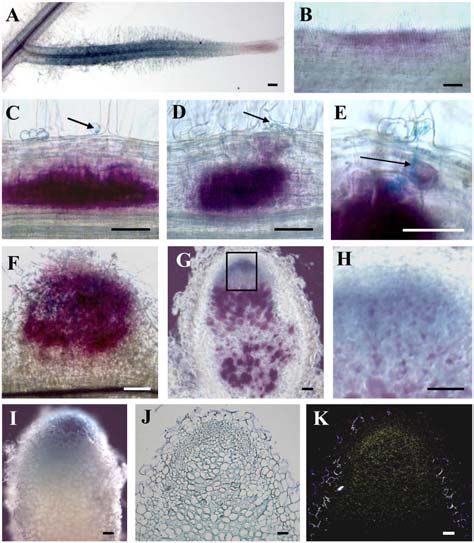
Medicago truncatula Lysine Motif-Receptor-Like Kinases

                                                                                          Figure 1. Amino acid alignment of
                                                                                          NFP (M. truncatula), SYM10 (pea),
                                                                                          and NFR5 (L. japonicus), showing con-
                                                                                          served domain structure and conserved
                                                                                          Cys residues in the predicted extracel-
                                                                                          lular parts (boxed). Sites where the
                                                                                          P-loops, activation loops, and the DFG
                                                                                          motifs are missing are indicated. In
                                                                                          NFP, positions of mutations found in
                                                                                          nfp-1 (residue no. 1) and nfp-2 (residue
                                                                                          no. 67) are boxed and indicated by
                                                                                          arrowheads. SP, Signal peptide; LysM1,
                                                                                          LysM2, and LysM3, three LysM do-
                                                                                          mains; TM, transmembrane segment.

pattern of the NFP gene using promoter-GUS fusions             reflected NFP expression. NFP transcripts were de-
and roots of M. truncatula transformed by A. rhizogenes.       tected in a relatively broad area of mature nodule
The 1.5-kb fragment in these constructions included            apices, with the highest level corresponding to the most
the same 1.1-kb promoter region used for the func-             distal part of the infection zone, as found with the
tional complementation described above.                        ProNFP-GUS fusion (Fig. 2, J and K). Consistent with
   Noninoculated roots showed strong GUS activity in           1.1 kb of this 1.5-kb promoter fragment being sufficient
root hair cells of lateral roots (Fig. 2A). Older regions of   for functional complementation of the nfp mutation, this
roots showed low or no GUS activity. One day after             indicates that the major cis-acting elements required
inoculation with S. meliloti, GUS activity was restricted      for cell-specific expression of NFP are present in the
to discrete areas of the epidermis (Fig. 2B). At 2 d, GUS      1.5-kb NFP promoter fragment. Furthermore, this shows
activity was associated with the inner cortex, forming         that NFP expression is associated with the infection
broad stained regions where cell divisions correspond-         process.
ing to the formation of nodule primordia could be
observed. GUS activity subsequently extended to the            Knockdown of NFP Suggests a Role for NFP during
middle cortex of regions where root hairs could be             Infection Thread Formation
seen to be undergoing root hair curling, the first step of
infection (Fig. 2C). At 3 d, GUS activity was detected in        We investigated the role of NFP in the infection
outer cortical cells directly underlying infected root         process using RNA interference (RNAi) constructs
hairs and through which infection threads were sub-            and A. rhizogenes-mediated root transformation of
sequently seen to pass (Fig. 2, D and E). At 5 d, GUS          M. truncatula. We expected to produce roots with a
staining was very intense in the central nodule tissue         range of knockdown levels, some of which could show
of young, emerging nodules, which mainly consists of           weak phenotypes for infection and nodulation. Two
cells undergoing infection by the highly ramified              independent NFP interference constructs were ana-
infection thread network (Fig. 2F). In mature nodules,         lyzed, NFPi1 and NFPi2, which correspond to the
GUS activity was restricted to the infection zone,             LysM and kinase domains, respectively.
showing strongest staining in cell layers directly adja-         The majority of NFP knockdown roots were Nod2
cent to the meristem (Fig. 2, G and H). Much lower             following inoculation with S. meliloti: 79% of NFPi1 and
expression was visible in interzone II/III and no ex-          90% of NFPi2 roots (i.e. a total of 21/24 Nod2 roots).
pression was detected in the nitrogen-fixing zone (Fig.        The rare nodules formed showed a wild-type structure
2, G and H). This is clearly illustrated in nodules in         when sectioned (data not shown). For control roots,
which bacteria were not stained (Fig. 2I).                     100% were efficiently nodulated with 11 nodules per
   For mature nodules, we verified by in situ hybrid-          root on average. Because the nodulation phenotype for
ization that this localization of GUS activity accurately      the NFP knockdown roots is, in most cases, similar to
Plant Physiol. Vol. 142, 2006                                                                                                 267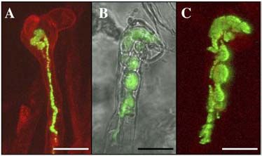
Arrighi et al.

                                                                                  Two lines of evidence support the assumption that
                                                                               silencing in NFP knockdown roots was specific for
                                                                               NFP transcripts. First, within the two regions used for
                                                                               NFP RNAi, there is no 21-bp region showing 100%
                                                                               identity between NFP and any known member of the
                                                                               M. truncatula LysM-RLK gene family described below.
                                                                               Second, RNAi performed on LYR1, the gene most
                                                                               homologous to NFP (see below), did not result in a
                                                                               Nod2 phenotype and infection threads were normal
                                                                               (data not shown).
                                                                                  Taken together, these results suggest a role for NFP
                                                                               in infection thread formation in agreement with NFP
                                                                               gene expression data.

                                                                               The NFP Kinase Domain Does Not Show
                                                                               Autophosphorylation Activity
                                                                                  In the NFP kinase domain, deviations were ob-
                                                                               served in the activation loop and in the P-loop, as well
                                                                               as in the DFG motif (Fig. 1), which are normally highly
                                                                               conserved in Ser-Thr kinases, and play important roles
                                                                               in positioning ATP and the Ser-Thr substrate. The NFP
                                                                               kinase domain might therefore be inactive. In contrast,
                                                                               the LYK3 kinase domain is fully conserved and pre-
Figure 2. Localization of NFP gene expression throughout nodulation            dicted to be fully active.
in M. truncatula. A to H, Expression of a ProNFP-GUS fusion in                    To gain further insight into these possibilities, we
transgenic roots, inoculated or not with S. meliloti. A, Whole, non-
                                                                               studied the autophosphorylation abilities of NFP and
inoculated lateral root showing constitutive GUS activity (blue) in root
hairs. B to F, Root segments showing GUS activity (magenta) in the
                                                                               LYK3. The RLK BAK1 was used as a positive control
epidermis 1 d postinoculation (dpi; B), in the inner and middle cortex         (Li et al., 2002). N-terminal glutathione S-transferase
where cell divisions are occurring under a curled root hair (arrow) at         (GST)-tagged intracellular domains of these three pro-
2 dpi (C), in the inner and middle cortex 2 dpi, and in outer cortical cells   teins (called GST-NFP, GST-LYK3, and GST-BAK1)
(D), in outer cortical cells through which an infection thread (arrow) is      were expressed in Escherichia coli and purified on
passing (E), and in an emerging nodule 5 dpi (F). G and H, Longitudinal        glutathione Sepharose columns. The purified proteins
section of an 18-d-old nodule showing GUS activity (blue) in the apical        were incubated with a kinase buffer containing [g-32]
region and bacteria (magenta) in infection threads and the nitrogen-           P-labeled ATP and separated by SDS-PAGE followed
fixing zone. H, Close-up of the box in G. I, GUS activity (blue) in an         by Coomassie Blue staining (Fig. 4A). Phosphorylated
18-d-old whole nodule, with bacteria not stained. J and K, In situ
                                                                               proteins were visualized by phosphor imaging (Fig.
hybridization of NFP gene expression on a sectioned 14-d-old nodule
using a 35S-UTP labeled antisense NFP probe. Bright-field image of
                                                                               4B). We detected phosphorylation of the GST-LYK3
nodule section with silver grains visible in black (J); epipolarization        and GST-BAK1 proteins, but not the GST-NFP protein,
image of I (K). Arrows indicate points of infection in root hair curls (C      indicating that the intracellular domain of LYK3, but
and D) and an infection thread (E). Bars 5 100 mm.

the knockout phenotype, it can be assumed that there is
efficient silencing in these roots, whereas incomplete
silencing has occurred in the three Nod1 roots.
   When rhizobia were visualized, 37 6 12 infection
threads were seen per control root. These were always
tubular structures (Fig. 3A), but about 10% stopped
abruptly in root hair cells. Infection structures were
not seen on nonnodulated roots of NFPi plants, but
were observed on nodulated NFPi1 roots in regions
adjacent to nodules: 12 and six infection threads for the
                                                                               Figure 3. Analysis of the role of NFP in controlling infection thread
two NFPi1 roots exhibiting 10 and nine nodules,
                                                                               formation. Roots of wild-type M. truncatula plants were transformed
respectively. These infection threads nearly always                            with either the vector alone (A) or an RNAi construct for NFP (B and C).
aborted in root hairs and often had aberrant morphol-                          Confocal images of root hairs in which GFP-expressing S. meliloti are
ogy, consisting of large sac-like structures (Fig. 3, B and                    seen in infection threads, which are tubular for the control and sac-like
C). This type of infection thread was never seen on                            in NFPi root hairs. Root hairs in A and C were counterstained by
control roots.                                                                 propidium iodide (0.2 mg/mL). Bars 5 10 mm.

268                                                                                                                   Plant Physiol. Vol. 142, 2006
Medicago truncatula Lysine Motif-Receptor-Like Kinases
All nine corresponding proteins are predicted to
have an N-terminal signal peptide, one or more LysM
domains in the predicted extracellular regions (Pfam
predictions), a transmembrane-spanning segment of
22 to 23 predominantly hydrophobic residues, and a
Ser-Thr kinase domain in the predicted cytoplasmic
part of the protein. Alignments with NFP and LYK1 to
Figure 4. Autophosphorylation tests on the intracellular kinase do-
7 (except LYK5 because LYK5 is probably a pseudo-
mains of NFP and LYK3. Purified GST-tagged intracellular domains of gene; Limpens et al., 2003) showed that certain
NFP, LYK3, and BAK1 (GST-NFP, GST-LYK3, and GST-BAK1) were residues are highly conserved in the extracellular
assayed for autophosphorylation activity using [g-32P]ATP. Reactions parts, including two repeats of Cys-X-Cys, which, for
were subjected to SDS-PAGE and proteins visualized with Coomassie NFP, are located first between LysM1 and LysM2
Blue (A). Phosphorylated proteins were visualized by phosphor imag- and second between LysM2 and LysM3 (Supplemental
ing (B). Molecular mass markers of 55 and 72 kD are shown. Fig. 1).
Sequence conservation is strongest in the putative
not of NFP, shows autophosphorylation activity in the intracellular kinase domains, but close inspection
conditions tested. revealed that, in addition to LYK3, LYR5 and 6 and
LYK1, 2, 4, and 6 to 9 have fully conserved kinase
domains, whereas those of LYK10, and LYR1 to 4 are
The M. truncatula Genome Contains a LysM-RLK
like NFP in having deviations in normally conserved
Family of at Least 17 Genes
residues (Supplemental Fig. 1). In LYK10, the con-
We were intrigued that, although NFP and LYK3 are served Thr-Ser residue in the activation loop is re-
both candidate Nod factor receptors, they differ in placed by a Glu residue. In LYR1 to 4 kinase domains,
their kinase domains and in the number of predicted the P-loop is missing and the DFG motif is substituted.
LysM domains (three and two, respectively). To de- LYR1 also lacks the activation loop. In addition, the
termine whether such variations are specific to these catalytic Asp residue is replaced by Asn in LYR1 and 4,
two proteins, we characterized the M. truncatula LysM- and the conserved Thr-Ser residue in the activation
RLK family. We first performed in silico searches in loops of LYR3 and 4 is replaced by Asp. Based on this,
M. truncatula EST and genomic databases and, given as well as sequence length, overall homology and
the very large number of kinase-encoding genes in plant structural predictions of the extracellular parts of these
genomes (more than 400 RLKs alone in Arabidopsis proteins (see below), four major groups of M. truncatula
[Arabidopsis thaliana]), we used LysM domains. Then, LysM-RLKs were distinguished and are represented
because LysM domains are not always associated with schematically in Figure 5.
kinase domains, we made a preselection among ESTs These results show that the LysM-RLK gene family
and only sequenced those most similar to LysM do- in M. truncatula consists of at least 17 genes, more than
mains of Arabidopsis LysM-kinase proteins. In addi- in either of the nonlegume plants Arabidopsis (five
tion to NFP and the previously identified LYK gene genes) or rice (Oryza sativa; six genes), and with six
cluster (LYK1–7; Limpens et al., 2003), nine new po- showing variation in conserved kinase residues.
tential LysM-RLK genes were identified and, as ex-
plained below, named LYR1 to 6 and LYK8 to 10. All The M. truncatula LysM-RLK Family Is Divided
except LYR1 were represented as ESTs and six genes into Three Subfamilies
(LYR1, 3–6, and LYK10) as genomic sequences with one
(LYR1, 3, and 4), two (LYR5 and 6), and 10 (LYK10) Based on kinase domain phylogeny, Arabidopsis
exons, and the LysM-encoding part is always devoid and rice LysM-RLK proteins form two clades, LysM-I
of introns. and LysM-II (Shiu et al., 2004). These were distinguished
Figure 5. Schematic representation of the domain structure of 16 M. truncatula LysM-RLKs, based on the amino acid alignment
presented in Supplemental Figure 1 and protein domain predictions explained in the text. SP, Signal peptide; LysM1, LysM2, and
LysM3, three LysM domains; CXC, Cys-X-Cys motif; TM, transmembrane segment; PL, P-loop; AL, activation loop; Ser-Thr kinase,
predicted intracellular kinase domain.
Plant Physiol. Vol. 142, 2006 269Arrighi et al.
when we explored the phylogenetic relationships M. truncatula genes is based on these important and
among kinase domains of the M. truncatula LysM- clear differences in gene structure. Thus, the previ-
RLK family, together with those of the five similar ously adopted LYK (LysM domain-containing RLK)
proteins of Arabidopsis and six of rice and NFR1 and has been used for genes having 10 to 12 exons, and
NFR5 of L. japonicus, with LYK3 and NFP belonging to other new genes are called LYR (for LYK-related).
the LysM-I and the LysM-II subfamilies, respectively Consistent with several deviations from the consensus
(Fig. 6). A third group, LysM-III, consisted only of the kinase sequence in NFP and LYR1 to 4, these proteins
M. truncatula LYR5/6 genes (Fig. 6). cluster together in the LysM-II clade, where NFP and
Comparison of intron/exon organization (known LYR1 (both lacking an activation loop) form a distinct
for all Arabidopsis and rice genes, NFR1 and NFR5, subgroup. In both of the other clades (LysM-I and
and 13 M. truncatula genes) with clades indicated that LysM-III), all kinases have classical conserved se-
LysM-I clade genes have 10 to 12 exons, whereas quences, except LYK10.
LysM-II and LysM-III genes have one and two exons, The same correlation was found between clade and
respectively. The nomenclature adopted for the new conservation of key kinase residues for Arabidopsis
Figure 6. Phylogenetic tree of kinase domains of LysM-RLK proteins of M. truncatula (16), rice (six), Arabidopsis (five), and NFR1
and NFR5 of L. japonicus, showing bootstrap values. NFP and LYK3 are underlined and the three groups are labeled I, II, and III.
270 Plant Physiol. Vol. 142, 2006Medicago truncatula Lysine Motif-Receptor-Like Kinases
and rice LysM-RLK kinase domains, with Os03g13080 in loop regions and there is a strongly electropositive
in LysM-II even missing the activation loop exactly as cavity in LysM2 of NFP, but not in LysM2 of LYK3
for NFP and LYR1. The only exceptions involved (Fig. 7; data not shown).
At1g51940 and Os01g36550 in LysM-I, which, like Close inspection of the amino acid alignment in
LYK10, lack a normally conserved Ser-Thr residue in Supplemental Figure 1 indicated a certain level of
their activation loops. This indicates that all LysM-II conservation among equivalent LysM domains of dif-
clade proteins either have substantially reduced ki- ferent proteins. To study this further, separate align-
nase activity or unusual functional characteristics dif- ments were made of domains in the same position of
ferent from classical kinases, whereas the majority of each protein to take into account structurally homol-
LysM-I and all LysM-III proteins have normal kinase ogous amino acids. Supplemental Figure 3 shows that
activity. LysM1, LysM2, and LysM3 of one protein have several
Finally, according to structural similarity between structurally homologous amino acids in common with
Ser-Thr kinases and the human Tyr kinase (PDB code the same domain of other LysM-RLK proteins, but not
IFIN; Jeffrey et al., 1995), in silico models were built for with other domains of the same protein. This suggests
LYK3 and NFP kinase domains as representatives of that, in addition to the overall conserved 3-D struc-
LysM-I and LysM-II kinases, respectively (Supplemen- tures, and the differences in charge between different
tal Fig. 2). Canonical three-dimensional (3-D) structures domains in NFP and LYK3, certain structural features
were predicted for both and the only significant dif- are unique to each domain.
ference concerned the region of the activation loop,
which, of course, is missing in the structure of NFP Seven New LysM-RLK Genes Are Expressed
(Supplemental Fig. 2). in Roots and Nodules
Three LysM Domains Are Structurally Predicted We performed quantitative reverse transcription
for Each M. truncatula LysM-RLK (RT)-PCR for the nine new M. truncatula genes, as
well as NFP, using RNA extracted from symbiotic
We analyzed extracellular domains of M. truncatula (roots and nodules) and nonsymbiotic (leaves and
LysM-RLK proteins by in silico modeling to determine stems) plant tissues (Table I). As a result, four classes
how many LysM domains are predicted within each of expression pattern could be distinguished. In the
protein and whether each LysM domain is likely to be first class, LYR2 and LYK10 were expressed specifically
equivalent in terms of potential ligand binding. in roots and nodules. In the second class, NFP, LYR3,
Based on positions of the Cys-X-Cys motifs, three and LYR6 were expressed in roots and nodules and at
potential LysM domains were identified for each pro- a much lower level in leaves or stems. In the third
tein and used to make 3-D structural models. Hydro- class, LYR1 and LYK8 are expressed mostly or only in
phobic cluster analysis (HCA) plots were generated roots. Finally, LYR4, LYR5, and LYK9 were expressed in
and compared to the HCA plot of the murein hydro- all tested tissues, with the highest levels in leaves.
lase from E. coli, for which there is an NMR-solved Table I also shows the relative expression levels in
LysM domain structure (Bateman and Bycroft, 2000; roots of new genes compared to NFP, indicating that
Supplemental Data 1). This E. coli LysM domain has a the majority are more highly expressed than NFP.
globular structure with a shallow groove on the sur- These results indicate that NFP, like other previously
face, thought to be the site for peptidoglycan binding. characterized symbiotic LysM-RLK genes (Limpens
The structural backbone of this domain is formed of et al., 2003; Madsen et al., 2003; Radutoiu et al., 2003),
two strands of a b-sheet (b1, b2) flanking two short is preferentially expressed in nonaerial parts of the
a-helices (a1, a2). Despite the moderate percentages of plant. Furthermore, six new genes (LYR1, LYR2, LYR3,
identity (approximately 25%) and similarity (approx- LYR6, LYK8, and LYK10) were expressed predomi-
imately 67%) between the E. coli and M. truncatula nantly in roots and nodules.
LysM domains, these structures were easily delineated
on the HCA plots of all the M. truncatula sequences LysM-RLKs Have Expanded in the M. truncatula
(illustrated for NFP in Fig. 7), indicating pronounced Genome by Gene Duplication and Segmental
3-D structural conservation and the presence of three Genome Duplication
LysM domains in each of the 16 proteins analyzed
(NFP, LYR1–6, LYK1–4, and LYK6–10). As for RLK gene expansion in Arabidopsis (Shiu
For NFP and LYK3, LysM domains differ from their and Bleecker, 2003), both tandem duplication and
E. coli counterparts in some insertions or deletions segmental genome duplication appear to have oc-
occurring in loop regions and by their charges. LysM1 curred for the M. truncatula LysM-RLK family. Thus,
domains are essentially electropositively charged (net LYK3 is in a cluster of seven LYK genes (Limpens et al.,
charge from 2–4) or neutral, but LysM2 (net charge 2003), which shows microsynteny to a second region of
from 22 to 24) and LysM3 (net charge from 21 to 25) chromosome 5 (Gualtieri and Bisseling, 2002). For new
are predominantly electronegative (illustrated for NFP genes, we identified two tandem repeats: LYR3/NFP
in Fig. 7). The electropositively (Arg, Lys) or electro- and LYR5/LYR6. Also, highly homologous pectate
negatively (Asp, Glu) charged residues mainly occur lyase genes were found adjacent to both LYR3/NFP
Plant Physiol. Vol. 142, 2006 271Arrighi et al.
Figure 7. Structural predictions for LysM domains of NFP. 1 and 3, HCA plots showing homologous hydrophobic residues
(circled orange) and secondary structural features (boxed; 1) and electropositively (circled blue) and electronegatively (circled
red) charged residues (3). 2, Ribbon diagrams showing the N- and C-terminal ends of the LysM domain polypeptides. 4, 3-D
models with electrostatic potentials displayed on the molecular surface of LysM domains at 25 kT and 15 kT levels; red
(electronegative), blue (electropositive), and white (neutral). A, LysM domain of E. coli murein hydrolase. B to D, Structural
predictions for NFP LysM domains built from the atomic coordinates of this E. coli LysM domain: LysM1 (B); LysM2 (C); LysM3 (D).
Note the strongly electropositive cavity in LysM2 of NFP (4C).
and LYR1 (but not to LYR5/LYR6), and remnants of colocalize, analysis of BAC contigs showed that they
LysM genes were found adjacent to LYR1, suggesting could be physically separated.
chromosomal segmental duplication followed by se- This analysis indicates that both tandem duplication
quence changes for the LYR3/NFP and LYR1 regions. and segmental genome duplication events are respon-
To further characterize genomic organization of sible for the large M. truncatula LysM-RLK gene family.
LysM-RLKs, we used the M. truncatula genetic map Furthermore, 11 of the 17 identified M. truncatula LysM-
(Thoquet et al., 2002) and positioned new LysM-RLK RLK genes are now known to be located in clusters or
genes on six of the eight chromosomes of M. truncatula tandem repeats of genes of the same subfamily.
(Fig. 8). The highest density of LysM-RLK genes is on
chromosome 5 (11 of 17 genes), where NFP and the DISCUSSION
LYK1 to 7 gene cluster map (Ben Amor et al., 2003;
Limpens et al., 2003). On this chromosome, although The discovery that a new class of plant receptors, the
LYR4 and the LYK1 to 7 gene cluster genetically LysM-RLKs, control Nod factor signaling provided a
272 Plant Physiol. Vol. 142, 2006Medicago truncatula Lysine Motif-Receptor-Like Kinases
Table I. Expression analysis by quantitative RT-PCR of 10 LysM-RLK genes of M. truncatula
mRNA levels were normalized against ACTIN2 and values were calculated as ratios relative to root
expression levels. The averages of two independent biological experiments are presented. Root samples
always have a value of 1 and SEMs are given for other samples. Bold text indicates preferential localization of
gene expression. ND, Not determined.
Expression Levels Relative to Roots Root Expression
Gene
Root Nodule Leaf Stem Relative to NFP
NFP 1 0.59 6 0 0 0.024 6 0.02 1
LYR1 1 0 0 0 ND
LYR2 1 1.75 6 0.035 0 0 0.37
LYR3 1 2.90 6 0.89 0.23 6 0.11 0.005 6 0.004 6.23
LYR4 1 2.27 6 0.59 10.50 6 5.78 1.70 6 0.95 21.56
LYR5 1 6.39 6 0.54 21.79 6 0.24 0.26 6 0.02 2.43
LYR6 1 3.80 6 3.95 0.26 6 0.032 0.008 6 0.004 12
LYK8 1 0.06 6 0 0.01 6 0.007 0 1.22
LYK9 1 0.74 6 0.12 2.33 6 0.76 0.28 6 0.085 38.32
LYK10 1 0.73 6 0.10 0 0 0.15
major breakthrough in our understanding of the The NFP Gene Controls Both Early Symbiotic Responses
Rhizobium symbiosis (for review, see Cullimore and and Infection
Dénarié, 2003; Parniske and Downie, 2003; Riely et al.,
2004; Stacey et al., 2006). Given that M. truncatula nfp NFP gene expression observed in root hairs before
mutants exhibit very similar phenotypes to L. japonicus inoculation with S. meliloti is consistent with the role of
and pea LysM-RLK mutants, it was likely that NFP NFP in controlling a rapid calcium flux, calcium
would also encode a LysM-RLK. Our confirmation of spiking, and inhibition of reactive oxygen efflux
this provides support for the genetic evidence that within minutes of Nod factor addition to root hairs
NFP could be a Nod factor receptor (Ben Amor et al., (Ben Amor et al., 2003; Shaw and Long, 2003). More-
2003), and we have now shown that NFP is implicated over, this constitutive expression of NFP was restricted
in nodule organogenesis and throughout the infection to growing and recently matured root hairs, cor-
process. The evidence that NFP has three LysM domains responding to the susceptible zone for Nod factor
with different potential properties for ligand recogni- responses and rhizobial infection. After rhizobial
tion and a kinase domain with no apparent autophos- inoculation, NFP expression was strongly linked to
phorylation activity provides new information on how nodule primordia development in the root cortex and
NFP might function in Nod factor perception and to infection in root hairs and underlying outer cortical
transduction. Furthermore, we show that M. truncatula cells. This latter type of cell has been described in
has a larger LysM-RLK gene family than Arabidopsis Medicago as being prepared for infection by the pres-
or rice, with other genes that may fulfill symbiotic roles. ence of preinfection threads (Timmers et al., 1999). In
Figure 8. Genetic map positions of
M. truncatula LysM-RLK genes. LYK and
LYR genes are positioned between mi-
crosatellite markers on six of the eight
M. truncatula linkage groups (LG). The
three cases of tandemly arranged genes
(NFP/LYR3, LYK1–7, and LYR5/LYR6) are
each indicated by a single map position.
LYR4 and LYK1–7 also have a single map
position, but are not physically linked (see
text). The drawing was made using Map-
Chart (Voorips, 2002). Distances are in cM
Kosambi.
Plant Physiol. Vol. 142, 2006 273Arrighi et al.
mature nodules, expression coincided again with in- outward wave of activation for nodule organogenesis
fection thread formation and with bacterial release is induced by Nod factors or Nod factor derivatives, or
into host cells. Using the RNAi approach, rare infec- whether secondary signals are transmitted following
tions were observed and characterized by abortive epidermal Nod factor perception. The strong accumu-
infection threads with sac-like structures. Infection lation of fluorescent Nod factor derivatives in root hair
thread growth and morphology is therefore appar- cell walls (Goedhart et al., 2003) does not exclude the
ently susceptible to a reduction in NFP levels. This, diffusion or transfer of a small proportion to inner
together with gene expression data, indicates that NFP tissues. The strong and rapid up-regulation of NFP
is implicated throughout the infection process, in expression in inner cortical cells is compatible with a
particular for infection thread development, and also long-range signal being Nod factors or Nod factor
possibly for bacterial release. derivatives. It will be interesting to elucidate the role of
Of the known orthologs of NFP, pea SYM10 but not a putative Nod factor receptor several cell layers
L. japonicus NFR5, is expressed in mature nodules, distant from the site of Nod factor-producing rhizobia.
which might reflect the different types of nodules
formed on M. truncatula and pea (indeterminate) com- LysM-RLKs Have a Conserved LysM Triplet in Their
pared to L. japonicus (determinate; Madsen et al., 2003). Extracellular Regions
A major difference between these two nodule types is
that only indeterminate nodules have a persistent LysM domains are probably of prokaryotic origin
meristem continually producing new nodule cells and are common modules of bacterial surface proteins
that can be infected by rhizobia. Therefore, this sup- (Bateman and Bycroft, 2000). The combination of LysM
ports important roles for NFP and pea SYM10 in Nod domains with a transmembrane helice and a kinase
factor signaling during the infection process in nod- domain is specific to the phylogenic lineage of plants
ules. Furthermore, the absence of expression of both and, interestingly, despite the two different gene struc-
NFR5 in mature L. japonicus nodules and NFP in the tures found for these genes, there is never an intron
central tissue of mature M. truncatula nodules indi- within the predicted extracellular LysM coding region.
cates that neither gene is likely to be involved in Whereas LYK3, LYK4, and L. japonicus NFR1 were
nitrogen fixation or assimilation. described as having two LysM domains each (Limpens
et al., 2003; Radutoiu et al., 2003), we could structur-
NFP Might Act Upstream of Distinct Nod Factor ally predict three such domains each time (data not
Signaling Pathways shown), as well as for the 14 other M. truncatula
proteins studied. This indicates that three LysM do-
Functional and expression analysis of DMI1, DMI2, mains are a general feature of plant LysM-RLKs. The
and DMI3, predicted to control Nod factor signaling Cys-X-Cys motif was always found between the three
downstream of NFP, suggests that Nod factor sig- predicted LysM domains of M. truncatula proteins and
naling occurs throughout infection in M. truncatula is present in the corresponding regions of Arabidopsis
(Bersoult et al., 2005; Limpens et al., 2005, Hogg et al., and rice proteins. This suggests that disulfide bridges
2006). However, the three DMI genes also control might participate in the spatial distribution of LysM
mycorrhization and dmi mutants have nonsymbiotic domains and that a LysM triplet of three LysM do-
phenotypes (Catoira et al., 2000; Esseling et al., 2004). mains separated by Cys-X-Cys motifs and linked to a
Because the NFP gene has not been shown to control kinase domain by a transmembrane helix appeared
anything other than Nod factor perception, our NFP early in plant history, probably predating the division
data more directly suggest that Nod factor perception of LysM-RLKs into the two major clades, LysM-I and
and signaling occur throughout the infection process. LysM-II.
The occurrence of Nod factor recognition in the infec- The LysM triplet in plant LysM-RLKs might have
tion zone of M. truncatula nodules is corroborated by originally consisted of the same LysM sequence re-
the expression pattern of LYK3 (Limpens et al., 2005). peated three times because each LysM domain has an
Also, in nodules, NFP expression coincides with the overall 3-D fold similar to that of a bacterial LysM
localization of rhizobial nod gene transcripts and Nod domain. Recent work by Mulder et al. (2006) also
factors, whereas the decline in NFP expression toward suggests conservation of structure between plant and
the nitrogen-fixing zone coincides with the decline of bacterial LysM domains. The differences in surface
nod gene transcripts, as well as with organelle redis- charge distribution and structurally homologous
tribution and cytoskeleton reorganization (Sharma and amino acids between LysM1, LysM2, and LysM3 of a
Signer, 1990; Schlaman et al., 1991; Timmers et al., given protein suggest that each domain has special-
1998). ized functions in terms of ligand recognition.
NFP expression is associated with two concurrent A LysM triplet might also be characteristic of plant
series of events described during establishment of nonkinase LysM proteins. During this work, we iden-
nodulation in M. truncatula (Timmers et al., 1999), tified two M. truncatula genes predicted to encode such
suggesting that NFP controls distinct Nod factor sig- proteins with three LysM domains structurally pre-
naling pathways, one for symbiotic infection and one dicted for each and showing strong homology to
for nodule organogenesis. It is not known whether the the glycosylphosphatidylinositol-anchored proteins
274 Plant Physiol. Vol. 142, 2006Medicago truncatula Lysine Motif-Receptor-Like Kinases
AtLYM1 (At1g21880) and AtLYM2 (At2g17120; Borner AtACR4 (Johnson and Ingram, 2005). Whether non-
et al., 2003). Both M. truncatula proteins are predicted classical kinases such as NFP associate with active
to be secreted and one to be membrane located. By kinase partners or have an unusual mechanism of
analogy with the role of proteins that resemble RLK signal transduction is an open question.
extracellular domains in RLK signaling (Jeong et al.,
1999), it will be interesting to investigate whether M. truncatula Has a Larger LysM-RLK Gene Family
nonkinase LysM proteins play roles in symbiotic sig- Than the Nonlegumes Arabidopsis and Rice
naling.
Approximately 60% of the gene-rich region of the
LysM-RLKs Have Either Classical or Nonclassical M. truncatula genome is sequenced and 16 expressed
Kinase Domains LysM-RLK genes have been identified. Compared to
Arabidopsis and rice, with five and six LysM-RLK
Compared to LysM domains, there is higher conser- genes, respectively, duplication of these genes in
vation among the kinase domains of LysM-RLKs, M. truncatula raises many questions. Some putative or-
illustrated by the conserved predicted 3-D structures thologs can be identified across plant species, suggest-
for NFP and LYK3. Despite this, we distinguished ing related functions, and there are some potentially
kinase domains of distinct evolutionary origin and M. truncatula-specific LYK and LYR proteins. By anal-
only LysM-RLKs in clades I and III were predicted to ogy with the role of LysM domains in recognizing
have classically functional kinase domains. In clade II, N-acetyl-glucosamine (Steen et al., 2003, 2005) and
accumulated mutations in generally conserved resi- their presence in both a chitinase of Volvox carteri
dues probably predated dicot/monocot divergence. (Amon et al., 1998) and putative Nod factor receptors,
For example, M. truncatula NFP, M. truncatula LYR1, all plant LysM-RLKs may recognize b1- to b4-linked
and Os03g13080 have key residues not conserved and N-acetyl-amino sugar ligand molecules such as chitin
no activation loop. A similar such gene was recently oligomers. Furthermore, because such molecules have
identified in Populus trichocarpa (Stacey et al., 2006). In never been reported in plants, all plant LysM-RLKs
contrast, the LYR5 and LYR6 genes may be of relatively might have roles in recognizing other organisms, for
recent origin because they form a distinct phylogenetic example, in the process of innate immunity.
group with no close relatives in Arabidopsis or rice. According to Shiu et al. (2004), plant RLKs involved
Close inspection of LYR5/6 indicates that their LysM in development have rarely been duplicated after the
domains are more similar to LYR than to LYK proteins, dicot-monocot split, whereas those involved in de-
whereas their kinase domains show 59% identity to fense/disease resistance have undergone many dupli-
those of Arabidopsis WAK-like proteins. Molecular cation events. Some M. truncatula LysM-RLKs are
complexity in the LysM-RLK family has thus appar- found as ESTs from elicitor/pathogen-treated tissue,
ently arisen by fusion of the same LysM triplet region but these are not highly represented in their cDNA
to different kinase domains, as for other plant RLKs libraries. As pointed out by Parniske and Downie
(Shiu and Bleecker, 2003). (2003), the clustering of Nod factor receptor genes
Amino acid replacements in conserved protein do- could facilitate recombination and hence participate in
mains can be expected to contribute to functional the recognition by closely related plants of different
diversity. Different plant LysM-RLKs may conse- Nod factor structures. Consistent with this, NFP and
quently have distinct modes of functioning for signal LYK3 are two of the 11 M. truncatula LysM-RLK genes
transduction, with some potentially having inactive or known to be located in clusters or tandem repeats.
so-called dead kinase domains. This is supported by The finding that many M. truncatula LysM-RLK
the absence of detectable in vitro autophosphorylation genes, like NFP and LYK3, are expressed in nodules
activity for the NFP kinase domain. Moreover, al- suggests that expansion of the LysM-RLK gene family
though we cannot exclude ligand-activated autophos- in M. truncatula is connected with nodulation. Seven
phorylation, the long-lasting conservation of this type newly identified genes (LYR2, LYR3, LYR4, LYR5,
of kinase with a predicted typical 3-D structure sup- LYR6, LYK9, and LYK10) are expressed in nodules,
ports an important role in signal transduction. A and six (LYR1, LYR2, LYR3, LYR6, LYK8, and LYK10)
similar conclusion can be drawn for NFP orthologs are expressed exclusively or almost exclusively in
in L. japonicus and pea, based on the null phenotypes roots and/or nodules. At least the first group of genes
of certain kinase domain mutants (Madsen et al., 2003). must fulfill roles in nodulation. The different expres-
Increasing evidence suggests that some dead kinase sion patterns of the tandemly repeated genes LYR5/6
receptor kinases are signal-transducing molecules suggest that specialization for symbiotic functions can
without kinase activity. Examples in animals include follow gene duplication.
ErbB3 and H-Ryk, which both form heterodimers with Large-scale genomic studies have explored why
kinase-active receptors (Kroiher et al., 2001). In plants, rhizobia nodulate legumes (Weidner et al., 2003;
kinase-dead RLKs are found in three families and Silverstein et al., 2006). Further analysis of LysM-RLK
include AtCRR1 and AtCRR2, which have no activa- gene families in different plant species should help
tion loops, are nearly inactive in autophosphorylation elucidate which preexisting functions were adapted
assays, and AtCRR2 can be phosphorylated by for Nod factor signaling. It will also be interesting to
Plant Physiol. Vol. 142, 2006 275Arrighi et al.
see whether LysM-RLK expansion is a characteristic In conclusion, it can be recalled that forward genet-
of all nodulating legumes or only of legumes like ics screens of model legumes for mutants exhibiting no
M. truncatula, which form indeterminate nodules. In- response to Nod factors identified NFR1 and NFR5 in
terestingly, the NFR1 gene of L. japonicus (determinate L. japonicus and NFP in M. truncatula. Comparative
nodules) is found within a cluster of only three LysM- genetics and RNAi led to the identification of LYK3
RLK genes compared to seven for the M. truncatula and LYK4 in M. truncatula. These genetic approaches
LYK3 gene cluster (Zhu et al., 2006). Also, for the rest of thus suggested that there are at least three Nod factor
the newly expanded M. truncatula family, only a po- receptor genes in M. truncatula. In this study, a ge-
tential ortholog of LYR3 has so far been identified in nomic approach identified seven new M. truncatula
L. japonicus (Zhu et al., 2006). LysM-RLK genes that are expressed in roots and
nodules, suggesting a higher number of Nod factor
LysM-RLKs and Nod Factor Perception receptor genes. This can be explained if different Nod
factor receptor complexes act at different steps of the
The molecular identity of NFP supports genetic symbiotic process in such a way that mutants in Nod
evidence that this protein is an essential part of a factor receptor genes not previously identified would
Nod factor receptor. In this scenario, Nod factor struc- not have null phenotypes, or if there is functional
ture would be recognized by the extracellular LysM redundancy, suggested by the fact that no mutants
domains. The identification of a new nfp allele, nfp-2, other than nfp mutants were found with the same null
mutated in the first LysM domain supports a crucial phenotype (equivalent to nfr1 mutants of L. japonicus;
role for this domain in Nod factor recognition. Fur- Geurts et al., 2005). The implication of new LysM-
thermore, this predicted amino acid change in LysM1 RLKs in the nodulation process in M. truncatula now
leads to strong defective phenotypes, suggesting no needs to be examined using RNAi and reverse genetics
redundancy among the three LysM domains of NFP. resources.
Instead, only LysM1 may be needed or, consistent
with the conservation of the three domains, all three
may be required for optimal activity as shown for an
MATERIALS AND METHODS
N-acetylglucosaminidase of Lactococcus lactis (Steen
et al., 2005). Plant Material and Growth Conditions
In L. japonicus, the ortholog of NFP, NFR5, is hypoth-
Medicago truncatula Jemalong A17 was used in expression and RNAi
esized to dimerize with NFR1, the probable ortholog of studies using growth conditions as described by Catoira et al. (2000). The nfp-2
LYK3 (Madsen et al., 2003). Until now, in M. truncatula, mutant came from ethyl methanesulfonate-mutagenized seeds (M. Harrison,
there was no evidence that NFP might associate with unpublished data) derived from L416, a transgenic A17 line carrying the
LYK3. However, the abnormal infection threads ob- pMtENOD11-gusA construct (Charron et al., 2004).
served on NFP knockdown roots resembled those
reported for LYK3 knockdown roots (Limpens et al., Plant Transformation and Rhizobial Inoculation
2003), suggesting that NFP and LYK3 act together at Plasmid introduction into Agrobacterium rhizogenes ARqua1, hairy root
this stage of the symbiotic process. LYK4 is also transformation, and inoculation with Sinorhizobium meliloti were carried out as
implicated in infection (Limpens et al., 2003) and, described by Lévy et al. (2004).
interestingly, whereas LYK3 and LYK4 have practically
identical extracellular LysM parts, suggesting recog- NFP Cloning
nition of the same ligand, their kinase domains differ,
The candidate NFP gene was identified by the EST BE204912 and the BAC
suggesting that they phosphorylate different targets. clone mth2-34m14 (Oklahoma University M. truncatula genome-sequencing
In the possible scenario of NFP-LYK3/4 heterodimers, project). For mapping, a cleaved-amplified polymorphic sequence (CAPS)
the differences in LysM domains between NFP and was made with the primers GGTTGTTCTCGGAATCTTCG and CTGCCAAA-
LYK3/4 suggest contribution of distinct ligand recog- GAAGCCAAACTT, and a PCR cycle of 94°C for 4 min, then 40 cycles of 94°C
nition properties to the receptor dimer. for 30 s, 50°C for 30 s, 72°C for 30 s, and, finally, 72°C for 6 min. The 456-bp
products were digested with DdeI before loading on 2% agarose gel. For in
Different LysM-RLK complexes might be implicated planta complementation, a 4.6-kb EcoRV fragment, containing the wild-type
at different symbiotic steps (Cullimore and Dénarié, NFP gene plus a 1,137-bp promoter region and a 1,665-bp 3# region was cloned
2003) and could possibly recognize different ligands. in the binary vector pCambia2201 (www.cambia.org).
In animals, binding different ligands can cause hetero-
oligomerization between different receptor combina- Spatiotemporal Expression Analysis of the NFP Gene
tions and stimulation of different responses. In plants,
For promoter-GUS fusions, the 1.5-kb region upstream of NFP was
heterooligomerization of RLKs is a recurring theme amplified (94°C for 6 min, 40 cycles at 94°C for 30 s, 55°C for 30 s, and 72°C
and there is preliminary evidence for combinatorial for 1 min, and then 72°C for 5 min) with the primers GTAGGTCGAC (SalI)
heteromeric pairing in response to different ligands AGAAGGGAGACGGAGAGAGG and AGCGGATCC (BamHI) TTGTTG-
(Johnson and Ingram, 2005). In M. truncatula, the TGAGGAAATGCAAA and cloned into the binary vectors pLP100 (Szabados
discovery of three Nod factor-binding sites, all inde- et al., 1995) and pBI101 (Jefferson et al., 1987). Resulting plasmids were
verified by sequencing. Identical expression profiles were obtained with both
pendent of NFP, is compatible with Nod factor signal- plasmids and results are presented for pLP100. In situ hybridization was
ing being more complex than suggested by current performed as described (Limpens et al., 2005) on 14-d-old nodules with two
models (Hogg et al., 2006). fragments of NFP as probes (28–260 and 425–645).
276 Plant Physiol. Vol. 142, 2006Medicago truncatula Lysine Motif-Receptor-Like Kinases
RNAi Analysis of NFP dopsis thaliana) proteins from Swiss-Prot TrEMBL and 28,657 M. truncatula
peptides predicted from EST clusters in the MENS database (http://medicago.
Two NFP inverted-repeat binary constructs were cloned into pREDROOT toulouse.inra.fr/MENS). The ehmmsearch program of the EMBOSS
as described in Limpens et al. (2004). Target regions for NFPi1 and NFPi2 of package was then used to search the database of plant peptides with the
399 (327–725) and 532 (893–1,424) bp, respectively, were amplified using the seed HMM Pfam LysM domain (accession no. PF01476). The 65 potential
following primer combinations: ACTAGTGGCGCGCCTTTCGCGAATAT- LysM domains thus identified were put into a fasta database, which was then
CACCTAC and GGATCCATTTAAATCTGCTTTTTCTTCCATTTG; ACTAGT- used to BLAST (BLASTX with parameters 2F F 2G 6 2E 2 2e 0.1), nucleic
GGCGCGCCTAAGCAAGCCAACAATGTAT and GGATCCATTTAAATAC- acid sequence databases of M. truncatula, both genomic (http://www.
CCCAAAAGCGAAAACAT, where the underlined sequences were used for genome.ou.edu/medicago.html) and EST clusters (http://medicago.toulouse.
restriction ligation cloning. Transgenic roots were selected based on red inra.fr/MENS). Domain structure was analyzed with Pfam (http://www.
fluorescence and subsequently inoculated with S. meliloti strains Sm2011 sanger.ac.uk/Software/Pfam) and SUPERFAMILY (http://supfam.mrc-lmb.
(pHC60) or GMI5622 (pHC60; a nodFE deletion mutant; Limpens et al., 2003, cam.ac.uk/SUPERFAMILY). Signal peptides and transmembrane domains
2004). Identical results were obtained with both strains and results are were predicted (using http://www.cbs.dtu.dk/services/SignalP and http://
presented for Sm2011 (pHC60). www.cbs.dtu.dk/services/TMHMM, respectively). Rice (Oryza sativa) and
Arabidopsis LysM-RLKs were identified by BLAST against http://www.
ncbi.nlm.nih.gov/BLAST/Genome/PlantBlast.shtml?7 using M. truncatula
Microscopic Methods LysM domains and standard parameters. Among the candidate LysM-RLKs
Roots were observed for bacterial infection by histochemical staining of identified, the occurrence of the conserved Cys-X-Cys motif twice in potential
b-galactosidase activity expressed by the plasmid pXLGD4 (Ardourel et al., extracellular domains was found to be a useful criterion for selection. Amino
1994) for ProNFP-GUS analysis, and by detection of green fluorescent protein acid sequence alignments were made with ClustalX (Thompson et al., 1997)
(GFP) from the plasmid pHC60 for RNAi lines (Limpens et al., 2003). GUS and minor manual adjustments were made by comparison with HCA plots to
activity in ProNFP-GUS analysis of uninoculated roots was detected using improve alignments.
X-gluc (Catoira et al., 2000). Inoculated roots and nodules were prefixed with
0.5% paraformaldehyde and nodules embedded in 4% agarose before making Phylogeny Studies
100-mm-thick sections with a Bio-Rad microcut H1200 vibratome. Double
detection of ProNFP-GUS expression and bacterial LacZ activity was carried out Amino acid sequence alignment was performed using ClustalX. Distances
as described (Bersoult et al., 2005) using either X-Gal and Magenta-gluc or between proteins were calculated by neighbor joining with the phylo_win
Magenta-Gal and X-gluc. Observations were made with an Axiophot micro- program using the algorithm of the PROTDIST program of the PHYLIP
scope by bright field (roots) or phase contrast (nodule sections). Digital images package. One thousand bootstrap replicates were calculated. TreeView was
were taken with a Leica camera and software. GFP was visualized with a Carl used to construct trees.
Zeiss LSM 510 confocal laser-scanning microscope.
EST and BAC Clones
Autophosphorylation Test
EST clusters corresponding to newly identified genes are MtD10160
The sequences encoding the predicted NFP and LYK3 intracellular regions (LYR2), MtD20757 (LYR3), MtC93221 (LYR4), MtD13882 (LYR5), MtC90998
were amplified by PCR using forward primers GAATTCAAAATGAAGA- (LYR6), MtD06512 (LYK8), MtD04912 (LYK9), and MtD15867 (LYK10). BAC
GATTGAATAGAAG and GAATTCAAGAAGGAAGAAGAGAAAACT and clones identified were mth2-34m14 (NFP and LYR3), mth2-14m21 (LYR1),
reverse primers GTCGACTTAACGAGCTATTACAGAAGT and CTCGAGT- mth4-1f18 (LYR4), and mth2-3c7 (LYR5 and LYR6). For MtLYM1, the EST
CATCTAGTTGACAACAGATT, using the NFP gene and LYK3 cDNA, re- MtD01600 and the BAC clone mth2-77f21 were identified. For MtLYM2, the
spectively, as templates. PCR products were digested with EcoRI/SalI for NFP EST MtC30180 was identified.
or EcoRI/XhoI for LYK3 and cloned in the pGEX-6P-1 vector (Amersham
Biosciences) to produce the GST-NFP and GST-LYK3 constructs. The GST-
BAK1 construct is described in Li et al. (2002). GST-LYK3, GST-NFP, and GST- Quantitative RT-PCR
BAK1 fusion proteins were expressed and purified with a method modified
from Horn and Walker (1994). A 10-mL Escherichia coli overnight culture was Plants of wild-type M. truncatula Jemalong A17 were grown aeroponically
transferred to 400 mL of Luria-Bertani medium and grown at 37°C to a density as described (Catoira et al., 2000). Total RNA was extracted from leaves, stems,
of about 0.8 OD600. Isopropylthio-b-galactoside was added to 1 mM and cells uninoculated roots, and 10-d-old isolated nodules using the TRIzol kit
were grown for 24 h at 16°C. The cells were collected by centrifugation, (Invitrogen), treated with DNase I, and reverse transcribed as described (El
washed two times with 20 mM HEPES buffer, pH 7.5, and resuspended in Yahyaoui et al., 2004). The quantitative RT-PCR reaction was performed in a
20 mL of lysis buffer (10 mM sodium phosphate buffer, pH 7.5, 500 mM NaCl, LightCycler (Roche) on 50-fold diluted cDNA using the Master SYBR green I
5 mM dithiothreitol, 10 mM EDTA, and a mix of protease inhibitors [1 mg/mL kit (Roche). Samples were preincubated at 95°C for 5 min, followed by 45
of leupeptin, aprotinin, antipaine, chymostatin, and pepstatin]) and sonicated cycles (95°C, 5 s; 65°C, 7 s; 72°C, 15 s). Specificity of PCR amplification was
on ice for 5 min. Insoluble material was removed by centrifugation, the cleared checked with a heat dissociation step (from 70°C–95°C) at the end of the run.
extract was added to 800 mL of glutathione Sepharose 4B (Amersham Biotech), Crossing points (cycle threshold) were determined with the second derivative
and incubated with gentle agitation overnight at 4°C. Beads were collected by maximum method and the arithmetic signal baseline adjustment. Amplifica-
centrifugation, washed two times with 4 mL of lysis buffer without EDTA, and tion efficiencies and cDNA relative abundance in each sample were deter-
covered with 800 mL of elution buffer (20 mM Tris-Cl buffer, pH 8.0, 120 mM mined from a standard curve established from a serial 3 5 dilution of cDNA.
NaCl, 20 mM glutathione, 1 mg/mL protease inhibitors). Recombinant proteins Primers (Supplemental Table I) were designed using Vector NTI (InforMax).
were eluted by overnight incubation at 4°C under agitation. The quality and Results were standardized to the constitutive ACTIN2 expression levels as
quantity of eluted protein were checked by SDS-PAGE and Bradford assay, described (Limpens et al., 2005).
respectively. For kinase autophosphorylation assays, 1 mg of protein was
incubated in 20 mL of kinase buffer (50 mM HEPES, pH 7.4, 10 mM MgCl2,
10 mM MnCl2, 1 mM dithiothreitol, 600 nM ATP, 110 nM [10 mCi] [g-32P]ATP) at
Mapping New LysM-RLK Genes on the M. truncatula
30°C for 1.5 h. The reaction was stopped by adding SDS-PAGE sample buffer Genetic Map
and boiling for 5 min. Reactions were analyzed by SDS-PAGE and, after
CAPS or microsatellite markers were generated after nucleotide sequence
staining with Coomassie Brilliant Blue R250, gels were dried and analyzed by
comparison of a PCR-amplified region from both parents of the mapping
phosphor imaging.
population, Jemalong and DZA315.16 (Supplemental Table II). For CAPS
markers, PCR products were digested with restriction enzymes (Supplemen-
Sequence Analysis tal Table II). Markers were analyzed on 199 F6 recombinant inbred lines
derived from the mapping population and positioned between bordering
To search for DNA sequences of M. truncatula LysM domains, a database of microsatellite markers by MAPMAKER (T. Huguet, unpublished data; Sup-
plant peptides was first compiled, consisting of 44,509 Arabidopsis (Arabi- plemental Table III; Lander et al., 1987). PCR conditions were 94°C for 4 min,
Plant Physiol. Vol. 142, 2006 277You can also read