Journal of Oral Medicine and Dental - Research Genesis-JOMDR-2(1)-S1 Open Access
←
→
Page content transcription
If your browser does not render page correctly, please read the page content below
1

Journal of Oral Medicine and Dental
Research                         Genesis-JOMDR-2(1)-S1
                                                                                                           Volume 2 | Issue 1
                                                                                                                Open Access

The COVID-19 Pathway: A Proposed Oral-
Vascular-Pulmonary Route of SARS-CoV-2
Infection and the Importance of Oral
Healthcare Measures
                         1*                      2                            3               4
Graham Lloyd-Jones , Shervin Molayem , Carla Cruvinel Pontes , Iain Chapple

1
  Consultant Radiologist, Salisbury District Hospital, United Kingdom, Director of Radiology Masterclass
2
  Periodontist, Director, Mouth-Body Research Institute, Los Angeles, California
3
  Periodontist, Researcher, Mouth-Body Research Institute, Cape Town, South Africa
4
  Professor, Periodontal Research Group, Institute of Clinical Sciences, College of Medical & Dental Sciences, The
University of Birmingham & Birmingham Community Health Trust, Birmingham, United Kingdom

*
Corresponding author: Graham Lloyd-Jones, Consultant Radiologist, Salisbury District Hospital, United Kingdom,
Director of Radiology Masterclass.

                                                                                    ©
Citation: Lloyd-Jones G, Molayem S, Pontes CC,                           Copyright      2021 by Lloyd-Jones G, et al. All rights
Chapple I. (2021) The COVID-19 Pathway: A Proposed                       reserved. This is an open access article distributed
Oral-Vascular-Pulmonary Route of SARS-CoV-2                              under the terms of the Creative Commons Attribution
Infection and the Importance of Oral Healthcare
                                                                         License, which permits unrestricted use, distribution,
Measures. J Oral Med and Dent Res. 2(1):1-25.
                                                                         and reproduction in any medium, provided the
Received: April 09, 2021 | Published: April 20, 2021                     original    author       and   source   are   credited.

    Abstract
    Since the emergence of severe acute respiratory syndrome coronavirus 2 (SARS-CoV-2) infections in late
    2019, the world has faced a major healthcare challenge. There remains limited understanding of the reasons
    for clinical variability of coronavirus disease 2019 (COVID-19), and a lack of biomarkers to identify individuals
    at risk of developing severe lung disease. This article aims to present a hypothesis on a vascular route of
    transfer of SARS-CoV-2 from the oral cavity to the lungs. Saliva is a reservoir of SARS-CoV-2, thus any breach
    in the immune defenses of the mouth may facilitate entrance of the virus to the vasculature through the
    gingival sulcus or periodontal pocket. From the oral vasculature, the virus would pass through veins of the
    neck and chest, and reach the heart, being pumped into pulmonary arteries, and to the small vessels in the

Research Article | Lloyd-Jones G, et al. J Oral Med and Dent Res. 2021, 2(1)-S1.
2
lung periphery. The binding of the virus to the angiotensin-converting enzyme 2 receptor (ACE2), present
on the endothelial surface of lung vessels, inactivates ACE2 and increases angiotensin-II levels, leading to
pulmonary vasoconstriction and immunothrombosis (inflammatory-mediated clotting). This leads to
vascular congestion, proximal vasodilatation, and subsequent lung parenchymal damage mediated by
endothelial dysfunction. The biological rationale for the oral-vasculo-pulmonary route of infection is
discussed in detail in this article, including pertinent radiological and oral cavity scientific findings. We
propose that dental plaque accumulation and periodontal inflammation would further intensify this
pathway. Therefore, it is suggested that daily oral hygiene and oral healthcare should be prioritized as
such measures could be potentially lifesaving for COVID-19 patients. If this proposed pathological pathway
is verified, it would be hugely significant in terms of understanding disease management. Simple low-cost
measures, such as use of specific mouthwashes, could decrease the salivary viral load, and help prevent or
mitigate the development of lung disease and severe COVID-19.
Keywords
Severe acute respiratory syndrome coronavirus 2 (SARS-CoV-2); Saliva; Periodontal inflammation; Oral
healthcare; Mouthwash; Lung disease; Disease management; COVID-19 pneumonia
Introduction
Since the first cases of SARS-CoV-2 infection emerged in China, the world has faced a major ongoing
healthcare challenge. Although there have been advances in treatments and vaccination in populations,
there remains limited understanding of the reasons for clinical variability of the disease. There is a lack
of effective biomarkers to identify individuals at risk of developing COVID-19 lung disease, or those who
might progress to severe disease leading to intensive care admission, mechanical ventilation, or death
[1]. In this article, we propose a novel understanding of SARS-CoV-2 transmission from the mouth to the
lungs and the development of COVID-19 lung disease (Figure 1).
Our hypothesis is based upon:
Radiological evidence for primary vascular pathological processes in the lungs;
An understanding of the upper respiratory tract as the initial site of infection;
The formation of a viral reservoir in the oral cavity (and saliva);
Potential for translocation of the virus from saliva to the gingival sulcus/periodontal pocket;
Survival of the virus within the sub-gingival plaque biofilm, thus evading the oral mucosal
immune response;
Subsequent direct vascular delivery to the pulmonary vessels;
A model of the biological processes associated with viral binding of the ACE2 receptor on the
endothelium of pulmonary vessels and how subsequent processes correlate with the
radiological features of a primary pulmonary vasculopathy.
Research Article | Lloyd-Jones G, et al. J Oral Med and Dent Res. 2021, 2(1)-S1.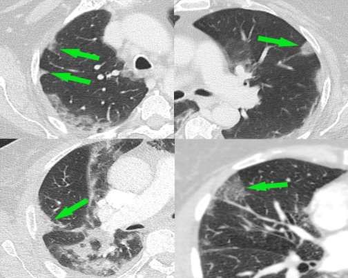
3

     Figure 1: The COVID-19 pathway: A hypothetical model for the oral-vascular-pulmonary route of infection.

If confirmed, this hypothetical model may provide a rationale for understanding why some individuals
develop COVID-19 lung disease and others do not. It would also fundamentally change the way COVID-
19 is managed, providing a new line of exploration into treatments targeted at the source of the viral
reservoir, the mouth.

Initial Upper Respiratory Tract Infection and Proposed Mechanism of
Transmission to the Lungs via Blood Vessels
Based on knowledge of the intensity of expression of the main SARS-CoV-2 binding receptor – the ACE2
receptor – the upper airways are considered the initial site of infection for SARS-CoV-2, rather than the
lower respiratory tract. Expression of the ACE2 receptor is reported to be between 200 to 700 times
more intense in the nasal airways, specifically on the surface of the olfactory neuroepithelial cells,
compared to the respiratory epithelial cells of the lower respiratory tract [1,2]. The conclusion from the
study by Chen and Shen et al. [2] that the initial site of infection is the upper airway, challenges the
notion that the SARS-CoV-2 virus is necessarily delivered to the lungs via the airways, where expression
of ACE2 receptors on respiratory epithelial cells is low [3].

Research Article | Lloyd-Jones G, et al. J Oral Med and Dent Res. 2021, 2(1)-S1.
4 The concept that the upper airways may be the predominant initial site of infection for viral transmission to the lungs requires further scrutiny. Here we present radiological evidence that raises the important possibility of a vascular viral delivery route to the lungs rather than via the respiratory airways. The proposed model describes the oral cavity as the reservoir of SARS-CoV-2, specifically in saliva, with transmission to the lungs mediated by a breach of the mucosal immune defense barrier of the periodontal tissues or oral mucosa, with subsequent intravascular carriage. If proven to be correct, this hypothesis would have significant implications for the understanding of how the disease should be managed. Simple antimicrobial oral healthcare measures could be implemented not only with the aim of reducing the risk of transmission between individuals but also with the aim of providing benefit to individuals who are COVID-19 positive. Specifically, these measures could be a means of mitigating the risk of developing lung disease, and therefore the most severe form of the disease. Radiological Perspective – COVID-19 Lung Disease 1. Pathological Distribution of Disease Pulmonary radiological findings in COVID-19 do not align with a model of SARS-CoV-2infection primarily causing disease of the airways of the lungs; the initial and dominant pathological features demonstrated radiologically are vascular in nature [4-6]. The distribution of lung disease does not favor an inhaled pathogen. No known inhaled infective pathogen has preferential tropism for the periphery of the lung bases. Rather, inhaled pathogens would be expected to present a uniform distribution to other areas of the lungs, including the mid or upper areas, and would not be expected to spare the perihilar or central areas [7,8]. It is important to appreciate the anatomy of the pulmonary arteries which dominantly deliver blood to the lung bases, bilaterally, symmetrically, peripherally, and posteriorly, matching the dominant distribution of disease in COVID-19. It has also been noted that many of the radiological findings typically associated with respiratory pneumonia, for example bronchial wall thickening, mucous secretion, and the ‘respiratory tree-in-bud’ opacification of small airways, are not features of COVID-19 [4,9]. Furthermore, if the airway findings typically associated with respiratory pneumonia are present on computed tomography (CT), they are considered inconsistent with the diagnosis of COVID-19 [10]. 2. Evidence of Pulmonary Vascular Phenomena Conversely, there are numerous studies within the radiological literature describing the pathogenesis of COVID-19 lung disease as driven by vascular phenomena [4-6,9-14]. Early in the pandemic period, the presence of “ground-glass opacities” visible on CT was reported to be the hallmark sign of COVID-19 lung disease [15]. However, these ground-glass opacities were acknowledged as a non-specific feature, and histological confirmation of their significance was required, with edema or hemorrhage suggested as possible causes [16]. Notably, the radiological literature now reports that these ground-glass opacities are accompanied by abnormally dilated blood vessels (Figure 2), which are thought to be responsible for the phenomenon of pulmonary arteriovenous vascular shunting and subsequent hypoxemia [4]. Research Article | Lloyd-Jones G, et al. J Oral Med and Dent Res. 2021, 2(1)-S1.
5

 Figure 2: CT images of patients with COVID-19 lung disease demonstrating ground-glass opacities (yellow arrows)
                         accompanied by abnormally dilated blood vessels (green arrows).

A specific vascular feature known as the ‘vascular tree-in-bud’ sign (not to be confused with ‘respiratory
tree-in-bud’ found in conventional respiratory pneumonia) is visible on CT as a distinct entity in 64% of
patients with COVID-19 lung disease [11]. This sign is thought to be a marker of the pathological process
of immunothrombosis and can be visible without lung parenchymal changes in the form of ground-glass
opacities. The presence of this sign correlates with the length of hospital stay [12].

Further evidence of vascular disease comes from studies of Dual-Energy CT which describe perfusion
defects in 100% of patients with COVID-19. These defects of blood flow are categorized by two distinct
patterns: a wedge-shaped pattern – analogous to pulmonary embolism; and a mottled/amorphous
pattern – analogous to chronic or idiopathic thromboembolic hypertension. Dilated blood vessels and
hyperperfusion are also described proximal to areas of ground-glass opacification [13].

3. Distinct Phenotype of Thromboembolic Disease
There has been much interest regarding the high incidence of pulmonary thromboembolic disease in
COVID-19 patients. When compared to conventional pulmonary thromboembolic disease, a different
distribution is described in patients with COVID-19. In COVID-19, the filling defects visible within

Research Article | Lloyd-Jones G, et al. J Oral Med and Dent Res. 2021, 2(1)-S1.
6
pulmonary arteries with CT pulmonary angiography (CTPA) are lower in volume and more peripheral.
This difference is thought to be related to the pathological process of immunothrombosis [14]. Indeed,
immunothrombosis is the main driver of disease in the lungs [15-17], and can even be considered as an
appropriate immune response, serving to trap pathogens in the affected area of tissue, thus preventing
escape into the systemic circulation [18]. This difference in the distribution of thromboembolic disease,
with smaller and more peripheral filling defects visible on CTPA, is significant because it is known that
smaller and more peripheral clots are more likely to result in pulmonary vascular occlusion when
compared to larger central filling defects [19]. Many peripherally-located areas of ground-glass
opacification are morphologically identical to pulmonary wedge-shaped infarcts (Figure 3). These are
visible regardless of the presence or absence of visible filling defects in adjacent pulmonary arteries [20].
Figure 3: CTPA scans of the lungs in patients with COVID-19 lung disease. Wedge-shaped areas of ground-glass
opacification located at the edges of the lungs resemble pulmonary infarcts. These can be present with or without
visible filling defects in the pulmonary arteries on CTPA examination.
4. Correlation of Radiological and Autopsy Findings
It is also important to note that both macroscopic and microscopic pulmonary vascular obstruction is
found on autopsy and that pulmonary infarcts are indeed present in the majority of individuals dying
with COVID-19 lung disease [21]. Viral elements have been detected in endothelial cells in autopsy
studies of those who have died with COVID-19, with evidence of endothelial cell inflammation and
inflammatory cell death [22,23]. Histologically, microangiopathy of lung vessels is described with
microthrombi visible within both pulmonary arterioles and peripheral lung venules. Thus, there is
Research Article | Lloyd-Jones G, et al. J Oral Med and Dent Res. 2021, 2(1)-S1.7 thrombosis on both sides of the capillary bed of the pulmonary vasculature, proximal and distal to the alveolar capillaries [24]. It is important to appreciate that this means that CTPA, the conventional imaging modality used to look for pulmonary thromboembolic disease, will underestimate the presence of thrombosis because some of the thrombosis is on the venous side which is not enhanced with intravenous contrast. This may be helpful in understanding the pathogenesis of some of the peripheral vasculitis mimics seen specifically in patients with COVID-19 lung disease which are thought to be mediated by microemboli arising from the venous side of the pulmonary vasculature (blood vessels returning to the heart) and disseminated systemically [25]. In summary, the radiological findings are not consistent with dominant or primary airways disease but rather are entirely consistent with a disease of the lung blood vessels occurring first. This vascular disease is mediated by the process of immunothrombosis which below is proposed to result from interaction with the pulmonary endothelial ACE2 receptor. The consequences of this interaction and subsequent effects of deregulated increase of angiotensin-II and vascular congestion could explain the other radiological features of proximal vascular dilatation and the vascular tree-in-bud sign. The development of ground-glass opacities could result from endothelial dysfunction [4]. Indeed, endothelial dysfunction is widely reported as a dominant pathological feature [26,27]. Periodontal Perspective – COVID-19 in the Oral Cavity 1. Entry factors for SARS-CoV-2 in oral and gingival tissues The invasion of host cells by SARS-CoV-2 is mediated by ACE2 receptors, furin, and trans membrane protease serine 2 (TMPRSS2). Viral spike proteins bind to ACE2 receptors on the surface of host cells, and TMPRSS2 mediates endocytosis. Furin is involved in the release of new viral particles to the extracellular compartment [28]. These mediators, which are key elements for infection, are expressed abundantly in the nasal airways and oral cavity, including gingival tissues, minor salivary glands, and tongue [29,30]. Although not all oral tissues express the three mediators of viral entry, cells of the sulcular epithelium do express ACE2, TMPRSS2, and furin. This indicates the potential for the gingival sulcus to be a target for SARS-CoV-2 infection. Thus, several niches in the oral cavity can become infected by the virus, including the gingival sulcus [30,31]. 2. Presence of SARS-CoV-2 in the oral cavity and periodontal tissues There is strong evidence from several studies confirming the presence of SARS-CoV-2 in saliva, minor salivary glands, tongue, and gingival crevicular fluid [32]. A recent study on autopsy tissues from deceased COVID-19 patients reported viral infection of the oral mucosa and salivary glands [29]. Another post-mortem study confirmed the presence of viral RNA in the periodontal tissues of 5 out of 7 patients who died of COVID-19 [33]. Together, these findings suggest that SARS-CoV-2 is abundant in saliva and may infect salivary gland tissue, gingival and oral mucosal cells. A study from Huang et al. [34] suggests that the virus can persist in saliva or in the nasopharynx for over two months. In asymptomatic individuals, viral clearance was observed after 0.5 to 3.5 weeks [34]. The salivary viral load has been linked to loss of taste, overall disease severity and mortality, being a better predictor of poor outcome than patient age or viral load in the nasopharynx [34,35]. This is a particularly Research Article | Lloyd-Jones G, et al. J Oral Med and Dent Res. 2021, 2(1)-S1.
8

important finding given that patient age is considered the most significant risk factor. [36]. Although it
could be speculated that the viral load in saliva is merely a marker for generalized high viral load, this
does not explain why the viral load in the nasopharynx is not a predictor of poor outcome. In the study
from Huang et al. [34] most asymptomatic individuals displayed only nasopharyngeal swab positivity.
Saliva viral load seems to be a specific predictor of poor outcome [35].

3. Periodontal pockets as a reservoir for viruses
Previous studies have reported the presence of human viruses in saliva, gingival crevicular fluid (GCF),
subgingival plaque, and gingival tissues, including human immunodeficiency virus, herpesviruses,
Epstein-Barr virus, cytomegalovirus, and Zika virus [37-41]. In a recent SARS-CoV-2 study, viral RNA was
detected in the GCF of 64% of COVID-19 positive patients [32]. It has been speculated that viral particles
in the oral cavity can migrate into the gingival sulcus/periodontal pockets, where the conditions are
favorable for their survival. Sub-gingival plaque biofilm can provide a unique environment for viruses,
and in periodontitis patients, the periodontal pocket epithelium develops micro-ulcerations that
facilitate the passage of microorganisms and viral particles to the underlying connective tissue and
gingival capillary complex, reaching the systemic circulation [31,33]. The area of exposed connective
tissue and associated blood vessels that are in direct contact with the sub-gingival biofilm ranges from 5
cm2 in mild disease to more than 20 cm2 in severe periodontitis [42]. Indeed, peripheral blood
neutrophils in periodontitis patients have been shown to exhibit a type-1 interferon gene expression
signature, consistent with intravascular exposure to periodontal microorganisms such as viruses [43].
Thus, periodontal pockets possibly present suitable conditions for viral replication, infection, and spread
to gingival capillaries.

Studies have shown that poor oral hygiene and periodontitis increase the risk for development of severe
COVID-19 with poor outcomes [31,33,34,45]. Specifically, the recent study by Marouf et al. [46]
examined dental X-rays of a large population sample (568 patients) with COVID-19. The study found an
increased risk of developing severe COVID-19 (defined as intensive care admission, need for mechanical
ventilation, or death) in those with periodontitis, with an overall odds ratio of 3.67 after confounders
were accounted for, including age, sex, smoking, BMI, diabetes and comorbidities [46].

4. Oral and nasal cavities as entry points for microorganisms
In periodontitis patients, the risk for viral invasion is likely to increase due to potential disruption of the
pocket epithelium resulting from local inflammation, which is the same principle that potentially
explains bacterial entrance to the systemic circulation [47]. Even in healthy patients, the permeable
nature of the junctional epithelium can facilitate viremia [48].

The presence of oral bacteria in the systemic circulation has been reported previously in studies on
bacteriemia and infective endocarditis of oral origin. These studies propose that oral bacteria can cause
damage elsewhere in the body, with the risk being higher with poor oral hygiene and periodontal
inflammation [49]. As bacteria can pass into the systemic circulation via breakdown of the immune
defenses of the mouth, then the same route could be open to viruses, including SARS-CoV-2, and
facilitated by periodontal disease. Other potential sources of transmucosal transfer include the floor of

Research Article | Lloyd-Jones G, et al. J Oral Med and Dent Res. 2021, 2(1)-S1.
9

the mouth (including salivary ducts) and Kiesselbach's plexus (Little’s area) of the nose, however
research evidence is currently lacking and nasal symptoms such as anosmia are associated with a lower
severity of coronavirus disease [50]. We propose that transfer across the gingival crevice is more likely
to be the most significant pathway, rather than a secondary route.

5. The potential role for viral-bacterial synergy in the periodontal environment
A higher prevalence of cytomegalovirus, Epstein-Barr virus, and other herpesviruses has been reported
in the subgingival plaque biofilm from periodontitis patients when compared to patients with gingivitis
or a healthy periodontium [51,52]. The presence of herpesviruses in periodontal pockets seems to
increase the risk for tissue destruction, suggesting a synergistic action between viruses and periodontal
pathogens [53]. Several Gram-negative anaerobic bacteria implicated in periodontal disease have been
associated with Epstein-Barr virus and cytomegalovirus, particularly Porphyromonas gingivalis and
Tannerella forsythia [52]. Thus, the co-presence of SARS-CoV-2 with periodontal bacteria may
exacerbate periodontal tissue damage, but the nature, extent, and consequences of this interaction are
currently unknown. In periodontitis patients, it can be speculated that i) a viral-bacterial synergy might
facilitate penetration of SARS-CoV-2 through the pocket epithelium, ii) such an interaction can help
viruses evade the immune response, thus enabling its entrance to gingival capillaries and endovascular
transmission directly to the pulmonary vessels. Co-infection in COVID-19 is also a possibility, given that
serious respiratory conditions are often associated with viral bacterial co-infections [54]. However, there
is a scarcity of data on SARS-CoV-2 bacterial co-infection [24]. Significantly, autopsy studies show a
surprising lack of bacterial super-infection in those who have died from COVID-19 [24]. Also, a report
relating to critical care patients did not find evidence of bacterial co-infection in blood, sputum, or
bronchoscopic sampling upon admission to intensive care [55].

It is also possible that periodontal inflammation can increase the risk for viral infection. A study on
Epstein-Barr virus found that gingival epithelial cells were frequently infected, and the level of viral
infection correlated with the level of periodontal inflammation [56]. Previous studies report that P.
gingivalis can facilitate the reactivation of latent Epstein-Barr and HIV-1 viruses [57,58]. Hence, a
synergistic relationship between SARS-CoV-2 and periodontal bacteria cannot be excluded.

In immunocompromised mice, cytomegalovirus and P. gingivalis co-infection resulted in the highest
mortality when compared to inoculation with the virus alone, P. gingivalis alone, or the combination of
the virus and Escherichia coli. Based on the observed lower systemic levels of gamma interferon and
lymphoid depletion observed in P. gingivalis and cytomegalovirus infection, it was suggested that this
periodontal bacteria can increase the viral impact on the host [59].

6. Potential role of local and systemic inflammatory response
In periodontitis, the host response to microorganisms in the subgingival biofilm is mediated by the
expression of pro-inflammatory cytokines, particularly tumor necrosis factor α (TNF-α), interleukin-1β
(IL-1β), and interleukin-6 (IL-6). These soluble proteins can change cellular functions to promote and
perpetuate inflammation and tissue destruction in the periodontal tissues and elsewhere in the body
[60,61]. The link between periodontitis and systemic diseases has been researched extensively in the

Research Article | Lloyd-Jones G, et al. J Oral Med and Dent Res. 2021, 2(1)-S1.
10

last decade, and findings from multiple studies point to the significance of elevated levels of pro-
inflammatory cytokines and acute-phase proteins [62-65]. Indeed, studies have demonstrated that
peripheral blood neutrophils of periodontitis patients are hyper-reactive with respect to cytokine
release (IL-1β, IL-8, IL-6, TNF-α) when FcγR and Toll-like R4 receptors are challenged, relative to non-
periodontitis controls [66]. Periodontal treatment has been shown to positively affect systemic
inflammation in healthy patients and in those who have chronic diseases, such as type-2 diabetes [67],
hypertension [68], coronary heart disease, and atherosclerosis [69-74].

Severity of COVID-19 has also been linked to systemic inflammation [75,76]. In COVID-19 patients, the
risk for respiratory failure was 22 times higher in patients who presented high IL-6 levels upon hospital
admission [77].

Although occult sources of infection in the body that perpetuate inflammation, such as periodontitis,
contribute to the systemic inflammatory burden, there is limited evidence of overspill of locally
produced inflammatory mediators in the periodontium into the systemic circulation, and no evidence
for such disseminating inflammation triggering the lung disease. A direct role for the periodontal
inflammatory response seems unlikely, especially in view of the potential for direct endothelial viral-
ACE2 interaction as described below.

7. Links between periodontitis and oral hygiene with other respiratory conditions
Evidence suggests that periodontitis can increase the risk for respiratory diseases such as pneumonia,
and chronic obstructive pulmonary disease (COPD) [78-80]. Studies also report on decreased lung
volume, airflow limitation, and worse pulmonary function in systemically healthy patients with
periodontitis [81,82], and patients with both periodontitis and COPD [83], as well as successful
periodontal treatment resulting in reduced exacerbations of COPD [84]. COPD has been suggested to be
low in prevalence in COVID-19 cases but conversely, if present, is described as a risk factor for poor
outcome in COVID-19 [85,86].

In hospital settings, adequate plaque control measures and dental treatment have been shown to
reduce the incidence and severity of pulmonary infection [87,88]. Aspiration and hematogenous spread
of oral microorganisms have been described as potential pathways for the connection between oral and
pulmonary conditions [89]. But findings from a study of the CT chest features in COVID-19 showed that
the presence of airway secretions is not typical in these patients [10], which further supports the notion
of a potential hematogenous route of transmission from the oral cavity to the lungs. It is also important
to consider the lack of bacterial super-infection found histologically, as reported above [24,55].

There is evidence that oral hygiene measures lower the incidence of aspiration pneumonia in elderly
patients in hospital and nursing homes, decreasing morbidity and mortality [90]. In the systematic
review from Sjogren et al. [91] the authors estimate that one in ten cases of death from pneumonia in
nursing home patients can be prevented through simple oral hygiene measures [91]. Given that
periodontitis and inadequate oral hygiene negatively impacts respiratory conditions and lung function,
particularly in hospitalized patients, their potential to worsen lung complications in hospitalized COVID-

Research Article | Lloyd-Jones G, et al. J Oral Med and Dent Res. 2021, 2(1)-S1.
11

19 patients should not be ignored. This also perhaps acts as an existing rationale for treating any patient
with symptomatic COVID-19 by implementing oral hygiene measures.

8. Shared risk factors between COVID-19 and periodontitis
Periodontitis and poor outcome in COVID-19 share many risk factors, such as patient age [92,93], male
sex [94,95], diabetes [96,97], cardiovascular disease [98,99], obesity [93,100], COPD [101,102], Down
syndrome [103,104], specific ethnic groups [105,106], type A blood group [107,108], chronic kidney
disease [109-111], physical disability or learning difficulty [112,113] and dementia [114,115].

Smoking is a recognized risk factor for periodontitis [116]. In a recent meta-analysis, smoking was
associated with increased risk for severe COVID-19 [117] but not all studies confirm this association
[118]. It is notable that if COVID-19 lung disease was mediated by airways pathology, smoking would be
considered a risk factor for poor outcome, as it is in influenza [119]. Rather, there is counterintuitive
evidence about the role of smoking and the suggestion that nicotine may have a therapeutic role [120-
122]. The harmful effects of smoking on gingival tissues are mediated, in part, by the vasoconstrictive
biological action of nicotine [123], which leads to significantly reduced gingival bleeding and decreased
diameter of gingival capillaries [124,125], Conceivably, the local vasoconstriction action of nicotine may
limit the transfer of microorganisms across the mucosal membrane of the oral cavity and periodontal
tissues to the venous drainage of the mouth.

A large population study showed increased COVID-19 symptoms in individuals who smoke [126].
Therefore, smoking may be considered yet another risk factor shared between COVID-19 and
periodontitis, however the exact mechanism of interaction of smoke inhalation and nicotine action is
complex and likely involves multiple toxins present within cigarette smoke vapor and tar fractions.

9. Higher severity of COVID-19 in patients with poor oral hygiene/periodontal disease
As mentioned previously, the case-control study on 568 COVID-19 patients showed an association
between periodontitis and COVID-19 severity [46]. This study found that periodontitis was associated
with complications for COVID-19 including death (odds ratio 8.81), intensive care admission (odds ratio
3.54), and need for assisted ventilation (odds ratio 4.57) [46]. Dental plaque could provide a constant
source of viral delivery to the vasculature during the acute phase of COVID-19. Thus, it is biologically
plausible that the ongoing delivery of the virus itself to the lung vessels could account for poorer
outcomes, rather than viral transmission via the airways or bacterial super-infection.

Biological Rationale for the Oral-Vascular-Pulmonary Route of Infection
Here we propose a model of direct viral delivery from the oral cavity via the venous drainage of the
mouth, neck (jugular veins), and chest (superior vena cava), through the right side of the heart, and then
to the pulmonary vessels. Such a route of transmission would be compounded by poor oral hygiene or
periodontal disease. This model of disease could be hugely significant in terms of a novel understanding
of the disease pathogenesis and its management.

Research Article | Lloyd-Jones G, et al. J Oral Med and Dent Res. 2021, 2(1)-S1.
12

Below is presented the biological plausibility for this oral-vascular-pulmonary route of SARS-CoV-2
transmission from the oral cavity to the lungs facilitating the development of COVID-19 lung disease. As
discussed above, periodontitis and poor oral health are associated with each of the following: age, male
sex, diabetes, cardiovascular disease, obesity, specific ethnic groups, disability, and type A blood group,
all of which are risk factors for severe COVID-19. Thus, periodontitis and poor oral hygiene are here
proposed as the converging and principal risk factor for severe COVID-19. It is proposed that the oral-
vascular-pulmonary anatomical route of transmission could explain why some patients develop lung
disease, and so are susceptible to severe disease, and others do not.

Anatomical Route – from the Oral Cavity to the Lungs
With high levels of SARS-CoV-2 in saliva, any breakdown of the primary immune barrier in the oral cavity
could facilitate viral entry to capillaries. Poor oral hygiene could further increase the risk of infection by
changing the physiologically permeable junctional and sulcular epithelium to a pocket lining epithelium,
which then ulcerates creating a co-called “periodontal wound”. The size of the periodontal wound, and
therefore the associated vascular access to plaque microorganisms, can be calculated and increases as
oral hygiene worsens. Through the venous drainage of the mouth and neck, the virus would reach the
superior vena cava, entering the right side of the heart, and then be pumped into the pulmonary
arteries. This route of hematogenous delivery of SARS-CoV-2 to the lungs, rather than via the lower
airways, would explain the vascular distribution of the lung disease in COVID-19 seen radiologically.

If other microorganisms can enter the systemic circulation via a breach of the mucosal defense barrier of
the mouth, it should be asked why they do not evidently cause lung disease. The specific pulmonary
vascular tropism of SARS-CoV-2 requires an explanation.

ACE2 Receptor-Virus Interaction
SARS-CoV-2 is known to bind to ACE2 receptors. These receptors are said to be expressed in respiratory
epithelial cells of the airways which has been used to explain the conventional model of viral interaction
with the lungs [127]. Although this is one way the virus could enter, the expression of ACE2 on
respiratory epithelial cells has been shown to be at much lower levels when compared to nasal
neuroepithelial cells [2,3], and some have suggested very low levels or even no expression in the normal
respiratory system [128].

Importantly, expression of ACE2 receptors has also been reported in endothelial cells of pulmonary
vessels and several other organs of the body [26,129,130]. This provides a specific model for the
interaction of SARS-CoV-2 with the endothelium of pulmonary vessels, unlike other viruses, which are
not known to bind to the ACE2 receptor. In view of the lack of airways disease visible radiologically, it
also helps explain the question of why the pulmonary vessels are dominantly affected compared to
other organs [131]. According to the model of the oral-vascular-pulmonary anatomical route of
transmission from the mouth to the lungs, the virus would be delivered to the pulmonary vessels first,
before it could reach other organs.

Research Article | Lloyd-Jones G, et al. J Oral Med and Dent Res. 2021, 2(1)-S1.
13

Additionally, as presented above, the radiologically observed dilated vessels and the specific vascular
tree-in-bud sign, which can be visible independent of ground-glass opacification or consolidation, are
consistent with a model of viral-ACE2 interaction and development of immunothrombosis and its
consequences.

Simply put, if the virus could pass from saliva or the sub-gingival periodontal environment into the
venous drainage of the mouth, it would first be delivered directly to the small vessels of the lung
periphery. This is exactly where immunothrombosis occurs, being accepted as the main driver of the
pathology [12,14,17,132].

On interacting with ACE2 receptors on pulmonary vascular endothelial surfaces, viral binding would lead
to an unregulated increase in local levels of angiotensin-II hormone, which has multiple biological
functions, including vasoconstriction, promotion of inflammation, and thrombosis [133]. These direct
actions of angiotensin-II have been proposed as a contributor to the development of
immunothrombosis in COVID-19 lung disease [134]. In this way, unregulated levels of angiotensin-II
could be the trigger for immunothrombosis and act as a key pathological step in the development of
lung disease and the systemic hypercoagulable state.

In COVID-19 autopsy studies, viral particles were detected in endothelial cells of the lungs, with
evidence of endothelial cell inflammation and inflammatory cell death [22,23,26]. The process of
immunothrombosis could be considered locally harmful because it prevents blood flow and, thus,
prevents gas exchange. However, it is suggested that the process of immunothrombosis serves to
contain and eliminate pathogens [18], which perhaps helps to explain the lack of viremia in the early
stages of COVID-19 [135]. It can be postulated that the viremia seen later in the disease process, which
is a predictor of death could arise when the mechanism of immunothrombosis is saturated and the lungs
can no longer trap the virus [136].

Clinical Significance
From the oral cavity, if SARS-CoV-2 can reach the lungs through the blood, causing immunothrombosis-
driven disease in the pulmonary vessels, then early measures to decrease transmission to the lungs in
this way must be considered in the management of COVID-19. This concept could influence the
development of new approaches with the aim of preventing or mitigating lung disease.

This concept potentially highlights the importance of active oral healthcare and adequate daily oral
hygiene measures in the management of COVID-19 [44]. Importantly, it is noted that readily available
mouthwashes containing cetylpyridinium chloride (CPC) or ethyl lauroyl arginate (ELA) can inactivate
SARS-CoV-2 with high efficacy in vitro [137-140]. Those containing povidone-iodine (PVP-I) have also
been shown to be effective [139,141-47]. Mouthwash products containing chlorhexidine or ethanol
alone displayed little or no ability to inactivate SARS-CoV-2 [138]. Given that mouthwash products
containing these specific ingredients are readily available across the world, they could provide a cheap
and effective treatment in those with COVID-19 with the additional potential benefit of reducing the risk

Research Article | Lloyd-Jones G, et al. J Oral Med and Dent Res. 2021, 2(1)-S1.
14

of transmission to others. These products should only be used as directed by the manufacturer, or as
advised by oral healthcare professionals as part of a population study or clinical trial. These specific
mouthwash ingredients may potentially play a role in mitigating development or worsening of the lung
disease at any stage, from those who are swab positive and asymptomatic in the community to those
who are hospitalized or even in intensive care.

Media outlets have reported the adoption of mouthwashes in some countries, as advised by
government officials, either officially or perhaps unofficially. In Japan, for example, the sales of
mouthwash increased substantially after a governor advised the population to use a gargling solution
[148]. COVID-19 outcomes in Japan are significantly better than in other G20 countries, such as the UK
and the US (as of February 19th, 2021 – data from the previous seven day period – 3.41 deaths per
million (Japan), 33.28 deaths per million (USA), and 46.28 deaths per million in the (UK) [149]. Although
there are likely to be many confounding reasons for the lower mortality in Japan, this difference of
approach is raised as a point of interest to be urgently researched by governmental and public health
officials.

Recommendations for Good General Oral Healthcare
Adequate daily home oral care habits are essential for oral and general health, as it decreases the risk
for dental caries, gingivitis, and periodontitis. If proven to be correct, the concept of the oral-vascular-
pulmonary infection route may mean that these simple measures could reduce the risk of developing
severe COVID-19 lung disease.

Although each patient has unique needs, the European Federation of Periodontology (EFP) provides
general recommendations [150]:
    - There is a universal recommendation to brush twice daily for at least 2 minutes with a
        fluoridated toothpaste.
    - For periodontitis patients 2 minutes is likely to be insufficient.
    - Manual or power toothbrushing is recommended as a primary means of reducing plaque and
        gingivitis. The benefits of toothbrushing outweigh any potential risks.
    - Daily inter-dental cleaning is strongly recommended to reduce plaque and gingival
        inflammation. When gingival inflammation is present, inter-dental cleaning, preferably with
        interdental brushes should be professionally taught to patients.
    - For the treatment of gingivitis and where improvements in plaque control are required,
        adjunctive use of antiplaque chemical agents may be considered. In this scenario, mouthwashes
        may offer greater efficacy but require an additional action to the mechanical oral hygiene
        regime.

Mouthwash products have shown potential to decrease the viral load in the oral cavity [151]. In the S3-
level evidence-based treatment guidelines provided by the European Federation of Periodontology (EFP)
in 2020, the use of mouthwash products is recommended as adjunctive agents in the treatment of
stages I-III periodontitis [152].

Research Article | Lloyd-Jones G, et al. J Oral Med and Dent Res. 2021, 2(1)-S1.
15
Considerations for Oral Healthcare in View of this Hypothesis
Despite the scarcity of clinical evidence for in vivo viral inactivation, well-established mouthwashes
containing specific ingredients, which inactivate SARS-CoV-2 in vitro, could potentially help mitigate
transmission and decrease the risk of severe lung disease in COVID-19 [138,139]. These ingredients
include:
0.05%-0.1% Cetylpyridinium Chloride (CPC): 15 ml for 30 seconds twice a day. In vitro and in
vivo studies indicate that mouthwash products containing CPC are able to inactivate SARS-CoV-
2 [137-140]. These products are generally considered to be safe, with staining of the tongue
and teeth being rarely reported [153].
0.147% Ethyl lauroyl arginate (ELA): 20 ml for 30 seconds twice a day. In vitro results suggest
virucidal activity of ELA against SARS-CoV-2 [138].
0.2%, 0.4% or 0.5% Povidone-Iodine (PVP-I): 10 ml for 30 seconds twice a day. The use of PVP-I
is supported by in vitro [141-43] and in vivo studies [139,144-46]. In one clinical trial, the use of
1% PVP-I mouth rinse resulted in temporary thyroid dysfunction in 42% of COVID-19 patients,
suggesting that lower concentrations should be preferred [144]. Contraindications: allergy,
hyperthyroidism, thyroid dysfunction, pregnancy, lactation, and treatment with radioactive
iodine [154,155].
The use of mouthwashes should not replace other daily oral hygiene measures. They should be used
after toothbrushing for limited periods due to potential side effects. Mouthwashes should never be
swallowed. In the context of the pandemic, it would seem logical to use mouthwash products both
before and after social interactions, but clinical trials are required to answer the critical question of the
potential effect of these products as a means of reducing the risk of transmission between individuals.
Clinical trials are also required to specifically address the potential for mouthwashes to mitigate the
development of COVID-19 lung disease, and hence the severest form of the disease.
Conclusion
The nasal and oral cavities are entry points into the body for microbial pathogens. The oral cavity
provides favorable conditions for viral replication, with saliva functioning as a reservoir for SARS-CoV-2.
Cells in different oral niches express receptors that make them potential targets for viral infection. The
gingival sulcus in healthy patients can allow the entrance of viral particles into gingival capillaries due to
the permeability of the sulcular epithelium. In periodontitis, the entrance of the virus into the circulation
could be facilitated by micro-ulcerations in the pocket epithelium. Poor oral hygiene and dental plaque
accumulation may further intensify this pathway. From gingival capillaries, the virus could reach the
lungs via a vascular route, where it would trigger the known main pathological driver of disease, that of
immunothrombosis. This proposed mechanism of transmission of the virus from the oral cavity to the
lungs may explain both the radiological appearances and the clinical variability of the disease. Although
to an extent the passage of the virus across the mucous membranes of the nasal passage and mouth
may occur in healthy individuals, the presence of poor oral health could act as a risk factor to identify
individuals more likely to develop COVID-19 lung disease, or those who might progress to severe disease
leading to intensive care admission, mechanical ventilation, or death. The co-morbid presence of
Research Article | Lloyd-Jones G, et al. J Oral Med and Dent Res. 2021, 2(1)-S1.16 periodontitis has been shown to represent an independent risk factor for other systemic inflammatory diseases that are characterized by hyper-inflammation and oxidative stress [67,103,105]. Given that – i) saliva is a reservoir of the SARS-CoV-2 virus; ii) high salivary viral load is a better predictor of poor outcome than patient age; iii) the virus initially interacts with the upper respiratory airways; iv) the lower respiratory tract is not dominantly involved radiologically until the airways are compromised secondary to the vascular disease; v) the pulmonary vessels are dominantly affected radiologically; and vi) the radiological manifestations align with the model of viral binding with pulmonary vessel endothelial ACE2 receptors and subsequent processes – it seems compelling that a breach in the mucosal defenses of the mouth could facilitate transmission of SARS-CoV-2 to the lungs via a hematogenous route. Studies are urgently required to confirm or refute this hypothesis. Determining viral load within blood samples taken simultaneously from the jugular vein and a peripheral site is suggested here as a possible means of corroborating the role of the proposed anatomical pathway. This method, by which the virus might be caught ‘red-handed’ as it passes down the jugular veins, has recently been offered with interest to laboratories and research teams in the UK, but is also offered here as an open proposal to researchers worldwide. It is not known if viable infectious virus could be transported by this route. This will need to be examined in future studies using a combination of electron microscopy to identify whether virus particles are being carried within or on particular cell types, for example macrophages, and functional assays to determine whether infectious virus can be rescued from these blood cells. If a differential viral load between samples taken simultaneously from a jugular vein and a peripheral site was established, then further corroboration of the theory could be gained by matching jugular viral load with salivary viral load, the severity of periodontitis, and post challenge following use of specific mouthwashes or other oral hygiene measures. Until proven or refuted, daily oral hygiene and other measures for plaque control, together with oral healthcare should be prioritized for the general public, since these measures not only improve oral health and wellbeing but could also be potentially lifesaving in the context of the pandemic. There is already a precedent for employing simple oral hygiene measures to improve outcomes in patients with pneumonia, and it seems unlikely that oral hygiene measures will cause harm, as long as the manufacturer’s instructions are followed carefully. In consideration of people in areas of the world where specific mouthwashes may not be available or affordable, there may be even simpler measures to consider. A recent study reported that saltwater rinsing can reduce gingival inflammation [156], and even mouth rinsing with boiled water (which has been allowed to cool) has shown positive effects on plaque and oral mucosa in hospitalized elderly patients [157]. This suggests that simple measures can help decrease the salivary viral load in areas of the world where mouthwash products are not readily available. It is sincerely hoped that, if proven correct, this concept provides a rationale for the use of oral healthcare measures which are cheap (or even free) and are available worldwide, and that these measures may help prevent the development of lung disease, mitigate deterioration to severe COVID-19, and reduce mortality. Research Article | Lloyd-Jones G, et al. J Oral Med and Dent Res. 2021, 2(1)-S1.
17

References
     1.    Hodges G, Pallisgaard J, Schjerning Olsen AM, McGettigan P, et al. (2020) Association between biomarkers
           and COVID-19 severity and mortality: A nationwide Danish cohortstudy. BMJ Open. 10(12):e041295.
     2.    Chen M, Shen W, Rowan NR, Kulaga H, Hillel A, et al. (2020) Elevated ACE-2 expression in the olfactory
           neuroepithelium: Implications for anosmia and upper respiratory SARS-CoV-2 entry and replication. Eur
           Respir J. 2020.
     3.    Sungnak W, Huang N, Bécavin C, Berg M, Queen R, et al. (2021) SARS-CoV-2 entry factors are highly
           expressed in nasal epithelial cells together with innate immune genes. Nat Med. 26(5):681-7.
     4.    Lang M, Som A, Carey D, Reid N, Mendoza DP, et al. (2020) Pulmonary Vascular Manifestations of COVID-
           19 Pneumonia. Radiol Cardiothorac Imaging, 2(3):e200277.
     5.    Oudkerk M, Kuijpers D, Oudkerk SF, van Beek EJ. (2020) The vascular nature of COVID-19. Br J Radiol.
           93(1113):20200718.
     6.    Oudkerk M, Buller HR, Kuijpers D, van Es N, Oudkerk SF, et al. (2021) Diagnosis, prevention, and treatment
           of thromboembolic complications in COVID-19: Report of the national institute for public health of the
           Netherlands. Radiology. 297(1):E216-22.
     7.    Nemec SF, Bankier AA, Eisenberg RL. (2021) Lower lobe-predominant diseases of the lung. Am J
           Roentgenol. 200(4):712-28.
     8.    Patwa A, Shah A. (2015) Anatomy and physiology of respiratory system relevant to anaesthesia. Indian J
           Anaesth. 2015 Sep; 59(9): 533-41.
     9.    Luo L, Luo Z, Jia Y, Zhou C, He J, et al. (2020) CT differential diagnosis of COVID-19 and non-COVID-19 in
           symptomatic suspects: A practical scoring method. BMC Pulm Med. 20(1):129.
     10.   Ufuk F, Savaş R. (2021) Chest CT features of the novel coronavirus disease (COVID-19). Turk J Med Sci.
           50(4):664-78.
     11.   Eddy RL, Sin DD. (2020) Computed Tomography Vascular Tree-in-Bud: A Novel Prognostic Imaging
           Biomarker in COVID-19? Am J Respir Crit Care Med. 202(5):642-44.
     12.   Patel B V, Arachchillage DJ, Ridge CA, Bianchi P, Doyle JF, et al. (2020) lmonary angiopathy in severe
           COVID-19: Physiologic, imaging, and hematologic observations. Am J Respir Crit Care Med. 202(5):690-9.
     13.   Ridge CA, Desai SR, Jeyin N, Mahon C, Lother DL, et al. (2021) Dual-Energy CT Pulmonary Angiography
           (DECTPA) Quantifies Vasculopathy in Severe COVID-19 Pneumonia. Radiol Cardiothorac Imaging.
           2(5):e200428.
     14.   van Dam LF, Kroft LJM, van der Wal LI, Cannegieter SC, Eikenboom J, et al. (2020) Clinical and computed
           tomography characteristics of COVID-19 associated acute pulmonary embolism: A different phenotype of
           thrombotic disease? Thromb Res. 193:86-9.
     15.   Bernheim A, Mei X, Huang M, Yang Y, Fayad ZA, et al. (2020) Chest CT findings in coronavirus disease 2019
           (COVID-19): Relationship to duration of infection. Radiology. 295:685-91.
     16.   Shi H, Han X, Jiang N, Cao Y, Alwalid O, et al. (2020) Radiological findings from 81 patients with COVID-19
           pneumonia in Wuhan, China: a descriptive study. Lancet Infect Dis. 20:425-34.
     17.   Nicolai L, Leunig A, Brambs S, Kaiser R, Weinberger T, et al. (2020) Immunothrombotic dysregulation in
           COVID-19 pneumonia is associated with respiratory failure and coagulopathy. Circulation. 142(12):1176-
           1189.
     18.   Nakazawa D, Ishizu A. (2020) Immunothrombosis in severe COVID-19. EBioMedicine. 59:102942.
     19.   Kirchner J, Obermann A, Stuckradt S, Tushaus C, Goltz J, et al. (2015) Lung infarction following pulmonary
           embolism: A comparative study on clinical conditions and CT findings to identify predisposing factors.
           Rofo. 187(6):440-4.
     20.   Martini K, Blüthgen C, Walter JE, Linh Nguyen-Kim TD, Thienemann F, et al. (2020) Patterns of organizing

Research Article | Lloyd-Jones G, et al. J Oral Med and Dent Res. 2021, 2(1)-S1.
18
pneumonia and microinfarcts as surrogate for endothelial disruption and microangiopathic
thromboembolic events in patients with coronavirus disease 2019. PLoS One. 15(10):e0240078
21. Lax SF, Skok K, Zechner P, Kessler HH, Kaufmann N, et al. (2020) Pulmonary Arterial Thrombosis in COVID-
19 With Fatal Outcome : Results From a Prospective, Single-Center, Clinicopathologic Case Series. Ann
Intern Med. 14:M20-2566.
22. Huertas A, Montani D, Savale L, Pichon J, Tu L, et al. (2020) Endothelial cell dysfunction: A major player in
SARS-CoV-2 infection (COVID-19)? Eur Respir J. 56(1):2001634.
23. Varga Z, Flammer AJ, Steiger P, Haberecker M, Andermatt R, et al.(2020) Endothelial cell infection and
endotheliitis in COVID-19. Lancet. 395(10234):1417-18.
24. Fox SE, Akmatbekov A, Harbert JL, Li G, Quincy Brown J, et al. Pulmonary and cardiac pathology in African
American patients with COVID-19: an autopsy series from New Orleans. Lancet Respir Med. 8(7):681-86.
25. McGonagle D, Bridgewood C, Ramanan AV, Meaney JFM, Watad A. (2021) COVID-19 vasculitis and novel
vasculitis mimics. Lancet Rheumatol. 3(3):e224-e233.
26. Ackermann M, Verleden SE, Kuehnel M, Haverich A, Welte T, et al. (2020) Pulmonary Vascular
Endothelialitis, Thrombosis, and Angiogenesis in Covid-19. N Engl J Med. 383(2):120-8.
27. Samavati L, Uhal BD. (2020) ACE2, Much More Than Just a Receptor for SARS-COV-2. Front Cell Infect
Microbiol. 10:317.
28. Murgolo N, Therien AG, Howell B, Klein D, Koeplinger K, et al. (2021) SARS-CoV-2 tropism, entry,
replication, and propagation: Considerations for drug discovery and development. PLoS Pathog.
17(2):e1009225.
29. Huang N, Perez P, Kato T, Mikami Y, Okuda K, et al. (2020) Integrated single-cell atlases reveal an oral
SARS-CoV-2 infection and transmission axis. medRxiv.
30. Sakaguchi W, Kubota N, Shimizu T, Saruta J, Fuchida S, et al. (2020) Existence of SARS-CoV-2 entry
molecules in the oral cavity. Int J Mol Sci. 21(17):6000.
31. Badran Z, Gaudin A, Struillou X, Amador G, Soueidan A. (2020) Periodontal pockets: A potential reservoir
for SARS-CoV-2? Med Hypotheses. 143:109907.
32. Gupta S, Mohindra R, Chauhan PK, Singla V, Goyal K, et al. (2021) SARS-CoV-2 Detection in Gingival
Crevicular Fluid. J Dent Res. 100(2):187-193.
33. Fernandes Matuck B, Dolhnikoff M, Maia GVA, Isaac Sendyk D, Zarpellon A, et al. (2020) Periodontal
tissues are targets for Sars-Cov-2: a post-mortem study. J Oral Microbiol. 13(1):1848135.
34. Huang N, Pérez P, Kato T, Mikami Y, Okuda K, et al. (2021) SARS-CoV-2 infection of the oral cavity and
saliva. Nat Med.
35. Silva J, Lucas C, Sundaram M, Israelow B, Wong P, et al. Saliva viral load is a dynamic unifying correlate of
COVID-19 severity and mortality. Prepr Serv Heal.
36. https://www.cdc.gov/coronavirus/2019-ncov/need-extra-precautions/older-adults.html
37. Pallos D, Ruivo GF, Ferrari-Junior SH, Pannuti CS, Perozini C, et al. (2020) Periodontal disease and
detection of human herpesviruses in saliva and gingival crevicular fluid of chronic kidney disease patients.
J Periodontol. 91(9):1139-47.
38. Contreras A, Nowzari H, Slots J. Herpesviruses in periodontal pocket and gingival tissue specimens. Oral
Microbiol Immunol. 15(1):15-8.
39. Musso D, Roche C, Nhan TX, Robin E, Teissier A, et al. (2015) Detection of Zika virus in saliva. J Clin
Virol.68:53-5.
40. Matičić M, Poljak M, Kramar B, Tomažić J, Vidmar L, et al. (2000) Proviral HIV-1 DNA in gingival crevicular
fluid of HIV-1-infected patients in various stages of HIV disease. J Dent Res. 79(7):1496-501.
41. Parra B, Slots J. (1996) Detection of human viruses in periodontal pockets using polymerase chain
reaction. Oral Microbiol Immunol [Internet]. 11(5):289-93.
Research Article | Lloyd-Jones G, et al. J Oral Med and Dent Res. 2021, 2(1)-S1.19
42. Hujoel PP, White BA, García RI, Listgarten MA. (2001) The dentogingival epithelial surface area revisited. J
Periodontal Res. 6(1):48-55.
43. Wright HJ, Matthews JB, Chapple ILC, Ling-Mountford N, Cooper PR. (2008) Periodontitis Associates with a
Type 1 IFN Signature in Peripheral Blood Neutrophils. J Immunol. 181(8):5775-84.
44. Sampson V, Kamona N, Sampson A. (2020) Could there be a link between oral hygiene and the severity of
SARS-CoV-2 infections? Br Dent J. 228(12):971-5.
45. Bao L, Zhang C, Dong J, Zhao L, Li Y, Sun J. (2020) Oral Microbiome and SARS-CoV-2: Beware of Lung Co-
infection. Front Microbiol. 11:1840.
46. Marouf N, Cai W, Said KN, Daas H, Diab H, et al. (2021) Association between periodontitis and severity of
COVID‐19 infection: A case–control study. J Clin Periodontol. 2021.
47. Bosshardt DD. (2018) The periodontal pocket: pathogenesis, histopathology and consequences.
Periodontology 2000. 76(1):43-50.
48. Shimono M, Ishikawa T, Enokiya Y, Muramatsu T, Matsuzaka KI, et al. (2003) Biological characteristics of
the junctional epithelium. J Electron Microsc. 52(6):627-39.
49. Ito H-O. (2006) Infective endocarditis and dental procedures: evidence, pathogenesis, and prevention. J
Med Investig. 53(3-4):189-98.
50. Foster KJ, Jauregui E, Tajudeen B, Bishehsari F, Mahdavinia M. (2020) Smell loss is a prognostic factor for
lower severity of coronavirus disease 2019. Ann Allergy, Asthma Immunol. 125(4):481-3.
51. Slots J. (2007) Herpesviral–bacterial synergy in the pathogenesis of human periodontitis. Curr Opin Infect
Dis. 20(3):278-83.
52. Slots J. (2000) Herpesviral-bacterial interactions in periodontal diseases. Periodontol 2000. 52(1):117-40.
53. Slots J. (2000) Herpesviruses in periodontal diseases. Periodontology 2000. 38:33-62.
54. Mizgerd JP. (2008) Acute Lower Respiratory Tract Infection. N Engl J Med. 358(7):716-27.
55. Bhatraju PK, Ghassemieh BJ, Nichols M, Kim R, Jerome KR, et al. (2020) Covid-19 in Critically Ill Patients in
the Seattle Region- Case Series. N Engl J Med. 382(21):2012-22.
56. Vincent-Bugnas S, Vitale S, Mouline CC, Khaali W, Charbit Y, et al. (2013) EBV Infection Is Common in
Gingival Epithelial Cells of the Periodontium and Worsens during Chronic Periodontitis. Glogauer M,
editor. PLoS One. 8(12):e80336.
57. Imai K, Ochiai K, Okamoto T. (2009) Reactivation of Latent HIV-1 Infection by the Periodontopathic
Bacterium Porphyromonas gingivalis Involves Histone Modification. J Immunol. 182(6):3688-95.
58. Imai K, Inoue H, Tamura M, Cueno ME, Inoue H, et al. The periodontal pathogen Porphyromonas gingivalis
induces the Epstein-Barr virus lytic switch transactivator ZEBRA by histone modification. Biochimie.
94(3):839-46.
59. Stern J, Shai E, Zaks B, Halabi A, Houri-Haddad Y, et al. (2004) Reduced expression of gamma interferon in
serum and marked lymphoid depletion induced by Porphyromonas gingivalis increase murine morbidity
and mortality due to cytomegalovirus infection. Infect Immun. 72(10):5791-8.
60. Pan W, Wang Q, Chen Q. (2019) The cytokine network involved in the host immune response to
periodontitis. Int J Oral Sci. 11:1-13.
61. Gemmell E, Marshall RI, Seymour GJ. (1997) Cytokines and prostaglandins in immune homeostasis and
tissue destruction in periodontal disease. Periodontol 2000. 14(1):112-43.
62. Cardoso EM, Reis C, Manzanares-Céspedes MC. Chronic periodontitis, inflammatory cytokines, and
interrelationship with other chronic diseases. Postgrad Med. 130(1):98-104.
63. Almaghlouth AA, Cionca N, Cancela JA, Décaillet F, Courvoisier DS, et al. (2014) Effect of periodontal
treatment on peak serum levels of inflammatory markers. Clin Oral Investig. 18(9):2113-21.
64. Gomes-Filho IS, Freitas Coelho JM, da Cruz SS, Passos JS, Teixeira de Freitas CO, et al. (2011) Chronic
Periodontitis and C-Reactive Protein Levels. J Periodontol. 82(7):969-78.
Research Article | Lloyd-Jones G, et al. J Oral Med and Dent Res. 2021, 2(1)-S1.You can also read