Pulmonary granulomatosis of genetic origin - European ...
←
→
Page content transcription
If your browser does not render page correctly, please read the page content below
EUROPEAN RESPIRATORY REVIEW
                                                                                                                           SERIES ARTICLE
                                                                                                                        S.F.N. BODE ET AL.

Pulmonary granulomatosis of genetic origin
Sebastian F.N. Bode 1, Jan Rohr1, Joachim Müller Quernheim2, Maximilan Seidl                                    3,4
                                                                                                                   ,
Carsten Speckmann5,6 and Andrea Heinzmann1

Number 6 in the Series “Rare genetic interstitial lung diseases”
Edited by Bruno Crestani and Raphaël Borie

1
 Dept of General Paediatrics, Adolescent Medicine and Neonatology, Medical Centre – University of Freiburg, Faculty of Medicine,
University of Freiburg, Freiburg, Germany. 2Dept of Pneumology, Medical Centre – University of Freiburg, Faculty of Medicine,
University of Freiburg, Freiburg, Germany. 3Institute for Surgical Pathology, Medical Centre – University of Freiburg, Faculty of
Medicine, University of Freiburg, Freiburg, Germany. 4Institute of Pathology, Heinrich-Heine University and University Hospital
Düsseldorf, Düsseldorf, Germany. 5Centre for Paediatrics and Adolescent Medicine, Medical Centre – University of Freiburg, Faculty of
Medicine, University of Freiburg, Freiburg, Germany. 6Institute for Immunodeficiency, Centre for Chronic Immunodeficiency (CCI),
Medical Centre – University of Freiburg, Faculty of Medicine, University of Freiburg, Freiburg, Germany.

                                                          Corresponding author: Sebastian F.N. Bode (Sebastian.Bode@uniklinik-freiburg.de)

                                      Shareable abstract (@ERSpublications)
                                      Pulmonary granulomatosis of genetic origin mostly occurs in immunodeficiency disorders and
                                      autoinflammatory conditions. In addition to specific approaches in this regard, the diagnostic
                                      workup needs to cover environmental and occupational aspects. https://bit.ly/31SqdHW

                                      Cite this article as: Bode SFN, Rohr J, Müller Quernheim J, et al. Pulmonary granulomatosis of genetic
                                      origin. Eur Respir Rev 2021; 30: 200152 [DOI: 10.1183/16000617.0152-2020].

                                      Abstract
 Copyright ©ERS 2021.                 Granulomatous inflammation of the lung can be a manifestation of different conditions and can be caused
                                      by endogenous inflammation or external triggers. A multitude of different genetic mutations can either
 This article is open access and
 distributed under the terms of the
                                      predispose patients to infections with granuloma-forming pathogens or cause autoinflammatory disorders,
 Creative Commons Attribution         both leading to the phenotype of pulmonary granulomatosis. Based on a detailed patient history, physical
 Non-Commercial Licence 4.0.          examination and a diagnostic approach including laboratory workup, pulmonary function tests (PFTs),
                                      computed tomography (CT) scans, bronchoscopy with bronchoalveolar lavage (BAL), lung biopsies and
 Received: 19 May 2020
                                      specialised microbiological and immunological diagnostics, a correct diagnosis of an underlying cause of
 Accepted: 27 Aug 2020
                                      pulmonary granulomatosis of genetic origin can be made and appropriate therapy can be initiated.
                                      Depending on the underlying disorder, treatment approaches can include antimicrobial therapy,
                                      immunosuppression and even haematopoietic stem cell transplantation (HSCT). Patients with
                                      immunodeficiencies and autoinflammatory conditions are at the highest risk of developing pulmonary
                                      granulomatosis of genetic origin. Here we provide a review on these disorders and discuss pathogenesis,
                                      clinical presentation, diagnostic approach and treatment.

                                      Introduction
                                      Granulomas are defined as focal, organised inflammatory infiltrates of epithelioid histiocytes
                                      (macrophages). They may contain multinucleated giant cells, lymphocytes and plasma cells, as well as
                                      necrotic areas [1, 2]. Granulomas are formed to encapsulate material or pathogens that cannot be
                                      eliminated otherwise, but in some cases the exact causes for granuloma formation are still unclear [1, 2].
                                      Granulomas can occur in different disorders and therefore warrant a careful evaluation of the clinical
                                      context [1]. Histologically, necrotising and non-necrotising granulomas are differentiated. Necrotising
                                      granulomas develop more commonly in association with an infectious cause [1]. Morphology, localisation
                                      and proof of infectious agents can give additional clues to the underlying diagnosis.

                                      Pulmonary granulomatous inflammatory conditions comprise a heterogeneous group of diseases with
                                      different pathologies, phenotypes and prognoses [1]. Infectious and noninfectious causes have to be
                                      differentiated. Mycobacterial infections (both Mycobacterium tuberculosis and nontuberculosis (non-TB)

https://doi.org/10.1183/16000617.0152-2020                                                                        Eur Respir Rev 2021; 30: 200152
European Respiratory Review RARE GENETIC ILDs | S.F.N. Bode et al.
mycobacteria) and fungal infections (histoplasma, cryptococcus, pneumocystis and aspergillus) are the
most common infectious triggers associated with pulmonary granuloma formation. Noninfectious
pulmonary granulomatous diseases encompass autoinflammatory conditions (e.g. sarcoidosis), granuloma
formation after environmental exposure (e.g. chronic beryllium disease), vasculitis (e.g. granulomatosis
with polyangiitis), autoimmune diseases and primary immunodeficiencies (see table 1 for an overview of
the conditions discussed in this review) [1, 2]. Non-necrotising granulomas more commonly develop in
cases without apparent infectious triggers, with the exception of granulomatosis with polyangiitis and
eosinophilic granulomatosis with polyangiitis (table 1) [1, 2].
Key for diagnosis of the underlying condition is not only the presence of pulmonary granulomas but a
combination of typical clinical features, laboratory parameters, pulmonary function tests (PFTs), histologic
assessment and imaging studies. Evaluation of affected lung tissue is the gold standard in accessing
pulmonary granulomatous disease and might even be essential in identifying infectious causes in some
conditions [1, 3]. Specimens can be obtained by bronchoscopy with bronchoalveolar lavage (BAL),
transbronchial biopsy, cryobiopsy, or video-assisted thoracoscopic surgical (VATS) biopsy. VATS biopsy
seems superior to transbronchial biopsy in establishing the correct diagnosis [4–6]; however, VATS should
only be performed if a histological result is paramount for treatment decisions, especially in paediatric
patients [3, 7, 8]. If granulomatous infiltration is present in organs that are more easily accessible for
biopsy (e.g. the skin) then these sites should be preferred for sampling. As lung biopsies are not always
readily available in the clinical setting, primary evaluation with high-resolution computed tomography
(HRCT) chest scans is used to appreciate suspected granulomatous inflammation of the lung [8, 9].
Imaging techniques such as positron emission tomography (PET)–computed tomography (CT) might allow
identification of additional sites with active lymphoproliferation that may be more easily accessible for
biopsy (e.g. peripheral lymph nodes) [8]. Additional laboratory workup may reveal immunological
abnormalities or hint at autoinflammatory conditions. PFTs are warranted to assess the clinical course of
pulmonary involvement; however, there is no consensus on the best diagnostic algorithm for all cases of
granulomatous lung disease and decisions need to be made on an individual basis.
This review focuses on genetic causes of pulmonary granulomatosis and respective disorders are presented
here with regard to genetic, clinical, diagnostic, histologic and therapeutic aspects.
Primary immunodeficiencies associated with pulmonary granulomatosis
Common variable immunodeficiency
Common variable immunodeficiency (CVID) comprises a heterogeneous group of primary
immunodeficiencies with hypogammaglobinaemia and variable T-lymphocyte/B-lymphocyte dysfunction
[10]. CVID typically manifests in young adulthood and is the most common primary immunodeficiency
with a prevalence of 0.7 per 10 000 [11, 12]. Monogenetic causes of CVID have been identified in up to
50% of cases, with mutations in nuclear factor κ light-chain-enhancer of activated B-cells 1 (NF-κB1) and
transmembrane activator and calcium-modulating ligand (CAML) interactor (TACI) being the most
common [13–18].
Diagnostic criteria for CVID include hypogammaglobulinaemia less than two standard deviations (SD)
below the mean for age. Additionally, a reduction of immunoglobulin (Ig)A/IgM, onset of symptomatic
immunodeficiency at greater than 2 years of age, absent isohaemagglutinins and/or poor response to
vaccination, as well as exclusion of other defined causes of hypogammaglobulinaemia, constitute the
criteria for CVID [19]. Patients typically suffer from recurrent bronchopulmonary infections and additional
autoimmune phenomena are present in up to 40% of cases [10, 20]. A complication of CVID is
granulomatous-lymphocytic interstitial lung disease (GLILD), characterised by granulomatous and
lymphoproliferative inflammation of the small airways and pulmonary interstitium (figure 1) [1]. GLILD is
considered to be a pulmonary manifestation of a systemic granulomatous disease that also includes other
organs. Dyspnoea and recurrent bronchopulmonary infections are apparent in affected patients [8] and a
restrictive pattern and reduced diffusing capacity of the lung for carbon monoxide (DLCO) can be observed
in PFTs [21]. Up to one third of patients with CVID can develop GLILD [8] and GLILD in patients with
CVID is associated with a poorer prognosis and higher mortality [22].
On histopathological evaluation, GLILD presents with lymphocytic interstitial pneumonitis, follicular
bronchiolitis and non-necrotising granulomas [8]. These changes are typically found in the lower lobes of
the lungs. A mix of both peribronchiolar and interstitial lymphoid infiltration is seen, predominantly by
CD4+ T-cells but also by B-cells [23]. GLILD is also associated with cryptogenic organising pneumonia
and interstitial fibrosis [23] and the latter may show a progressive course [22, 24].
https://doi.org/10.1183/16000617.0152-2020 2European Respiratory Review RARE GENETIC ILDs | S.F.N. Bode et al.
TABLE 1 Immunodeficiencies and autoinflammatory disorders associated with pulmonary granulomatosis
Condition Exemplary clinical features Exemplary laboratory Radiologic presentation Histology
features
CVID Recurrent bronchopulmonary Hypogammaglobulinaemia GLILD Interstitial pneumonitis
Infections Small and large nodules Follicular bronchiolitis
Lymphoproliferation Round infiltrates Non-caseating granulomas
(“reversed halo” or Lymphoid infiltration
“atoll” signs)
GGOs (mainly in the
lower lobes)
Lymphadenopathy
LRBA/CTLA-4 Autoimmunopathies Hypogammaglobulinaemia See CVID See CVID
deficiency (lymphoproliferation, Cytopenias
enteropathy, cytopenias) Functional T-cell defects
Respiratory infections
CNS involvement
CGD Recurrent infections Reduced “respiratory burst” Infiltrates Changes associated with
Skin and organ abscesses Abscesses inflammatory infiltrates,
Pneumatoceles abscesses and fibrosis
Bronchiectasis Non-caseating granulomas
obliterative bronchiolitis
Chronic fibrosis
STAT3 loss of function Dermatitis, skin abscesses ↑ IgE (>2.000 units·mL−1) Abscesses Changes associated with
(AD HIES) Tooth retention ↓ Th17 cells Pneumatoceles inflammatory infiltrates,
Coarse facial features ↓ Memory B cells Chronic pneumothorax abscesses and fibrosis
Bronchopulmonary infections ↓ Production of inflammatory Granulomas Non-caseating granulomas
cytokines (IL-17/IL-22)
STAT3 gain of function Autoimmunopathies Cytopenias Lymphocytic Lymphocytic interstitial
(cytopenias, Hyperglycaemia interstitial pneumonia
lymphoproliferation, Impaired T-cell signalling pneumonitis Non-necrotising
enteropathy, diabetes) Granulomas granulomas
Pulmonary fibrosis Fibrosis
Cryptogenic
organising
pneumonia
MSMD Mycobacterial infections Reduced IFN-γ production Focal infiltration of Necrotising granulomas
Infection by intracellular upper lobes, middle in Mycobacterium
pathogens lobes, or lingula tuberculosis
Chronic mucocutaneous Pulmonary granulomas Varying histology in
candidiasis as in mycobacterial non-M. tuberculosis
infection mycobacteria
Cavernas (necrotising and
Lymphadenopathy non-necrotising)
Random or
bronchocentric location
of granulomas
Blau syndrome/early Classical triad ↑ Inflammatory cytokines GGOs Non-necrotising
onset sarcoidosis (granulomatous ↑ ACE, sCD25, neopterin Micronodules granulomas
polyarthritis, dermatitis, BAL (lymphocytosis, CD4:CD8 Granulomas
uveitis) ratio >3.5:1)
50% fever,
lymphadenopathy,
vasculitis
NOD2-associated Recurrent fever Anaemia GGOs Non-necrotising
autoinflammatory Weight loss Leukocytosis Micronodules granulomas
disease Non-erosive arthritis Elevated inflammatory Granulomas
Granulomatous dermatitis cytokines
Granulomatous colitis
Chronic beryllium Dry cough Positive beryllium Nodules Non-necrotising
disease Shortness of breath lymphocyte proliferation GGOs, thickened septal granulomas
Malaise test lines Mononuclear cell infiltrates
Fatigue Bronchial wall
thickening
Lymphadenopathy
Continued
https://doi.org/10.1183/16000617.0152-2020 3European Respiratory Review RARE GENETIC ILDs | S.F.N. Bode et al.
TABLE 1 Continued
Condition Exemplary clinical features Exemplary laboratory Radiologic presentation Histology
features
SAVI Constant fever in infancy Constantly ↑ acute phase/ Nodules Mixed lymphocytic
Vasculitis inflammation parameters Cavities infiltrate
Lymphadenopathy Fixed infiltrates Interstitial fibrosis
Cutaneous manifestations Emphysema
Granulomatosis with Rhinitis, otitis ↑ Cytoplasmic ANCAs Nodules/granulomas Necrotising granulomas
polyangiitis Cough Cavities Necrotising vasculitis
Stridor Pleural effusions
Obstruction Lymphadenopathy
Dyspnoea
Eosinophilic Asthma ↑ Perinuclear ANCAs Nodules/granulomas Necrotising
granulomatosis with Vasculitis Eosinophilia GGOs granulomatous
polyangiitis Cutaneous, intestinal, cardial Bronchial wall inflammation
granulomas thickening Eosinophilic infiltration
Consolidations
Hypersensitivity Acute onset (fever, cough, ↑ Specific IgGs against organic Centrilobular GGOs Poorly-formed
pneumonitis tachydyspnea) compounds Nodular opacities non-caseating
Subacute/chronic (productive air-trapping granulomas
cough, fatigue, malaise, Mosaic attenuation Bronchiolitis with
chronic cyanosis) Septal thickening lymphocytic infiltration
Bronchiectasis Fibrotic nonspecific
Honeycombing interstitial inflammation
Lymphocytic infiltrates
Poorly-formed granulomas
CVID: common variable immunodeficiency; GLILD: granulomatous-lymphocytic interstitial lung disease; GGO: ground-glass opacity; LRBA:
lipopolysaccharide-responsive beige-like anchor protein; CTLA-4: cytotoxic T-lymphocyte-associated protein 4; CNS: central nervous system; CGD:
chronic granulomatous disease; STAT-3: signal transducer and activator of transcription 3; Ig: immunoglobulin; AD: autosomal-dominant; HIES:
hyper IgE syndrome; IL: interleukin; MSMD: Mendelian-susceptibility to mycobacterial disease; IFN: interferon; ACE: angiotensin-converting enzyme;
BAL: bronchoalveolar lavage; NOD2: nucleotide-binding oligomerisation domain-containing protein 2; STING: stimulator of interferon genes; SAVI:
STING-associated vasculopathy with onset in infancy; ANCA: anti-neutrophil cytoplasmic antibody.
Small and large nodules, consolidations and ground-glass abnormalities can be found on HRCT;
additionally, a lymphadenopathy is often present [21]. Lung biopsies are recommended to establish a
definitive diagnosis of GLILD and to rule out differential diagnoses including infectious causes, interstitial
pneumonia, sarcoidosis, cryptogenic organising pneumonia, lymphoma and others [8].
In CVID, Ig replacement therapy is indicated to reduce susceptibility to bronchopulmonary infections. In
GLILD, corticosteroids alone do not lead to durable improvement [8]; however, combined targeting of
T-cells and B-cells with azathioprine and rituximab improves both radiographic pathology and PFTs in
patients with CVID and GLILD [25]. Long-term therapy is needed to establish stable remission and
several trials are currently exploring alternative immunosuppressive treatment strategies, including the use
T-cell activation blocking agents like Abatacept.
Combined immunodeficiencies
Combined immunodeficiencies (CIDs) are a heterogeneous group of disorders with reduced but not absent
T-cell immunity. CID-associated mutations have been found in Caspase10, PI3KCD, ITK, Dock8 and
others [26]. There is considerable overlap with both severe combined immunodeficiencies (SCIDs) on one
end of the spectrum and CVID on the other [19, 26]. Patients with CID may therefore present with
SCID-like phenotypes, which require urgent haematopoietic stem cell transplantation (HSCT), but milder
phenotypes can also occur, making treatment decisions difficult. If susceptibility to infection or
autoimmunity are present, patients can be classified as having profound combined immunodeficiencies
(PCIDs). Some 7% of patients with PCID have been found to suffer from chronic lung disease, including
from GLILD [26]. Three disorders are discussed here in more detail, as follows: 1) null mutations in
recombinase activating gene 1 or gene 2 (RAG 1 or RAG 2). These mutations cause SCID; however,
hypomorphic mutations with residual RAG activity can lead to a PCID classified as atypical SCID
[26–28]. Systemic granulomatous inflammation is a hallmark of the disorder. Interstitial pneumonia with
noncaseating epithelioid-cell granulomas has also been reported in RAG deficiency [28]; 2) cytotoxic
https://doi.org/10.1183/16000617.0152-2020 4European Respiratory Review RARE GENETIC ILDs | S.F.N. Bode et al.
a) b)
*
c) d)
e) f)
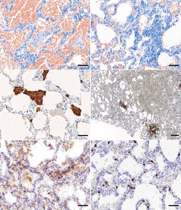
FIGURE 1 Alveolar haemorrhage: partly epithelioid cellular interstitial inflammation in the affected lung,
suggestive of a mild granulomatous-lymphocytic interstitial lung disease (GLILD). Airspaces are filled with
erythrocytes (a), as indicated by an exemplar asterisk), some cluster forming alveolar macrophages (a) and c),
as indicated by arrowheads, the latter using CD68+ stain) and small non-necrotising interstitial granulomas (b),
as indicated by arrowheads) consisting of a few epithelioid cells and lymphocytes. Interstitial lymphocytic
infiltrate mainly consists of CD4+ T-cells, partly clustered d) and partly scattered e). CD8+ T-cells are scattered
f ) and form the minority of the lymphocytic infiltrations. Scale bars: a–c, e, f ) 50 μm; d) 200 μm.
T-lymphocyte-associated protein 4 (CTLA-4) deficiency; and 3) lipopolysaccharide-responsive beige-like
anchor protein (LRBA) deficiency [20]. These latter two disorders are clinically very similar conditions
that have been classified as CVID but may show some aspects of CID. LRBA regulates intracellular
trafficking of CTLA-4 [29]. Both LRBA and CTLA-4 deficiency lead to impaired downregulation of
immune processes [20, 29]. The clinical phenotype of both disorders is similar: autoimmune cytopenias as
well as autoimmune inflammation in the intestines, the central nervous system (CNS) and other organs has
been reported [30–32]. GLILD has been found in up to two thirds of patients with CTLA-4 deficiency and
also in patients with LRBA deficiency (figures 2a–2c) [30, 31]. Diagnostic workup should follow the
standards outlined above.
https://doi.org/10.1183/16000617.0152-2020 5European Respiratory Review RARE GENETIC ILDs | S.F.N. Bode et al.
a) b)
c) d)
FIGURE 2 Granulomatous-lymphocytic interstitial lung disease (GLILD). a) A 9-year old girl with atypical and
confluent, partly spot-shaped infiltrates on chest computed tomography (CT) scan. b) A 20-year old woman
showing nodules with adjacent ground-glass infiltrates. c) A chest CT scan of a 6-year old boy with cytotoxic
T-lymphocyte-associated protein 4 deficiency. Multiple round infiltrates resembling “reversed halo” (or “atoll”)
signs are evidence of GLILD. d) The same patient as in c) but 6 months after allogenic haematopoietic stem
cell transplantation. Marked regression of GLILD is shown with mild residual, streaky interstitial consolidations.
Scale bars: a, c, d) 15 mm; b) 20 mm.
CTLA-4 fusion proteins (e.g. abatacept) and mammalian target of rapamycin (mTOR) inhibitors have been
used successfully in both LRBA and CTLA-4 deficiency and these treatments have had positive effects on
GLILD as well [29, 30, 33]. Severe infectious or autoimmune complications in CIDs that cannot be
controlled by Ig replacement or immunosuppressive therapy can be an indication for HSCT [26].
Improvement of GLILD after stem cell transplantation (SCT) has been demonstrated in CTLA-4 deficiency
(figure 2d) [30]. Symptoms of mild RAG deficiency can be controlled by immunosuppression but
long-term remission cannot be achieved in all patients. Some cases of partial RAG deficiency, especially
atypical SCID, can only be cured with SCT [27, 28]. The diverse phenotype of (P)CIDs and the individual
clinical course has therefore to be taken into account to decide whether SCT might be a viable option for
affected patients [26, 27].
Chronic granulomatous disease
Chronic granulomatous disease (CGD) is caused by defects in superoxide-generating nicotinamide adenine
dinucleotide phosphate (NADPH) oxidase of phagocytes and lymphocytes [34, 35]. Currently, five
different affected genes are known, while the most common findings are mutations in gp91phox (CYBB,
https://doi.org/10.1183/16000617.0152-2020 6European Respiratory Review RARE GENETIC ILDs | S.F.N. Bode et al.
cross-linked). Autosomal recessive forms involve mutations in p22phox (CYBA), p47phox (NCF1),
p67phox (NCF2) and p40phox (NCF4) [34–36]. Both impaired killing of bacteria and fungi, as well as
prolonged autoinflammatory reactions with granuloma formation, can occur [34, 35]. About one to three in
200 000 newborns are affected by CGD [36].
Pulmonary manifestations represent the most common organ involvement in CGD. Infectious
complications are found in 80% of CGD patients, mostly in the form of pneumonia [37, 38]. Formation of
abscesses, bronchiectasis and pulmonary granulomas has also been reported (figure 3) [3, 39].
Increased secretion of pro-inflammatory mediators or decreased production of anti-inflammatory cytokines
causes inflammatory or auto-inflammatory manifestations in CGD [40]. This can lead to development of
granulomas in multiple organs, such as the brain, liver, gastrointestinal tract, spleen, or lung. Granulomas
are noncaseating and contain multinucleated giant cells [37]. Whether or not an infection is essential to
trigger this inflammatory cascade is still debated [34]. Pulmonary symptoms in patients with CGD include
dyspnoea, cough and reduced exercise tolerance [34, 35].
CT scans are important for visualisation of pulmonary pathologies in CGD (table 1); however, lung
biopsies might be essential to correctly identify infectious triggers of disease exacerbation (as has been
shown for Burkholderia species [3]). PFTs may show obstructive or restrictive patterns depending on the
extent of lung involvement [34, 35].
CGD is curable by HSCT and appropriate evaluations should be initiated as soon as the diagnosis is made
[41, 42]. Until HSCT can be performed, control and prevention of infection is paramount in CGD.
Removal of severely damaged pulmonary tissue is sometimes indicated and some experience exists in the
use of steroids in combination with anti-infective drugs [43]. Some authors have suggested the use of
tumour necrosis factor (TNF) inhibitors to control inflammation and granuloma formation [9].
Mendelian susceptibility to mycobacterial disease
Defects in the interferon-γ (IFNγ)/interleukin-12 (IL-12) pathway lead to Mendelian susceptibility to
mycobacterial disease (MSMD). In MSMD, effective killing of intracellular pathogens is impaired [44,
45]. Currently, mutations in 15 different genes (IFNGR1, IFNGR2, STAT1, JAK1, IRF8, SPPL2A, IL12B,
IL12RB1, IL12RB2, IL23R, ISG15, TYK2, RORC, CYBB and NEMO) have been reported in MSMD,
FIGURE 3 Pulmonary granulomas in a 4-year old boy with chronic granulomatous disease. A subpleural
granuloma with a surrounding fuzzy rim is indicated (arrow). Scale bar: 10 mm.
https://doi.org/10.1183/16000617.0152-2020 7European Respiratory Review RARE GENETIC ILDs | S.F.N. Bode et al.
although these account for only around 50% of cases [44]. Prevalence is estimated to be around one in 50
000 [46]. Patients present with mycobacterial infections, classically from Bacillus Calmette–Guérin (BCG)
vaccine or non-TB mycobacteria. Infections by M. tuberculosis and other intracellular pathogens are
common, as is chronic mucocutaneous candidiasis (for specific genetic defects) [44, 46]. Clinical
presentation varies from localised to systemic manifestations and via an acute or chronic course.
Pulmonary granuloma formation develops as frequently as in pulmonary mycobacterial infection and both
necrotising and non-necrotising granulomas are found [44, 46]. Impaired cytokine secretion after leukocyte
stimulation and reduced cell-surface/intracellular expression of receptors/proteins involved in the IFNγ
pathway, can help in identifying specific targets for genetic analysis [45]. Prolonged treatment with
antimycobacterial antibiotics is essential to control infections and additional IFNγ therapy can be helpful.
In some cases, surgical removal of pulmonary lesions is indicated and in severe cases HSCT has been
successful [46, 47].
STAT3 loss of function
Loss of function mutations in the signal transducer and activator of transcription 3 (STAT3) cause STAT3
autosomal-dominant (AD) hyper IgE syndrome (HIES) [48–51]. In STAT3 HIES, impaired neutrophil
chemotaxis, reduced production of inflammatory cytokines and defective repair mechanisms of bronchiolar
and alveolar epithelial cells are pathogenic factors [52–55]. STAT3 HIES is a rare immunodeficiency with
an incidence of less than one in 1 000 000 [56].
STAT3 HIES patients present with typical coarse facial features, eczematous dermatitis, retention of
primary teeth, scoliosis and joint hyperextensibility, as well as with immunodeficiency and recurrent
bronchopulmonary infections [55]. Pulmonary granuloma formation as well as bronchiectasis,
pneumatoceles and cavernas in these patients have been attributed to recurrent infections and to impaired
lung remodelling mechanisms [55].
Imaging studies, especially chest CT scans, are essential in establishing the extent of pulmonary
involvement [55]. Therapeutic options include antimicrobial prophylaxis with trimethoprimsulfamethoxazole,
as well as early aggressive treatment of infections with antibiotics [52]. Ig replacement therapy has been
shown to reduce pulmonary complications if IgG levels are low or serological responses to vaccination are
missing [57]. Surgical excision of large pneumatoceles might be indicated [52].
STAT3 gain of function
Gain of function mutations in STAT3 cause early onset immunodeficiency with additional features such as
autoimmune enteropathy, diabetes mellitus, autoimmune cytopenias, lymphadenopathy, splenomegaly, short
stature and interstitial lung disease (ILD) [58–61]. Currently, less than 50 cases have been described [58].
Granulomatous lung disease has been reported in at least two affected individuals, but other forms of ILD
seem to be a more common feature in patients with gain of function STAT3 mutations [58, 62, 63].
Therapeutic options include immunosuppression with anti-IL-6 agents and Janus kinase ( JAK) inhibitors,
while SCT has not yielded convincing results [58, 64]. STAT3 inhibitors are currently being tested clinically
and might become a therapeutic option for patients with STAT3 gain of function in the future [58].
Other immunodeficiencies
Patients with XIAP and GATA2 deficiencies have been shown to develop GLILD, as have patients with
Kabuki syndrome and those with IgA/IgG2 deficiencies [65–67]. GLILD was successfully treated with
rituximab and azathioprine in at least one patient with XIAP deficiency [67].
Patients with caspase 8 deficiency, Rhoh deficiency, TAP1/TAP2 deficiency and Good’s syndrome have
been shown to develop pulmonary granulomas [66, 68–71]. Granulomatous lung disease has also been
reported in some cases of haemophagocytic lymphohistiocytosis (HLH) [72–74]. Treatment decisions have
to be individualised based on the specific defect and the clinical situation of the patient.
Autoimmune and autoinflammatory diseases associated with pulmonary granulomatosis
Sarcoidosis
Sarcoidosis is an inflammatory disorder of unknown cause that is characterised by granuloma formation in
the affected organs, most often in the lungs [75] although any organ can be affected. The incidence and
prevalence of sarcoidosis, as well as its clinical presentation, vary greatly across geographical regions and
between the sexes, aw well as between different ethnicities and age groups. Its prevalence varies between
two and 1160 per 100 000 and is highest in Scandinavia and in African-American populations [76–78].
https://doi.org/10.1183/16000617.0152-2020 8European Respiratory Review RARE GENETIC ILDs | S.F.N. Bode et al.
The disease develops in genetically predisposed individuals with exposure to an as-yet unknown antigen.
Genome-wide association studies have identified human leukocyte antigen (HLA) class II alleles and
several non-HLA genes as susceptibility factors [79–82]. Most interestingly, a polymorphism in the TNF
gene confers resistance to anti-TNF therapy [83]. Recently, defects in autophagy, JAK STAT signalling and
mTOR pathways have been identified as playing a crucial role in ineffective clearance of infectious agents
or nonorganic particles, triggering granuloma formation due to macrophage and T-cell dysfunction [84, 85].
Familial aggregation is known and having a family member with the disease is associated with a
two-to-four-fold increased risk of developing sarcoidosis [86]. Although these findings are significant,
there is no application of this genetic knowledge in everyday clinics.
The pathological hallmark of sarcoidosis is the presence of compact, epithelioid, non-necrotising
granulomas with varying degrees of lymphocytic inflammation. This is used, in combination with a
compatible clinical disease manifestation and typical radiological presentation, as a diagnostic parameter.
Nevertheless, other causes of granuloma need to be excluded [87]. These inflammatory processes attract
mononuclear and polymorphic nuclear cells to the lower respiratory tract, which can be probed by BAL.
An increase in lymphocytes with an elevated CD4/CD8 ratio heralds a spontaneous resolution or a
desirable course under therapy, but an increase in neutrophils is associated with progressive disease
requiring therapy [88]. PFTs may reveal reduced diffusion capacity and a restrictive pattern with loss of
vital capacity, but also obstructive changes [89].
Corticosteroids still constitute the first line treatment in cases with progressive organ damage [75, 87].
Corticosteroid-sparing agents, such as azathioprine or methotrexate, are frequently used when prolonged
therapy is necessary [90, 91]. Studies and case series demonstrate the successful use of newer agents
which manipulate the cytokine network, such as infliximab, rituximab, or JAK-inhibitors such as
tofacitinib [92–97]. None of these are approved but off-label therapy is often initiated [75].
Blau syndrome/early-onset sarcoidosis
Both Blau syndrome and early-onset sarcoidosis are rare disorders caused by gain of function mutations in
the nucleotide-binding oligomerisation domain-containing protein 2 (NOD2) pattern recognition receptor
(also known as the caspase-recruitment domain-containing protein 15 (CARD-15)) [98–100]. NOD2 is
involved in innate immune responses and the inflammative cascade after viral or bacterial infections (via
NF-κB and TNF receptor-associated factor 3 (TRAF3)) [101, 102]. Gain of function mutations in NOD2
are associated with granulomatous inflammation of affected tissues, though a triggering infection is possibly
essential [100, 103, 104]. Histologic evaluation reveals epithelioid cell-rich, noncaseating granulomas [105].
Blau syndrome is the inherited form of the disease and early-onset sarcoidosis is caused by de novo
mutations in NOD2. The clinical course of the two entities is phenotypically indistinguishable and patients
show a triad of granulomatous polyarthritis, dermatitis and uveitis [99, 103, 106–108] (figure 4). Most
patients present under the age of 2 years [109] and about one third to one half of patients have additional
manifestations. These include fever, lymphadenopathy, vasculitis, arterial hypertension, transient
neuropathies, granulomatous kidney disease and granulomatous liver disease, as well as pulmonary
embolisms [107, 110]. At least four patients with Blau syndrome and interstitial/granulomatous lung
disease have been described [100, 107, 111]. Clinical signs were mild or not present and the pulmonary
changes were found by chance on CT scan [111]. Pulmonary involvement in early-onset sarcoidosis is less
frequent than in later-onset sarcoidosis. One patient has been reported with bronchial granulomas [112],
one with bronchial granuloma and subsequent pulmonary haemorrhage [113] and one with pulmonary
micronodules [114].
Parameters that have been helpful in diagnosing sarcoidosis are serum levels of angiotensin-converting
enzyme (ACE), soluble IL-2 receptor and serum amyloid A [103, 115]. For patients with high suspicion of
Blau syndrome/early-onset sarcoidosis, genetic analysis should be performed (figure 4) [107]. A HRCT
chest scan is paramount in identifying pulmonary involvement. If patients undergo bronchoscopy, the
cellular composition of bronchoalveolar fluid should be analysed, as a lymphocytosis of >15% (as well as a
CD4/CD8 ratio of >3.5: 1) suggests pulmonary sarcoidosis or pulmonary involvement in Blau syndrome [116].
Therapeutic approaches with corticosteroids, anti-TNF agents and anti-IL-1 therapy have yielded positive results
in halting inflammation; however, there is no consensus regarding optimal therapy so far [109, 110, 117].
NOD2-associated autoinflammatory disease
A similar clinical picture to that in Blau syndrome can be found in NOD2-associated autoinflammatory
disease (NAID), which is caused by the IVS8+ NOD2 variant or the heterozygous p.T189M and p.R703C
https://doi.org/10.1183/16000617.0152-2020 9European Respiratory Review RARE GENETIC ILDs | S.F.N. Bode et al.
AgeEuropean Respiratory Review RARE GENETIC ILDs | S.F.N. Bode et al.
other immunosuppressive agents, such as methotrexate, azathioprine, or TNF antagonists, although
evidence for this is limited [130, 137].
STING-associated vasculopathy of infancy
Gain of function mutations in the stimulator of IFN genes (STING) cause STING-associated vasculopathy
with onset in infancy (SAVI) [138]. SAVI is considered rare with less than 20 cases reported so far [138–141].
Activation of STING by viral or bacterial triggers causes upregulation of IFNβ transcription, as well as
upregulation of expression of other IFN-regulated genes leading to STAT1 phosphorylation. Patients with
SAVI show uncontrolled STING activation, which causes early-onset constant fever, capillary vasculitis,
lymphadenopathy, chronic anaemia, failure to thrive, interstitial and granulomatous lung disease, or
pulmonary fibrosis [139, 140, 142]. Pulmonary involvement especially differentiates patients with SAVI
from other interferonopathies [141].
Chronic cough and tachypnoea manifest in the first weeks of life. Cutaneous manifestations typically show
within the first 6 months. They include teleangiectatic, pustular or blistering exanthemas, mostly on acral
sites like the fingers, nose and ears, but also on the cheeks [138]. ILD is found on CT scan (sometimes
with nodular infiltrates) and restrictive patterns in PFTs have also been demonstrated [143]. Histologically,
a mixed lymphocytic infiltrate, interstitial fibrosis and emphysema, as well as vasculitis, have been found
in lung biopsies [138]. Pulmonary involvement is life-limiting in a significant number of patients [138, 139].
Different immunosuppressive therapies, including corticosteroids, cyclophosphamide, azathioprine,
methotrexate, rituximab and infliximab, have shown only moderate beneficial effects [139]. As JAK
inhibitors target the phosphorylation of STAT1/STAT2 [144], this pharmacological approach might be
helpful in the future for patients with SAVI [138, 145].
Granulomatosis with polyangiitis
Granulomatosis with polyangiitis is an autoinflammatory systemic vasculitic disorder linked to
polymorphisms in HLA-DPB1, HLA-DPA1, PRTN3 and SERPINA1 [146]. Patients show elevated
anti-neutrophil cytoplasmic antibodies (ANCAs) and, in 70–90% of cases, ANCAs that target proteinase-3
are detected [147]. The pathogenesis of granulomatosis with polyangiitis is not fully understood; however,
a combination of genetic susceptibility factors and environmental triggers may lead to a dysregulation of
innate and adaptive immune responses [148].
Cytoplasmic ANCA associated vasculitis has an incidence of about 13–20·(1 000 000)−1·year−1 in adults and
0.45–6.39·(1 000 000)−1·year−1 in children [149, 150]. The diagnosis of granulomatosis with polyangiitis can
be established following the established classification criteria with a combination of histological, serological
and clinical findings [151, 152]. Pulmonary involvement is common in granulomatosis with polyangiitis
[152, 153]. Typical symptoms include rhinitis, persistent otitis media, cough, stridor, obstruction, dyspnoea
and haemoptysis. Subglottic or bronchial stenosis might develop secondary to inflammation.
Pulmonary nodules, cavities or fixed infiltrates are found in imaging studies [149, 154] and
fluorodeoxyglucose (FDG)-PET/CT scan can help to appreciate the extent of the disease and to identify
occult sites of inflammation [155]. Bronchoscopy is indicated if tracheal or bronchial stenosis is suspected
and to obtain biopsies [105]. On histologic examination of affected tissues, necrotising granulomas with
necrotising vasculitis are found [1]. Infectious causes contribute to a high morbidity and mortality in
granulomatosis with polyangiitis and need to be ruled out and treated aggressively [105].
Control of autoinflammation can be achieved with a combination of high-dose corticosteroids and other
immunosuppressive agents, such as cyclophosphamide, azathioprine, methotrexate, mycophenolate mofetil
or rituximab [152].
Eosinophilic granulomatosis with polyangiitis
Eosinophilic granulomatosis with polyangiitis (Churg–Strauss syndrome) is associated with polymorphisms
of the Fcγ receptor 3B that is expressed on neutrophils and contributes to the clearance of immune
complexes [156]. ANCA antibodies, which usually target myeloperoxidase ( perinuclear (p-)ANCAs), can
be detected in 30–75% of adult patients and around 30% of affected children [157, 158].
The incidence in adults is around 1–3·(100 000)−1·year−1 and less than 100 paediatric cases have been reported
[157]. Asthma and eosinophilia are present in almost all patients with eosinophilic granulomatosis with
polyangiitis and the most common misdiagnosis is therapy-refractive asthma. Vasculitis and granuloma
https://doi.org/10.1183/16000617.0152-2020 11European Respiratory Review RARE GENETIC ILDs | S.F.N. Bode et al.
formation in the lungs, skin, digestive tract and heart are common in eosinophilic granulomatosis with
polyangiitis [157, 159]. The condition has different phases of disease activity: a prodromal phase can be
asymptomatic and is followed by an eosinophilic phase (where most paediatric patients are diagnosed); whereas
adults are mainly diagnosed in the final vasculitic phase [157]. HRCT chest scan typically shows ground-glass
opacities (GGOs), bronchial wall thickening, (micro)nodules and consolidations [157, 159]. Cytologic
evaluation of BAL can demonstrate a mean eosinophilia of up to 33%, although patients may also present
without eosinophilia [157, 159]. The histologic hallmark of eosinophilic granulomatosis with polyangiitis is
necrotising granulomatous inflammation with eosinophilic infiltration, mainly of the small vessels of the upper
and lower airways as well as the surrounding tissue, with formation of extravascular granulomas [159, 160].
Systemic corticosteroids, possibly in combination with azathioprine or cyclophosphamide, are recommended
to control eosinophilic granulomatosis with polyangiitis [157, 161]. The IL-5 antibody mepolizumab has
also yielded positive results in ANCA-positive patients with an eosinophilia of >150 cells·µL−1 [162].
Other conditions
Apart from the immunodeficiencies and autoinflammatory diseases presented here, there is growing
evidence that other conditions that have some genetic background might predispose to pulmonary
granulomatous inflammation. One example is histiocytosis X/Langerhan’s cell histiocytosis (LCH), a
neoplastic process with inflammatory characteristics. Somatic mutations in BRAF and NRAS have been
described in most patients with pulmonary LCH [163, 164]. Incidence of pulmonary LCH is around 0.27
per 100 000 and mostly affects young adults who smoke [165]. Clinical presentation is unspecific,
involving dry cough, dyspnoea and fatigue [165]. Chest CT scans typically reveal stellate nodules, nodular
opacities, cysts, or honeycombing [166] and these changes mainly occur in the middle and upper lobes of
the lungs. BAL may yield CD1a and CD207 positive cells supporting the diagnosis [167]. Cessation of
smoking is essential. Glucocorticoid therapy is successful only in some patients and others might need
more aggressive chemotherapy (e.g. cytarabine or vinblastine) [167].
Conclusions and further directions
In this review we discuss the broad clinical and genetic spectrum of pulmonary granulomatosis of genetic
origin. The diversity of conditions that can manifest in infants, children, adolescents and adults, based on
specific genetic defects, is evident in the varying amount and type of granulomatous inflammation as well
as the accompanying symptoms. These symptoms may give clues to the underlying disorder but can be
nonspecific in many cases. Pathophysiology of granuloma formation is well understood in some of the
diseases presented here and may be based on endogenous inflammation, impaired control of environmental
pathogens, or pathological immune responses to harmless antigens. In other conditions, the pathways
leading to pulmonary granuloma formation are less clear. Nevertheless, in all cases of suspected
pulmonary granulomatosis it is paramount to exclude infectious causes before considering the rarer,
noninfectious reasons for their development, while simultaneously keeping in mind that some of the
genetic defects discussed here specifically predispose to infections that lead to the pulmonary granuloma.
Certain immunodeficiencies and autoimmune disorders have been classically associated with pulmonary
granulomatosis. As the understanding of the genetic basis of many disorders is expanded, so is the possible
list of differential diagnoses of pulmonary granulomatosis of genetic origin. Decisive indicators of the
underlying disorder can be elicited by pulmonologists, immunologists, rheumatologists, radiologists and
pathologists; therefore, a multidisciplinary approach is paramount in correctly diagnosing affected patients.
Early identification of individuals at risk for pulmonary granulomatosis of genetic origin can help to avoid
environmental or infectious triggers, support aggressive antimicrobial or anti-inflammatory therapy in
patient subgroups and might identify individuals eligible for SCT. A combined diagnostic approach taking
clinical presentation, laboratory workup, imaging techniques, histologic findings and genetics results into
consideration can therefore help to shape the way to personalised diagnosis and treatment. With the
advance of new sequencing techniques, the genetic background of other causes of pulmonary
granulomatous inflammation might be discovered in the future.
Previous articles in the Series: No. 1: Daccord C, Good J-M, Morren M-A, et al. Brit–Hogg–Dubé syndrome.
Eur Respir Rev 2020; 29: 200042. No. 2: Hadchouel A, Drummond D, Abou Taam R, et al. Alveolar proteinosis of
genetic origins. Eur Respir Rev 2020; 29: 200187. No. 3: Cazzato S, Omenetti A, Ravaglia C, et al. Lung involvement
in monogenetic interferonopathies. Eur Respir Rev 2021; 30: 200001. No. 4: Yokoyama T, Gochuico BR.
Hermansky–Pudlak syndrome pulmonary fibrosis: a rare inherited interstitial lung disease. Eur Respir Rev 2021; 30:
200193. No. 5: van Moorsel CHM, Van der Vis JJ, Grutters JC. Genetic disorders of the surfactant system: focus on
adult disease. Eur Respir Rev 2021; 30: 200085.
https://doi.org/10.1183/16000617.0152-2020 12European Respiratory Review RARE GENETIC ILDs | S.F.N. Bode et al.
Provenance: Commissioned article, peer reviewed.
Author contributions: S.F.N. Bode conceptualised the review and prepared the first draft of the
manuscript. M. Seidl provided the histological figures and descriptions. All authors critically revised and edited the
manuscript and agreed to the submitted version.
Conflict of interest: S.F.N. Bode has nothing to disclose. J. Rohr has nothing to disclose. J.M. Müller-Quernheim
reports grants and non-financial support from Bristol Myers Squibb; personal fees from Roche and Novartis;
personal fees and non-financial support from Boehringer Ingelheim; and non-financial support from CLS Behring,
outside the submitted work. In addition, J.M. Müller-Quernheim has a patent “Use of inhaled VIP for treatment of
immune checkpoint inhibitor-induced pneumonitis” pending and is co-founder of AdVita Lifescience GmbH, a
spin-off of the University of Freiburg. Advita reports a pending intellectual property concerning the use of Aviptadil
for the treatment of immune checkpoint inhibitor (ICI) pneumonitis. M. Seidl has nothing to disclose.
C. Speckmann has nothing to disclose. A. Heinzmann has nothing to disclose.
References
1 Ohshimo S, Guzman J, Costabel U, et al. Differential diagnosis of granulomatous lung disease: clues and
pitfalls. Eur Respir Rev 2017; 26: 170012.
2 Mukhopadhyay S, Gal AA. Granulomatous lung disease: an approach to the differential diagnosis. Arch
Pathol Lab Med 2010; 134: 667–690.
3 Greenberg DE, Goldberg JB, Stock F, et al. Recurrent Burkholderia infection in patients with chronic
granulomatous disease: 11-year experience at a large referral center. Clin Infect Dis 2009; 48: 1577–1579.
4 Lieberman S, Gleason JB, Ilyas MIM, et al. Assessing the safety and clinical impact of thoracoscopic lung
biopsy in patients with interstitial lung disease. J Clin Diagn Res 2017; 11: OC57–OC59.
5 Blackhall V, Asif M, Renieri A, et al. The role of surgical lung biopsy in the management of interstitial lung
disease: experience from a single institution in the UK. Interact Cardiovasc Thorac Surg 2013; 17: 253–257.
6 Lee YC, Wu CT, Hsu HH, et al. Surgical lung biopsy for diffuse pulmonary disease: experience of 196
patients. J Thorac Cardiovasc Surg 2005; 129: 984–990.
7 Bush A, Cunningham S, de Blic J, et al. European protocols for the diagnosis and initial treatment of
interstitial lung disease in children. Thorax 2015; 70: 1078–1084.
8 Baumann U, Routes JM, Soler-Palacin P, et al. The lung in primary immunodeficiencies: new concepts in
infection and inflammation. Front Immunol 2018; 9: 1837.
9 Mahdaviani SA, Rezaei N. Pulmonary manifestations of primary immunodeficiency diseases. Basel, Springer,
2019.
10 Cunningham-Rundles C, Bodian C. Common variable immunodeficiency: clinical and immunological features
of 248 patients. Clin Immunol 1999; 92: 34–48.
11 Selenius JS, Martelius T, Pikkarainen S, et al. Unexpectedly high prevalence of common variable
immunodeficiency in Finland. Front Immunol 2017; 8: 1190.
12 Al-Mousa H, Al-Saud B. Primary immunodeficiency diseases in highly consanguineous populations from
Middle East and North Africa: epidemiology, diagnosis, and care. Front Immunol 2017; 8: 678.
13 Aggarwal V, Banday AZ, Jindal AK, et al. Recent advances in elucidating the genetics of common variable
immunodeficiency. Genes Dis 2020; 7: 26–37.
14 Grimbacher B, Hutloff A, Schlesier M, et al. Homozygous loss of ICOS is associated with adult-onset
common variable immunodeficiency. Nat Immunol 2003; 4: 261–268.
15 Castigli E, Wilson SA, Garibyan L, et al. TACI is mutant in common variable immunodeficiency and IgA
deficiency. Nat Genet 2005; 37: 829–834.
16 van Zelm MC, Reisli I, van der Burg M, et al. An antibody-deficiency syndrome due to mutations in the CD19
gene. N Engl J Med 2006; 354: 1901–1912.
17 Abolhassani H, Hammarstrom L, Cunningham-Rundles C. Current genetic landscape in common variable
immune deficiency. Blood 2020; 135: 656–667.
18 Tuijnenburg P, Lango Allen H, Burns SO, et al. Loss-of-function nuclear factor κB subunit 1 (NFKB1) variants
are the most common monogenic cause of common variable immunodeficiency in Europeans. J Allergy Clin
Immunol 2018; 142: 1285–1296.
19 European Society for Immunodeficiencies (ESID). Diagnostic criteria for primary immunodeficiencies. Geneva,
ESID, 2020. https://esid.org/Working-Parties/Clinical-Working-Party/Resources/Diagnostic-criteria-for-PID2
20 Sun D, Heimall J. Disorders of CTLA-4 expression, how they lead to CVID and dysregulated immune
responses. Curr Opin Allergy Clin Immunol 2019; 19: 578–585.
21 Hurst JR, Verma N, Lowe D, et al. British Lung Foundation/United Kingdom Primary Immunodeficiency
Network consensus statement on the definition, diagnosis, and management of granulomatous-lymphocytic
interstitial lung disease in common variable immunodeficiency disorders. J Allergy Clin Immunol Pract 2017;
5: 938–945.
https://doi.org/10.1183/16000617.0152-2020 13European Respiratory Review RARE GENETIC ILDs | S.F.N. Bode et al.
22 Bates CA, Ellison MC, Lynch DA, et al. Granulomatous-lymphocytic lung disease shortens survival in
common variable immunodeficiency. J Allergy Clin Immunol 2004; 114: 415–421.
23 Rao N, Mackinnon AC, Routes JM. Granulomatous and lymphocytic interstitial lung disease: a spectrum of
pulmonary histopathologic lesions in common variable immunodeficiency–histologic and immunohistochemical
analyses of 16 cases. Hum Pathol 2015; 46: 1306–1314.
24 Maglione PJ, Overbey JR, Cunningham-Rundles C. Progression of common variable immunodeficiency
interstitial lung disease accompanies distinct pulmonary and laboratory findings. J Allergy Clin Immunol
Pract 2015; 3: 941–950.
25 Chase NM, Verbsky JW, Hintermeyer MK, et al. Use of combination chemotherapy for treatment of
granulomatous and lymphocytic interstitial lung disease (GLILD) in patients with common variable
immunodeficiency (CVID). J Clin Immunol 2013; 33: 30–39.
26 Speckmann C, Doerken S, Aiuti A, et al. A prospective study on the natural history of patients with profound
combined immunodeficiency: an interim analysis. J Allergy Clin Immunol 2017; 139: 1302–1310.
27 Bulkhi AA, Dasso JF, Schuetz C, et al. Approaches to patients with variants in RAG genes: from diagnosis to
timely treatment. Expert Rev Clin Immunol 2019; 15: 1033–1046.
28 Schuetz C, Huck K, Gudowius S, et al. An immunodeficiency disease with RAG mutations and granulomas.
N Engl J Med 2008; 358: 2030–2038.
29 Lo B, Zhang K, Lu W, et al. Patients with LRBA deficiency show CTLA4 loss and immune dysregulation
responsive to abatacept therapy. Science 2015; 349: 436–440.
30 Schwab C, Gabrysch A, Olbrich P, et al. Phenotype, penetrance, and treatment of 133 cytotoxic
T-lymphocyte antigen 4-insufficient subjects. J Allergy Clin Immunol 2018; 142: 1932–1946.
31 Schubert D, Bode C, Kenefeck R, et al. Autosomal dominant immune dysregulation syndrome in humans
with CTLA4 mutations. Nat Med 2014; 20: 1410–1416.
32 Lopez-Herrera G, Tampella G, Pan-Hammarstrom Q, et al. Deleterious mutations in LRBA are associated with
a syndrome of immune deficiency and autoimmunity. Am J Hum Genet 2012; 90: 986–1001.
33 Lee S, Moon JS, Lee CR, et al. Abatacept alleviates severe autoimmune symptoms in a patient carrying a
de novo variant in CTLA-4. J Allergy Clin Immunol 2016; 137: 327–330.
34 Segal BH, Leto TL, Gallin JI, et al. Genetic, biochemical, and clinical features of chronic granulomatous
disease. Medicine (Baltimore) 2000; 79: 170–200.
35 Winkelstein JA, Marino MC, Johnston RB, Jr, et al. Chronic granulomatous disease. Report on a national
registry of 368 patients. Medicine (Baltimore) 2000; 79: 155–169.
36 Rider NL, Jameson MB, Creech CB. Chronic granulomatous disease: epidemiology, pathophysiology, and
genetic basis of disease. J Pediatric Infect Dis Soc 2018; 7: Suppl. 1, S2–S5.
37 Mahdaviani SA, Mohajerani SA, Rezaei N, et al. Pulmonary manifestations of chronic granulomatous disease.
Expert Rev Clin Immunol 2013; 9: 153–160.
38 Mansouri D, Adimi P, Mirsaedi M, et al. Primary immune deficiencies presenting in adults: seven years of
experience from Iran. J Clin Immunol 2005; 25: 385–391.
39 van den Berg JM, van Koppen E, Ahlin A, et al. Chronic granulomatous disease: the European experience.
PLoS One 2009; 4: e5234.
40 De Ravin SS, Naumann N, Cowen EW, et al. Chronic granulomatous disease as a risk factor for autoimmune
disease. J Allergy Clin Immunol 2008; 122: 1097–1103.
41 Gungor T, Teira P, Slatter M, et al. Reduced-intensity conditioning and HLA-matched haemopoietic stem-cell
transplantation in patients with chronic granulomatous disease: a prospective multicentre study. Lancet
2014; 383: 436–448.
42 Morillo-Gutierrez B, Beier R, Rao K, et al. Treosulfan-based conditioning for allogeneic HSCT in children with
chronic granulomatous disease: a multicenter experience. Blood 2016; 128: 440–448.
43 Yamazaki-Nakashimada MA, Stiehm ER, Pietropaolo-Cienfuegos D, et al. Corticosteroid therapy for refractory
infections in chronic granulomatous disease: case reports and review of the literature. Ann Allergy Asthma
Immunol 2006; 97: 257–261.
44 Bustamante J. Mendelian susceptibility to mycobacterial disease: recent discoveries. Hum Genet 2020; 139:
993–1000.
45 Esteve-Sole A, Sologuren I, Martinez-Saavedra MT, et al. Laboratory evaluation of the IFN-γ circuit for the
molecular diagnosis of Mendelian susceptibility to mycobacterial disease. Crit Rev Clin Lab Sci 2018; 55:
184–204.
46 Rosain J, Kong XF, Martinez-Barricarte R, et al. Mendelian susceptibility to mycobacterial disease: 2014–2018
update. Immunol Cell Biol 2019; 97: 360–367.
47 Reed B, Dolen WK. The child with recurrent mycobacterial disease. Curr Allergy Asthma Rep 2018; 18: 44.
48 Hagl B, Heinz V, Schlesinger A, et al. Key findings to expedite the diagnosis of hyper-IgE syndromes in
infants and young children. Pediatr Allergy Immunol 2016; 27: 177–184.
49 Holland SM, DeLeo FR, Elloumi HZ, et al. STAT3 mutations in the hyper-IgE syndrome. N Engl J Med 2007;
357: 1608–1619.
https://doi.org/10.1183/16000617.0152-2020 14European Respiratory Review RARE GENETIC ILDs | S.F.N. Bode et al.
50 Minegishi Y, Saito M, Tsuchiya S, et al. Dominant-negative mutations in the DNA-binding domain of STAT3
cause hyper-IgE syndrome. Nature 2007; 448: 1058–1062.
51 Renner ED, Torgerson TR, Rylaarsdam S, et al. STAT3 mutation in the original patient with Job’s syndrome.
N Engl J Med 2007; 357: 1667–1668.
52 Mogensen TH. STAT3 and the hyper-IgE syndrome: clinical presentation, genetic origin, pathogenesis, novel
findings and remaining uncertainties. JAKSTAT 2013; 2: e23435.
53 Tadokoro T, Wang Y, Barak LS, et al. IL-6/STAT3 promotes regeneration of airway ciliated cells from basal
stem cells. Proc Natl Acad Sci USA 2014; 111: E3641–E3649.
54 Yan C, Naltner A, Martin M, et al. Transcriptional stimulation of the surfactant protein B gene by STAT3 in
respiratory epithelial cells. J Biol Chem 2002; 277: 10967–10972.
55 Kroner C, Neumann J, Ley-Zaporozhan J, et al. Lung disease in STAT3 hyper-IgE syndrome requires intense
therapy. Allergy 2019; 74: 1691–1702.
56 Freeman AF, Holland SM. The hyper-IgE syndromes. Immunol Allergy Clin North Am 2008; 28: 277–291.
57 Chandesris MO, Melki I, Natividad A, et al. Autosomal dominant STAT3 deficiency and hyper-IgE syndrome:
molecular, cellular, and clinical features from a French national survey. Medicine (Baltimore) 2012; 91:
e1–e19.
58 Fabre A, Marchal S, Barlogis V, et al. Clinical aspects of STAT3 gain-of-function germline mutations:
a systematic review. J Allergy Clin Immunol Pract 2019; 7: 1958–1969.
59 Flanagan SE, Haapaniemi E, Russell MA, et al. Activating germline mutations in STAT3 cause early-onset
multi-organ autoimmune disease. Nat Genet 2014; 46: 812–814.
60 Haapaniemi EM, Kaustio M, Rajala HL, et al. Autoimmunity, hypogammaglobulinemia, lymphoproliferation,
and mycobacterial disease in patients with activating mutations in STAT3. Blood 2015; 125: 639–648.
61 Hillmer EJ, Zhang H, Li HS, et al. STAT3 signaling in immunity. Cytokine Growth Factor Rev 2016; 31: 1–15.
62 Maffucci P, Filion CA, Boisson B, et al. Genetic diagnosis using whole exome sequencing in common variable
immunodeficiency. Front Immunol 2016; 7: 220.
63 Gutierrez M, Scaglia P, Keselman A, et al. Partial growth hormone insensitivity and dysregulatory immune
disease associated with de novo germline activating STAT3 mutations. Mol Cell Endocrinol 2018; 473:
166–177.
64 Milner JD, Vogel TP, Forbes L, et al. Early-onset lymphoproliferation and autoimmunity caused by germline
STAT3 gain-of-function mutations. Blood 2015; 125: 591–599.
65 Schussler E, Beasley MB, Maglione PJ. Lung disease in primary antibody deficiencies. J Allergy Clin Immunol
Pract 2016; 4: 1039–1052.
66 Seppänen M, Rezaei N. General considerations of pulmonary manifestations of primary immunodeficiency
diseases. In:Mahdaviani SA, Rezaei N, eds. Pulmonary manifestations of primary immunodeficiency diseases.
1st Edn. Basel, Springer, 2019; pp. 1–36.
67 Steele CL, Dore M, Ammann S, et al. X-linked inhibitor of apoptosis complicated by granulomatous
lymphocytic interstitial lung disease (GLILD) and granulomatous hepatitis. J Clin Immunol 2016; 36: 733–738.
68 Gadola SD, Moins-Teisserenc HT, Trowsdale J, et al. TAP deficiency syndrome. Clin Exp Immunol 2000; 121:
173–178.
69 Crequer A, Troeger A, Patin E, et al. Human RHOH deficiency causes T cell defects and susceptibility to
EV-HPV infections. J Clin Invest 2012; 122: 3239–3247.
70 Lee SH, Lee SM, Yang SC, et al. A case of granulomatous lung disease in a patient with Good’s syndrome.
Korean J Intern Med 2008; 23: 219–222.
71 Niemela J, Kuehn HS, Kelly C, et al. Caspase-8 deficiency presenting as late-onset multi-organ lymphocytic
infiltration with granulomas in two adult siblings. J Clin Immunol 2015; 35: 348–355.
72 Fitzgerald NE, MacClain KL. Imaging characteristics of hemophagocytic lymphohistiocytosis. Pediatr Radiol
2003; 33: 392–401.
73 Jin YK, Xie ZD, Yang S, et al. Epstein–Barr virus-associated hemophagocytic lymphohistiocytosis: a
retrospective study of 78 pediatric cases in mainland of China. Chin Med J 2010; 123: 1426–1430.
74 Rohr J, Beutel K, Maul-Pavicic A, et al. Atypical familial hemophagocytic lymphohistiocytosis due to
mutations in UNC13D and STXBP2 overlaps with primary immunodeficiency diseases. Haematologica 2010;
95: 2080–2087.
75 Grunewald J, Grutters JC, Arkema EV, et al. Sarcoidosis. Nat Rev Dis Primers 2019; 5: 45.
76 Arkema EV, Grunewald J, Kullberg S, et al. Sarcoidosis incidence and prevalence: a nationwide
register-based assessment in Sweden. Eur Respir J 2016; 48: 1690–1699.
77 Dumas O, Abramovitz L, Wiley AS, et al. Epidemiology of sarcoidosis in a prospective cohort study of U.S.
women. Ann Am Thorac Soc 2016; 13: 67–71.
78 Cozier YC, Berman JS, Palmer JR, et al. Sarcoidosis in black women in the United States: data from the
Black Women’s Health Study. Chest 2011; 139: 144–150.
79 Hofmann S, Franke A, Fischer A, et al. Genome-wide association study identifies ANXA11 as a new
susceptibility locus for sarcoidosis. Nat Genet 2008; 40: 1103–1106.
https://doi.org/10.1183/16000617.0152-2020 15You can also read