2021 TSOC Expert Consensus on the Clinical Features, Diagnosis, and Clinical Management of Cardiac Manifestations of Fabry Disease
←
→
Page content transcription
If your browser does not render page correctly, please read the page content below
Acta Cardiol Sin 2021;37:337-354
Review Article                                                                            doi: 10.6515/ACS.202107_37(4).20210601A

Consensus

            2021 TSOC Expert Consensus on the Clinical
          Features, Diagnosis, and Clinical Management of
              Cardiac Manifestations of Fabry Disease
        Chung-Lieh Hung,1,2 Yen-Wen Wu,3,4 Chih-Chan Lin,5,6 Chih-Hung Lai,7 Jimmy Jyh-Ming Juang,8,9 Ting-Hsing Chao,5,6
                    Ling Kuo,10 Kuo-Tzu Sung,1 Chao-Yung Wang,11,12,13 Chun-Li Wang,11,12 Chun-Yuan Chu,14
                                        Wen-Chung Yu4,10* and Charles Jia-Yin Hou1,15*

     Fabry disease (FD) is an X-linked, rare inherited lysosomal storage disease caused by a-galactosidase A gene variants
     resulting in deficient or undetectable a-galactosidase A enzyme activity. Progressive accumulation of pathogenic
     globotriaosylceramide and its deacylated form globotriaosylsphingosine in multiple cell types and organs is proposed
     as main pathophysiology of FD, with elicited pro-inflammatory cascade as alternative key pathological process.
     The clinical manifestations may present with either early onset and multisystemic involvement (cutaneous,
     neurological, nephrological and the cardiovascular system) with a progressive disease nature in classic phenotype,
     or present with a later-onset course with predominant cardiac involvement (non-classical or cardiac variant; e.g.
     IVS4+919G>A in Taiwan) from missense variants. In either form, cardiac involvement is featured by progressive
     cardiac hypertrophy, myocardial fibrosis, various arrhythmias, and heart failure known as Fabry cardiomyopathy
     with potential risk of sudden cardiac death. Several plasma biomarkers and advances in imaging modalities along
     with novel parameters, cardiac magnetic resonance (CMR: native T1/T2 mapping) for myocardial tissue characterization
     or echocardiographic deformations, have shown promising performance in differentiating from other etiologies of
     cardiomyopathy and are presumed to be helpful in assessing the extent of cardiac involvement of FD and in guiding or
     monitoring subsequent treatment. Early recognition from extra-cardiac red flag signs either in classic form or red
     flags from cardiac manifestations in cardiac variants, and awareness from multispecialty team work remains the
     cornerstone for timely managements and beneficial responses from therapeutic interventions (e.g. oral chaperone
     therapy or enzyme replacement therapy) prior to irreversible organ damage. We aim to summarize contemporary
     knowledge based on literature review and the gap or future perspectives in clinical practice of FD-related
     cardiomyopathy in an attempt to form a current expert consensus in Taiwan.

     Key Words:         Cardiac magnetic resonance (CMR) · Cardiac variant · Chaperone · Enzyme replacement
                        therapy (ERT) · Fabry disease (FD) · Globotriaosylsphingosine (Lyso-Gb3) ·
                        Globotriaosylceramide (Gb3) · IVS4+919G>A · a-galactosidase A (GLA) gene

Received: May 16, 2021            Accepted: June 1, 2021
1
  Division of Cardiology, Departments of Internal Medicine, MacKay Memorial Hospital, Taipei; 2Institute of Biomedical Sciences, Mackay
Medical College; 3Division of Cardiology, Cardiovascular Medical Center, Far Eastern Memorial Hospital, New Taipei City; 4School of Medicine,
National Yang Ming Chiao Tung University, Taipei; 5Division of Cardiology, Department of Internal Medicine, National Cheng Kung University
Hospital; 6Department of Internal Medicine, College of Medicine, National Cheng Kung University, Tainan; 7Cardiovascular Center, Taichung
Veterans General Hospital, Taichung; 8Division of Cardiology, Department of Internal Medicine and Cardiovascular Center, National Taiwan
University Hospital; 9College of Medicine, National Taiwan University; 10Division of Cardiology, Taipei Veterans General Hospital, Taipei;
11
   Division of Cardiology, Chang Gung Memorial Hospital, Linkou Medical Center; 12School of Medicine, College of Medicine, Chang Gung
University, Taoyuan; 13Institute of Cellular and System Medicine, National Health Research Institutes, Zhunan; 14Division of Cardiology,
Kaohsiung Medical University Hospital, Kaohsiung; 15Department of Medicine, Mackay Medical College, New Taipei City, Taiwan.
Corresponding author: Dr. Wen-Chung Yu, Division of Cardiology, Department of Medicine, Taipei Veterans General Hospital, Taipei, Taiwan.
Tel: 886-2-2871-2121 ext. 2997; Fax: 886-2-2877-1746; E-mail: wcyu@vghtpe.gov.tw
* These authors contributed equally to the study.

                                                                    337                                     Acta Cardiol Sin 2021;37:337-354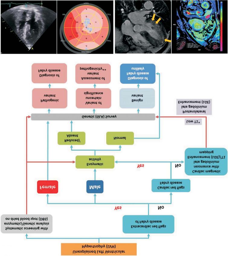
Chung-Lieh Hung et al.
GENERAL INTRODUCTION AND PREVALENCE pain, anhidrosis or hypohidrosis, and skin lesions known
as angiokeratoma 8 with the heart and kidneys being
Fabry disease (FD) has an estimated prevalence of 1 more prominent by the third decade of life, eventually
in 40,000 to 1 in 117,000 live births for males, 1-3 and leading to premature death from renal failure, stroke
presents as an abnormal accumulation of glycosphingo- and cardiac disease.1,9 Some missense genetic variants
lipids within lysosomes due to deficient or absent a- have been identified, including p.N215S in North Amer-
galactosidase A (a-GAL A) enzyme activity in various cell ica and Europe, p.F113L in Portugal, and IVS4+919G®A
types and organs (Figure 1). As a rare X-linked inherited in Taiwan. These variants have been associated with re-
metabolic disease, FD has been increasingly recognized sidual a-GAL A activity and may manifest with late-on-
in recent years.4,5 To date, more than 1,000 GLA gene set FD affecting the heart as the predominant clinical
variants have been identified and are categorized as picture (cardiac variant) (Figure 2).7,10,11
pathogenic, benign without clinical relevance, or of un- Random X-chromosome inactivation likely results in
clear significance.6,7 Nonsense, missense variants, and mosaicism in female patients, with some cells expressing
premature stop codons that lead to absent or low a-GAL the normal allele and others the mutated allele. This
A enzyme activity are usually associated with classic likely causes heterogeneous manifestations 12 ranging
early-onset FD, presenting with a male predominance as from an asymptomatic or mild symptomatic course later
a multisystemic disorder with earlier onset, and progres- in life to a more severe phenotype resembling classic
sive clinical picture (in childhood or adolescence) as the FD. With advances in cardiac imaging techniques and
classic FD phenotype (Figure 2). Initial clinical manifesta- enhanced awareness of cardiomyopathy, the actual pre-
tions may involve multiple organs including neuropathic valence of FD in high-risk patients may not be as rare as
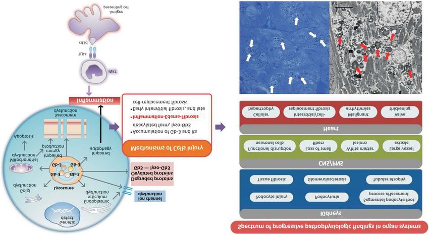
A B C
Figure 1. (A) Typical pathophysiology of Fabry disease (FD) as a storage disease with cardiac involvement and recently reported secondary path-
ways operating in FD. Figure was adapted and modified from reference 30. (B) Endomyocardial biopsy from a 86-year-old female Fabry patient of
cardiac variant (IVS4+919G>A mutation) presenting with hypertrophic cardiomyopathy. Toluidine blue staining demonstrating abundant granular in-
clusions (white arrows) caused by Gb3 accumulation within cardiomyocytes. (C) Electron microscopy showing enlarged secondary lysosomes packed
with lamellated membrane structures (zebra bodies) and myofibrillar loss in cardiomyocytes. CNS, central nervous system; Gb3, globotriao-
sylceramide; iNKT, invariant natural killer T; lyso-Gb3, globotriaosylsphingosine; PNS, peripheral nervous system; TLR4, toll-like receptor-4. (Pathology
shown by courtesy of Dr. Chen, Tung-Ying. MacKay Memorial Hospital, Division of Pathology).
Acta Cardiol Sin 2021;37:337-354 338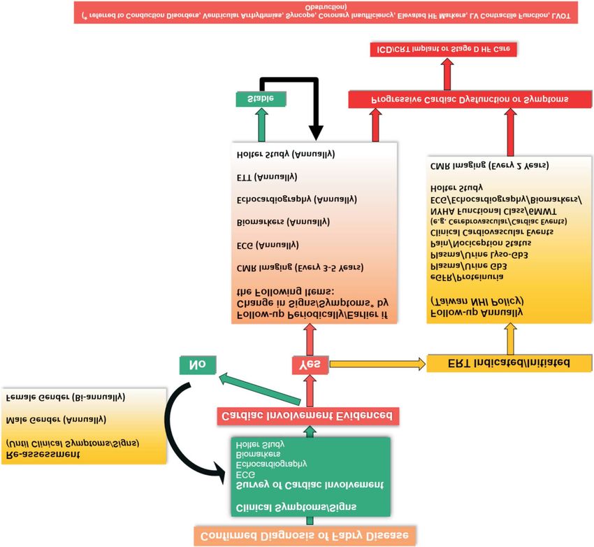
2021 TSOC Expert Consensus for Fabry Disease

     A

     B
Figure 2. (A) Several key GLA mutations associated with classic or later-onset (or cardiac variant) Fabry disease phenotype, variants of unclear sig-
nificance (VUS), and benign variants. The triangular shape presented illustrates higher frequency of GLA mutations involving benign or benign vari-
ants with such mutations probably seen during screening yet less related to Fabry-related clinical manifestations. c.936+919G>A refers to
IVS4+919G>A. (B) Proposed stages of Fabry disease (FD) of cardiac involvement evolution along with clinical progression in relation to expected en-
zyme replacement treatment (ERT) efficacy. Figure was adapted and modified from references 30 and 47.

previously reported,4 especially for late-onset (as a non-                    ramide (Gb3). A combination of cardiogenic embolism
classic phenotype) or atypical FD8,13 with predominant                        formation or vascular wall changes due to Gb3 accu-
cardiac involvement (phenotypes) (e.g. in certain Asian                       mulation and abnormal reactivity/signaling contributes
regions) (Table 1). One European multicenter cross-sec-                       to the activation of coagulation pathways predisposing
tional Anderson-Fabry survey by Elliott et al. including                      to cerebrovascular events. 20,21 These complex patho-
1386 hypertrophic cardiomyopathy (HCM) patients re-                           physiological conditions likely explain why juvenile and/
ported a 0.5% prevalence rate of pathogenic GLA muta-                         or cryptogenic transient ischemic attack and cerebro-
tions.14 In comparison, another study reported a patho-                       vascular complications are the major causes of morbid-
genic mutation rate of around 1% during screening tri-                        ity and early mortality in both male and female patients
als,15 and a newborn screening survey conducted in Italy                      with FD.22-24
suggested a prevalence of up to 1 in 8,800 newborns.6,16                           Owing to the high prevalence of cardiovascular in-
There is a high prevalence of the cardiac variant (IVS4+                      volvement, the leading cause of death in FD is cardio-
919G®A) (»1 in 1600 males) of FD in Taiwan as re-                             vascular death in both men and women.25 Cardiac in-
vealed by newborn screening programs17,18 and patients                        volvement is a common clinical manifestation in FD pa-
with idiopathic HCM.11                                                        tients, with hallmark features including increased myo-
     As a disease with multisystemic involvement, early                       cardial inflammation, myocardial fibrosis,26-28 and left
pathological changes in FD are thought to predomi-                            ventricular hypertrophy (LVH). Several cellular compo-
nantly involve the microvasculature19 (endothelial dys-                       nents of the heart have been shown to be involved due
function) due to affected clearance of globotriaosylce-                       to chronic intracellular accumulation of Gb3, including

                                                                        339                                       Acta Cardiol Sin 2021;37:337-354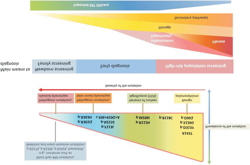
Chung-Lieh Hung et al.

Table 1. Fabry disease red flags for differential diagnosis
                                            Cardiac red flags                                                     Extra-cardiac red flags
                                         Family history of LVH, particularly if no         Family history of renal failure and/or stroke       Any time
                   History

                                         evidence of male-to-male transmission
                                                                                           Neuropathic pain                                      1-2

                                         Short PQ interval#                                Gastrointestinal symptoms                             1-2
                   Electrocardiography

                                         Bradycardia                                       Angiokeratomas                                        1-2

                                         Chronotropic incompetence                         Cornea verticillata*                                  1-2

                                                                                                                                                          Presenting decades of age
                                                                   #
                                         Atrioventricular blocks                           Hypohidrosis, heat/cold, and exercise intolerance     1-2
 Diagnostic tool

                                         LVH with normal systolic function                 Albuminuria                                           1-2

                                         Reduced global longitudinal strain                Juvenile and/or cryptogenic TIA/stroke                3-4
                   2D-Echo

                                         Mild-to-moderate aortic root dilation             Hearing loss (either progressive or sudden)           3-4

                                         Mitral and aortic valve thickening with           Dolichoectrasia of the basilar artery, chronic        3-4
                                         mild-to-moderate regurgitation                    white matter hyperintensities at brain MRI
                                         Hypertrophy of papillary muscles                  Proteinuria                                           3-4
                   CMR Imaging

                                         Mid-layer posterolateral late gadolinium          Renal failure                                         3-4
                                         enhancement
                                         Low native T1                                     Lymphedema                                            3-4

Fabry disease red flags for differential diagnosis of patients with idiopathic left ventricular hypertrophy (LVH) and/or hypertrophic
cardiomyopathy.
                                                                     #
* In the absence of iatrogenic causes (chloroquine/amiodarone). Short PQ interval in early stages; atrioventricular and bundle
branch blocks are more common in advanced disease.
2D-Echo, 2-dimensional echocardiography; CMR, cardiac magnetic resonance; TIA, transient ischemic attack.
Table was adapted and modified from reference 30.

endothelial cells, vascular smooth muscle cells, car-                                      to chest pain, heart failure symptoms and arrhythmic
diomyocytes, conduction system cells and valvular fibro-                                   events (Figure 2) (~60%).34 Various cardiac arrhythmias
blasts.4 Excessive Gb3 deposits within cardiomyocytes                                      have been identified, including bradyarrhythmia, con-
may also elicit sarcomeric myofilament dysfunction and                                     duction block, and ventricular tachycardia. The degree
myofibrillolysis.29 Given the relatively low percentage of                                 of LVH and the presence of myocardial fibrosis are risk
complex lipids (1-2%) in total cardiac mass, accumulat-                                    factors for more malignant form of ventricular tachyar-
ing data suggest that pathological cardiac involvement                                     rhythmia or sudden cardiac death in both FD and non-
in FD typically begins with in Gb3 deposition as the ini-                                  FD HCM.35
tial step, followed by provoked alternative pathological                                       Renal involvement is one of the main complications
signaling of subsequent inflammation, hypertrophy, and                                     of FD and may affect podocytes, glomerular, vascular
interstitial fibrotic processes over time (Figure 1). 4,30                                 smooth muscle and tubular cells at an early stage,36,37
Furthermore, coronary microvascular ischemia from en-                                      possibly since early childhood and early adolescence.38,39
dothelial dysfunction, impaired endocytosis/autophagy                                      High amounts of Gb3 deposits have been found in se-
processes and mitochondrial dysfunction may also play                                      veral types of renal cells,40 with segmental podocyte foot
a role in progressively diseased myocardium,31-33 leading                                  process effacement in young classic FD, which is an early

Acta Cardiol Sin 2021;37:337-354                                                     340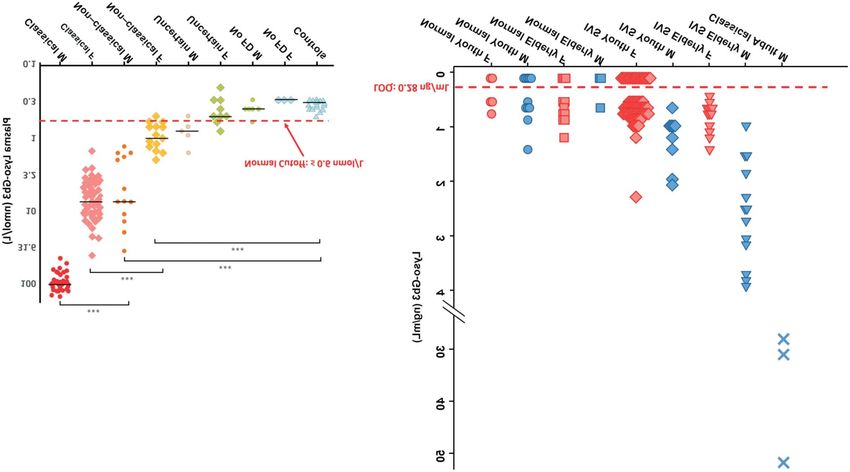
2021 TSOC Expert Consensus for Fabry Disease

marker of nephropathy prior to albuminuria or decline                           more patients are likely to have late onset (non-classic)
of estimated glomerular filtration rate. Podocyturia due                        than the classic type of FD manifesting with first presen-
to continuous podocyte loss may antedate proteinuria                            tations of unexplained LVH, especially in men.9 Cardiac
in FD, whereas proteinuria likely indicates a more ad-                          involvement in FD may present with unexplained LVH,
vanced stage with glomerular involvement. 41 A study                            preserved ejection fraction heart failure and ventricular
from a US dialysis registry reported that FD may account                        arrhythmias starting from the third decade of life (> 30
for 0.01% of patients with end-stage kidney disease,42                          years in males, > 40 years in females.11,30,44 Therefore,
probably due to continued and irreversible cellular loss                        routine screening of a-GAL A enzyme activity (in males)
of both glomerulus and podocytes without early en-                              and GLA sequencing (in females) for individuals with un-
zyme replacement therapy (ERT).43                                               explained LVH older than 40 years and with unknown
                                                                                family background may be recommended (Figure 3).44 In
                                                                                classic FD, confirmation of severely reduced or absent
SCREENING CRITERIA IN CARDIOLOGY                                                plasma or leukocyte a-GAL A activity is often sufficient
                                                                                for a diagnosis in males. 45 Apart from several typical
     As a multisystemic disease with relatively rare clini-                     clinical manifestations or markers as cardiac red flags,
cal prevalence, multiple diagnostic approaches are help-                        identifying the unique features of extra-cardiac red flags
ful for the early recognition and referral in highly sus-                       relevant to the pathophysiological findings of FD may
pected subjects. Apart from the typical clinical manifes-                       also be helpful.30 One large prospective, multidisciplin-
tations of extra-cardiac red flag picture in classic FD with                    ary, multicenter screening program conducted in multi-
multisystemic involvement (Table 1) (e.g. skin, periph-                         specialty clinics (including cardiology, neurology, ne-
eral/central nervous system, renal dysfunction or stroke),                      phrology, pediatrics, ophthalmology, dermatology, gas-

  A                                                                   B
Figure 3. (A) Plasma globotriaosylsphingosine (lyso-Gb3) levels stratified by disease phenotype and sex on a logarithmic scale. Dotted horizontal
red line refers to the upper limit of normal cutoff defined as 2 SDs above the mean in healthy controls (£ 0.6 nmol/L, mean 0.4 nmol/L, SD: 0.1, n =
20). Male subjects (M) are depicted as circles, female subjects (F) are depicted in diamonds, and controls depicted as triangles. Horizontal line per
group represents the median group value. Figure was adapted and modified from reference 48. *** p < 0.01. Of note, plasma lyso-Gb3 in classical M
and F subjects differed significantly from healthy controls (both p < 0.01). (B) Lyso-Gb3 levels in adults with the later-onset FD (GLA IVS4+919G>A mu-
tation) stratified by sex and disease phenotypes from ethnic Taiwanese population. Adapted and modified from reference 46. with permission
granted from Dr. Chien, Yin-Hsiu. FD, Fabry disease; SD, standard deviation.

                                                                          341                                      Acta Cardiol Sin 2021;37:337-354
Chung-Lieh Hung et al.
troenterology, internal medicine, and genetics clinics) IMAGING FOR FABRY DISEASE
reported that 37 (1.8%) patients who fulfilled the inclu-
sion criteria incorporating clinical, genetic, and bioche- Cardiac ultrasonography is a convenient bedside im-
mical indices were carriers of GLA mutations among aging modality and remains a fundamental tool in deter-
2,034 probands.45 The most commonly involved organ mining the precise geometry (e.g. concentric hypertro-
systems were the heart (69%), peripheral nerves (46%), phy), phenotypic LVH or the presence of HCM. Conven-
kidneys (45%), eyes (37%), brain (34%), skin (32%), gas- tional geometric echocardiography measures in FD in-
trointestinal tract (31%), and auditory system (19%). clude wall thickness, ventricular dimension, atrial/ven-
The male patients with late-onset FD had higher resid- tricular volumes by using linear measurements and se-
ual a-GAL A activity compared to those with classic FD, veral functional hemodynamic assessments, standard 2D
although its level is far below normal. In comparison, and hemodynamic Doppler measurements.9,11,30,27,28,57 A
plasma or leukocyte a-GAL A activity may be normal or maximum LV wall thickness of 15 mm not explained by
slightly deficient in heterozygous females.45,46 Hence all pressure overload indicates typical phenotypic HCM,
FD diagnoses should be confirmed by genetic testing, whereas in those manifesting a borderline value (13-14
with cascade family genetic screening according to X- mm) of maximum wall thickness, other demographic
linked inheritance. 47 Assessment of more highly spe- elements (including family history, other clinical sys-
cific biomarkers of FD, such as circulating Gb3 and temic manifestations such as renal, neurological, or der-
globotriaosylsphingosine (lyso-Gb3) levels in plasma matological involvement, and electrocardiographic find-
and Gb3 levels in urine, can be useful and highly spe- ings) should be used to guide the clinical diagnosis.58 A
cific in establishing the diagnosis and clinical pheno- recently published study combining several key clinical
type (e.g. high levels in classical patients and lower le- indices (including PR interval on ECG, arrhythmia his-
vels in non-classical patients) of FD, particularly in fe- tory, LVH pattern on echocardiography, and autonomic
male patients in whom partial enzyme activity is pre- dysfunction) reported high28 sensitivity for classifying
sent (Figure 4).48-50 patients at high risk of FD in a Korean HCM cohort.
Right ventricular hypertrophy (defined as RV wall thick-
ness > 5 mm using echocardiography) is not uncommon
ELECTROCARDIOGRAPHY (ECG) (71% reported by Niemann et al.; 59 31% reported by
Graziani et al.60) in Fabry cardiomyopathy.
A variety of ECG parameters have been reported in- Advanced echocardiographic imaging modalities us-
cluding macroscopic myocardial changes compatible ing tissue Doppler imaging, strain and strain rate th-
with pathological ventricular hypertrophy including left rough either tissue Doppler-based imaging or speckle-
ventricular (LV) strain pattern, T-wave inversion and left tracking techniques, may allow for the identification of
atrial enlargement in precordial leads.51 These features the early stages of FD prior to the development of LVH.61
are also common in other forms of advanced and pro- In this regard, echocardiography is particularly useful in
gressive HCM.52 The presence of posterolateral fibrosis identifying diastolic dysfunction (as prevalent as 79% in
may cause ST-T segment depression and T-wave inver- phenotypic LVH compared to 7% with normal LV wall
sion in the inferolateral leads in some FD patients.53 Ad- thickness), impaired pre-clinical systolic function prior
ditionally, recent studies have shown multiple ECG ab- to LVH, 61 and impaired atrial compliance. 62 Doppler-
normalities in FD.54 A shortened PR interval has been re- based myocardial strain and strain rate are promising to
ported in the earlier stage of FD,55 probably due to al- allow for early detection for cardiomyopathy progres-
tered cellular conductive properties from intracardiac sion in FD, which may be able to guide early ERT in such
Gb3 deposition.56 In contrast, due to the pathological and patients.63,64 Novel speckle tracking-based echocardio-
progressive burden of Gb3, PR prolongation and subse- graphy, irrespective of degree of LV remodeling, has
quent AV block is not uncommon in the later stage of greater sensitivity and specificity than conventional ec-
FD,57 making these novel findings helpful in identifying hocardiographic parameters65 and can be used to assess
FD. regional myocardial longitudinal dysfunction which cor-
Acta Cardiol Sin 2021;37:337-354 3422021 TSOC Expert Consensus for Fabry Disease
A
B C
Figure 4. (A) Proposed flowchart and red flag signs for diagnosis of Fabry disease (FD) in patients with idiopathic, unexplained left ventricular hy-
pertrophy (LVH). * Low native non-contrast cardiac magnetic resonance (CMR) imaging T1 values reinforce or generate suspicion of FD, however, nor-
mal T1 values do not exclude FD which can occasionally and rarely be seen in untreated patients with mild LVH (mostly females) or in more advanced
disease stage due to “pseudo-normalization”. With normal native T1 values, genetic analysis remains indicated if other clinical findings are in favor of
FD. ** By lyso-Gb3 levels assessment and endomyocardial biopsy. Figure was adapted and modified from reference 30. (B) Echocardiography includ-
ing 2D (Left, 4 chamber view) and severely deteriorated global longitudinal strain (-6.8%) (Right, bull eye view) from a 61-year-old, male cardiac vari-
ant FD (GLA IVS4+919G>A mutation) patient presenting clinical HFpEF. (C) In same patient, CMR imaging showed diffuse myocardial fibrosis including
basal lateral segment using LGE technique (Left) and native T1 imaging demonstrated relatively low T1 values (808.7 ms) at septal region. (CMR im-
aging by courtesy of Dr. Yun, Chun-Ho. MacKay Memorial Hospital, Division of Radiology).
relates with segmental fibrosis by late gadolinium en- longitudinal systolic function despite preserved LV ejec-
hancement (LGE) using cardiac magnetic resonance (CMR) tion fraction (Figure 4).66 This pattern is distinct from
imaging. 66 Marked global reduction in deformational amyloid cardiomyopathy which mainly affected longitu-
strain measures, especially longitudinal strain, has been dinal strain in the basal and mid regions leading to apical
observed in FD which may be either due to a regional sparing.67 Furthermore, a segmental myocardial longitu-
(e.g. basal inferolateral segment) decrease caused by dinal strain worse than -12.5% has been shown to be an
myocardial fibrotic changes or a deterioration in global excellent indicator of region-specific fibrosis, while a
343 Acta Cardiol Sin 2021;37:337-354Chung-Lieh Hung et al.
segmental strain better than -16.5% excludes the pres- BIOMARKERS
ence of fibrosis. Compared to FD, HCM patients tend to
have reduced regional longitudinal strain in segments Circulating biomarkers including N terminal pro B
of greatest hypertrophy (e.g. septal region).68 Interest- type natriuretic peptide (NT-proBNP) and cardiac tro-
ingly, decreased global circumferential strain accompa- ponin I/T (cTNI/cTNT) have been proposed to be alter-
nied by loss of the normal base-to-apex gradient may native surrogate markers of cardiac involvement in FD.
be able to differentiate FD from other types of HCM.69 NT-proBNP level has been shown to correlate with over-
In hypertensive cardiomyopathy, the worst longitudinal all disease severity in FD,81 given the high prevalence of
strain has been observed in basal septal segments of LV dysfunction and myocardial damage from multiple
the hypertrophic part, or in most of the basal regions,70 pathological mechanisms as mentioned, resulting in an
although a more heterogeneous reduced global longi- excessive degree of LV remodeling or the presence of
tudinal strain pattern has been observed in aortic ste- impaired diastolic mechanics.82 Overall, NT-proBNP has
nosis.71 been shown to be meaningful and useful both in experi-
CMR imaging is a novel imaging modality that fea- mental models and clinical scenario earlier than overt
tures myocardial tissue characteristics and is increas- pathological cardiac remodeling stage, suggesting its
ingly being used in FD. As a more accurate non-invasive pre-clinical screening role in subjects with suspicious
imaging tool in determining ventricular volume, wall profiles.83,84 Likewise, cardiac troponins reflecting car-
thickness and LV mass, CMR imaging also allows for diac muscular necrosis and damage, can be helpful in
more clear characterization of pathologic myocardial assessing active and continuous cardiac involvement in
processes (e.g. infiltration, replacement fibrosis, or in- FD. cTNT/cTNI have been correlated with the presence
flammation) and enables assessment of the extracel- of LGE by CMR independently of coronary lesions,85 as a
lular volume fraction and differentiation of pathologi- predictor of progressive cardiomyopathy, and thereby
cal cardiomyocyte and myocardial interstitial involve- helpful in clinical staging in FD.86,87 Several biomarkers
ment (Figure 4).72,73 These features may be helpful in or cytokines, such as interleukin-6 and high sensitivity
differentiating FD from LVH with different underlying C-reactive protein, reflecting elicited pro-inflammatory
etiologies.72,73 In particular, CMR imaging can supple- pathways/signaling in FD,88 have also been reported to
ment echocardiography in determining the extent of be markers in the evaluation, monitoring and prognosis
sphingolipid accumulation, edema, and interstitial fi- of FD.89-91 The utilization of these biomarkers were listed
brosis (presence of LGE), and thereby is able to evalu- in Table 2.
ate changes in the stages of FD. Low native T1 myocar-
dial values may represent a higher burden of sphingo-
lipid storage from Gb3 accumulation, and a lower native TREATMENT EFFECTS AND MONITORING OF FD
T1 value has been observed in patients with FD com-
pared to controls or patients with concentric LVH from Several treatment options are currently available for
other etiologies. In addition, it has been shown to be FD. Table 3 shows detailed information of these thera-
correlated with ECG changes and likely serves as a novel pies along with their advantages and disadvantages.
predictor of worsening disease, especially in pre-hyper- Among them, ERT remains the main therapeutic inter-
trophic FD.74-76 A reduction in native T1 value prior to vention for FD.30,92,93 Other potential and novel therapies
the development of LVH has thus been correlated with for FD are evolving and currently under development,
early LV functional changes detected by echocardio- including chaperone therapy, substrate reduction ther-
graphy,77 and more closely associated with the extent of apy, and stem cell-, gene-, and messenger ribonucleic
LV remodeling compared to strain measure.78 By assess- acid-based therapies. 92 Lack of effective treatment in
ing increased T2 myocardial relaxation time in FD, the this patient population may result in reduced life expec-
presence of myocardial edema can be evaluated and de- tancy of nearly 20 years in male carriers and 15 years in
termined, especially in segments with LGE, which is com- female carriers compared to the general population.94,95
mon in basal inferolateral segments.30,79,80 Two recombinant enzyme preparations of a-galactosi-
Acta Cardiol Sin 2021;37:337-354 3442021 TSOC Expert Consensus for Fabry Disease
Table 2. Biomarkers and their potential clinical utilities in Fabry disease
Organ
Biomarkers Potential clinical utilization (organ specificity)
Involvement
Systemic Plasma and leukocytes a-galactosidase A (a-Gal) plasma Diagnosis, disease activity, organ damage
activity
Plasma and urinary Gb3 Diagnosis, disease activity, efficacy of ERT, monitor
Plasma and urinary Lyso-Gb3 disease progression, organ damage
Neutralizing antibodies Prognosis, disease severity
MiRNA (miR21, miR210, miR29, miR200, miR21-5p, Diagnosis, disease activity, response to treatment,
miR19a-3p, etc.) prognosis
Kidney Proreinuria, albuminuria, eGFR Renal involvement
Uromodulin, N-acety-b-D-glucosaminidase, Renal involvement (under investigation)
beta2 microglobulin
Urinary lyso-Gb3 analogues Diagnosis (detectable in case not in control)
Cystatin C Early renal involvement
Heart 3-NT Cardiac and vascular involvement
Longitudinal strain distribution Cardiac involvement, prognosis
TNF, IL-6, TNFR1, TNFR2 Cardiac involvement, prognosis
Cardiac-specific scores, left ventricular hypertrophy, Cardiac involvement, prognosis
diastolic dysfunction
Late gadolinium enhancement on CMR imaging, Cardiac involvement (detect pre-hypertrophic stages),
non-contrast T-1 mapping prognosis
NT-proBNP, BNP, MRproANP, MMP2, MMP9, galectin-1, Cardiac involvement (remodeling. diastolic dysfunction),
galectin-3 prognosis
BNP, B-type natriuretic peptide; CMR, cardiac magnetic resonance; eGRF, estimated glomerular filtration rate; ERT, enzyme
replacement therapy; IL, interleukin; Lyso-Gb3, globotriaosylsphingosine; MiRNA, microRNA; MMP, matrix metalloproteinases;
MRproANP, mid-regional pro-atrial natriuretic peptide; NT-proBNP, N-terminal pro-B-type natriuretic peptide; TNF, tumor necrosis
factor; TNFR, tumor necrosis factor receptor; 3-NT, 3-nitrotyrosine.
Table was adapted and modified from reference 106.
dase A (r-aGAL A) are commercially available (agal- foundly changed or modulated the natural history of FD
sidase-alfa [Replagal, Takeda, Japan] at a dose of 0.2 and improved the patients’ quality of life by decreasing
mg/kg biweekly, and agalsidase-beta [Fabrazyme, Sanofi neurological symptoms (e.g. neuropathic pain), gastroin-
Genzyme, France] at a dose of 1.0 mg/kg biweekly) for testinal manifestations, as well as heat and exercise in-
the treatment of FD for over 15 years, with variations in tolerance.44,47,100,101 Close following of the treatment re-
the efficacy, partly due to the timing of treatment initia- sponse is essential to assess disease progression and re-
tion, duration and distinct clinical phenotypes.92-95 De- quires a multidisciplinary approach. Recently, expert
spite the lack of head-to-head comparison from ran- consensus on the management and monitoring of FD
domized control trials, long-term observational clinical has been documented.44 A multiparametric clinical scor-
studies have shown a slightly better ERT on both cardio- ing system for the treatment effects has also been pro-
vascular and renal events with agalsidase-beta at higher posed and well validated.102
dose compared to agalsidase-alfa.96,97 The effects ob- Lyso-Gb3 is a degradation product of Gb3 and a sur-
served were probably dose-dependent, with benefits rogate biomarker of FD, and it has been shown to be an
mainly in those who received treatment before irrevers- effective monitoring marker for ERT in FD treatment and
ible organ damage,98,99 since presence of decreased re- to serve as a useful indicator for treatment outcomes.,
nal function, proteinuria and/or cardiac fibrosis at the although its role in the late-onset cardiac variant (e.g.
time of treatment initiation are associated with disease IVS4 + 919G > A) remains less clear.103-105 Furthermore,
progression despite treatment with ERT.93 ERT has pro- some new biomarkers, including microRNAs and lyso-
345 Acta Cardiol Sin 2021;37:337-354Chung-Lieh Hung et al.
Table 3. Contemporary approved and under development therapies for Fabry disease
A. ERT/SRT and Chaperone Therapy
Approved
Mechanism of
ERT Pharmacological chaperone
action
Drug name Agalsidase (Alfa) Agalsidase (Beta) Migalastat
Route of IV IV Oral
Administration
Dose 0.2 mg/kg/every other week 1.0 mg/kg/every other week 123 mg/every other day
Notes Agalsidase alfa is the human Agalsidase beta is a recombinant form of Indicated only for adult patients with
protein a-galactosidase A human a-galactosidase A and is produced migalastat-amenable a-galactosidase
produced in a human cell by recombinant DNA technology using a variants (i.e., a GLA variant translating
line by genetic engineering mammalian Chinese hamster ovary cell into a-Gal A proteins that may be
technology.* culture. The amino acid sequence of the stabilized by migalastat, thereby
recombinant form, as well as the restoring their trafficking to lysosomes
nucleotide sequence that encoded it, are and their intralysosomal activity).
§
identical to the natural form of a- No food 2 h before and after intake.
†
galactosidase A.
Ongoing or under development (phase III trials)÷÷*
Mechanism of
ERT SRT
action
Drug name Pegunigalsidasealfa Moss-aGal Venglustat Lucerastat
Route of IV IV Oral Oral
Administration
Dose 1 mg/kg/every other week Being tested as 0.2 mg/kg 15 mg/once daily 1.0 g/twice daily (dose
to measure pharmacokinetics adjusted for renal function)
and safety
Notes Produced in tobacco cells Produced in moss. Phase I Ongoing long-term, phase Ongoing phase III trial for
and chemically modified trial completed. Plans for II trial. patients with Fabry disease
with polyethylene glycol phase II and III studies in Plans for phase III trials in with neuropathic pain.
Three ongoing phase III progress. progress.
clinical trials.
ERT, enzyme replacement therapy; IV, intravenous; SRT, substrate reduction therapy.
‡
Adapted and modified from references 30 and 47 .
†
* Shire Pharmaceuticals Limited. Agalsidase alfa. Summary of product characteristics. Sanofi Genzyme. Agalsidase beta. Summary
§
of product characteristics. Amicus Therapeutics UK Limited. Migalastat hydrochloride. Summary of product characteristics.
÷÷
Information taken from ClinicalTrials.gov.
Note: These therapies are not recommended in those patients with well-characterized benign a-galactosidase benign variants.
‡
In the absence of demonstrable Fabry disease related tissue pathology or clinical symptoms, ERT may not be appropriate,
particularly in heterozygous female patients; however, these patients should be monitored regularly by a multidisciplinary care
‡
team. In patients with late-onset Fabry disease, ERT should be considered in the presence of laboratory, histological, or imaging
evidence of injury to the heart, kidney, or central nervous system, even in the absence of typical Fabry symptoms. In Taiwan, the
information about ERT for late-onset Fabry disease (e.g. IVS4+919G®A) can be obtained at https://pse.is/3hg2wz, or Supplement
information as English version.
B. Gene-based Therapy
Experimental animal model
Mechanism of gene transference
In vivo Ex vivo
Types of administration Systemic or local administration of viral vectors Cultures of extracted and patient stem cells
carrying GLA gene. transfected using virus vectors and
AAV (adeno-associated viruses) intramuscular reimplanted.
injection.
Virus used for vectors Adenovirus or Lentivirus
Mechanism of gene transference Human Trials
Ex vivo (HSC) transduced stem cell with in vivo transplant.
Types of administration CD34+ stem cell from the patients infected with autologous cell transplant.
Virus used for vectors Lentivirus
These data were adapted and modified from references 113-116.
Acta Cardiol Sin 2021;37:337-354 3462021 TSOC Expert Consensus for Fabry Disease
Gb3 isoforms, are currently under investigation. Several dosage of ERT, and antidrug antibody development
other proposed markers, including clinical manifesta- against exogenous a-GAL A.11,92,107 In one longitudinal
tions, systemic, urine or circulating plasma, and cardiac Fabry Registry study (by Germain et al.108) conducted to
remodeling biomarkers have been widely used to moni- determine the effect of Fabrazyme on the progression
tor the treatment effect and response to ERT in FD (Ta- of LVH in male patients, untreated group had a 3.4-fold
ble 2).106 higher risk of having faster increases in LV mass com-
Long-term follow-up studies and registry data have pared to treated group [odds ratio: 3.43, 95% confidence
shown that ERT may alter and delay cardiac disease pro- interval (CI): 1.05-11.22, p = 0.042] after more than 2
gression and reduce the cardiovascular event rate.44,47,92,100 years of treatment. In the same study, higher baseline
Excessive LV remodeling or LVH is an important pheno- age (³ 40 years) was also associated with LVH progres-
type in FD, and it has gained attention as a potential sion (odds ratio: 5.03; 95% CI: 1.03-24.49; p = 0.046)
therapeutic monitoring index or marker in ERT. Prior re- compared to men younger than 30 years. Attenuated T1
ports have shown that early ERT may prevent the devel- lowering after ERT has been shown to be accompanied
opment of LVH, and regression at the earlier stage of by small reductions in maximum wall thickness and sta-
LVH has been reported in patients with both classic and bilization of the LV mass index.109 In one prospective
cardiac phenotypes, although relatively few studies have long-term CMR imaging study, long-term therapy with
reported on late-onset cardiac FD variants. 30,44,47,92,100 agalsidase-beta at 1 mg/ kg every 2 weeks was effective
Notably, the therapeutic response to ERT regarding in significantly reducing LVH and myocardial T2 relax-
myocardial fibrosis and LVH progression is uncertain and ation times and improving overall cardiac performance.110
probably less prominent in advanced cardiac FD. Several The summary of cardiac imaging in monitoring response
factors have been proposed to predict the cardiac re- to therapy in FD was listed in Table 4. The flowchart of
sponse to ERT, including phenotype, sex, timing and cardiac monitoring in treated and untreated FD patients
Table 4. The role of cardiac imaging including echocardiography and cardiac magnetic resonance in monitoring response to therapy
in Fabry disease
The role of echocardiography in monitoring response to therapy in Fabry disease
Echocardiographic parameter Response to Treatment Comments
LV wall thickness or mass Variable More reduction seen in patients with baseline LVH and
little or no LGE
Diastolic function No change or minor improvement E/e¢ may be more sensitive than mitral inflow alone
Tissue Doppler imaging Improvement in strain and strain rate Improvement only seen in patients without LGE
Tei index No change
Speckle tracking strain Improvement in LA strain LA strain improvement correlates with decreased left
High quality LV strain data not available atrial volume index
LV inferolateral regional strain predicted progress of LGE
The role of cardiac magnetic resonance (CMR) imaging in monitoring response to therapy in Fabry disease
CMR parameter Response to treatment Comments
LV wall thickness/mass Variable More reduction seen in patients with baseline LVH and
little/no LGE
LGE No change (ERT)* Absence predicts regression of LV mass
T1 mapping Reduction of lowering T1 value Correlates with small reduction in wall thickness and LV
mass index
T2 mapping Reduction in T2 relaxation time Correlates with reduction in LV mass
RV mass No change
CMR, cardiac magnetic resonance; E/e¢, ratio of early mitral inflow to early diastolic tissue Doppler e¢ velocity; ERT, enzyme
replacement therapy; LA, left atrial; LGE, late gadolinium enhancement; LV, left ventricular; LVH, left ventricular hypertrophy; RV,
right ventricular.
Table was adapted and modified from reference 80, 109.
* Pending results of the Effect of Migalastat on Cardiac Involvement in Fabry Disease (MAIORA) study (ClinicalTrials.gov Identifier:
NCT03838237).
347 Acta Cardiol Sin 2021;37:337-354Chung-Lieh Hung et al.
is further illustrated in Figure 5. cardiomyocytes) and kidney (mainly podocytes) may
Despite huge efforts, several limitations have been only take up very limited amounts of recombinant en-
observed in ERT for FD. For example, even though pro- zymes, leading to inefficient bio-distribution.37,111,112
gressive LV mass remodeling has been shown to be par-
tially ameliorated in FD patients who receive ERT, mean-
ingful events including sudden cardiac death continue to SOCIAL RESOURCES AND REFERRAL
develop even after long-term treatment.62,109 Therefore,
risk stratification for the need of implantable cardio- The National Health Insurance (NHI) program was
verter-defibrillator (ICD) in patients with FD presenting launched in Taiwan to provide universal health coverage
with overt hypertrophic phenotype is indicated.58 Fur- more than two decades ago. Once the diagnosis of a
thermore, as most of the administered recombinant en- rare disease is confirmed by physicians and the Health
zyme may end up in the liver, two of the most severely Promotion Administration of the Ministry of Health and
affected cell types in FD including the heart (mainly Welfare is informed, the patient will receive a major ill-
Figure 5. Flowchart for clinical management and treatment monitoring approach for confirmed FD with or without cardiac involvement. Figure
was adapted and modified from reference 44. CAD, coronary artery disease; CMR, cardiac magnetic resonance; CRT, cardiac resynchronization ther-
apy; ECG, electrocardiogram; ERT, enzyme replacement therapy; ETT, exercise treadmill test; ICD, implantable cardioverter-defibrillator; LV, left ven-
tricle; LVOT, left ventricular outflow tract.
Acta Cardiol Sin 2021;37:337-354 3482021 TSOC Expert Consensus for Fabry Disease
Table 5. Recommended requirements on medical subdivisions/subspecialty, technical and hardware facilities/equipment for the
clinical care of FD patients
Suggested facility and taskforce Potential clinical utilization
Capacity/capability and markers Optional
to become hub for main centers (organ subspecialty)
Laboratory Plasma and Leukocytes a-galactosidase A Diagnosis, disease activity, organ damage
(a-Gal) plasma activity
Plasma/urine Lyso-Gb3 Diagnosis, disease activity
DBS Diagnosis
6MWT Cardiopulmonary functional capacity Prognosis, disease severity
CMR Imaging Cine Chamber/structure assessments
LGE Cardiac fibrosis
Native T1 mapping Glycolipid storage
ECV Diffuse fibrosis/inflammation V
T2 mapping (relaxation time) Myocardial edema
Echocardiography 2D Chamber/structure assessment
Color Doppler imaging Hemodynamic assessment
Tissue Doppler imaging s¢, e¢, and strain/strain rate assessment
Speckle tracking techniques Chamber-specific strain/strain rate
assessment
Exercise Stress Test Functional study during exercise V
Biomarkers NT-proBNP/BNP Cardiac involvement (remodeling.
Diastolic dysfunction), response to
treatment and prognosis
EMB Myocardial Gb3/Lyso-Gb3 staining Diagnosis, disease activity, organ damage
Genetic counseling Clinical genetics Diagnosis/genotyping V
BNP, B-type natriuretic peptide; CMR, cardiac magnetic resonance; DBS, dry blood smear; ECV, extracellular volume fraction; EMB,
endomyocardial biopsy; FD, Fabry disease; LGE, late gadolinium enhancement; NT-proBNP, N-terminal pro-B-type natriuretic
peptide; 6MWT, 6-minute Walking Test.
ness certificate which exempts them from co-payments. Table 5, we recommend requirements (including rele-
As the classic FD phenotype is a rare disease, the NHI vant medical specialties, techniques and hardware) for
Administration reimburses these patients through spe- the care of patients with FD in detail.
cially earmarked funds to receive ERT at NHI-contracted
healthcare institutions. Additional criteria are required
to apply for reimbursements for ERT in those with non- CONFLICT OF INTEREST
classic FD (Table 3). Additionally, the Taiwan Foundation
for Rare Disorders has established social networks en- All the authors declare no conflict of interest.
gaged in funding, counseling, and research of rare disor-
ders in Taiwan (http://www.tfrd.org.tw/tfrd/). Further,
as an evolving rare disease with high diagnostic and ACKNOWLEDGEMENT
management threshold with multiple co-morbid condi-
tions, multidiscipline teamwork is essential for the clini- We thank Sanofi Taiwan Co., Ltd. for providing sup-
cal diagnosis, management and follow-up of FD. The port.
emerging need for a systematic approach, subspecialty
care, technical requirement, and advanced equipment
for diagnostic accuracy, management and disease moni- REFERENCES
toring is rapidly growing, hence an adequate referral
system is thus required to improve the clinical care. In 1. Mehta A, Ricci R, Widmer U, et al. Fabry disease defined: base-
349 Acta Cardiol Sin 2021;37:337-354Chung-Lieh Hung et al.
line clinical manifestations of 366 patients in the Fabry Out- gosine accumulation and not alpha-galactosidase-A deficiency
come Survey. Eur J Clin Invest 2004;34:236-42. causes endothelial dysfunction in Fabry disease. PLoS One 2012;
2. Meikle PJ, Hopwood JJ, Clague AE, Carey WF. Prevalence of 7:e36373.
lysosomal storage disorders. JAMA 1999;281:249-54. 20. Moore DF, Kaneski CR, Askari H, Schiffmann R. The cerebral
3. Germain DP. Fabry disease. Orphanet J Rare Dis 2010;5:30. vasculopathy of Fabry disease. J Neurol Sci 2007;257:258-63.
4. Linhart A, Elliott PM. The heart in Anderson-Fabry disease and 21. Mehta A, Ginsberg L. Natural history of the cerebrovascular
other Lysosomal storage disorders. Heart 2007;93:528-35. complications of Fabry disease. Acta Paediatr Suppl 2005;94:
5. O’Mahony C, Elliott P. Anderson-Fabry disease and the heart. 24-7.
Prog Cardiovasc Dis 2010;52:326-35. 22. Rolfs A, Böttcher T, Zschiesche M, et al. Prevalence of Fabry dis-
6. Fabry Database. The Fabry mutants list. 2020. Available at: ease in patients with cryptogenic stroke: a prospective study.
http://fabry-database.org/mutants. Accessed September 19, Lancet 2005;366:1794-6.
2020. 23. Wozniak MA, Kittner SJ, Tuhrim S, et al. Frequency of unrecog-
7. Germain DP, Brand E, Burlina A, et al. Phenotypic characteristics nized Fabry disease among young European-American and Afri-
of the p.Asn215Ser (p.N215S) GLA mutation in male and female can-American men with first ischemic stroke. Stroke 2010;41:
patients with Fabry disease: a multicenter Fabry Registry study. 78-81.
Mol Genet Genomic Med 2018;6:492-503. 24. Kolodny E, Fellgiebel A, Hilz MJ, et al. Cerebrovascular involve-
8. Mehta A, Hughes DA. Fabry disease. 2002 Aug 5 [Updated 2017 ment in Fabry disease: current status of knowledge. Stroke
Jan 5]. In: Adam MP, Ardinger HH, Pagon RA, et al., editors. 2015;46:302-13.
GeneReviewsâ [Internet]. Seattle (WA): University of Washing- 25. Waldek S, Patel MR, Banikazemi M, et al. Life expectancy and
ton, Seattle; 1993-2021. Bookshelf URL: https://www.ncbi. cause of death in males and females with Fabry disease: find-
nlm.nih.gov/books/ ings from the Fabry registry. Genet Med 2009;11:790-6.
9. Zarate YA, Hopkin RJ. Fabry’s disease. Lancet 2008;372:1427-35. 26. Kampmann C, Baehner F, Whybra C, et al. Cardiac manifesta-
10. Azevedo O, Gal A, Faria R, et al. Founder effect of Fabry disease tions of Anderson-Fabry disease in heterozygous females. J Am
due to p.F113L mutation: clinical profile of a late-onset pheno- Coll Cardiol 2002;40:1668-74.
type. Mol Genet Metab 2020;129:150-60. 27. Yousef Z, Elliott PM, Cecchi F, et al. Left ventricular hypertrophy
11. Hsu TR, Hung SC, Chang FP, et al. Later onset Fabry disease, car- in Fabry disease: a practical approach to diagnosis. Eur Heart J
diac damage progress in silence: experience with a highly pre- 2013;34:802-8.
valent mutation. J Am Coll Cardiol 2016;68:2554-63. 28. Seo J, Kim M, Hong GR, et al. Fabry disease in patients with hy-
12. Echevarria L, Benistan K, Toussaint A, et al. X chromosome inac- pertrophic cardiomyopathy: a practical approach to diagnosis. J
tivation in female patients with Fabry disease. Clin Genet 2016; Hum Genet 2016;61:775-80.
89:44-54. 29. Chimenti C, Hamdani N, Boontje NM, et al. Myofilament degra-
13. Nakao S, Takenaka T, Maeda M, et al. An atypical variant of dation and dysfunction of human cardiomyocytes in Fabry dis-
Fabry’s disease in men with left ventricle hypertrophy. N Engl J ease. Am J Pathol 2008;172:1482-90.
Med 1995;333:288-93. 30. Pieroni M, Moon JC, Arbustini E, et al. Cardiac involvement in
14. Elliott P, Baker R, Pasquale F, et al. Prevalence of Anderson- Fabry disease: JACC review topic of the week. J Am Coll Cardiol
Fabry disease in patients with hypertrophic cardiomyopathy: 2021;77:922-36.
the European Anderson-Fabry Disease Survey. Heart 2011;97: 31. Lucke T, Hoppner W, Schmidt E, et al.. Fabry disease: reduced
1957-60. activities of respiratory chain enzymes with decreased levels of
15. Doheny D, Srinivasan R, Pagant S, et al. Fabry disease: pre- energy-rich phosphates in fibroblasts. Mol Genet Metab 2004;
valence of affected males and heterozygotes with pathogenic 82:93-7.
GLA mutations identified by screening renal, cardiac and stroke 32. Elliott PM, Kindler H, Shah JS, et al. Coronary microvascular dys-
clinics, 1995-2017. J Med Genet 2018;55:261-8. function in male patients with Anderson-Fabry disease and the
16. Burlina AB, Polo G, Salviati L, et al. Newborn screening for effect of treatment with alpha galactosidase A. Heart 2006;92:
lysosomal storage disorders by tandem mass spectrometry in 357-60.
North East Italy. J Inherit Metab Dis 2018;41:209-19. 33. Ivanova M. Altered sphingolipids metabolism damaged mito-
17. Lin HY, Chong KW, Hsu JH, et al. High incidence of the cardiac chondrial functions: lessons learned from Gaucher and Fabry
variant of Fabry disease revealed by newborn screening in the diseases. J Clin Med 2020;9:1116.
Taiwan Chinese population. Circ Cardiovasc Genet 2009;2:450-6. 34. Linhart A, Kampmann C, Zamorano JL, et al. Cardiac manifesta-
18. Hwu WL, Chien YH, Lee NC, et al. Newborn screening for Fabry tions of Anderson-Fabry disease: results from the international
disease in Taiwan reveals a high incidence of the later-onset Fabry outcome survey. Eur Heart J 2007;28:1228-35.
GLA mutation c.936+919G>A (IVS4+919G>A). Hum Mutat 2009; 35. Baig S, Edward NC, Kotecha D, et al. Ventricular arrhythmia and
30:1397-405. sudden cardiac death in Fabry disease: a systematic review of
19. Namdar M, Gebhard C, Studiger R, et al. Globotriaosylsphin- risk factors in clinical practice. Europace 2018;20:f153-61.
Acta Cardiol Sin 2021;37:337-354 3502021 TSOC Expert Consensus for Fabry Disease
36. Gubler MC, Lenoir G, Grünfeld JP, et al. Early renal changes in 53. Namdar M. Electrocardiographic changes and arrhythmia in
hemizygous and heterozygous patients with Fabry’s disease. Fabry disease. Front Cardiovasc Med 2016;3:7.
Kidney Int 1978;13:223-35. 54. Birket MJ, Raibaud S, Lettieri M, et al. A human stem cell model
37. Thurberg BL, Rennke H, Colvin RB, et al. Globotriaosylceramide of Fabry disease implicates LIMP-2 accumulation in cardiomyo-
accumulation in the Fabry kidney is cleared from multiple cell cyte pathology. Stem Cell Rep 2019;13:380-93.
types after enzyme replacement therapy. Kidney Int 2002;62: 55. Namdar M. Electrocardiographic changes and arrhythmia in
1933-46. Fabry disease. Front Cardiovasc Med 2016;3:7.
38. Ries M, Ramaswami U, Parini R, et al. The early clinical pheno- 56. Namdar M, Steffel J, Vidovic M, et al. Electrocardiographic
type of Fabry disease: a study on 35 European children and ado- changes in early recognition of Fabry disease. Heart 2011;97:
lescents. Eur J Pediatr 2003;162:767-72. 485-90.
39. Tøndel C, Bostad L, Hirth A, Svarstad E. Renal biopsy findings in 57. Ikari Y, Kuwako K, Yamaguchi T. Fabry’s disease with complete
children and adolescents with Fabry disease and minimal al- atrioventricular block: histological evidence of involvement of
buminuria. Am J Kidney Dis 2008;51:767-76. the conduction system. Br Heart J 1992;68:323-5.
40. Tøndel C, Kanai T, Larsen KK, et al. Foot process effacement is an 58. Authors/Task Force Members; Elliott PM, Anastasakis A, et al.
early marker of nephropathy in young classic Fabry patients 2014 ESC Guidelines on diagnosis and management of hyper-
without albuminuria. Nephron 2015;129:16-21. trophic cardiomyopathy: the Task Force for the Diagnosis and
41. Trimarchi H, Canzonieri R, Schie A, et al. Podocyturia is signifi- Management of Hypertrophic Cardiomyopathy of the Euro-
cantly elevated in untreated vs treated Fabry adult patients. J pean Society of Cardiology (ESC). Eur Heart J 2014;35:2733-79.
Nephrol 2016;29:791-7. 59. Niemann M, Breunig F, Beer M, et al. The right ventricle in Fabry
42. Thadhani R, Wolf M, West ML, et al. Patients with Fabry disease disease: natural history and impact of enzyme replacement
on dialysis in the United States. Kidney Int 2002;61:249-55. therapy. Heart 2010;96:1915-9.
43. Warnock DJ. Fabry disease: diagnosis and management, with 60. Graziani F, Laurito M, Pieroni M, et al. Right ventricular hyper-
emphasis on the renal manifestations. Curr Opin Nephrol Hy- trophy, systolic function, and disease severity in Anderson-
perten 2005;14:87-95. Fabry disease: an echocardiographic study. J Am Soc Echo-
44. Linhart A, Germain DP, Olivotto I, et al. An expert consensus cardiogr 2017;30:282-91.
document on the management of cardiovascular manifesta- 61. Pieroni M, Chimenti C, Ricci R, et al. Early detection of Fabry
tions of Fabry disease. Eur J Heart Fail 2020;22:1076-96. cardiomyopathy by tissue Doppler imaging. Circulation 2003;
45. Favalli V, Disabella E, Molinaro M, et al. Genetic screening of 107:1978-84.
Anderson-Fabry disease in probands referred from multispe- 62. Boyd AC, Lo Q, Devine K, et al. Left atrial enlargement and re-
cialty clinics. J Am Coll Cardiol 2016;68:1037-50. duced atrial compliance occurs early in Fabry cardiomyopathy. J
46. Chien YH, Bodamer OA, Chiang SC, et al. Lyso-globotriaosyl- Am Soc Echocardiogr 2013;26:1415-23.
sphingosine (lyso-Gb3) levels in neonates and adults with the 63. Weidemann F, Niemann M, Stork S, et al. Long-term outcome of
Fabry disease later-onset GLA IVS4+919G>A mutation. J Inherit enzyme-replacement therapy in advanced Fabry disease: evi-
Metab Dis 2013;36:881-5. dence for disease progression towards serious complications. J
47. Ortiz A, Germain DP, Desnick RJ, et al. Fabry disease revisited: Intern Med 2013;274:331-41.
management and treatment recommendations for adult pa- 64. Zamorano J, Serra V, Perez de Isla L, et al. Usefulness of tissue
tients. Mol Genet Metab 2018;123:416-27. Doppler on early detection of cardiac disease in Fabry patients
48. Smid BE, van der Tol L, Biegstraaten M, et al. Plasma globo- and potential role of enzyme replacement therapy (ERT) for
triaosylsphingosine in relation to phenotypes of Fabry disease. avoiding progression of disease. Eur J Echocardiogr 2011;12:
J Med Genet 2015;52:262-8. 671-7.
49. Arends M, Wanner C, Hughes D, et al. Characterization of classi- 65. Shanks M, Thompson RB, Paterson ID, et al. Systolic and dia-
cal and nonclassical Fabry disease: a multicenter study. J Am Soc stolic function assessment in fabry disease patients using
Nephrol 2017;5:1631-41. speckle-tracking imaging and comparison with conventional
50. Nowak A, Mechtler TP, Hornemann T, et al. Genotype, pheno- echocardiographic measurements. J Am Soc Echocardiogr 2013;
type and disease severity reflected by serum LysoGb3 levels in 26:1407-14.
patients with Fabry disease. Mol Genet Metab 2018;2:148-53. 66. Kramer J, Niemann M, Liu D, et al. Two-dimensional speckle
51. Namdar M, Steffel J, Jetzer S, et al. Value of electrocardiogram tracking as a non-invasive tool for identification of myocardial
in the differentiation of hypertensive heart disease, hypertro- fibrosis in Fabry disease. Eur Heart J 2013;34:1587-96.
phic cardiomyopathy, aortic stenosis, amyloidosis, and Fabry 67. Phelan D, Collier P, Thavendiranathan P, et al. Relative apical
disease. Am J Cardiol 2012;109:587-93. sparing of longitudinal strain using two-dimensional speckle-
52. Kampmann C, Wiethoff CM, Martin C, et al. Electrocardio- tracking echocardiography is both sensitive and specific for the
graphic signs of hypertrophy in fabry disease-associated hyper- diagnosis of cardiac amyloidosis. Heart 2012;98:1442-8.
trophic cardiomyopathy. Acta Paediatr Suppl 2002;91:21-7. 68. Biswas M, Sudhakar S, Nanda NC, et al. Twoand three-dimen-
351 Acta Cardiol Sin 2021;37:337-354You can also read