Spectrum, risk factors and outcomes of neurological and psychiatric complications of COVID-19: a UK-wide cross-sectional surveillance study
←
→
Page content transcription
If your browser does not render page correctly, please read the page content below
doi:10.1093/braincomms/fcab168                                                                                   BRAIN COMMUNICATIONS 2021: Page 1 of 15                          | 1

Spectrum, risk factors and outcomes of
neurological and psychiatric complications of

                                                                                                                                                                                          Downloaded from https://academic.oup.com/braincomms/article/3/3/fcab168/6325504 by Catherine Sharp user on 05 October 2021
COVID-19: a UK-wide cross-sectional
surveillance study
Amy L. Ross Russell,1,2,† Marc Hardwick,2,3,† Athavan Jeyanantham,3 Laura M. White,4
Saumitro Deb,5 Girvan Burnside,6 Harriet M. Joy,7 Craig J. Smith,8,9 Thomas A. Pollak,10
Timothy R. Nicholson,10 Nicholas W. S. Davies,11 Hadi Manji,12,13 Ava Easton,14,15
Stephen Ray,15,16 Michael S. Zandi,13 Jonathan P. Coles,17 David K. Menon,17
Aravinthan Varatharaj,2,3 Beth McCausland,2,3,18 Mark A. Ellul,15,16,19 Naomi Thomas,20,21
Gerome Breen,22 Stephen Keddie,23,24 Michael P. Lunn,12,13 John P. S. Burn,25
Graziella Quattrocchi,26 Luke Dixon,27 Claire M. Rice,28,29 George Pengas,2
Rustam Al-Shahi Salman,30 Alan Carson,30 Eileen M. Joyce,13 Martin R. Turner,31
Laura A. Benjamin,15,16,32 Tom Solomon,15,16,19 Rachel Kneen,15,33 Sarah Pett,34,35
Rhys H. Thomas,20,2136,‡ Benedict D. Michael15,16,19,‡ and Ian Galea2,3,‡; on behalf of the
CoroNerve Studies Group§

†    These authors are joint primary authors.
‡    These authors are joint senior authors.
§    Members are listed in the Appendix.

SARS-CoV-2 is associated with new-onset neurological and psychiatric conditions. Detailed clinical data, including factors associated
with recovery, are lacking, hampering prediction modelling and targeted therapeutic interventions. In a UK-wide cross-sectional surveil-
lance study of adult hospitalized patients during the first COVID-19 wave, with multi-professional input from general and sub-specialty
neurologists, psychiatrists, stroke physicians, and intensivists, we captured detailed data on demographics, risk factors, pre-COVID-19
Rockwood frailty score, comorbidities, neurological presentation and outcome. A priori clinical case definitions were used, with cross-
specialty independent adjudication for discrepant cases. Multivariable logistic regression was performed using demographic and clinical
variables, to determine the factors associated with outcome. A total of 267 cases were included. Cerebrovascular events were most fre-
quently reported (131, 49%), followed by other central disorders (95, 36%) including delirium (28, 11%), central inflammatory (25,
9%), psychiatric (25, 9%), and other encephalopathies (17, 7%), including a severe encephalopathy (n ¼ 13) not meeting delirium crite-
ria; and peripheral nerve disorders (41, 15%). Those with the severe encephalopathy, in comparison to delirium, were younger, had
higher rates of admission to intensive care and a longer duration of ventilation. Compared to normative data during the equivalent
time period prior to the pandemic, cases of stroke in association with COVID-19 were younger and had a greater number of conven-
tional, modifiable cerebrovascular risk factors. Twenty-seven per cent of strokes occurred in patients 60 years old, the younger stroke patients presented with delayed onset from respiratory symptoms, higher rates of multi-vessel occlu-
sion (31%) and systemic thrombotic events. Clinical outcomes varied between disease groups, with cerebrovascular disease conferring
the worst prognosis, but this effect was less marked than the pre-morbid factors of older age and a higher pre-COVID-19 frailty score,

Accepted May 24, 2021. Advance Access publication July 22, 2021
C The Author(s) (2021). Published by Oxford University Press on behalf of the Guarantors of Brain.
V
This is an Open Access article distributed under the terms of the Creative Commons Attribution License (http://creativecommons.org/licenses/by/4.0/), which permits unrestricted reuse,
distribution, and reproduction in any medium, provided the original work is properly cited.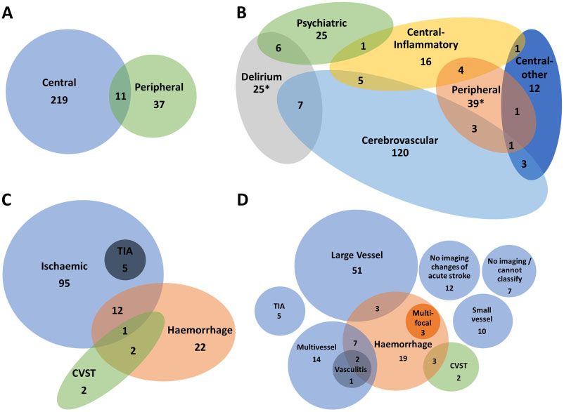
2   | BRAIN COMMUNICATIONS 2021: Page 2 of 15                                                                        A. L. Ross Russell et al.

and a high admission white cell count, which were independently associated with a poor outcome. In summary, this study describes the
spectrum of neurological and psychiatric conditions associated with COVID-19. In addition, we identify a severe COVID-19 encephal-
opathy atypical for delirium, and a phenotype of COVID-19 associated stroke in younger adults with a tendency for multiple infarcts
and systemic thromboses. These clinical data will be useful to inform mechanistic studies and stratification of patients in clinical trials.

1 NIHR Southampton Clinical Research Facility and Biomedical Research Centre, University Hospital Southampton NHS Foundation
    Trust, Southampton SO16 6YD, UK
2 Department of Neurology, Wessex Neurological Centre, University Hospital Southampton NHS Foundation Trust, Southampton

                                                                                                                                                 Downloaded from https://academic.oup.com/braincomms/article/3/3/fcab168/6325504 by Catherine Sharp user on 05 October 2021
    SO16 6YD, UK
3 Clinical Neurosciences, Clinical and Experimental Sciences, Faculty of Medicine, University of Southampton, Southampton SO16 6YD, UK
4 Liverpool University Hospitals NHS Foundation Trust, Liverpool, L9 7AL, UK
5 Liverpool Clinical Trials Centre, University of Liverpool, Liverpool, L3 5TR, UK
6 Department of Health Data Science, University of Liverpool, Liverpool, L69 3BX, UK
7 Neuroradiology Department, Wessex Neurological Centre, University Hospital Southampton NHS Foundation Trust, Southampton,
    SO16 6YD, UK
8 Manchester Centre for Clinical Neurosciences, Geoffrey Jefferson Brain Research Centre, Manchester Academic Health Science
    Centre, Salford Royal Foundation Trust, Salford, M6 8HD, UK
9 Division of Cardiovascular Sciences, Lydia Becker Institute for Immunology and Inflammation, University of Manchester,
    Manchester, M13 9PL, UK
10 Institute of Psychiatry, Psychology, and Neuroscience, King’s College London, London, SE5 8AF, UK
11 Chelsea and Westminster NHS Foundation Trust, London, London, SW10 9NH, UK
12 MRC Centre for Neuromuscular Diseases, National Hospital for Neurology, London, WC1N 3BG, UK
13 UCL Queen Square Institute of Neurology, University College London, London, WC1N 3BG, UK
14 Encephalitis Society, Malton, Malton, YO17 7DT, UK
15 Department of Clinical Infection Microbiology and Immunology, Institute of Infection, Veterinary, and Ecological Sciences,
University of Liverpool, Liverpool, L7 3EA, UK
16 The National Institute for Health Research Health Protection Research Unit for Emerging and Zoonotic Infections, University of
Liverpool, Liverpool, L69 7BE, UK
17 Division of Anaesthesia, Department of Medicine, University of Cambridge, Cambridge, CB2 0SP, UK
18 Memory Assessment and Research Centre, Moorgreen Hospital, Southern Health Foundation Trust, Southampton, SO40 2RZ, UK
19 Department of Neurology, The Walton Centre NHS Foundation Trust, Liverpool, L9 7LJ, UK
20 Translational and Clinical Research Institute, Newcastle University, Newcastle, NE1 7RU, UK
21 Wellcome Centre for Mitochondrial Research, Newcastle University, Newcastle, NE2 4HH, UK
22 Department of Social Genetic and Developmental Psychiatry, King’s College London, London, SE5 8AF, UK
23 Department of Neuromuscular Diseases, University College London, London, WC1N 3BG, UK
24 National Hospital for Neurology and Neurosurgery, University College London Hospitals NHS Foundation Trust, London, WC1N 3BG, UK
25 Rehabilitation Department, Poole Hospital, University Hospitals Dorset NHS Foundation Trust, Poole, BH15 2JB, UK
26 Department of Neurology, North Middlesex University Hospital NHS Trust, London, N18 1QX, UK
27 Department of Neuroradiology, Imperial College NHS Healthcare Trust, London, W2 1NY, UK
28 Department of Neurology, Southmead Hospital, North Bristol NHS Trust, Bristol, S10 5NB, UK
29 Translational Health Sciences, Bristol Medical School, University of Bristol, Bristol, BS8 1TH, UK
30 Centre for Clinical Brain Sciences, University of Edinburgh, Edinburgh, EH16 4SB, UK
31 Nuffield Department of Clinical Neurosciences, University of Oxford, Oxford, OX3 9DU, UK
32 Laboratory of Molecular and Cell Biology, UCL, Gower St, King’s Cross, London, London, WC1E 6BT, UK
33 Department of Neurology, Alder Hey Children’s NHS Foundation Trust, Liverpool, Liverpool, L14 5AB, UK
34 Medical Research Council Clinical Trials Unit, Institute of Clinical Trials and Methodology, University College London, London,
WC1V 6LJ, UK
35 Institute for Global Health, University College London, London, WC1N 1EH, UK
36 Department of Neurology, Royal Victoria Infirmary, Newcastle, NE1 4LP, UK

Correspondence to: Ian Galea
Clinical Neurosciences, Clinical & Experimental Sciences, Faculty of Medicine
University of Southampton, Mailpoint 806, Level D, Southampton General Hospital
Southampton SO16 6YD, UK
E-mail: I.Galea@soton.ac.uk

Keywords: COVID-19; SARS-CoV-2; encephalopathy; stroke; neurology
Abbreviations: CRFs ¼ Case Record Forms; IQR ¼ interquartile range; mRS ¼ modified Rankin score; SSNAP ¼ Sentinel Stroke
National Audit Programme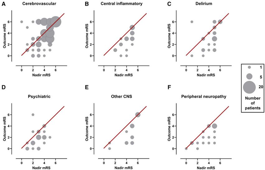
COVID-19 neurology                                                       BRAIN COMMUNICATIONS 2021: Page 3 of 15    | 3

Graphical Abstract

                                                                                                                           Downloaded from https://academic.oup.com/braincomms/article/3/3/fcab168/6325504 by Catherine Sharp user on 05 October 2021
Introduction                                                 information. This is further hampered by a lack of
                                                             unified diagnostic criteria and under-appreciation of over-
COVID-19 causes a multi-system disorder associated with      lapping presentations. Consequently, the factors predict-
a broad spectrum of neurological and neuropsychiatric        ing recovery remain poorly understood.
complications.1,2 Mild disease has been associated with        To address these gaps, we conducted a UK-wide
neurological symptoms, such as headache, anosmia and         surveillance study of neurological and psychiatric compli-
ageusia1,3 without major neurological complications.4        cations of COVID-19 (March–October 2020). National
Approximately 10–25% of patients hospitalized with           and cross-specialty recruitment was conducted to identify
COVID-19 present with or develop a significant neuro-        common and rarer presentations, and incorporated rigor-
logical disorder,4–8 the risk of which may increase with     ous clinical case definitions to evaluate overlapping
disease severity.1,9 Complications may reflect para- or      neurological presentations and determine the factors asso-
post-infectious central and peripheral immune-mediated       ciated with recovery. In this paper, we first deliver an
syndromes, or rarely direct CNS infection.10,11 We are at    overview of the main neurological and psychiatric mani-
the early stages of understanding the impact of these        festations encountered. Then we present more detail on
neurological complications of COVID-19.                      each category of disorder and perform analyses to try to
  As neurological complications are varied and occur         deliver insight into prognosis and underlying disease
throughout the disease course, multiple mechanisms have      mechanisms.
been proposed. These may include direct viral infection
of endothelium via angiotensin converting enzyme-2
receptors, systemic inflammation resulting in coagulop-
athy, cytokine toxicity, blood–brain barrier disruption,     Materials and methods
antibody and cell-mediated autoimmunity and consequen-
ces of prolonged severe illness.2,12–15 These suggested
                                                             Study design
pathological processes may co-exist, act synergistically     Physicians were invited to complete standardized electron-
and occur simultaneously in different parts of the nervous   ic Case Record Forms (CRFs) by the five major profes-
system, causing overlapping clinical presentations.          sional neuroscience associations in the UK (Association of
  Studies reporting neurological complications of COVID-     British Neurologists, British Association of Stroke
19 have successfully met the pressing need to disseminate    Physicians, Royal College of Psychiatrists, the Neuro
data rapidly to inform pandemic management and re-           Anaesthesia and Critical Care Society, and the Intensive
search efforts. However, this speed has limited geograph-    Care Society). This study was approved by the University
ical reach, so there is a paucity of nationwide studies      of Liverpool (UoL #7725/2020) and the University of
and limited detailed clinical diagnostic and prognostic      Southampton (ERGO #56504). The British Peripheral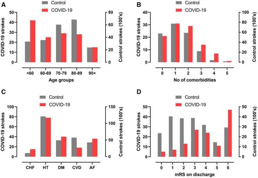
4   | BRAIN COMMUNICATIONS 2021: Page 4 of 15                                                         A. L. Ross Russell et al.

Nerve Society’s surveillance study for Guillain–Barré        protein > 0.45 g/dl, and/or neuroimaging consistent in-
syndrome was performed independently,16 but the case          flammation and/or demyelination).2 For psychiatric disor-
definitions and data fields were aligned to enable inclu-     ders, CRFs were assessed by a sub-specialty team of
sion. Four cases were published as single case studies        senior psychiatrists (co-authors TN and TP). Delirium
(Supplementary Table 1). The UK Health Research               was defined in accordance with the DSM-5 and the Ten
Authority advised that the study did not require review       Societies position statement20: (i) new-onset disturbance
by a NHS Research Ethics Committee as this was a sur-         in attention, awareness and cognition, developing over
veillance study with non-identifiable information.            hours or days, with some fluctuation, not in the context
   The CRF included demographics, evidence of SARS-           of a severely reduced level of arousal, such as coma, and

                                                                                                                                  Downloaded from https://academic.oup.com/braincomms/article/3/3/fcab168/6325504 by Catherine Sharp user on 05 October 2021
CoV-2 infection, neurological and non-neurological clinic-    not secondary to medication or substance misuse; and (ii)
al features, pre-morbid Rockwood frailty score,17 comor-      encephalopathy attributable to fever/sepsis, and/or hyp-
bidities and medications on admission, risk factors for       oxia–ischaemia. Therefore, severe encephalopathy was
stroke, respiratory disease course, requirement for inten-    defined as those with a severely reduced level of arousal
sive care, laboratory/imaging results and modified Rankin     (a Glasgow coma score 13/15 and/or seizures).
score (mRS).18 The mRS was captured at two time               Psychiatric presentations were considered a primary diag-
points: at nadir and at discharge from hospital or the        nosis if there was no evidence of an explanatory neuro-
first follow-up assessment visit. The mRS was selected for    logical disorder (e.g. psychosis without encephalitis/
several reasons. In view of the expected heterogeneity of     delirium). When multiple psychiatric diagnoses were
neurological conditions, no single scale would have been      reported, the primary diagnosis was ascertained in ac-
considered optimal, and the consensus view was that the       cordance with Bedford’s hierarchical model,21 which pla-
anticipated high proportion of strokes, familiarity of most   ces psychiatric conditions in the following order of
clinicians with the mRS and the ease of its administration    primacy: organic disorders (including neurocognitive dis-
made the mRS the best candidate. The CRF was hosted           order), followed by psychotic disorders, followed by
on ALEA through the Clinical Information Research Unit        mood disorders, followed by anxiety disorders, and final-
at the University of Southampton. Data lock was 14            ly personality/behavioural disorders. Peripheral neuropa-
October 2020.                                                 thies were cases involving the peripheral nervous system
                                                              and categorized as inflammatory and non-inflammatory,
                                                              on the basis of the reported diagnosis and whether in-
Inclusion criteria                                            flammation is the sole recognized pathophysiological
Physicians were invited to complete a CRF for any adult       cause of this diagnosis; for example, Guillain–Barré syn-
patient (18 years) hospitalized with a neurological or       drome is an archetypal inflammatory neuropathy, when
psychiatric presentation and COVID-19, or else develop-       compared to critical illness neuromyopathy.
ing these conditions whilst in hospital with COVID-19.           When cases met multiple clinical case definitions, the
Using World Health Organization criteria, cases were          primary definition was determined by blinded adjudica-
defined as ‘confirmed COVID-19’ if polymerase chain re-       tion of the CRF data by three groups of senior authors
action (PCR) of respiratory samples or CSF was positive,      representing neurology, psychiatry and stroke. Discrete
or serology was positive for anti-SARS-CoV-2 antibodies.      clinical case definitions reported in the same patient were
Cases were defined as ‘probable COVID-19’ if a chest          considered ‘overlapping syndromes’, for example,
radiograph or CT was consistent with COVID-19 but             Guillain–Barré syndrome and an ischaemic cerebrovascu-
PCR and serology were negative or not done. Finally,          lar event. When complications were consistent with the
cases were defined as ‘possible COVID-19’ if suspected        primary clinical case definition, such as haemorrhage in
on clinical grounds by the notifying clinician but PCR,       acute haemorrhagic leukoencephalopathy, the primary
serology and chest imaging were negative or not done,2        diagnosis sufficed.
or if these data were unavailable. Cases of nosocomial in-       Patients with stroke were compared with those from
fection following admission with a primary neurological       the national stroke audit [Sentinel Stroke National Audit
presentation were excluded.                                   Programme (SSNAP)] over a comparable period in the
                                                              preceding year (April—June 2019). Patients presenting
                                                              with cerebrovascular events below the age of 60 were
Clinical case definitions                                     compared with those presenting above the age of 60.
Patients were classified using standardized clinical case
definitions .2,19 Cerebrovascular events were defined as
symptoms, signs and/or neuroimaging consistent with
                                                              Statistical analysis
transient ischaemic attack, ischaemic or haemorrhagic         Statistical analysis was performed using SAS software
stroke, or intracranial venous thrombosis. Central inflam-    (version 9.4; SAS Institute, Cary, NC, USA), SPSS v26
matory conditions were defined as those involving the         (IBM) and GraphPad Prism v8.4.3 (GraphPad Software,
CNS, with evidence of meningeal, parenchymal or vascu-        LLC). Normality of distribution was assessed using
lar inflammation (CSF white cell count > 4/mm3, and/or        Kolmogorov–Smirnov tests. Data were analysed using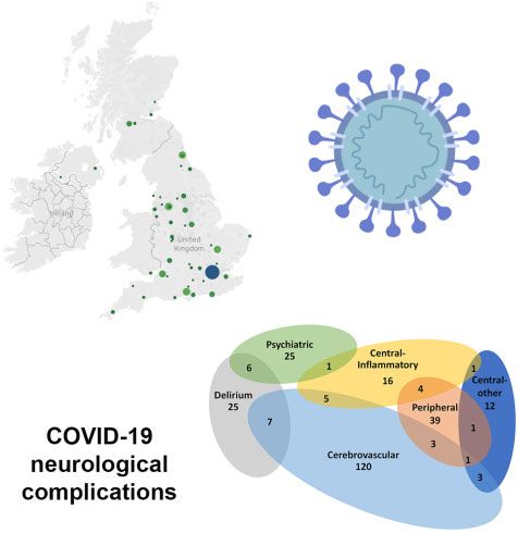
COVID-19 neurology                                                         BRAIN COMMUNICATIONS 2021: Page 5 of 15    | 5

descriptive statistics, group comparison tests, chi-squared    comprising 131 (49%) patients (Figs 1 and 2). The se-
tests, z-tests for independent proportions, and univariable    cond most common CNS groups were delirium (28,
logistic regression. A good outcome was defined as mRS         11%) and central inflammatory conditions (25, 9%); the
2 (reflecting no symptoms, slight disability, but independ-   latter comprising mostly demyelination and leukoence-
ent) and a poor outcome as mRS >2 (moderate disability         phalopathy, but also vasculitis, encephalitis, and opsoclo-
requiring assistance, or worse, including death).              nus–myoclonus syndrome (Figs 1 and 2). Psychiatric
Multivariable logistic regression models were developed        presentations (25, 9%) were most commonly new diagno-
using baseline pre-COVID-19 variables with >80% data           ses (19, 76%) but included six patients with an exacerba-
availability. Two sensitivity analyses were carried out for    tion of an underlying condition (24%). Those remaining

                                                                                                                              Downloaded from https://academic.oup.com/braincomms/article/3/3/fcab168/6325504 by Catherine Sharp user on 05 October 2021
each model, one adjusting for diagnostic categories, and       were all other encephalopathies (17, 7%), including 13
one using multiple imputation to account for the potential     with severe encephalopathy and four with posterior re-
effect of missing data. The imputation model used a fully      versible encephalopathy syndrome. The peripheral ner-
conditional specification and included the auxiliary varia-    vous system was primarily involved in 41 (15%) cases,
bles weight and mRS at nadir. All hypothesis testing was       of which 35 (85%) were inflammatory and six (15%),
two-tailed with alpha
6 | BRAIN COMMUNICATIONS 2021: Page 6 of 15 A. L. Ross Russell et al.
Table 1 Patient demographics and clinical characteristics
All patients
Demographics
Age in years, n (%) 20–29 6 (2)
30–39 15 (6)
40–49 35 (13)
50–59 57 (21)
60–69 51 (19)
70–79 50 (19)
Downloaded from https://academic.oup.com/braincomms/article/3/3/fcab168/6325504 by Catherine Sharp user on 05 October 2021
80–89 36 (14)
>90 17 (6)
Sex, n (%) Male 172 (64)
Female 95 (36)
Ethnicity, n (%) Asian 23 (9)
Black 21 (8)
White 196 (73)
Mixed 3 (1)
Unknown 24 (9)
COVID diagnosis, n (%) Confirmed or probable 239 (90)
Possible 28 (10)
Clinical characteristics
ICU admission, n (%) Yes 76 (28)
No 171 (64)
Unknown 20 (8)
Ventilation required, n (%) None 165 (62)
NIV 15 (6)
Invasive 67 (25)
Unknown 20 (7)
Pre-COVID-19 frailty score, median (IQR) 3 (2–5)
At least one co-morbidity, n (%) 196 (81)
Type of co-morbidity, n (%) Any neurological 66 (28)
Any psychiatric 22 (10)
Hypertension 125 (48)
Diabetes mellitus 63 (24)
Atrial fibrillation 43 (18)
Congestive heart failure 19 (10)
Previous TIA/stroke 25 (13)
Number of co-morbidities, median (IQR) 2 (1–4)
Admission GCS, median (IQR) 15 (14–15)
Fever, n (%) 172 (73)
Admission WCC, median (IQR) 8 (6–12)
Admission CRP, median (IQR) 41 (9–140)
Any non-neurological, non-respiratory systemic complication, n (%) 101 (42)
mRS at nadir, median (IQR) 4 (3–5)
mRS at outcome, median (IQR) 3 (2–5)
Improvement in mRS score, n (%) 125 (53)
Admission length in days, median (IQR) 23 (7–48)
Death, n (%) 57 (24)
mRS refers to modified Rankin Scale. Pre-COVID-19 frailty score refers to Rockwood frailty score. For definition of medically significant co-morbidities, see Supplementary
methods. Improvement in mRS score was defined as mRS at outcome < mRS at nadir, or mRS score of 0 at both nadir and outcome.
versus 11, 15%) and more non-neurological thrombotic CSF was positive for SARS-CoV-2. Nine cases (43%)
events (6, 18% versus 8, 8%) (Supplementary Table 3). needed ventilation and had acute kidney injury, of which
seven (78%) required renal replacement therapy.
Central nervous system
inflammatory conditions Delirium
The most common complication in the central inflamma- Delirium had a bimodal age distribution, the first peak at
tory group was leukoencephalopathy, affecting 13 (52%) 30–39 years (4, 14%) (Supplementary Table 4). Relative
cases. Encephalitis was reported in three; in one PCR of to the rest of the cohort delirium was not significantlyCOVID-19 neurology BRAIN COMMUNICATIONS 2021: Page 7 of 15 | 7
Downloaded from https://academic.oup.com/braincomms/article/3/3/fcab168/6325504 by Catherine Sharp user on 05 October 2021
Figure 1 Classification of main neurological diagnoses.
associated with established risk factors, such as age, median (IQR) duration of ventilation of 11 (0–36) versus
markers of systemic inflammation and intensive care 0 (0–13) days.
(Supplementary Table 5). There were six cases that met
both delirium and psychiatric diagnostic criteria, of which
three were8 | BRAIN COMMUNICATIONS 2021: Page 8 of 15 A. L. Ross Russell et al.
Downloaded from https://academic.oup.com/braincomms/article/3/3/fcab168/6325504 by Catherine Sharp user on 05 October 2021
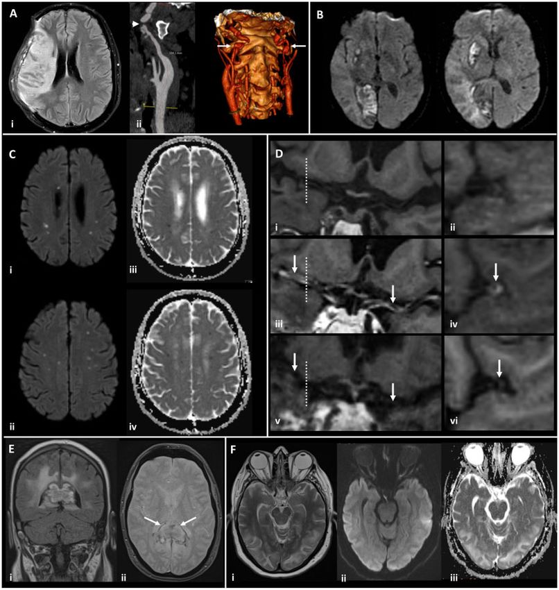
Figure 2 Magnetic resonance imaging demonstrating the range of neurological complications seen in this study. (A) Territorial
infarct, secondary to internal carotid artery (ICA) dissection in a middle-aged previously fit male: Axial fluid-attenuated inversion recovery image
(i) showing a right middle cerebral artery (MCA) territory infarct following decompressive craniectomy for malignant MCA syndrome despite
treatment with thrombolysis. Reformatted images from a CT angiogram (ii) showing irregularity of the extracranial segment of both internal
carotid arteries, consistent with dissection (arrows), with tight stenosis of the true lumen on the right (arrowhead). (B) Multiple territorial
infarcts in a female >60 years old with hypertension and dyslipidaemia: Diffusion-weighted images (DWI) demonstrate recent infarcts in the right
medial occipital lobe and lentiform nucleus, involving the territories of the right posterior cerebral artery and lenticulo-striate perforators of the
right MCA respectively. (C) Acute lacunar infarcts due to small vessel vasculopathy in a male > 60 years old, with a background of hypertension
and type 2 diabetes: B1000 images (i, ii) and corresponding apparent diffusion coefficient (ADC) maps (iii, iv) from DWI showing multiple tiny
foci of restricted diffusion. (D) Vasculitis in a male >60 years old, with a background of type 2 diabetes, hypertension and hypercholesterolaemia:
T1-weighted SPACE vessel wall imaging of both distal ICAs and proximal MCAs, with curved multiplanar coronal reconstructions along the
course of both proximal MCAs (first column) and perpendicular to the right MCA (second column, at the position of the dotted line).
Pre-treatment pre-contrast (i, ii) and post-contrast images (iii, iv) demonstrate abnormal concentric, long segment vessel wall enhancement
(arrows) of both proximal MCAs. Post-contrast images after treatment with prednisolone and tocilizumab (v, vi) demonstrate treatment
response with resolution of the previous abnormal mural MCA enhancement (arrows). (E) Acute encephalomyelitis with haemorrhage in a
middle-aged male, with a history of chronic obstructive pulmonary disease, who required intensive care and haemofiltration: Coronal FLAIR (i)
and axial gradient echo (ii) images showing focal heterogeneous signal abnormality and swelling of the splenium of the corpus callosum, with
peripheral low signal indicative of haemosiderin staining (arrows). Confluent high signal is present in periventricular and deep white matter of the
parieto-occipital region. (F) Typical imaging appearances of posterior reversible encephalopathy syndrome in a normotensive middle-aged female:
Axial T2 image (i) demonstrating hyperintense signal in subcortical white matter of both occipital lobes, with B1000 image (ii) and ADC map (iii)
from DWI showing no corresponding restricted diffusion.COVID-19 neurology BRAIN COMMUNICATIONS 2021: Page 9 of 15 | 9
Downloaded from https://academic.oup.com/braincomms/article/3/3/fcab168/6325504 by Catherine Sharp user on 05 October 2021
Figure 3 Venn diagrams showing overlap of diagnostic groups. The numbers shown here are when all diagnoses were considered, in
addition to the primary neurological diagnosis. The total numbers for several groups are larger in this Figure than the primary diagnosis
flowchart (Fig. 1) due to coexisting diagnoses. (A) Central and peripheral nervous system disease. (B) Primary diagnostic categories (*two cases
of Guillain–Barré syndrome with delirium were not possible to accommodate on this diagram). (C) Stroke group subtypes. (D) Specific stroke
group subtypes. CVST, cerebral venous sinus thrombosis; TIA, transient ischaemic attack.
Cerebrovascular events were associated with the earliest (39%, P < 0.001), while central inflammatory conditions
onset, with median (IQR) time from respiratory symp- improved most (77%, P < 0.03).
tom onset to cerebrovascular event of 7.5 (2–16) days. Multivariable analysis using baseline variables easily
Interestingly, longer time to onset was observed in the available at admission, demonstrated a higher probability
central inflammatory, psychiatric and peripheral neur- of a poor outcome (mRS 2) with older age, a higher
opathy diagnostic categories. Rockwood frailty score and higher white cell count on
admission. In comparison, the association of outcome
with individual neurological diagnostic categories was
negligible. A similar pattern was observed with mortality
Clinical outcome and risk factors (Table 2).
Outcome mRS was assessed at a median (IQR) follow-up
time of 30 days (7–60). This was at hospital discharge
(48%), as an inpatient (22%) or an outpatient (29%).
Patients in this study were substantially disabled, since
Discussion
131 (56%) had an outcome mRS of 2–5; moreover, 57 Through a nationwide surveillance study of adults hospi-
(24%) patients died. Outcome was assessed in three talized with COVID-19, conducted through a cross-spe-
ways: whether mRS improved (mRS at outcome versus cialty collaboration spanning six national physician
mRS nadir), mRS at outcome and death. associations, we present the broad spectrum of potential
Improvement in outcome mRS relative to the mRS neurological and psychiatric complications of COVID-19,
score at nadir of illness was seen in all primary diagnos- across central and peripheral nervous systems. Our results
tic categories other than cerebrovascular events (Fig. 6). build on existing knowledge,1,4–8,10–12,22 by applying
There was a significant difference in mRS improvement standardized, internationally agreed, a priori clinical case
across diagnostic groups (Supplementary Table 7, definitions and independent, blinded case adjudication to
P < 0.001). Cerebrovascular events improved the least determe specific diagnostic group membership, and by10 | BRAIN COMMUNICATIONS 2021: Page 10 of 15 A. L. Ross Russell et al.
Downloaded from https://academic.oup.com/braincomms/article/3/3/fcab168/6325504 by Catherine Sharp user on 05 October 2021
Figure 4 COVID-19 strokes versus historical controls. Comparison between strokes associated with COVID-19 in this study and strokes
from a national UK audit in 2019. (A, B) total number of co-morbidities which are risk factors for stroke (atrial fibrillation, hypertension,
diabetes mellitus, congestive heart failure and previous TIA or stroke). (C) Age distributions. (D) mRS (modified Rankin scale) scores on
discharge from hospital (or death).
presenting detail on the overlap between clinical presenta- encephalopathy, in settings where asymptomatic testing is
tions. We provide further evidence of a coagulopathy pre- not routine. The later presentation of central inflamma-
cipitating stroke in young patients, occurring in the para- tory and peripheral nerve presentations, after respiratory
infectious phase of illness, and suggest this group is dis- recovery, and the high rates of improvement seen in these
tinct to older patients with multiple conventional risk fac- groups, supports a post-infectious process, driven by an
tors. Nevertheless, despite a younger cohort of patients adaptive immune response.
with COVID-19 associated stroke compared to non-
COVID-19 stroke patients, conventional, often modifi-
able, risk factors were more frequent even in younger Possible mechanisms underlying
patients. peri-COVID-19 stroke
Our comparison with pre-COVID-19 SSNAP data
Timing of onset identified higher rates of young stroke in our COVID-19
Onset of neurological disease, in days relative to respira- cohort, despite reports of a reduction in overall stroke
tory symptoms, varied across different diagnostic catego- admissions during the pandemic.24 The underlying mech-
ries. In 29% of cases, neurological symptoms preceded anisms leading to stroke may differ between younger and
respiratory symptoms, suggesting occurrence during the older cases, as younger strokes had a significantly delayed
virological, or para-infectious phase, the early part of presentation, were associated with fewer comorbidities,
which is usually asymptomatic.23 This supports early and demonstrated higher rates of both multi-vessel occlu-
mechanisms, such as activation of the innate immune sys- sion and of thrombotic complications outside of the
tem and direct viral effects on endothelial cells. Within CNS. These findings are supportive of a para-infectious
the context of a pandemic, neurological syndromes thrombo-inflammation, potentially driven by endothelitis
described in this study could be a sentinel sign of and subsequent cytokine release, and in line with
COVID-19, and we encourage SARS-CoV-2 testing of previous reports of elevated serum markers of coagulop-
patients with neurological presentations, including acute athy in stroke patients.25 Early administration ofCOVID-19 neurology BRAIN COMMUNICATIONS 2021: Page 11 of 15 | 11
illness in the absence of severe hypoxia, and suggests
COVID-19 confers additional risk compared to other
infections. In addition to delirium, we identified distinct
aetiological groups, including posterior reversible enceph-
alopathy syndrome and a severe encephalopathy outside
the accepted definition of delirium.20 This latter syn-
drome may represent excitotoxic injury, such as is seen
following seizures, metabolic disturbance, or an underly-
ing inflammatory or microvascular process. The greater
Downloaded from https://academic.oup.com/braincomms/article/3/3/fcab168/6325504 by Catherine Sharp user on 05 October 2021
need for intensive care in this group may represent both
the cause (exposure to potentially ictogenic medications)
and the consequence of seizures. Indeed, multiple overlap-
ping disease mechanisms may be apparent, even within
an individual patient, and further studies are underway
to evaluate this (COVID-CNS).
Clinical outcome
Age and a higher Rockwood frailty score were much
more indicative of outcome than the neurological or psy-
chiatric disorder. It is interesting that the adjusted hazard
ratio of age for outcome (death or hospitalisation) is
10-fold that of neurological disorders.31 Individual disease
groups were heterogeneous and did not demonstrate sig-
nificantly different outcomes, but this requires further
Figure 5 Timing of onset of neurology. Violin plot study in larger cohorts. Ongoing assessment of the pre-
demonstrating distributions of time intervals in days between onset
dictive power of premorbid frailty will be important as
of respiratory symptoms and onset of neurological symptoms for
we see increasing numbers of young people affected. Poor
each primary diagnostic category. Patients whose neurological
symptoms preceded COVID-19 symptoms were arbitrarily outcome also associated with a high admission white cell
assigned a value of minus seven days. The Kruskal–Wallis test was count, which might be a useful predictor given that this
used to determine any significant difference in time intervals is usually normal in the early stages of COVID-19.32
between groups (P < 0.0001). Dunn’s multiple group comparison
test showed a significant difference between stroke and central
inflammatory primary diagnostic groups (P ¼ 0.001), stroke and Limitations
psychiatric groups (P ¼ 0.037), and stroke and peripheral groups This study has several limitations. It concerns a specific
(P ¼ 0.003). population of COVID-19 positive patients requiring ad-
mission to hospital because of neurological or psychiatric
conditions or else developing these complications whilst
in hospital with COVID-19. Hence it would not have
anti-inflammatory therapy has potential benefit, and our
captured neurological symptoms, such as headache, anos-
data strengthen the case for trials to consider stroke out-
mia, dysgeusia and mild cognitive dysfunction, generally
comes so that this effect can be evaluated. In older cases,
looked after in the community or in outpatient services.
where the highest risk is in the initial days of symptoms,
Although this study captured data from a breadth of dif-
it is likely that COVID-19 is precipitating stroke similarly
ferent specialties, there was still under-representation of
to other acute respiratory infections, through interaction
psychiatrists, primary care and internal medicine which
with existing cerebrovascular risk factors.26
may have skewed the study population to more severe
cases. The mRS was used as a clinical outcome measure
but this scale has not been validated for its measurement
Encephalopathy properties across the wide spectrum of conditions studied
Encephalopathy is widely reported in COVID-197,27,28 here. We did not review primary clinical data held locally
and, in an undifferentiated form, has been demonstrated by the referring physician, and could not perform an
to be an independent predictor of death and poorer func- independent review of data submission quality. Where
tional recovery in survivors.9,29 However, there is a lack possible the original syndromic classification from the
of consensus as to the distinct underlying pathophysi- referring team was presumed to be correct. On rare occa-
ology.30 The presentation of delirium in younger patients, sions, there was a reclassification of cases based on
seen frequently in our cohort, is unusual for a respiratory senior author panel review.12 | BRAIN COMMUNICATIONS 2021: Page 12 of 15 A. L. Ross Russell et al.
Downloaded from https://academic.oup.com/braincomms/article/3/3/fcab168/6325504 by Catherine Sharp user on 05 October 2021
Figure 6 Recovery from neurological condition. Bubble plots displaying the relationship between mRS (modified Rankin scale) at nadir of
illness whilst in hospital and mRS at outcome assessment, within individual diagnostic categories. Bubble area corresponds to patient number.
Line of equivalence is shown in red: cases below the line improved, cases above the line got worse, while cases on the line stayed the same.
Participation by physicians occurred during an unprece- our findings were similar to those of the SETICOS
dented healthcare and social emergency, during which study,33 which used a case–control design, provides some
clinical service and research teams were stretched. This validation of the approach used here.
has the potential for reporting bias, in particular under-
reporting of mild disease, and potential over-representa-
tion of unusual presentations. These circumstances are
also likely to have contributed to missing clinical data
Conclusions
fields (Supplementary Table 8). Although not the largest In summary, this nationwide, cross-specialty study of
study to date,6 this study provides both granularity of de- neurological and psychiatric manifestations of COVID-19,
tail and breadth of sub-specialty input. We included cases has identified older age and a higher pre-COVID-19
from the very beginning of the pandemic and PCR con- frailty score to be associated with poor outcome, and the
firmation of COVID-19 was not always present, though effect of these baseline characteristics overshadowed the
COVID-19-associated neurology was an inclusion effects of specific neurological diagnoses. Presentations
criterion. spanned pre-symptomatic, early and later phases of
The SSNAP database is a high-quality rolling national COVID-19, implying different pathophysiological proc-
audit, which captures routine, consecutive, unselected esses may occur, and these may act synergistically in
stroke cases and has a low risk of bias; there is good driving neurological complications. Cerebrovascular
overlap with essential data fields in our study since events were the most common complication and, in
SSNAP is so comprehensive. We selected a SSNAP data- young as opposed to older patients, COVID-19-associated
base time period from the year preceding the pandemic, events occurred later after respiratory symptom onset,
which was comparable both in season and duration to supportive of thrombo-inflammation and systemic coagul-
our study. However, the comparability of these two opathy, and this requires further study. A severe enceph-
cohorts has to be guarded with caution, due to the po- alopathy beyond the clinical definition of delirium occurs
tential for selection biases in our cohort. The fact that during COVID-19. Future work must focus on longerCOVID-19 neurology BRAIN COMMUNICATIONS 2021: Page 13 of 15 | 13
Table 2 Clinical outcome
Complete case model: variables Complete case model: adjusting Multiple imputation
available at admission for diagnostic variables model
OR (95% CI) P-value OR (95% CI) P-value OR (95% CI) P-value
Outcome variable: MRS score at outcome >2
Age (10-year age groups) 1.67 (1.26, 2.22)14 | BRAIN COMMUNICATIONS 2021: Page 14 of 15 A. L. Ross Russell et al.
Z/20/Z). T.S. is supported by the National Institute for Health Marcia McDougall, Caroline Mcinnes, David McKee, Isabel
McMullen, Brian Menezes, Stephanie Miers, Thomas Minton, Amulya
Research Health Protection Research Unit (NIHR HPRU) in
Misra, Dipak Mistry, James Mitchell, Hartmann Monika, Mireia
Emerging and Zoonotic Infections at University of Liverpool
Moragas, Hamish Morrison, Walied Mowafi, Paul Mudd, Louis
(IS-HPU-1112–10117 and NIHR200907), and National Murphy, Anna Nagy, Edward Newman, Choo Ng, Sam Nightingale,
Institute for Health Research Global Health Research Group Khin Nyo, Suzanne O’Neil, Richard O’Brien, Ivy Ong, Matt Oram,
on Brain Infections (17/63/110), and the European Union’s Belgin Ozalp, Stella-Marie Paddick, Kevin Pankhurst, Nehal Parmar,
Horizon 2020 research and innovation program ZikaPLAN Carmen Parr, Kath Pasco, Sarah Pearce, Gordon Plant, David Price,
(Preparedness Latin America Network; 734584). S.P. receives Gary Price, Jane Pritchard, Nicholas Pritchard, Harald Proeschel,
salary support from Medical Research Council core funding David Protheroe, Terence Quinn, Akansha Rajan, Jessica Redgrave,
Downloaded from https://academic.oup.com/braincomms/article/3/3/fcab168/6325504 by Catherine Sharp user on 05 October 2021
(MC_UU_12023/23 and MC_UU_12023/26). Rebecca Redwood, Chris Rickards, Christina Roffe, Jonathan Rogers,
Alia Roof, Amrit Sachar, Neshika Samarasekera, Amal Samaraweera,
Stephen Sawcer, Shona Scott, Lakshmanan Sekaran, Jordi Serra-
Competing interests Mestres, Kanchan Sharma, Pamela Shaw, Mohammed Siddiqui, Emily
Simon Thomas, Fai Lam Chun Chiang Sin, Andrew Sissons, Mara
T.S. was Chair/Co-Chair of the United Kingdom Research Sittampalam, Anushta Sivananthan, Thandar Soe, Mohamed Soliman,
and Innovation / National Institute for Health Research Andreas Sotiriou, Aginor Spanoulis, Jon Stone, James Sun, Peter
COVID-19 Rapid Response and Rolling Funding Initiatives, Swann, Hafiz Syed, Konrad Szewczyk-Krolikowski, Leonie Taams,
was an Advisor to the UK COVID- 19 Therapeutics Amr Tageldin, Maryam Talaei, Emma Tallantyre, Riffat Tanveer, Lisa
Advisory Panel and is a member of the UK Medicines and Templeton, Philip Thomas, Catherine Trezise, David Turner, Jaap van
der Boom, Elizabeth Varghese, Angela Vincent, Briony Waddell, Tom
Healthcare Products Regulatory Agency COVID-19
Walker, Stephen Webb, Nic Weir, Chris Wharton, Lou Wiblin,
Vaccines Benefit Risk Expert Working Group.
Malcolm Wiggam, Tim Williams, Greta Wood, Nicholas Wood,
Charmaine Yam.
Appendix
For the purposes of this paper, the CoroNerve Studies Group includes
all the authors and Nisha Abraham-Thomas, Katja Adie, Claire Allen,
References
Nabeel Amiruddin, Heather Angus-Leppa, Fahim Anwar, Neil 1. Mao L, Jin H, Wang M, et al. Neurologic manifestations of hospi-
Archibald, James Arkell, James Armitage, Cherie Armour, Peter talized patients with coronavirus disease 2019 in Wuhan, China.
Arthur-Farraj, Marc Atkin, Mark Baker, Nawar Bakerly, Rajaram JAMA Neurol. 2020;77(6):683–690.
Bathula, Alexandra Belcher, Benson Benjamin, Viraj Bharambe, 2. Ellul MA, Benjamin L, Singh B, et al. Neurological associations of
Gordon Blair, Catrin Blank, Jim Bolton, Michael Bonello, Iryna COVID-19. Lancet Neurol. 2020;19(9):767–783.
Boubriak, Claire Boynton, David Breen, Robert Brenner, Dennis 3. Lechien JR, Chiesa-Estomba CM, Place S, et al. COVID-19 Task
Briley, Fiona Brodie, Helga Brown, Stefania Bruno, Ed Bullmore, Force of YO-IFOS. Clinical and epidemiological characteristics of
1420 European patients with mild-to-moderate coronavirus dis-
Angus Butchart, Hannah Castell, Kah Lok Chan, Bharath Cheripelli,
ease 2019. J Intern Med. 2020;288(3):335–344.
Patrick Chinnery, James Choulerton, Philip Clatworthy, Joanne
4. Cagnazzo F, Arquizan C, Derraz I, et al. Neurological manifesta-
Clements, Jonathon Coates, Alasdair Coles, Ceryce Collie, Gwen tions of patients infected with the SARS-CoV-2: A systematic re-
Collin, Peter Cottrell, Charles Coughlan, Sarah Crisp, Mazen Daher, view of the literature. J Neurol. 2021;268(8):2656–2665.
Jane Dale, Ruth Davies, Sylviane Defres, Sofia Dima, Katherine Dodd, 5. Romero-Sánchez CM, Dı́az-Maroto I, Fernández-Dı́az E,
Liam Dodge, Fergus Doubal, Cordelia Dunai, Ahilanadan et al. Neurologic manifestations in hospitalized patients with
Dushianthan, Dipankar Dutta, Richard Ellis, Salwa Elmamoun, COVID-19: The ALBACOVID registry. Neurology. 2020;95(8):
Hannah Emerson, Bethany Facer, Patricia Fearon, Peter Fernandes, e1060–e1070.
Barnaby Fiddes, James Firth, Emma Fisher, Leonora Fisniku, Alasdair 6. Frontera JA, Balcer L, Galetta S. A prospective study of neurologic
Fitzgerald, Enrico Flossman, Caroline Fornolles, Andrew Gallagley, disorders in hospitalized COVID-19 patients in New York City.
Neurology. 2021;96(11):549–550.
Brian Gallen, Garcia del Carrizo Fernando, Alan Gemski, Jackie
7. Meppiel E, Peiffer-Smadja N, Maury A, et al. contributors to the
Gilbert, Effrossyni Gkrania-Klotsas, Farhad Golestani, Bruno Gran,
NeuroCOVID registry. Neurologic manifestations associated with
Andrew Gratrix, Susan Green, Helen Grote, Rebecca Grue, Sabine COVID-19: A multicentre registry. Clin Microbiol Infect. 2021;
Grundler, Alexander Grundmann, Savini Gunatilake, Mahir Hamad, 27(3):458–466.
Khalid Hamandi, Hisham Hamdalla, Kirsty Harkness, Neil Harrison, 8. Rifino N, Censori B, Agazzi E, et al. Neurologic manifestations in
Timothy Harrower, Jennifer Hartman, Ahamad Hassan, Ghaniah Zeb 1760 COVID-19 patients admitted to Papa Giovanni XXIII
Hassan-Smith, Alison Hatfield, Catherine Hatfield, Charles Hillier, Hospital, Bergamo, Italy. J Neurol. 2021;268(7):2331–2338.
Akram Hosseini, Gary Hotton, Jack Hubbett, Saif Huda, Nathan 9. Liotta EM, Batra A, Clark JR, et al. Frequent neurologic manifes-
Huneke, Caroline Hutchison, Anne-Catherine Huys, Thomas Ian, tations and encephalopathy-associated morbidity in Covid-19
Hajira Iftikhar, Ihmoda Ihmoda, Muhammad Ilyas, Sissi Ispoglou, patients. Ann Clin Transl Neurol. 2020;7(11):2221–2230.
10. Paterson RW, Brown RL, Benjamin L, et al. The emerging spec-
Tom Jenkins, Nicola Jones, Ingrid Kane, Ryan Keh, Hind Khalifeh, Jeff
trum of COVID-19 neurology: Clinical, radiological and labora-
Kimber, Amit Kishore, Martin Knolle, Christopher Kobylecki, Sander
tory findings. Brain. 2020;143(10):3104–3120.
Kooij, Anita Krishnan, Matthew Lambert, Gavin Langlands, Phil 11. Varatharaj A, Thomas N, Ellul MA, et al. Neurological and
Laws, Charles Leek, Lucia Li, George Lilly, Rebecca Luxton, Graham neuropsychiatric complications of COVID-19 in 153 patients: A UK-
Mackay, Barbara Madigan, Melissa Maguire, Azer Majeed, Arshad wide surveillance study. Lancet Psychiatry. 2020;7(10):875–882.
Majid, Gauhar Malik, Mark Manford, Richard Marigold, Sarah 12. Wang L, Shen Y, Li M, et al. Clinical manifestations and evidence
Marrinan, Monica Marta, Paul Matthews, Michael McCormick, of neurological involvement in 2019 novel coronavirus SARS-COVID-19 neurology BRAIN COMMUNICATIONS 2021: Page 15 of 15 | 15
CoV-2: A systematic review and meta-analysis. J Neurol. 2020; Presymptomatic SARS-CoV-2 infections and transmission in a
267(10):2777–2789. skilled nursing facility. N Engl J Med. 2020;382(22):2081–2090.
13. Violi F, Pastori D, Cangemi R, Pignatelli P, Loffredo L. 24. Kansagra AP, Goyal MS, Hamilton S, Albers GW. Collateral effect
Hypercoagulation and antithrombotic treatment in coronavirus of Covid-19 on stroke evaluation in the United States. N Engl J
2019: A new challenge. Thromb Haemost. 2020;120(6):949–956. Med. 2020;383(4):400–401.
14. Beyrouti R, Adams ME, Benjamin L, et al. Characteristics of is- 25. Li Y, Li M, Wang M, et al. Acute cerebrovascular disease follow-
chaemic stroke associated with COVID-19. J Neurol Neurosurg ing COVID-19: A single center, retrospective, observational study.
Psychiatry. 2020;91(8):889–891. Stroke Vasc Neurol. 2020;5(3):279–284.
15. Bastard P, Rosen LB, Zhang Q, et al. COVID Human Genetic 26. Smeeth L, Thomas SL, Hall AJ, Hubbard R, Farrington P,
Effort. Autoantibodies against type I IFNs in patients with life- Vallance P. Risk of myocardial infarction and stroke after acute in-
threatening COVID-19. Science. 2020;370(6515):eabd4585. fection or vaccination. N Engl J Med. 2004;351(25):2611–2618.
Downloaded from https://academic.oup.com/braincomms/article/3/3/fcab168/6325504 by Catherine Sharp user on 05 October 2021
16. Keddie S, Pakpoor J, Mousele C, et al. Epidemiological and cohort 27. Helms J, Kremer S, Merdji H, et al. Delirium and encephalopathy
study finds no association between COVID-19 and Guillain-Barré in severe COVID-19: A cohort analysis of ICU patients. Crit Care.
syndrome. Brain. 2021;144(2):682–693. 2020;24(1):491.
17. Rockwood K, Song X, MacKnight C, et al. A global clinical meas- 28. Agarwal P, Ray S, Madan A, Tyson B. Neurological manifesta-
ure of fitness and frailty in elderly people. CMAJ. 2005;173(5): tions in 404 COVID-19 patients in Washington State. J Neurol.
489–495. 2020;268(3):770–772.
18. Quinn TJ, Dawson J, Walters MR, Lees KR. Reliability of the 29. Khan SH, Lindroth H, Perkins AJ, et al. Delirium incidence, dur-
modified rankin scale: A systematic review. Stroke. 2009;40(10): ation, and severity in critically ill patients with coronavirus disease
3393–3395. 2019. Crit Care Explor. 2020;2(12):e0290.
19. Sacco RL, Kasner SE, Broderick JP, et al. Council on Nutrition, 30. Varatharaj A, Pollak TA, Nicholson TR, et al.; study authors.
Physical Activity and Metabolism. An updated definition of stroke Characterising neuropsychiatric disorders in patients with COVID-
for the 21st century. Stroke. 2013;44(7):2064–2089. 19 - Authors’ reply. Lancet Psychiatry. 2020;7(11):934–935.
20. Slooter AJC, Otte WM, Devlin JW, et al. Updated nomenclature 31. Clift AK, Coupland CAC, Keogh RH, et al. Living risk prediction
of delirium and acute encephalopathy: Statement of ten societies. algorithm (QCOVID) for risk of hospital admission and mortality
Intens Care Med. 2020;46(5):1020–1022. from coronavirus 19 in adults: National derivation and validation
21. Foulds GA, Bedford A. Hierarchy of classes of personal illness. cohort study. BMJ. 2020;371:m3731.
Psychol Med. 1975;5(2):181–192. 32. Han R, Huang L, Jiang H, Dong J, Peng H, Zhang D., 2214/
22. Fraiman P, Godeiro Junior C, Moro E, Cavallieri F, Zedde M. AJR.20. Early clinical and CT manifestations of coronavirus dis-
COVID-19 and cerebrovascular diseases: A systematic review and ease 2019 (COVID-19) pneumonia. AJR Am J Roentgenol. 2020;
perspectives for stroke management. Front Neurol. 2020;11: 215(2):338–343.
574694. 33. Perry RJ, Smith CJ, Roffe C, et al. Characteristics and outcomes of
23. Arons MM, Hatfield KM, Reddy SC, et al. Public Health–Seattle COVID-19-associated stroke: A UK multicentre case-control study.
and King County and CDC COVID-19 Investigation Team. J Neurol Neurosurg Psychiatry. 2020;92(3):242–248.You can also read


