Imaging Inflammation and Infection in the Gastrointestinal Tract - MDPI
←
→
Page content transcription
If your browser does not render page correctly, please read the page content below
International Journal of
            Molecular Sciences

Review
Imaging Inflammation and Infection in the
Gastrointestinal Tract
Alex N. Frickenstein 1,2,† , Meredith A. Jones 1,2,† , Bahareh Behkam 3 and Lacey R. McNally 1,2,4, *
 1    Stephenson School of Biomedical Engineering, University of Oklahoma, Norman, OK 73019, USA;
      africk256@ou.edu (A.N.F.); meredith.jones@ou.edu (M.A.J.)
 2    Stephenson Cancer Center, University of Oklahoma, Oklahoma City, OK 73104, USA
 3    Department of Mechanical Engineering, Virginia Tech University, Blacksburg, VA 24061, USA;
      behkam@vt.edu
 4    Department of Surgery, University of Oklahoma, Oklahoma City, OK 73104, USA
 *    Correspondence: lacey_mcnally@hotmail.com
 †    These authors contributed equally to this work.

 Received: 20 October 2019; Accepted: 25 December 2019; Published: 30 December 2019

 Abstract: A variety of seemingly non-specific symptoms manifest within the gastrointestinal (GI)
 tract, particularly in the colon, in response to inflammation, infection, or a combination thereof.
 Differentiation between symptom sources can often be achieved using various radiologic studies.
 Although it is not possible to provide a comprehensive survey of imaging gastrointestinal GI tract
 infections in a single article, the purpose of this review is to survey several topics on imaging of GI
 tract inflammation and infections. The review discusses such modalities as computed tomography,
 positron emission tomography, ultrasound, endoscopy, and magnetic resonance imaging while
 looking at up-an-coming technologies that could improve diagnoses and patient comfort. The
 discussion is accomplished through examining a combination of organ-based and organism-based
 approaches, with accompanying selected case examples. Specific focus is placed on the bacterial
 infections caused by Shigella spp., Escherichia coli, Clostridium difficile, Salmonella, and inflammatory
 conditions of diverticulitis and irritable bowel disease. These infectious and inflammatory diseases
 and their detection via molecular imaging will be compared including the appropriate differential
 diagnostic considerations.

 Keywords: gastrointestinal tract; inflammation; infection; molecular imaging

1. Introduction
      Gastrointestinal (GI) tract disorders are comprised of a wide variety of infectious, inflammatory,
and malignant diseases, many of which manifest with similar clinical symptoms including abdominal
pain and diarrhea. Abdominal pain is the most common reason for an emergency department visit,
is on the rise and comprises 8.6% of adult and 14.0% in pediatric emergency department visits [1–4].
Identification of the etiology of abdominal pain is based primarily on structural differences with
little knowledge about the molecular characteristics of the affected tissues, resulting in high rates of
misdiagnosis and improper treatment. Along with similar physical presentations, many GI diseases also
have similar bio-clinical symptoms such as, leukocytosis and fevers which are extremely nonspecific,
providing little to no insight for a proper diagnosis [5]. At present, the ability to locally identify
inflammation and infection within the GI tract is confined to anatomic methods. While molecular
imaging may fill this gap in the future, its ability to identify a specific genus of bacterial organism,
separate Gram-positive/-negative bacteria, or recognize specific bacterial strains may be limited largely
due to a lack of development or translation of bacterial strain specific contrast agents. Likewise, the

Int. J. Mol. Sci. 2020, 21, 243; doi:10.3390/ijms21010243                        www.mdpi.com/journal/ijms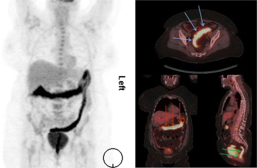
Int. J. Mol. Sci. 2020, 21, 243                                                                                      2 of 20

use of laboratory and ex vivo methods, mostly blood or stool tests, have the capability to identify
specific species with potential subspecies or strain, but also generally lack the capability to localize the
site of infection.
      There is currently no gold standard of care for a patient presenting with symptoms consistent with
a GI illness. Cross sectional imaging techniques have started being used to aid in GI malady analysis,
optimizing treatment plans through earlier and more accurate detection of disease [6]. Drastically
different treatment protocols must be followed for GI infections, inflammation, and cancer, so it is
imperative that disease’s causative agents are identified rapidly and accurately.
      Diarrhea is another common and serious symptom of gastrointestinal disorders that causes up to
500,000 deaths per year [7] and is the second leading cause of death in children under the age of 5 [8].
Developing nations see a higher rate of deaths due to GI diseases than developed nations which may
be attributed to the difference in standard of care and available treatments. Bacterial infections are
frequently the causative agents of diarrhea, all which require highly specific in vitro procedures that
require a lab and expensive equipment. These facilities may not be readily available in developing
areas, leading to a higher misdiagnosis rate and improper treatment of diseases. Inflammation is
often associated with these bacterial infections, making it even more difficult to differentiate between
infections and inflammatory conditions that both cause abdominal pain and diarrhea.
      Imaging is a promising next step for the differentiation of nonspecific infectious and inflammatory
symptoms in the GI tract. For this review of GI imaging, prevalent infections and inflammatory
conditions of the GI tract are discussed. To provide an appropriate background on each disease, their
symptoms and epidemiology are briefly detailed. The prevalence and usefulness of various imaging
modalities for each disease is then considered. A summary of the specificity and selectivity results
obtained from a variety of human and animal studies on is listed in Table 1. Finally, identification and
differentiation of discussed maladies from GI malignant tumors is reviewed before analyzing future
directions for the field.

                Table 1. Sensitivity and specificity values of different modalities in reviewed studies.

         Modality                   Subjects       Sensitivity           Specificity               Study
                                                       Diverticulitis
     CT vs. contrast
                                   542 humans      98% vs. 92%          Not Reported   Ambrosetti et al. (2002) [9]
         enema
     CT vs. contrast
                                   56 humans       93% vs. 80%               100%          Cho et al. (1990) [10]
         enema
             CT                    150 humans          97%                   100%          Rao et al. (1997) [11]
                                  684 humans vs.
   CT vs. ultrasound                               94% vs. 92%          99% vs. 90%     Lameris et al. (2007) [12]
                                   630 humans
        Ultrasound                 161 humans         98.6%                  96.5%      Schwerk et al. (1993) [13]
            MRI                    55 humans           95%                   88%       Heverhagen et al. (2008) [14]
            MRI                    40 humans           86%                   92%           Ajaj et al. (2005) [15]
                                                   Irritable Bowel Disease
            MRI                    50 humans           87%                   88%          Ordás et al. (2013) [16]
        Endoscopy                   Unknown            91%                   92%          Long et al. (2011) [17]
    Ultrasonography                60 humans         75–96%                  100%        Civitelli et al. (2014) [18]
             CT                    23 humans          76.9%                  90%        Wold et al. (2003) [19,20]
          PET/CT                   65 humans           98%                   68%        Lemberg et al. (2005) [21]
Int. J. Mol. Sci. 2020, 21, 243 3 of 20
2. Infection
Early or chronic infections are difficult to detect. Non-invasive diagnosis of infectious and
in particular discrimination between infection and other sources would have a profound impact
on disease management and patient outcomes. To this end different imaging techniques such as
radiograph, ultrasonography, magnetic resonance imaging (MRI), positron emission tomography (PET),
computed tomography (CT), and single photon emission computed tomography (SPECT) have been
explored [22–26]. The morbidity and mortality rate associated with diarrhea in immunocompromised
patients is much higher than that in a standard adult due to their suppressed immune systems.
Ninety-five percent of AIDS patients in developing countries had acquired chronic diarrhea [27]. Along
with immunocompromised patients, children are also at a higher risk for contracting GI infections due
to underdeveloped immune system and frequent oral contact with foreign, unsanitary objects. The
prevalence of these infectious diseases in underdeveloped nations due to poor sanitation and over
crowdedness is a major public health problem [28].
MRI enables diagnosis of microbial infection and has also been used for longitudinal bacteria
tracking in vivo. MRI diagnostics have potential for translation into clinical use. Application of MRI
for diagnosis of bacterial and viral infectious diseases is currently limited to the detection of the
resulting local inflammation, edema formation and the resulting changes in the local tissue properties
(e.g., relation time, water content, and diffusivity), as well as other manifestations of the immune
response, and not the direct detection of the causative infectious agent [29].
PET detects the dissemination of pathogens indirectly via detection of changes in cellular processes
and metabolic turn over, therefore, it cannot detect between infection and inflammation [26,30].
Exogenously radiolabeled patient leukocytes, as well as 99m Tc, 67 Ga, and 18 F-FDG tracers have been
utilized for clinical PET imaging due to their absorbance by cells exhibiting high metabolic rates [31].
However, specific radiolabeled antimicrobial peptides, antibodies, and antibiotics have been proposed
for specific detection of infection via SPECT. Although given the low resolution of this technique,
further improvements are needed [32].
Ultrasonography (US) is used to detect pathological changes in tissue due to infection. Similar to
the previous imaging modalities, US is unable to detect the causative infectious agent. For instance,
ultrasonography has been used to detect salmonella enterocolitis through detection of pathological
changes in bowel, ascites, and colonic wall thickening which may provide insight into the severity of
infection [33].
White light imaging (WLI) and linked color imaging (LCI) endoscopy have also used for diagnosis
of active infections. For example, Helicobacter pylori infection identification using WLI was shown to
have a sensitivity and specificity of 81.7%, and 66.7%, respectively, while significantly higher sensitivity
and specificity of 93.3%, and 78.3% were reported for LCI [34].
2.1. Shigella spp.
Shigella spp. is a Gram-negative bacterial pathogen, that is primarily transmitted via the oral–fecal
route [28,35–38]. Out of 165 million cases of Shigella annually, 1.5 million cases resulted in fatalities
with 98% being in underdeveloped nations [39] and approximately 500,000 cases reported in the United
States alone [40]. Pathogenesis of Shigella causes dysentery accompanied by vomiting, dehydration,
and abdominal pain. Colonic inflammation is seen in shigellosis, but this inflammation alone is not
specific enough to diagnose the patient. Blood and mucous in the feces is a good indication of Shigella,
but further testing is required to identify the correct pathogen and administer proper treatment [41].
The presence or absence of the Shigella pathogen is traditionally identified using a variety of techniques
in stool (Table 2), a difficult, time consuming, and expensive process [28,35,41]. Alternative in vitro
techniques have been utilized to identify Shigella in stool cultures.Int. J. Mol. Sci. 2020, 21, 243 4 of 20
Table 2. Potential infectious agents of gastrointestinal tract.
Infectious Agent Clinical Evaluation Screening Study
Lin et al. (2016) [42];
Shigella LPS O-Antigen ELISA/Serotyping
Gentle et al. (2015) [35]
Shigella and Escherichia coli Stool blot DNA Probing Sethabutr et al. (1985) [43]
Shigella and Escherichia coli Culture Media NMR Rautureau et al. (2019) [44]
Salmonella Stool Sample Selective Media Gorski (2012) [45]
Toxigenic Culture
Clostridium difficile Stool Sample Mirzaei (2018) [46]
Testing, PCR
The Shigella species has 4 subtypes, S. dysenteriae, S. flexneri, S.boydii, and S. sonnei. All four types
contain a virulent plasmid that is around 120–140 mDal in size. Shigella specific DNA probes have
been derived and used successfully to identify the presence of the pathogen [47]. The DNA is either
extracted from a stool sample and amplified by polymerase chain reaction to accumulate quantities
that can be detected by DNA probes or stool blots can be treated with the DNA probes [28,41]. An
alternative to DNA probing is the use of an enzymatically-linked immunosorbent assay (ELISA) to
identify the pathogen in vitro. The Shigella plasmid encodes for virulent antigens, including invasion
plasmid antigen (Ipa) proteins and a covalently linked extracellular lipopolysaccharide O-antigen,
both of which can be identified by an ELISA with an appropriate antiserum [35,42]. The combination
of DNA probes and ELISA can provide an accurate diagnosis of Shigella, but are not readily available
from a clinical standpoint [28].
2.2. Escherichia coli
While Escherichia coli has many beneficial functions in the GI tract, there are many different
diarrheagenic forms of the bacteria that are very dangerous in. Enteroinvasive Escherichia coli (EIEC)
is the pathogenic form that is indistinguishable from Shigella on the species level and induces the same
clinical symptoms as Shigella. Like Shigella, EIEC is also transmitted via the fecal-oral route and is
responsible for up to 40% of diarrhea in children under the age of 5 [30]. The taxonomical relatedness
of the two pathogens makes it very difficult to differentiate between them, thus identify the correct
etiological agent without a gold-standard test (Table 2) [30,41].
Both EIEC and Shigella spp. contain a large virulence plasmid that codes for similar invasion
plasmid antigen proteins and O-antigens [48]. Serotyping of the O-antigens had been used as the gold
standard to differentiate between E. coli serotypes, but this procedure is often erroneous and require
extensive lab technique. DNA probing and multiplex PCR assays of stool samples targeting the O and
H antigens along with the virulent plasmids are more specific ways that have been successfully utilized
to positively identify the pathogen [49]. A recent study done by Pautureau et al. explored nuclear
magnetic resonance in order to differentiate between Shigella and EIEC. An analysis of untargeted
proton NMR metabolomics was able to successfully differentiate between the two bacteria based on
the metabolic footprint produced [44]. NMR’s characterization of the metabolites used by the Shigella
bacteria and EIEC pathogen can also provide insight into better ways to identify and treat the infection.
2.3. Clostridium Difficile
Usually spread through the fecal-oral route, Clostridium difficile causes such nonspecific symptoms
as diarrhea, colitis, abdominal pain, and possibly fever or shock. The diarrhea from the disease is
often associated with antibiotic therapy in the weeks preceding infection [50]. Annually, it is estimated
that there are 453,000 infections and 29,000 deaths associated with C. difficile [51–53]. From these
figures alone, the morbidity of C. difficile infection (CDI) is apparent, made even more so by a study
that revealed that 9% of patients admitted to hospitals for CDI will die [54]. The startling mortality
of CDI is due in part to greater prevalence of fulminant colitis [51,53,55]. Rapid identification and
treatment, usually by antibodies, is necessary to increase likelihood of patient survival. At present,Int. J. Mol. Sci. 2020, 21, 243 5 of 20
Int. J. Mol. Sci. 2019, 20, x FOR PEER REVIEW 5 of 20
identification and treatment, usually by antibodies, is necessary to increase likelihood of patient
stool collection and testing is a common method for CDI diagnoses. Specifically, the gold standard
survival. At present, stool collection and testing is a common method for CDI diagnoses. Specifically,
is to test for the presence of toxins A and B in patient feces in vitro (Table 2) [51,52,56]. Testing for
the gold standard is to test for the presence of toxins A and B in patient feces in vitro (Table 2)
the presence of C. diff alone is not sufficient for diagnoses as a) C. diff is innately present in the gut
[51,52,56]. Testing for the presence of C. diff alone is not sufficient for diagnoses as a) C. diff is innately
microbiomes of 4% of healthy adults and b) some strains of C. diff do not produce toxins [52]. Toxin
present in the gut microbiomes of 4% of healthy adults and b) some strains of C. diff do not produce
presence can be determined in vitro through multiple protocols, though the current best practice is to
toxins [52]. Toxin presence can be determined in vitro through multiple protocols, though the current
use toxigenic culture testing [46,52]. While highly accurate, the test can take between 2 and 5 days to
best practice is to use toxigenic culture testing [40,52]. While highly accurate, the test can take between
complete as C. to
2 and 5 days diffcomplete
needs toasbeC.isolated andtogrown
diff needs from and
be isolated patient
grownstool samples
from and
patient requires
stool facilities
samples and
with the appropriate instrumentation [46,50].
requires facilities with the appropriate instrumentation [40,50].
Faster
Faster methods
methods of of identification
identification include
include enzyme immunoassays (EIAs)
enzyme immunoassays (EIAs) and and nucleic
nucleicacid
acid
amplification
amplification (NAAT). These methods bring their own complications. EIAs sensitivity values have a a
(NAAT). These methods bring their own complications. EIAs sensitivity values have
wide
widerange,
range,and
andboth
bothEIAsEIAsandand NAAT
NAAT possess difficulty in
possess difficulty in differentiating
differentiatingbetween
betweentoxictoxicand
andnontoxic
nontoxic
C.C.diff [51,52,57,58]. Fine-tuning of these methods may yield greater accuracy
diff [51,52,57,58]. Fine-tuning of these methods may yield greater accuracy in CDI diagnoses. in CDI diagnoses.
Radiologic
Radiologicimaging,
imaging,primarily
primarilyby bycomputed
computed tomography
tomography (CT) (CT) with
withoral
oralcontrast
contrast(e.g.,
(e.g.,barium
bariumsulfate)
sulfate)
(Figure
(Figure1 1[59]),
[59]),may
mayserve
serveas asan
anadditional
additional diagnostic tool. CT CTimages
imagesofofconfirmed
confirmedCDI CDIpatients
patientsshow
show
signs of bowel wall thickening, pericolonic
signs of bowel wall thickening, pericolonic stranding, and low-attenuation mural thickening,
and low-attenuation mural thickening, among among
others
others[52,59–61]. Ultrasonographymay
[52,59–61]. Ultrasonography mayserve
serve as as another
another imaging
imaging modality
modality to aidtoinaid in confirming
confirming CDI
CDI diagnoses,
diagnoses, according
according to Wiener-Well
to Wiener-Well et et
al. al. [62].
[62]. Further
Further testing
testing ofofthese
theseimaging
imagingmodalities
modalitiesasas
diagnostic
diagnostictools
toolsfor
forCDI
CDIisisrecommended
recommended before
before clinical
clinical action is taken.
taken.
Figure1.1.CT
Figure CTimaging
imagingusedused to identify C. diff. with
to identify with an
anaxial
axialCT
CTscan
scandemonstrates
demonstrateslow-attenuation
low-attenuationwallwall
thickening
thickeninginvolving
involvingthe theentire
entire length
length of the colon
colon with
with intense
intensemucosal
mucosalenhancement
enhancement(white(white arrow,
arrow,
(a));
(a));the
theenhancing
enhancingmucosa
mucosaisisstretched
stretched over
over the
the edematous haustral folds,
edematous haustral folds,resembling
resemblingan anaccordion
accordion
even
even in the absence of intra-luminal contrast (black arrow, (b)). A large amount of ascites ispresent
in the absence of intra-luminal contrast (black arrow, (b)). A large amount of ascites is also also
(dashed
presentarrow, (c))arrow,
(dashed [59]. (c)) [59].
2.4.
2.4.Salmonella
Salmonella
As
Aswith
withmost
mostdiseases
diseasesof ofthe
the GI
GI tract,
tract, infection
infection by by nontyphoid Salmonellaenterica
nontyphoidSalmonella enterica(NTS),
(NTS),referred
referred
totoasassalmonellosis, presents with enterocolitis and symptoms such as abdominal
salmonellosis, presents with enterocolitis and symptoms such as abdominal pain, diarrhea, and pain, diarrhea,
and
feverfever [62–64].
[62–64]. MostMost often,
often, thisthis bacterial
bacterial infection
infection is is acquired
acquired throughconsuming
through consuminginfected
infectedfoods,
foods,
primarily
primarilyconsisting
consisting of of
meats, eggs,
meats, andand
eggs, dairy
dairyproducts. Globally,
products. NTSNTS
Globally, is estimated to result
is estimated in 90 million
to result in 90
illnesses and 155,000 deaths per year [64]. In the United states, 40,000 infections
million illnesses and 155,000 deaths per year [64]. In the United states, 40,000 infections and 450 and 450 deaths are
reported
deaths areannually
reportedas aannually
result ofasNTS infection
a result of NTS [45,64]. Children
infection and
[39,64]. immunocompromised
Children and immunocompromised youths have
a youths
higher risk
haveofainfection,
higher risk with the latter being
of infection, with the more susceptible
latter being morefor more severe for
susceptible disease
moremanifestations
severe diseaseor
death [64]. Treatment
manifestations or deathof Salmonella infection
[64]. Treatment often includes
of Salmonella lessening
infection of symptoms
often includes and administration
lessening of symptoms
ofand
antibiotics [65]. Toofprovide
administration accurate
antibiotics diagnosis
[65]. To provideofaccurate
salmonellosis,
diagnosis theofcurrent practicethe
salmonellosis, is to collect
current
practice
stool is tofrom
samples collect stool and
patients samples from
culture them patients and culture
in Salmonella themmedia
selective in Salmonella
[45,64,65]. selective media
In this method,
[39,64,65]. In
enrichment this method,
broths suited forenrichment
enhanced broths suitedoffor
isolation enhancedmay
Salmonella isolation of Salmonella
be used to increase may be used
diagnostic
to increase
accuracy diagnostic
[65,66]. accuracy [65,66].
After isolation, Aftercan
cell samples isolation, cell samples
be serotyped, usingcanthebe serotyped,
Widal test, to using
betterthe Widal
administer
test, to better
treatment administer treatment
and understand and understand
infection source [67,68]. Use infection source [67,68].
of polyvalent Use ofspecific
and antisera, polyvalent andO
for the
antisera, specific for the O and H antigens of individual Salmonella serotypes,
and H antigens of individual Salmonella serotypes, on isolated bacteria provide necessary biochemical on isolated bacteria
provide necessary
verification of exact biochemical verification
pathogen identity [65]. of exact pathogen identity [65].Int. J. Mol. Sci. 2020, 21, 243 6 of 20
Int. J. Mol. Sci. 2019, 20, x FOR PEER REVIEW 6 of 20
Other methodsofofsalmonellosis
Other methods salmonellosisidentification
identification areare possible.
possible. ForFor example,
example, a study
a study by Hennedige
by Hennedige et al.
et al. reviewed
reviewed computed computed tomography
tomography (CT) images
(CT) images for confirmation
for confirmation of salmonellosis
of salmonellosis patients topatients
examine to
examine
radiologic radiologic manifestations
manifestations of infectionof infection
[69]. Of[69].theOf the manifestations
manifestations observed,
observed, they they concluded
concluded that
that thickened
thickened terminal
terminal ileum ileum or thickened
or thickened proximal
proximal colon colon
withwith mesenteric
mesenteric lymphadenopathy
lymphadenopathy could
could be
be specific
specific radiologic
radiologic indicators
indicators ofof Salmonella
Salmonella infection[69].
infection [69].Further
Furthertesting
testingofofthese
these finding’s
finding’s accuracy
is recommended before clinical diagnoses on shigellosis by CT CT are
are performed.
performed. Due Due toto a lack of
standardization,
standardization, use useofofELISA
ELISA testing
testing forfor Salmonella
Salmonella is currently
is currently nonspecific
nonspecific and requires
and requires furtherfurther
tuning
tuning before effective use as a diagnostic
before effective use as a diagnostic tool [67,70]. tool [67,70].
In summary,
summary, the bacterial agents responsible for GI illness along with corresponding corresponding metabolites
have been successfully identified using in vitro vitro biochemical
biochemical techniques,
techniques, (Table
(Table 2.)2.) providing more
information
information aboutaboutthe thepathogens
pathogens onon a molecular
a molecular level.level. Future
Future implications
implications of the of the inwork
in vitro vitroinclude
work
include
the use ofthe use of imaging
molecular molecular imagingthe
to identify to pathogen
identify in thevivo.
pathogen in vivo.
Fluorescently Fluorescently
labeled labeled
antibody/peptide
antibody/peptide
(as contrast agent)(as for contrast
molecular agent)
imagingfor endoscopy
molecular imaging
for rapid endoscopy
detection and for diagnosis
rapid detection and
of bacterial
diagnosis of bacterial
infection [71]. Ultrasounicinfection [71]. Ultrasounic
assessment of Salmonellaassessment
spp. has of been
Salmonella spp. in
successful has been successful
children with the
in children
ability with the
to identify ability to identify
pathological changes pathological
in the bowel changes in the bowel and
and intra-abdominal intra-abdominal
spaces with thickeningspaces
of
with thickening of the colonic wall (Figure 2) [72]. Elimination of the need
the colonic wall (Figure 2) [72]. Elimination of the need for stool cultures and biochemical laboratory for stool cultures and
biochemicalwould
procedures laboratory procedures
drastically decrease would
the timedrastically decrease
take to reach the timeMolecular
a diagnosis. take to reach
imaginga diagnosis.
in the GI
Molecular
tract is still imaging
relativelyinnew,the but
GI has
tractbeen
is still relatively
successful new, but has
in identifying been successful
malignancies in identifying
and is progressing in
malignancies and is progressing
diagnosing inflammatory in diagnosing
and infectious diseases inflammatory and infectious
through the development and diseases
testing ofthrough
endoscopicthe
development
molecular probes and [44,71–73].
testing of endoscopic molecular probes [38,71–73].
Figure 2.2.Evaluation
Figure Evaluationof of
lower abdomen
lower of pediatric
abdomen of pediatric with Salmonella
patientpatient enterocolitis
with Salmonella using transverse
enterocolitis using
ultrasound.ultrasound.
transverse There is thickening
There is of the sigmoid
thickening colon
of the whichcolon
sigmoid results predominantly
which from submucosal
results predominantly from
edema (arrow) observed as the thick hyperechoiclayer. S = sigmoid colon [72].
submucosal edema (arrow) observed as the thick hyperechoiclayer. S = sigmoid colon [72].
3. Inflammation
3. Inflammation
3.1. Diverticulitis
3.1. Diverticulitis
Diverticulitis is characterized by the presence of inflamed diverticula in the GI tract, most
Diverticulitis
commonly the largeis intestine.
characterized by thepercent
Sixty-five presence of inflamed
of the diverticulaand
elderly population in the
5% GI tract, most
of individuals
commonly the large intestine. Sixty-five percent of the elderly population and 5%
40 years old or younger will have diverticula, 25% of those individuals will have diverticulitisof individuals 40
years old
while the or youngerare
remaining willasymptomatic
have diverticula, 25%Symptomatic
[74,75]. of those individuals will have
diverticulitis diverticulitis
is frequently whilewith
coupled the
remaining are asymptomatic [74,75]. Symptomatic diverticulitis is frequently
complications including anal bleeding, fistulas, perforation, and abscess formation [74,76–78]. Thecoupled with
complications including
clinical misdiagnosis rate anal bleeding, fistulas,
of diverticulitis perforation,
is upwards and abscess
of 50%, indicating theformation [74,76–78].
need for better The
diagnostic
clinical misdiagnosis rate of diverticulitis is upwards of 50%, indicating the need for better
techniques [79]. Recently there has been a spark in the investigation of potential biomarkers as safe diagnostic
techniques [79]. Recently there has been a spark in the investigation of potential biomarkers as safe
and effective ways to diagnosis and assist physicians in treating diverticulitis [74]. C-reactive proteinInt. J. Mol. Sci. 2020, 21, 243 7 of 20
and effective ways to diagnosis and assist physicians in treating diverticulitis [74]. C-reactive protein
levels >50 mg/L, in addition to left lower quadrant abdominal tenderness and absences of vomiting is
indicative of diverticulitis [79,80]. Fecal calprotectin is derived from neutrophils, so its presence in
the stool is a good indication of inflammation in the GI tract. A study done by Tursi et al. showed
increased levels of fecal calprotectin concentrations in diverticulitis patients when compared to IBS
and control patients [81]. This provides an appealing clinical tool for differentiation between IBS and
diverticulitis, although it must be noted that IBS and diverticulitis can exist simultaneously. Increased
levels of these biomarkers in the blood and stool strongly support a clinical diagnosis of diverticulitis
but are not specific enough to fully replace initial imaging techniques.
3.1.1. X-ray
Contrast enemas have been used in an attempt to diagnose cases of diverticulitis [82,83]. Initially,
barium had been used as the contrast agent, but the shift to water soluble contrast agents was made to
minimize risk to the patient. Water soluble contrast agents eliminated the risk of barium peritonitis,
decreased the wait time between injection and scan, and allowed for other imaging modalities to
be utilized after the enema [76,84,85]. While water-soluble contrast enemas are extremely beneficial
at showing morphological changes of the colon, they are limited in that they cannot indicate active
inflammation which is a key marker of diverticulitis. Therefore, the enema is then incapable of making
a complete diagnosis of diverticulitis without the assistance of another imaging modality or it may
result in a misdiagnosis indicating a low sensitivity of contrast enemas (Table 1) [86].
3.1.2. Computed Tomography
Computed tomography (CT) has many advantages when compared to the traditional contrast
enema, which is why it is now accepted as the primary imaging modality for patients that present with
abdominal pain. A CT scan will identify inflamed diverticula, bowel wall inflammation, pericolic fat
stranding, and corresponding complications [9–11,83,87,88]. CT is capable of visualizing pericolonic
and colonic complications which results in a more accurate diagnosis for the patient, along with
better standard of care. Thirty percent of cases of diverticulitis are accompanied by complications
including, fistulas, perforations, and abscesses. Accurate assessment of complications is crucial in the
development of a treatment plan. CT is particularly beneficial in the management of abscesses since it
can be used for percutaneous drainage of the abscesses. Such drainage eliminates the need for multiple
surgeries, making CT more cost effective and lower risk for the patients [9,10,77,82,83,86,88–91].
Contrast agents are utilized in CT scans to achieve maximum colonic distension along with colonic
opacification in order to better identify inflammatory wall thickening consistent with diverticulitis [90].
Opacification of the colon is necessary to differentiate between intra and extra luminal air and fluid
cavities, indicating the presence of abscesses [11,90,92]. Oral contrast agents are beneficial to the
visualization of loops within the small bowel, but lack in completely covering the colon. Oral contrast
agents are limited by inconsistent opacification of the colon and a large waiting period, resulting in the
infrequent use of these types of agents [11,92]. Intravenous contrast agents are advantageous due to
their capability of enhancing inflammation in the abdominal and pelvic regions. Such enhancement
permits a more accurate identification of other illnesses that may have similar clinical symptoms as
diverticulitis [83,87]. The use of an intravenous contrast agent is associated with a higher risk to the
patient, at no significant benefit in comparison to rectally administered contrast agents [11,92]. Rectally
administered contrast agents are the safest and most efficient way of obtaining uniform opacification of
the colon with maximum distention [90]. Gastrografin-based contrast agents are the most widely used
agents when imaging the gastrointestinal tract [9,11]. The risks associated with rectal contrast agents
are similar to those associated with performing an enema: there is a higher chance of exacerbating
perforations or extravasation of contrast material [92].Int. J. Mol. Sci. 2020, 21, 243 8 of 20
3.1.3. Ultrasonography
Ultrasonography (US) is an extremely low cost and low risk imaging modality that is not frequently
used for the diagnosis of diverticulitis due to its reliance on a technician and inferiority to CT. While
ultrasound is non-invasive, provides real-time images, and is useful in identifying inflammation,
abscesses, and bowel wall thickening, it is limited in that it cannot be used on obese patients and that is
can be impeded by gas bubbles [77,87]. A meta-analysis of 6 CT studies and 6 US studies showed that
CT was no better or worse than ultrasound at diagnosing diverticulitis (Table 1), but CT still remains
the modality of choice due to its ability to detect a multitude of complications [12,77,91].
3.1.4. Magnetic Resonance Imaging
While magnetic resonance imaging (MRI) is not traditionally used when a patient presents with
abdominal pain, it has proven to have a high sensitivity and specificity in the diagnosis of diverticulitis,
especially with the introduction of an intravenous gadolinium-based contrast agent (Table 1) [14,15,87].
MRI is superior to CT in that it lacks associated harmful ionizing radiation, thus an appealing and
a safe alternative to CT, but gadolinium-based contrast agents remain somewhat controversial with
potential Gadolinium toxicity [93,94]. However, the use of MRI in diagnosing diverticulitis is limited
not only by its cost and time, but also by the motion of other organs to continue breathing while
the scan is taking place [14]. Due to these obstacles, MRI remains in the shadow of other imaging
modalities for diagnosis of diverticulitis.
3.1.5. Endoscopy
Many features that are indicative of a CT scan are also indicators of colorectal cancer. After
a positive diagnosis is made using CT, it is standard procedure for the patient to get a follow up
colonoscopy around 6 weeks after being diagnosed [9,95]. This is a preventative measure to ensure
that the diagnosis of diverticulitis did not miss a diagnosis of colon cancer [77,82,87,91,95–97]. In a
study conducted by Lau et al., 34% of patients’ follow up colonoscopies came back positive for further
complications or misdiagnosis of the initial diverticulitis [97]. While there is nothing to indicate that a
positive diagnosis for diverticulitis is correlated with a higher risk for colorectal cancer, it has been
observed that patients who present with diverticular complications have been more likely to have a
positive colonoscopy for colorectal cancer sometime after the diverticulitis diagnosis [77,97].
3.2. Irritable Bowel Disease
Irritable bowel disease (IBD) is expressed in two major forms: Crohn’s disease (CD) and ulcerative
colitis (UC). For both manifestations, IBD symptoms are similar to those of other GI disorders and
include abdominal pain or discomfort, weight loss, bloody stool, diarrhea, and nausea [98]. In the
intestines, the small and large bowel walls thicken and abscesses, collections of fluid surrounded in the
inflamed intestinal tissue, may form. Unlike other disorders, the intensity of IBD symptoms changes
with time. Periods of high intensity symptoms are defined as “flare,” while times of low intensity
symptoms indicate “remission” [99].
Incidence of IBD in the United States and Europe is increasing, with current estimates of the
affected population in those countries exceeding 1.6 million and 3 million, respectively [99–102]. For
the different manifestations of IBD, CD’s prevalence in children and adults is 58 and 241 cases per
100,000, respectively, while UC prevalence in children and adults is 34 and 263 cases per 100,000,
respectively [102–104]. In the United States, CD incidence is currently estimated to be 3.1–14.6 cases per
100,000 person-years and UC incidence is estimated at 2.2–14.3 cases per 100,000 person-years [102,103].
IBD occurrence is spreading across the world as well, with observed incidences in developing
countries [100]. Approximately 20% of IBD patients are diagnosed during their childhood [99,104].
The effects of IBD have long-term impacts on children, namely growth failure or delays in puberty
onset [63,100]. In 10–15% of IBD cases, CD and UC cannot be distinguished based on how they presentInt. J. Mol. Sci. 2020, 21, 243 9 of 20
in the patient, who is given an “IBD-unclassified” diagnosis. IBD patients are also at an increased
risk of developing colorectal cancer, primarily due to the chronic intestinal inflammation inherent to
CD and UC [105,106]. Eaden et al., in their meta-analysis, showed that patients with UC developed a
cumulative risk to develop colorectal cancer of 2%, 8%, and 18% at 10, 20, and 30 years, respectively,
after disease development [105–107]. Another study by Beaugerie and Itzkowitz demonstrated that in
North America and some countries in Europe, the risk of colorectal cancer in IBD patients is up to two
times higher than the risk of the general public [108].
The need for rapid and accurate imaging of IBD and its manifestations is apparent given its
increasing prevalence and worldwide impact. Various imaging methods are practiced for IBD
identification and monitoring [99]. with potential new and more effective modalities entering the
clinical landscape as time progresses.
3.2.1. Endoscopy
Endoscopy is usually among the first steps carried out in diagnosing IBD [109–111]. The most
common endoscopic technique is colonoscopy. In this procedure, clinicians insert an endoscope,
comprised of a white light and a camera, into the patient via the anus for direct visualization of the
colon. Colonoscopy is a highly invasive procedure, proving undesirable for patients. Other endoscopic
techniques that offer minimal invasion are practiced, such as single-balloon and double-balloon
enteroscopy [111].
3.2.2. Chromoendoscopy
To improve the surveillance of dysplasia, lesions, and other abnormalities in mucosal topography,
chromoendoscopy (CE) may be used [112–114]. In this technique, dilute dye (indigo carmine or
methylene blue) is sprayed, within appropriate guidelines, onto the lumen of the colon using a
dye spray catheter [113]. This dye better reveals the location and pattern of lesions present in the
colon, aiding clinician analysis, biopsy, or removal of present tissue. Compared to standard white
light colonoscopy, CE has proved to be superior in multiple studies on per-patient and per-lesion
analysis [113,114]. Given this, in order to increase accuracy of analysis and dysplasia detection, most
international subspecialty societies recommend the use of CE when examining IBD patients [112]. This
accuracy is particularly beneficial for early detection of colorectal cancer in patients.
3.2.3. Computed Tomography
Given its ease-of-access, non-invasive nature, ability to scan intraluminal and extraluminal effects,
and not requiring anesthesia due to short scan times, computed tomography (CT) is a highly favorable
method for IBD imaging [115]. The rapid image acquisition is particularly beneficial for pediatric
patients, who may be more susceptible to the harmful effects of anesthesia. IBD evaluation via
CT typically requires application of both oral and IV contrast agents, such as the gastrogafin-based
agents described above [99,116,117]. These agents distend and opacify the bowel to reveal where
extraluminal fluids are collected and to best characterize the abnormalities (e.g., thickening) in the
bowel wall. To detect mucosal enhancement, associated with inflammation of or lesions in the
intestines, CT enterography (CTE) may be used, which requires application of neutral contrast
agents (e.g., water) [99,117]. Cross-sectional images of patients reveal the type and extent of their
intestinal inflammation. The location and degree of inflammation and wall thickening determines the
manifestation of IBD [118]. Further, these factors are heavily considered when diagnosing patients
with IBD as opposed to other diseases. Specificity and sensitivity values for CT studies of IBD are
shown in Table 1.
As stated, CT images can be acquired quickly and does not require use of anesthetics. Further,
they are easy to reproduce and possess high spatial resolution, presenting clearer images to clinicians.
These factors alone make it a strong modality for imaging IBD patients. CTE may boast greater
sensitivity and specificity over magnetic resonance enterography (MRE) [99,119,120]. CT is notInt. J. Mol. Sci. 2020, 21, 243 10 of 20
without its disadvantages. The greatest apparent drawback with CT usage is the required patient
Int. J. Mol. Sci. 2019, 20, x FOR PEER REVIEW 10 of 20
exposure to radiation [99,121,122]. Given the chronic nature of IBD, patients need frequent imaging
for monitoring disease progress and symptom flare. Repeated exposure to ionizing radiation is not
ideal, especially given the increased risk of cancer produced in patients. This is especially undesirable
ideal, especially given the increased risk of cancer produced in patients. This is especially undesirable
for pediatric patients, of which there is a significant number. Other studies have shown that CT
for pediatric patients, of which there is a significant number. Other studies have shown that CT
imaging, when compared to US and MRI, may actually possess lower specificity and sensitivity for
imaging, when compared to US and MRI, may actually possess lower specificity and sensitivity for
IBD diagnoses, decreasing its overall reliability [19,123]. Another potential disadvantage is that CT
IBD diagnoses, decreasing its overall reliability [19,123]. Another potential disadvantage is that CT
cannot be performed in patients who are allergic to contrast agents used.
cannot be performed in patients who are allergic to contrast agents used.
It should be noted that the prevalence of CT usage, and thus associated regional clinician skill,
It should be noted that the prevalence of CT usage, and thus associated regional clinician skill, is
is much higher in the United States as compared to Europe, where US and MRI are favored [19]. For
much higher in the United States as compared to Europe, where US and MRI are favored [19]. For all
all these modalities, this disparity can affect the specificity and sensitivity results when looking at a
these modalities, this disparity can affect the specificity and sensitivity results when looking at a global
global as opposed to regional scale.
as opposed to regional scale.
3.2.4. Positron
3.2.4. Emission Tomography
Positron Emission Tomography
For analysis
For analysisof ofIBD,
IBD,particularly
particularlyininsymptom
symptom flare
flare situations
situations or or pediatric
pediatric diagnostic
diagnostic work-ups,
work-ups, CT
scanning is paired with positron emission tomography (PET) [21,123–127]. PET molecularmolecular
CT scanning is paired with positron emission tomography (PET) [21,123–127]. PET imaging
imaging
tracks tracks accumulation
accumulation of molecular
of molecular radiopharmaceutical
radiopharmaceutical 18 F-FDG to 18F-FDG to determine inflamed areas.
determine inflamed areas. 18 F-FDG
18F-FDG is used as it is similar to glucose and experiences significant uptake by leukocytes activated
is used as it is similar to glucose and experiences significant uptake by leukocytes activated by tissue
by tissueand
damage damage and inflammation.
inflammation. In pairing In PET
pairing
withPETCT,with CT, three-dimensional
three-dimensional CT reconstructions
CT reconstructions follow
follow PET molecular imaging to confirm anatomical and structural information in
PET molecular imaging to confirm anatomical and structural information in vivo [124]. Many studies vivo [124]. Many
studies have confirmed the usefulness of PET/CT imaging, particularly in cases where
have confirmed the usefulness of PET/CT imaging, particularly in cases where IBD affects the small IBD affects the
small bowel [123,128–133]. Further, PET/CT is reported to have better sensitivity
bowel [123,128–133]. Further, PET/CT is reported to have better sensitivity than endoscopy, though athan endoscopy,
thoughspecificity
lower a lower specificity than ultrasound
than ultrasound [21,123,125].[21,123,125].
More studies More studies are recommended
are recommended to better
to better characterize
the effectiveness of this joint modality [125,128]. An example of PET/CT imaging is shown in Figure is
characterize the effectiveness of this joint modality [125,128]. An example of PET/CT imaging 3.
shown in Figure 3.
Figure 3. 18 F-FDG PET/CT scan of ulcerative colitis (UC) patient. Extent of disease is seen on the left
Figure 3. 18F-FDG PET/CT scan of ulcerative colitis (UC) patient. Extent of disease is seen on the left
and in areas indicated by arrows in the right panel [124].
and in areas indicated by arrows in the right panel [124].
3.2.5. Immuno-PET
A relatively new modality for IBD imaging is immuno-PET. Using fragments of monoclonal
antibodies (mAbs), innate immune cells, especially present during symptom flare of IBD, are targeted
to track inflammation [134,135]. While testing of this modality is primarily still in the preclinical stage,
murine model results are promising for translation to clinical applications [134,135]. mAb-therapeuticInt. J. Mol. Sci. 2020, 21, 243 11 of 20
3.2.5. Immuno-PET
A relatively new modality for IBD imaging is immuno-PET. Using fragments of monoclonal
antibodies (mAbs), innate immune cells, especially present during symptom flare of IBD, are targeted
to track inflammation [134,135]. While testing of this modality is primarily still in the preclinical stage,
murine model results are promising for translation to clinical applications [134,135]. mAb-therapeutic
response can be measured with this imaging method as well [136]. This modality may also be
useful in characterizing cancers of the GI tract, such as colon cancer, given the specificity of mAb
technologies [136].
3.2.6. Ultrasonography
Using sound waves, ultrasonography (US) provides real-time images of the body’s interior. For
determining IBD presence and extent, bowel wall thickness is the primary factor considered with US
modalities [99,137–139]. Given its non-invasive nature and ability to render images in real-time, US is
heavily favored for IBD imaging and analysis. US modalities do not expose patients to any radiation,
are widely available, and remain generally inexpensive [99,140]. Studied specificity and selectivity
values for US usage in IBD cases can be seen in Table 1.
There are several drawbacks to US imaging for IBD. For one, US has been primarily used with
CD patients, limiting its effectiveness for UC cases [137]. Further, one meta-analysis indicates US is
used more in Europe than in the United States [19]. This indicates that the accuracy for US techniques,
given clinician exposure and usage, may be lower in the United States in comparison to Europe. The
time required and accuracy of the imaging are heavily dependent on the experience of the clinician
and size of the patient.
3.2.7. Contrast-Enhanced US
Some IBD patients may still exhibit wall thickening without active inflammation [141–144]. In
these cases, US could lead to inappropriate therapy for the patient as the symptom intensity misaligns
with the proposed treatment. To increase the accuracy of US in these and general IBD cases, contrast
enhanced ultrasonography (CEUS) may be used. CEUS uses a intravenously applied microbubble
contrast agent, such as sulfur hexafluoride, to show bowel wall enhancement and mesentery [144,145].
CEUS provides real-time information on the vascularity of the scanned area. Since IBD causes vascular
alterations in affected areas, this information gives clinicians an understanding on IBD activity in the
patient [144–148]. In general, CEUS appears to provide greater clinical certainty in evaluating IBD
in patients.
3.2.8. Magnetic Resonance Imaging
Magnetic resonance imaging (MRI) produces three-dimensional anatomical images of patients. In
imaging the GI tract, especially for small bowel analysis, MR enterography (MRE), MR enteroclysis, and
MR fistulography are the commonly used procedures [99,149]. For both the small and large bowel, intra
and extra mural involvement can be measured with the appropriate contrast agents such as barium
suspensions [99,150]. These agents induce bowel wall distension, ensuring easier detection of bowel
wall complications, and fat suppression for ease of image interpretation [99,150,151]. Fat stranding,
wall thickening, and intestinal strictures are all evidence for IBD found in MRI analyses [99,152]. MRI
techniques do not use ionizing radiation to acquire images, making them favorable over CT modalities,
especially for young patients. Additionally, MRI produces high resolution images while permitting
significantly improved soft tissue contrast as compared to CT [149].
As with previously analyzed modalities requiring contrast agents, MRI cannot be used with patients
who are allergic to contrast agents. Additionally, the long scan time (compared to US, for example) is
logistically inconvenient for patients, and may require sedation [99]. Further, reference information
and clinician or technology-induced bias can lead to improper image assessment [19,119,143].Int. J. Mol. Sci. 2020, 21, 243 12 of 20
3.2.9. Multispectral Optoacoustic Tomography
By stimulating patient tissues with lasers, ultrasound waves are generated. These waves can
Int. read
be J. Mol.and
Sci. 2019, 20, x FOR to
interpreted PEER REVIEW tissue characteristics inside patients. This phenomenon
understand 12isofthe
20
basis for multispectral optoacoustic tomography (MSOT) technologies. MSOT is purely non-invasive
in nature
in nature andand allows
allows for for real-time
real-time imaging
imaging of of patient
patient tissue
tissue in
in vivo
vivo [66,153,154].
[66,153,154]. Images
Images gathered
gathered by by
MSOT show the distribution of such molecules as hemoglobin and melanin in patient tissue, duedue
MSOT show the distribution of such molecules as hemoglobin and melanin in patient tissue, to
to the
the difference
difference in how in these
how these molecules
molecules absorbabsorb light This
light [155]. [155]. This distribution
distribution is indicative
is indicative of diseaseofpresence
disease
presence and behavior in patients. A study evaluating MSOT imaging of murine
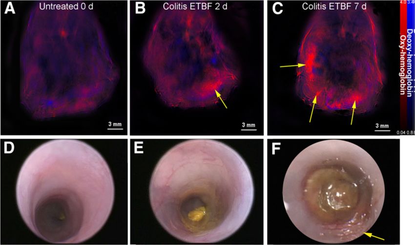
and behavior in patients. A study evaluating MSOT imaging of murine colitis showed the correlation colitis showed the
correlation between inflammation level and concentration of oxy-hemoglobin
between inflammation level and concentration of oxy-hemoglobin (Figure 4) [66]. In a separate study, (Figure 4) [66]. In a
separatetrials
clinical study, on clinical trialshave
CD patients on CD patients
shown have
similar shownIncreased
results: similar results: Increasedlevels
oxy-hemoglobin oxy-hemoglobin
with active
levels with active CD compared to remission CD [153,156]. MSOT
CD compared to remission CD [153,156]. MSOT technologies are still undergoing clinical technologies are still undergoing
trials for
clinical trials for widespread use and are primarily seen in Europe. Preliminary
widespread use and are primarily seen in Europe. Preliminary results reveal significant potential results reveal
for
significant potential for MSOT in
MSOT in rapidly diagnosing and monitoring IBD.rapidly diagnosing and monitoring IBD.
Figure 4. Multispectral
Figure 4. optoacoustic tomography
Multispectral optoacoustic tomography (MSOT)
(MSOT) (A–C)
(A–C) and
and colonoscopic (D–F) images
colonoscopic (D–F) images of
of
murine colitis progression. Images were taken at t = 0, 2, and 7 days after bacterial inoculation. Yellow
murine colitis progression. Images were taken at t = 0, 2, and 7 days after bacterial inoculation. Yellow
arrows indicate areas of inflammation correlating with colitis [67].
arrows indicate areas of inflammation correlating with colitis [67].
In summary, inflammatory conditions such as diverticulitis and IBD can be diagnosed and
In summary, inflammatory conditions such as diverticulitis and IBD can be diagnosed and
analyzed by a wide range of imaging modalities. Further work is required to verify the accuracy
analyzed by a wide range of imaging modalities. Further work is required to verify the accuracy and
and refine the safety or comfortability of some of the mentioned modalities. Emerging technologies
refine the safety or comfortability of some of the mentioned modalities. Emerging technologies such
such as immuno-PET and MSOT could prove increasingly beneficial to imaging and diagnostic
as immuno-PET and MSOT could prove increasingly beneficial to imaging and diagnostic accuracy
accuracy while increasing patient compliance. Since inflammation is also consistent with infection, it is
while increasing patient compliance. Since inflammation is also consistent with infection, it is
important for patients with diverticulitis and IBD to be regularly monitored for alternative infectious
important for patients with diverticulitis and IBD to be regularly monitored for alternative infectious
or malignant diseases.
or malignant diseases.
4. Conclusions
4. Conclusion
Nonspecific GI symptoms can be attributed to a broad span of infectious and inflammatory
Nonspecific
conditions leadingGItosymptoms
high ratescan be attributed to
of misdiagnosis anda improper
broad span of infectious
patient and inflammatory
care. Presently, anatomic
conditions leading to high rates of misdiagnosis and improper patient care. Presently, anatomic
imaging modalities have the capability to localize areas of inflammation with limitations in sensitivity
imaging modalities have the capability to localize areas of inflammation with limitations in sensitivity
and specificity, but lack the ability to localize specific bacterial species, albeit largely due to a lack of
and specificity, but lack the ability to localize specific bacterial species, albeit largely due to a lack of
bacterial strain specific contrast agents. While in vitro biochemical techniques often can recognize
infectious agents to correctly identify bacterial pathogens, blood or stool-based techniques often must
be paired with anatomical imaging to arrive at the correct diagnosis. Future development of bacterial
specific or inflammatory specific contrast agents would dramatically improve the ability to
specifically and locally identify disease within the GI tract to allow for differentiation betweenInt. J. Mol. Sci. 2020, 21, 243 13 of 20
bacterial strain specific contrast agents. While in vitro biochemical techniques often can recognize
infectious agents to correctly identify bacterial pathogens, blood or stool-based techniques often must
be paired with anatomical imaging to arrive at the correct diagnosis. Future development of bacterial
specific or inflammatory specific contrast agents would dramatically improve the ability to specifically
and locally identify disease within the GI tract to allow for differentiation between inflammatory and
autoimmune diseases in order to better diagnose and treat patients.
Funding: This work was supported by NIH CA205941, CA212350, and EB020125.
Acknowledgments: This work was supported by NIH CA205941, CA212350, and EB020125.
Conflicts of Interest: The authors declare no conflict of interest.
Abbreviations
GI Gastrointestinal tract
ELISA Enzymatically-linked immunosorbent assay
Ipa Invasion plasmid antigen
EIEC Enteroinvasive Escherichia coli
CDI Clostridium difficile infection
EIA Enzyme immunoassay
NAAT Nucleic acid amplification
CT Computed tomography
NTS Nontyphoid Salmonella enterica
US Ultrasonography
MRI Magnetic resonance imaging
IBD Irritable bowel disease
CD Crohn’s disease
UC Ulcerative colitis
CE Chromoendoscopy
CTE Computer tomography enterography
MRE Magnetic resonance enterography
PET Positron emission tomography
mAb Monoclonal antibodies
CEUS Contrast enhanced ultrasonography
MSOT Multispectral optoacoustic tomography
References
1. Meltzer, A.C.; Pines, J.M.; Richards, L.M.; Mullins, P.; Mazer-Amirshahi, M. US emergency department visits
for adults with abdominal and pelvic pain (2007-13): Trends in demographics, resource utilization and
medication usage. Am. J. Emerg. Med. 2017, 35, 1966–1969. [CrossRef] [PubMed]
2. Pant, C.; Deshpande, A.; Sferra, T.J.; Olyaee, M. Emergency department visits related to functional abdominal
pain in the pediatric age group. J. Investig. Med. Off. Publ. Am. Fed. Clin. Res. 2017, 65, 803–806. [CrossRef]
[PubMed]
3. Macaluso, C.R.; McNamara, R.M. Evaluation and management of acute abdominal pain in the emergency
department. Int. J. Gen. Med. 2012, 5, 789–797. [CrossRef] [PubMed]
4. Boendermaker, A.E.; Coolsma, C.W.; Emous, M.; Ter Avest, E. Efficacy of scheduled return visits for emergency
department patients with non-specific abdominal pain. Emerg. Med. J. 2018, 35, 499–506. [CrossRef]
5. Schiller, L.R.; Pardi, D.S.; Sellin, J.H. Chronic Diarrhea: Diagnosis and Management. Clin. Gastroenterol.
Hepatol. 2017, 15, 182–193. [CrossRef]
6. Ady, J.; Fong, Y. Imaging for infection: From visualization of inflammation to visualization of microbes. Surg.
Infect. 2014, 15, 700–707. [CrossRef]
7. Centers for Disease Control and Prevention. Global Diarrhea Burden. Available online: https://www.cdc.
gov/healthywater/global/diarrhea-burden.html (accessed on 8 October 2019).You can also read


