Vasculoprotective Effects of Vildagliptin. Focus on Atherogenesis - MDPI
←
→
Page content transcription
If your browser does not render page correctly, please read the page content below
International Journal of
            Molecular Sciences

Review
Vasculoprotective Effects of Vildagliptin. Focus
on Atherogenesis
Michał Wiciński 1 , Karol Górski 1, * , Eryk Wódkiewicz 1 , Maciej Walczak 1 ,
Magdalena Nowaczewska 2 and Bartosz Malinowski 1
 1    Department of Pharmacology and Therapeutics, Faculty of Medicine, Collegium Medicum in Bydgoszcz,
      Nicolaus Copernicus University, M. Curie 9, 85-090 Bydgoszcz, Poland; wicinski4@wp.pl (M.W.);
      eryk.wodkiewicz09@gmail.com (E.W.); maciej.walczak5@hotmail.com (M.W.);
      bartosz.malinowski@cm.umk.pl (B.M.)
 2    Department of Otolaryngology, Head and Neck Surgery, and Laryngological Oncology, Faculty of Medicine,
      Ludwik Rydygier Collegium Medicum in Bydgoszcz, Nicolaus Copernicus University, M. Curie 9,
      85-090 Bydgoszcz, Poland; magy_mat@by.onet.pl
 *    Correspondence: karolgorski-2@gazeta.pl

 Received: 17 February 2020; Accepted: 23 March 2020; Published: 25 March 2020

 Abstract: Vildagliptin is a representative of Dipeptidyl Peptidase-4 (DPP-4) inhibitors, antihyperglycemic
 drugs, approved for use as monotherapy and combination therapy in type 2 diabetes mellitus.
 By inhibiting enzymatic decomposition, DPP-4 inhibitors increase the half-life of incretins such as GLP-1
 (Glucagon-like peptide-1) and GIP (Gastric inhibitors polypeptide) and prolong their action. Some
 studies present results suggesting the anti-sclerotic and vasculoprotective effects of vildagliptin reaching
 beyond glycemic control. Vildagliptin is able to limit inflammation by suppression of the NF-κB
 (nuclear factor kappa-light-chain-enhancer of activated B cells) signaling pathway and proinflammatory
 agents such as TNF-α (tumor necrosis factor α), IL-1β (Interleukin-1β), and IL-8 (Interleukin 8).
 Moreover, vildagliptin regulates lipid metabolism; attenuates postprandial hypertriglyceridemia;
 and lowers serum triglycerides, apolipoprotein B, and blood total cholesterol levels. This DPP-4
 inhibitor also reduces macrophage foam cell formation, which plays a key role in atheromatous
 plaque formation and stability. Vildagliptin reduces vascular stiffness via elevation of nitric oxide
 synthesis, improves vascular relaxation, and results in reduction in both systolic and diastolic blood
 pressure. Treatment with vildagliptin lowers the level of PAI-1 presenting possible antithrombotic
 effect. By affecting the endothelium, inflammation, and lipid metabolism, vildagliptin may affect the
 development of atherosclerosis at its various stages. The article presents a summary of the studies
 assessing vasculoprotective effects of vildagliptin with special emphasis on atherogenesis.

 Keywords: vildagliptin; endothelium; inflammation; atherogenesis; pathways; pharmacology

1. Introduction
     The prevalence of diabetes mellitus type 2 (DM2) is constantly increasing worldwide at an
alarming rate. According to the World Health Organization, nowadays, about 422 million people
globally are suffering from this condition [1]. Diabetes confers about a two-fold excess risk for a wide
range of vascular diseases such as arterial stiffness, hypertension, and atherogenesis which are the
causes of stroke, heart attack, retinopathy, and diabetic foot [2]. In recent years, there have appeared
interesting postulates that vildagliptin may display a vasculoprotective properties beyond glycemic
control (Table 1). Vildagliptin is a representative of Dipeptidyl Peptidase-4 (DPP-4) inhibitors which
are registered and effectively used in DM2 therapy. These class of drugs was designed to increase the
half-life of incretins such as GLP-1 (Glucagon-like peptide-1) and GIP (Gastric inhibitors polypeptide)

Int. J. Mol. Sci. 2020, 21, 2275; doi:10.3390/ijms21072275                         www.mdpi.com/journal/ijms
Int. J. Mol. Sci. 2020, 21, 2275 2 of 17
and to prolong their action [3]. Under hyperglycemic state, they stimulate glucose-dependent insulin
secretion from pancreatic β-cells and also inhibit the secretion of glucagon from α-cells [4]. Furthermore,
they slow down the rate of absorption of nutrients into the bloodstream by delaying gastric emptying.
It prolongs the satiety time and, in consequence, restriction of food intake [5]. Interestingly, GLP-1 is also
responsible for the activation of some pro-satiety factors such as peptide YY [6]. The abovementioned
antihyperglycemic action of vildagliptin lead to a reduction in fasting blood glucose, postprandial
glycemia, and HbA1c plasma levels [7].
Vildagliptin (1-((3-hydroxy-1-ada-mantyl) amino) acetyl)2-cyano-(S)-pyrrolidine) at a dose 50 mg
is rapidly and well absorbed with an absolute oral bioavailability of 85% [8]. Peak plasma concentration
after oral administration in the fasting state is observed at an average of 1.5h [9]. High-fat meals delay
the absorption rate by 45 min [10]. Excretion is rapid with over 90% excreted within 48 h. After a single
oral administration, 100 mg dose of vildagliptin as 85% of the dose was excreted in the urine and 15%
in the feces [11]. Cytochrome P450 enzymes do not metabolize vildagliptin to any quantifiable extent,
and vildagliptin inhibitor does not induce these enzymes. No dose adjustment is necessary in elderly
patients. Vildagliptin should not be used in patients with hepatic impairment. Moderate or severe
renal dysfunction requires dose adjustment [12].
Table 1. Summary of reviewed results.
Authors Subject of Study Dose of Vildagliptin Results
LPS stimulated varied at every stage of ↓iNOS, ↓ NF-κB, ↓pJNK,
Lee et al. (2016) [13]
RAW264.7 cells the experiment ↓TLR-2, ↓TLR-4,
Dei et al. (2017) [14] rBMVECs 2.5–5 mg/day for 4–12 months ↓ SDF-1α ↓ EPC
↓ LDH,
↓ ROS, ↓TNF-α, ↓IL-8,
Zhang et al. (2018) [15] HAECs 5 and 10 µM for 24–72 h.
↓ICAM-1, ↓MCP-1,
↓TLR-4, ↓NF-κB
↓ LDH, ↓ NAPHD,
Qi et al. (2019) [16] HUVECs 2.5 and 5 µM for 24h ↓AMPK, ↓IL-1β, ↓IL-18,
↑eNOS, ↑GSH
HUVECs and diabetic varied at every stage of
Liu et al. (2019) [17] ↓mtROS, ↑ATP, ↓Drp1
mice the experiment
varied at every stage of
Seo al. (2019) [18] rabbit aortic rings ↑VD
the experiment
Oeseburg et al. (2010) HUVECs and diabetic varied at every stage of ↑cAMP, ↑PKA,
[19] fatty rats the experiment ↑CREB, ↑HO-1
Terasaki et al. (2012 and varied at every stage of
Diabetic Apoe (–/–) mice ↓MFCF
2013) [20,21] the experiment
STZ-induced diabetic ↓ TLC, ↓ TGL,↓ CRP
Khan et al. (2015) [22] 10 or 20 mg/kg/day for 3 weeks
rats ↓TNF-α, ↑aPTT, ↑NO
varied at every stage of ↑NO, ↑EDR, ↓ROS,
Jain et al. (2015) [23] diabetic rats
the experiment ↓MPO, ↑GSH
Koyama et al. (2016) [24] rabbits 10 mg/kg/day for 5 weeks ↑eNOS, ↑VGIH
10 or 20 mg/kg/day for ↓ TCL, ↓ ED, ↓Angptl3,
Zhang et al. (2018) [25] diabetic rats
12 weeks ↓Bhmt,↓ Pon1
Ji et al. (2019) [26] diabetic mice 35 mg/kg/day for 4 weeks ↓ ERS, ↓ NF-κB,
van Poppel et al. (2011)
DM2 patients 50 mg /day for 4 weeks ↑EDR
[27]
Noguchi et al. (2015) [28] normoglycemic patients 50 mg once ↓ TGL, ↓EDs
Tani et al. (2015) [29] DM2 patients 50 mg/day for 8 weeks ↓ PAI-1
50 mg once or twice/day ↓SBP, ↓ DBP, ↓TGL,
Evans et al. (2016) [30] DM2 patients
for 24 weeks ↓VLDL,↓LDL, ↑HDLInt. J. Mol. Sci. 2020, 21, 2275 3 of 17
Table 1. Cont.
Authors Subject of Study Dose of Vildagliptin Results
↓TLC,↓LDL, ↓hsCRP,
Duvnjak et al. (2016) [31] DM2 patients 100 mg/day for 12 weeks
↓AS, ↓CBP
Park et al. (2017) [32] DM2 patients 1 mg/twice a day for 12 weeks ↓SDF-1α
patients with DM2 and Metformin + vildagliptin 25 or
Younis et al. (2017) [33] ↓ IL-1β, ↓hsCRP
CAD 50 mg/day
El-Naggar et al. (2019) DM2 patients with 50 mg/twice a day + 25 mg/day
↓BP,↓VEGF
[34] hypertension captopril for 24 weeks
Note: ↓ = reduction, ↑ = increase, p- = phosphorylation, LPS = Lipopolysaccharides, iNOS = inducible nitric oxide
synthase, NF-κB = nuclear factor kappa-light-chain-enhancer of activated B cells, JNK = c-Jun N-terminal kinase,
TLR2 = Toll-like receptor 2, TLR4 = Toll-like receptor 4, rBMVECs = rat brain microvascular endothelial cells,
SDF-1α = stromal cell-derived factor 1α, EPC – endothelial progenitor cells, HAECs = Human aortic endothelial
cells, LDH = lactate dehydrogenase, ROS = reactive oxygen species, TNF-α = tumor necrosis factor α, IL-8 =
Interleukin 8, ICAM-1 = intercellular adhesion molecule 1, MCP-1 = Monocyte chemoattractant protein-1, HUVECs
= Human umbilical vein endothelial cells, NADPH = Nicotinamide adenine dinucleotide phosphate, AMPK =
5’AMP-activated protein kinase, IL-1β = Interleukin 1 beta, IL-18 = Interleukin 18, eNOS = endothelial nitric oxide
synthase, GSH = glutathione, mtROS –mitochondrial reactive oxygen species, ATP = adenosine triphosphate, Drp1 =
Dynamin related protein 1, VD = vascular dilatation, cAMP = 3’,5’-cyclic adenosine monophosphate, PKA = protein
kinase A, CREB = cAMP response element-binding protein, HO-1 = Heme oxygenase 1, MFCF = macrophages foam
cells formation, STZ = streptozocin, TLC = total cholesterol level, TGC = triglycerides, CRP = C-reactive protein,
aPTT = activated partial thromboplastin time, NO = nitric oxide, EDR = endothelial-dependent relaxation, MPO =
myeloperoxidase, VGIH – vein intimal grafts hyperplasia, ED = endothelium dilatation, Angptl3 = Angiopoietin-like
3, Bhmt = betaine-homocysteine S-methyltransferase, Pon1 = paraoxonase 1, ERS = endoplasmic reticulum stress,
DM2- diabetes mellitus type 2, EDs = endothelial dysfunction, PAI-1 = plasminogen activator inhibitor-1, SBP =
systolic blood pressure, DBP = diastolic blood pressure, VLDL = very low density lipoprotein, LDL = low density
lipoprotein, HDL = high density lipoprotein, hs CRP = high specific C-reactive protein, AS = arterial stiffness, CBP
= central blood pressure, SDF-1α = stromal cell-derived factor 1, CAD = coronary artery disease, VEGF = vascular
endothelial growth factor.
2. Vascular Endothelium
Vascular endothelium is the continuous cellular lining of inner layer of the vascular wall.
It constitutes a physical barrier between blood and tissues and plays a crucial role in providing
the proper hemostatic balance [35]. Under physiological conditions, it prevents thrombosis via
anticoagulant and antiplatelet mechanisms. Endothelial cells synthesize nitric oxide and other factors
involved in maintaining proper vascular tone, blood pressure, and local blood flow [36]. The layer
is a rich source of ROS (reactive oxygen species), which play a dual role in the cardiovascular
system. In moderate concentrations, they have an important signaling function; however, their
overproduction is detrimental to the body [37]. Excessive increase of mitochondrial ROS is involved in
the development of electrical instability events responsible for sudden cardiac death and mediates
heart remodeling in chronic heart failure [38]. Normally functioning endothelium participates in
the body’s inflammatory and immunological reactions and regulates cell growth [39]. Various
conditions such as hyperglycemia, hypertension, hyperlipidemia, inflammation, hypercholesterolemia,
hyperhomocysteinemia, or tobacco addiction disturb endothelial homeostasis and contribute to the
development of endothelial dysfunction [40]. Progression of these risk factors underlie the development
of atherosclerosis and cardiovascular diseases [41]. Liu et al. (2019) demonstrated that vildagliptin
promotes NO (nitric oxide) production and restores physiological ROS synthesis deregulated under
diabetic conditions. Vildagliptin also improves diabetes-induced mitochondrial dysfunction. Drp1
and Fis1 (dynamin-related proteins) are essential regulators of mitochondrial fission. Diabetic state
is characterized by significantly increased expression of Drp1 and Fis1 [17]. The usage of Drp1
inhibitor mdivi¬1 (mitochondrial division inhibitor 1) () inhibits cardiomyocyte cell death after cardiac
arrest. Mdivi-1 preserves the postischemic LV function by preventing mitochondria fragmentation
and increased cell viability [42]. Animal studies [43–45] showed that DPP-4 inhibitors like vildagliptin
reduce infarct size and have a protective effect on heart function after HF. However, human studies have
not yet confirmed these observations [46]. Vildagliptin has the ability to reduce the expression of Drp1Int. J. Mol. Sci. 2020, 21, 2275 4 of 17
and Fis1 and, as a result, mitigates mitochondrial fragmentation induced by hyperglycemia. Decreased
expression and activation of Drp1 has been probably caused by increased phosphorylation of AMPK
(5’AMP-activated protein kinase) and its target acetyl-CoA carboxylase induced by vildagliptin [17]. In
another study performed by Oeseburg et al. (2010), the DPP-4 inhibitor significantly increased GLP-1
levels and limited reactive oxygen species-induced senescence of endothelial cells. GLP-1 induces
cAMP (3’,5’-cyclic adenosine monophosphate) production and downstream PKA (Protein Kinase A)
phosphorylation. GLP-1 activates the cAMP response element-binding transcription factor (CREB) in
cAMP/PKA-dependent manner. Molecular analysis revealed that GLP-1 acting through CREB can
induce the oxidative defense genes HO-1 (Heme oxygenase 1) and NQO1 (NAD(P)H dehydrogenase
(quinone 1)) [19]. HO-1 provides cytoprotective effects and plays an important role in the development
of oxidative and age-related disorders [47]. Using the experimental pancreatectomy-induced vascular
dementia model, Jain et al. (2015) presented a wide range of endothelial protective effects caused by
vildagliptin. DPP-4 inhibitors significantly attenuated a decrease in serum NO level and improved
endothelial dependent relaxation. Moreover, it significantly reduced ROS production by mitigation
of the increased levels of calcium, myeloperoxidase (MPO), and aortic superoxide anion as well as
a considerable decrease in glutathione (GSH). Administration of vildagliptin by restoring the balance
of oxidants/antioxidants improves endothelial function and significantly attenuates impairment of
learning and memory [23]. It seems interesting to further evaluate the possible benefits of treatment
with selective DPP inhibitors in vascular dementia. Apart from ROS overproduction, further factors
correlating with vascular injury are diminishment of circulating endothelial progenitor cells (EPCs)
and increased plasma (C-term) stromal cell-derived factor 1α (SDF-1α) [48,49]. SDF-1α is a substrate of
DPP-4; hence, DPP-4 inhibitors may increase SDF-1α level by restraining DPP-4-mediated enzymatic
decomposition [50]. SDF-1α is a pivotal mediator of stem cell mobilization, which takes part in the
homing of endothelial progenitor cells from bone marrow to areas of vascular injury and ischemic
sites in order to promote angiogenesis and repair [51]. The effects of SDF-1α appeared to be beneficial
in ischemia and restenosis [52,53]. On the other hand, the role of SDF-1α in atherogenesis might
be completely different. SDF-1α released by the atherosclerotic plaques stimulates receptors of
inflammatory cells such as monocytes and macrophages [54]. As a consequence, the plaque formation
process accelerates [55]. In the Framingham Heart Study of 3359 participants (10% of individuals
with diabetes), high plasma SDF-1α were directly and independently associated with the risk of
heart failure and 10-year all-cause mortality [56]. Two independent randomized double-blind control
studies [14,32] in small groups of DM2 patients provide interesting evidence for the effect of vildagliptin
on SDF-1α. Although DPP-4 degrades SDF-1α and its blockade should be associated with an increase
in SDF-1α, the administration of vildagliptin resulted in a significant decrease in circulating SDF-1α.
We can speculate that decreased circulating stem cells in DM2 might induce plasma SDF-1α, and
administration of DPP-4 inhibitors decreased it by stem cell mobilization. Dei et al. (2017) noted an
increase in the number of EPCs after vildagliptin treatment, what may support this hypothesis [14].
In summary, by lowering SDF-1α levels and by stimulating EPCs, vildagliptin may have an impact
on slowing the progression of atherosclerosis by reducing inflammation and by repairing endothelial
injuries. However, the ambiguous effect of DPP-4 inhibitors on SDF-1α requires further evaluation.
3. Inflammation
Recently, several studies implicate subclinical chronic inflammation as an important pathogenetic
factor in the development of insulin resistance and diabetes type 2 [57]. The mechanisms involved in the
progression of inflammation include overproduction of proinflammatory cytokines, hyperactivation of
vessels via NF-κB (nuclear factor kappa-light-chain-enhancer of activated B cells), increased expression
of cyclooxygenase and inducible nitric oxide synthase and unbalanced pro-/anti-inflammatory
expression microRNA [58]. Systemic inflammation leads to neutrophil extracellular trap activation
and release and may initiate damage to the endothelium. Monocytes and T cells infiltrate the lesion.
After that, monocytes differentiate into macrophages. These cells proliferate to sustain their populationInt. J. Mol. Sci. 2020, 21, 2275 5 of 17 and secrete pro-inflammatory mediators such as TNF-α (tumor necrosis factor α), IL-6 (Interleukin 6), and IL-18 (Interleukin 6) and thus lead to vascular damage [59–61]. Lee et al. (2016) demonstrated an alleviation of inflammatory response after administration of vildagliptin. By downregulation of TRLs (toll-like receptors), it suppressed the increased expression of inducible nitric oxide synthase (iNOS) and phosphorylated JNK (pJNK), activation of the NF-κB pathway, and the resulting production of pro-inflammatory cytokines [13]. NF-κB is considered to be the main intracellular inflammatory pathway mediating the majority of vascular inflammatory responses [62]. High-glucose-induced NF-κB activity inhibits the migration of endothelial cells, which delays the wound healing process and repair lesion in atherosclerotic arteries [63]. Inhibition of the NF-κB pathway attenuates the proliferation and growth of vascular smooth muscle cells, of which the hyperactivity promotes the formation of atherosclerotic plaques [64]. Moreover, suppression of EP2 (prostaglandin E receptor subtype 2) and TNF-α, the NF-κB activators, reduced macrophage infiltration and intracranial aneurysm formation and progression [65]. Elevation of some NF-κB activators, such as osteoprotegerin are associated with elevated mortality, especially due to cardiovascular diseases [66]. Ji et al. (2019) observed a decrease of endoplasmic reticulum (ER) stress markers GRP78 and CHOP after vildagliptin administration to diabetic mice. ER stress/NF-κB pathway suppression inhibits vascular smooth muscle cells proliferation and reduces stenosis of the injured carotid artery [26]. Zhang and colleagues (2018) reveal that the cellular protective effects mediated by vildagliptin involve suppression of NF-κB signaling. They noted decreased production of the intercellular cell adhesion molecule-1 (ICAM-1) and monocyte chemotactic protein 1 (MCP-1), which may stimulate leukocyte recruitment and adhesion to the endothelial wall under pro-inflammatory state. Furthermore, administration of a DPP-4 inhibitor leads to a reduction of vascular inflammatory factors, including TNF-α and IL-8 (interleukin-8) [15]. This molecules act proinflammatory and proatherogenic, increase vascular permeability, stimulate adhesion molecules, and promotes leukocyte arrest [67]. The TNF-α/NF-κB/IL-8 pathway is involved in the regulation of neoangiogenesis [68,69]. Neoangiogenesis of atherogenic arteries increases the local flow of nutrients and oxygen and may thereby promote plaque progression and remodeling. New, immature, leaky, and fragile neocapillaries promote leukocytes infiltration and increase the risk of intraplaque hemorrhages, which can lead to plaque instability and rupture [70]. Plaque rupture unleashes the fatty core, and its high content of tissue factor provide a powerful substrate for the activation of the coagulation cascade. Depending on the size, the formed thrombus may be clinically silent or may cause a critical restriction of the blood flow and acute ischemic event [71]. Coagulation cascade disorders, especially hyperreactive platelets and impaired fibrinolysis, are also a significant problem in patients with DM2 [72]. Interestingly, Khan et al. (2015) reported a significant increase of coagulation biomarkers, including activated partial thromboplastin time (aPTT); prothrombin time (PT); and decrease of proinflammatory factors concentration such as NO, C-reactive protein (CRP), and TNF-α in DM2 rats after 3 weeks of vildagliptin treatment. The most favorable results were obtained when vildagliptin and pioglitazone were used simultaneously. However, vildagliptin alone might prove as a promising therapy for DM-linked thrombosis marked by inflammation and hypercoagulation. Vildagliptin also caused a reduction in cholesterol and triglyceride concentration [22]: Free fatty acids (FFAs) arising mainly from triglycerides and cholesterol. FFAs induce expression of proteins of the NLRP3 inflammasome to enhance production of interleukin-1β (IL-1β) and interleukin-18 (IL-18), which impair endothelial function [73,74]. AMPK (5’ AMP-activated protein kinase) plays a critical role regulating dyslipidemia and improving endothelial function. Activation of AMPK inhibits the production of reactive oxygen species, ER stress, and Nicotinamide adenine dinucleotide phosphate (NADPH) oxidase and increase the bioavailability of nitric oxide. Consequently, it limits pro-inflammatory factors production induced by dyslipidemia and hyperglycemia [75]. Furthermore, AMPK may downregulate FFA-induced increases in NF-κB transactivation [76]. Treatment of endothelial cells with vildagliptin reverses FFA induction: reduced levels of GSH (glutathione), elevated expression of NAPHD oxidase protein, released cellular LDH (lactate dehydrogenase), and generated ROS. Qi et al. (2019) showed that vildagliptin suppresses FFA-induced expression of proteins of the NLRP3 inflammasome complex and
Int. J. Mol. Sci. 2020, 21, 2275                                                                      6 of 17

mitigates inactivation of the AMPK pathway. It inhibits the production of two major cytokines of the
NLRP3 inflammasome: IL-1β and IL-18 [16]. Patients with diabetes and atherosclerosis are especially
prone to high levels and persistent increase in IL-1β [77]. Interleukin-1β stimulates the synthesis of
numerous secondary inflammatory mediators and induces its own production, which is the key step in
the pathogenesis of many auto-inflammatory disease [78]. Ridker et al. (2017) conducted double-blind,
randomized study involving 10,061 patients with previous myocardial infarction and a high-sensitivity
C-reactive protein. The researchers assessed the effect of canakinumab (IL-1β antibody) on reducing
the risk of cardiovascular disease by ameliorating inflammation. Three months of treatment with
cenakinumab 150 mg daily resulted in a significantly lower rate of recurrent cardiovascular events than
placebo [79]. Interestingly, Younis et al. (2017) observed IL-1β-suppressing effect after treatment with
vildagliptin. The addition of vildagliptin to metformin treatment in patients with DM2 and coronary
artery disease leads to reduction of hsCRP concentration and suppression of the IL-1β elevation
compared to only metformin treatment [33]. The emerging role of inflammation in atherogenesis
pathophysiology inclines to increasing interest in targeting inflammation to improve prevention and
control of the disease. It seems to be reasonable that future research should focus on a model of
combined suppression for various inflammatory response pathways [80].

4. Lipids Metabolism Disorders
      Lipids metabolism disorders have been shown to be strongly related to the development of
atherosclerosis [81]. Population studies have demonstrated that elevated levels of low-density
lipoprotein (LDL) cholesterol and apolipoprotein B (Apo B) 100, the main structural protein of
LDL, are directly associated with risk for atherosclerotic cardiovascular events [82]. Hyperlipidemia
and hyperglycemia lead to increase oxidative damage. Oxidized LDL (Ox-LDL) becomes a strong
chemoattractant and enhances the accumulation of massive intracellular cholesterol deposits. Ox-LDL
is attracted by monocyte derived macrophages. They are involved in the formation of foam cells
constituting the core of core of the atherosclerotic plaque [83]. Clinical studies prove that lipid lowering
therapy by statins, ezetimibe, or PCSK9 inhibitors significantly improve CV outcomes [84]. Multicenter,
randomized, double-blind control study “REDUCE-IT” carried out on 8179 patients with elevated
fasting triglyceride level revealed that addition 2 g of icosapent ethyl (triglicerydes lowering drug) to
statin therapy significantly lowered the risk of major ischemic events, including cardiovascular death
than with placebo [85]. Noguchi et al. (2015) demonstrated that a single administration of vildagliptin
in dose of 50 mg attenuates postprandial endothelial dysfunction and hypertriglyceridemia in healthy,
normoglycemic individuals. Postprandial hypertriglyceridemia impairs endothelial function and
plays an important role in the development of atherosclerosis [28]. To explain the mechanism of
vasculoprotective properties of vildagliptin related to lipid lowering effect and improved endothelial
function, Zhang et al. (2018) performed the experimental study on diabetic rats. Twelve weeks
of treatment with vildagliptin significantly reduced blood total cholesterol and blood glucose and
attenuated endothelial dysfunction. Vildagliptin downregulated the angiopoietin-like 3 (Angptl3) and
betaine-homocysteine S-methyltransferase (Bhmt) expression and activated paraoxonase-1 (Pon1) in
the aorta of diabetic rats [25]. Angptl3 is a key regulator which can inhibit lipoprotein lipase (LPL)
responsible for hydrolyzes triglycerides (TGL) to free fatty acids (FFA) [86]. Angptl3 levels are increased
in obesity and T2D patients compared with healthy individuals [87]. Patients with a loss-of-function
mutation of Angptl3 are characterized by low plasma total cholesterol (TCL), TGL, HDL, and LDL [88].
Using Angptl3-specific antibodies in mice and monkey lead to reduced plasma TGL [89]. Bhmt by
re-methylation can reduce homocysteine levels, of which the high concentration has been recognized
as an independent risk factor for the development of cardiovascular disease [90]. It was assumed
that Bhmt expression should increase after using vildagliptin; therefore, this phenomenon requires
insightful analysis in the future. The last findings presented by the researchers are related to the elevated
Pon1 expression. Pon1 is an enzyme possessing antioxidant functions which take part in removal of
harmful Ox-LDL [91]. Decreased Ox-LDL concentration lowers the cholesterol accumulation, foam
Int. J. Mol. Sci. 2020, 21, 2275 7 of 17
cell formation, and atherosclerotic lesions [92]. Overexpression of Pon1 decreases the oxidative stress
and atherosclerotic lesions [93], whereas Pon1 deficiency promotes LDL oxidation and atherosclerotic
lesions [94]. These findings may demonstrate the vasoprotective pathway of vildagliptin in vivo.
Based on the mentioned study, we can assume that Angptl3 and Pon1 may constitute proposal
of novel molecular targets for antiatherogenic drugs. Nevertheless, the lipids-lowering properties
of vildagliptin are not unequivocal. Terasaki et al. (2012 and 2013) in both studies conducted on
E-null apolipoprotein mice (Apoe (–/–)) did not observe any changes in serum cholesterol levels
after vildagliptin treatment [20,21]. Nonetheless, vildagliptin is able to suppress the macrophage
foam cell formation. Wang et al. (2016) indicated that reduced macrophage foam cell formation by
TLR-4 (toll like receptor 4) inhibitor suppresses the progression of atherosclerosis in apolipoprotein
E-deficient mice [95]. Macrophage foam cells play a key role in plaque formation and stability. They
promote the recruitment of more monocytes and immune cells and proliferation of smooth muscle
cells and fibsroblasts into the subendothelial space [96,97]. Moreover, foam cells produce a number
of chemoattractant and proliferation factors [98,99]. Proinflammatory macrophage foam cells exhibit
increased secretion of proinflammatory cytokines and susceptibility to apoptosis and significantly
reduced secretion of anti-inflammatory cytokines [100]. Impaired atheroprotective functions, including
the cholesterol efflux and efferocytosis of apoptotic macrophages, leads to secondary necrosis and
enlargement of the necrotic core rich in oxidative and inflammatory components. The progressive
thinning of the fibrous cap makes it more prone to rupture [101–103]. Exposure of thrombotic
components to platelets and procoagulant factors leading to thrombus formation and clinical events.
The progression of atherothrombotic disease is characterized by the elevation of numerous coagulation
parameters, including the plasminogen-1 activator inhibitor (PAI-1) [104]. PAI-1 is mainly produced
by the endothelium but may be also secreted by adipose tissue [105]. Increase in the serum level of
PAI-1 has been reported to be related to increase in the serum levels of triglyceride strongly related to
progression of arteriosclerosis [106]. PAI-1 deficiency protects against atherosclerosis progression in the
mouse carotid artery [107]. Research of Tani et al. (2015) in DM2 patients indicated significant decreased
PAI-1 level, serum TGL, and Apo B concentration after vildagliptin treatment [29]. A systemic review
of 38 articles evaluating PAI-1 levels in 11,557 patients revealed that elevated plasma PAI-1 antigen
levels are associated with higher risk of death, myocardial infarction, or cerebrovascular accident [108].
The contradictory results regarding lipid-lowering properties of vildagliptin may result from
significant different methodologies. Therefore, it seems to be obvious that the lipid-lowering properties
of vildagliptin require multidimensional verification regarding especially both the dosage and the time
of treatment.
5. Vascular Dilatation and Blood Pressure
Hypertension is the most important modifiable risk factor for all-cause morbidity and mortality
worldwide and is associated with an increased risk of coronary heart disease, arrythmias, heart failure,
cerebrovascular disease, peripheral artery disease, and renal failure [109,110]. Hypertension occurs
in more than 50% of patients with DM2 and contributes to the development of both micro- and
macro-vascular diseases [111]. Elevated blood pressure (BP) causes mechanical injury to endothelial
cells. Concomitant endothelial dysfunction caused by other risk factors, e.g., persistent inflammation
and hypercholesterolemia, leads to vasoconstriction and to an increase in BP [112]. Double-blind,
randomized, controlled trials performed on over 2000 previously drug-naive DM2 patients revealed
multidirectional benefits of vildagliptin treatment. After 24 weeks of treatment at doses of 50 mg once
or twice daily, Evans et al. (2016) observed significant reduction of systolic and diastolic BP. They also
noted the decrease of fasting triglycerides, very low-density lipoprotein cholesterol, and low-density
lipoprotein cholesterol whereas high-density lipoprotein cholesterol increased [30]. The recent study
showed that lowering BP and lipid levels together significantly reduces the incidence of cardiovascular
events and is more favorable compared to the normalization of only one parameter [113]. Duvnjak
et al. (2016) revealed that, apart from affecting lipid profile and blood pressure, vildagliptin mayInt. J. Mol. Sci. 2020, 21, 2275 8 of 17
improve hsCRP levels and augmentation Index (AI), which is effective marker of arterial stiffness
(AS) [31]. The obtained results are consistent with Ott et al. (2014), who also postulated that
vildagliptin may improve AS [114]. On the other hand, Zografou et al. (2015) did not observe any
effect of vildagliptin on the improvement of AS but noted a decrease in hsCRP and additionally an
improvement in β-cell function after treatment [115]. Arterial stiffness impairs relaxation of blood
vessels and contributes to an augmentation of the blood pressure. Increased AS has been shown to
correlate with cardiovascular morbidity and mortality [116,117]. Decreased vasodilatation in coronary
arteries with established atherosclerosis induced vasoconstriction may result in reduced myocardial
perfusion and myocardial ischemia [118]. Research suggests that vildagliptin is able to improve
vasodilation in a variety of ways. In the van Poppel et al. (2011) study, 4 weeks of treatment with
vildagliptin results in greater vasodilatation response to intra-arterially acetylcholine administration
compared to acarbose [27]. Acetylcholine stimulates endothelial muscarinic receptors, thereby
activating NO synthase. This results in the endothelial release of NO, causing vasodilatation [119].
Interestingly, administration of sodium nitroprusside, which also stimuli NO synthesis, did not cause
any significant response of endothelium [120]. Koyama et al. (2016) also observed that acetylcholine
induces endothelium-dependent relaxation only in the vildagliptin group but did not modify the
endothelial Ca2+ level [24]. Additionally, vildagliptin reduces intimal hyperplasia in vein grafts,
which may affect enhanced reactivity of vein bypass grafts and may prolong long-term success of
vascular interventions [121]. However, the vasodilatory effect is not limited to regulating NO levels.
Seo et al. (2019) analyzed the molecular mechanisms of vildagliptin-induced vasodilation. They
observed that application of the voltage-dependent K+ channel inhibitor or sarcoplasmic/endoplasmic
reticulum Ca2+ -ATPase (SERCA) inhibitor significantly reduced the vasodilatory effects of vildagliptin.
Pretreatment with Ca2+ -activated K+ channel blocker; ATP-sensitive K+ channel blocker; or adenylyl
cyclase, guanylyl cyclase, protein kinase A (PKA), and protein kinase G (PKG) inhibitors does not
influence vasodilatory effects of vildagliptin. Based on obtained results, the authors concluded that
vildagliptin induced vasodilation via activation of Kv channels and the SERCA pump; however, other
K+ channels and PKA/PKG-related signaling are not involved in this process [18]. Last but not least,
vildagliptin demonstrated an antihypertensive effect through modulating serum vascular endothelial
growth factor (VEGF) in diabetic hypertensive patients. Elevating VEGF levels improve physiological
angiogenesis and vasculature [34]. VEGF signaling pathway inhibitors (VSP) have been approved for
treatment of a number of different malignancies. One quarter of patients starting VSP therapy struggle
with hypertension, and nearly every patient receiving VSP has an increase in blood pressure [122]. Low
levels of VEGF may predict poor clinical outcome of CHD patients after PCI treatment [123]. As we
mentioned in a previous paragraph, VEGF may increase the risk of plaque rupture, so the influence
of elevated VEGF level requires detailed research, especially for patients with both atherosclerosis
and hypertension.
6. Cardiovascular Outcome
Patients with type 2 diabetes are two-fold to six-fold more prone to cardiovascular disease (CVD)
than nondiabetics, making it a leading cause of death in the population. [124] Therefore, the main
objective of glycemic control should be preventing death and limiting morbidity due to CVD and
microvascular diseases. The earlier registered antidiabetic agents such as biguanides, sulfonylureas
(SU), and thiazolidinediones have not been tried for CV safety in large outcome trials. Metformin
presented a reduction in CV events in an inadequately powered subgroup with a small number of
patients in the The UK Prospective Diabetes Study trial [125]. On the other hand, groups such SGLT-2
inhibitors and GLP-1 agonists revealed themselves as cardioprotective. The SLGT2 inhibitors reduced
the risk for a major cardiac event by 11% (hazard ratio, 0.89; 95% CI, 0.83–0.96) in patients with
CVD. [126,127]. Metanalysis of seven randomized controlled trials of GLP-1 agonists versus placebo
shows that the treatment was associated with a significant 12% lower risk for MACE (cardiovascularInt. J. Mol. Sci. 2020, 21, 2275 9 of 17
death, stroke, or myocardial infarction), a 12% reduction in all-cause mortality risk, and a significant
9% reduction in rates of hospitalization for heart failure [128,129].
According to directives of regulatory agencies, it has been required since 2008 that all new
antihyperglycemic agents, including DPP-4 inhibitors and GLP-1 agonist, undergo long-term CV safety
assessments [130]. Although vildagliptin still lacks established or ongoing randomized controlled trials
for CV outcomes, unlike the other DPP-4 inhibitors [131–136], a meta-analysis reporting vildagliptin CV
safety based on a large pool of double-blind, randomized-controlled phase III and IV vildagliptin studies
shows that vildagliptin is not associated with an increased risk of MACE relative to comparators. [137]
However, there are reports of anti-sclerotic and vascular protective effects in experimental conditions,
clinical efficacy of vildagliptin in prevention of major adverse CV events remains debatable. Patil et al.
(2012) comprising 18 randomized trials (n = 8544) where patients were randomized to DPP-4 inhibitors
(n = 4998), including vildagliptin, and other antidiabetic agents such as metformin, SU, and/or placebo
(n = 3546) showed that only 1 of the DPP4 inhibitors, sitagliptin, presented a significant risk decrease
(RR 0.37, p = 0.001). Risk decreases achieved with saxagliptin and vildagliptin, although similar in
magnitude to that noted with sitagliptin, were statistically insignificant. [138] In another meta-analysis
(2013), 70 randomized controlled trials (n = 41,959) with DPP-4 inhibitors versus other comparators
(oral hypoglycemic agents, insulin, or both) were analyzed and revealed a statistically significant
lower risk of MACE, which was consistent across different drugs, although statistical significance was
reached only for saxagliptin and vildagliptin [139]. Additional studies are crucial to verify if there is
primus inter pares among incretin-based therapies or if this is only a class effect.
7. Conclusions
The presented results suggest that the benefits of vildagliptin extend far beyond glycemic control.
Significant improvement of the impaired endothelial function, reduction of inflammation, and balancing
of lipid metabolism disorders allow to regulate the process of atherosclerosis in various stages of
its development. Alleviating arterial stiffness, improving endothelium-dependent relaxation, and
lowering blood pressure can have an impact in the management of hypertension. If vildagliptin is
demonstrated to have clinically meaningful activity in humans, one potential application may be to
reduce the burden of certain cardiovascular disorders (Figure 1). However, the presented studies
have many limitations. Some of the obtained results like effect of vildagliptin on lipid metabolism
are inconclusive. The contradictory results may be due to significant different methodologies such
as differences in dosage and time of treatment and a summary of experimental studies, animal
models, and human studies together. Promising, vasculoprotective properties of vildagliptin require
multidimensional verification in randomized, multicenter studies on large groups of participants.Int. J. Mol. Sci. 2020, 21, 2275 10 of 17
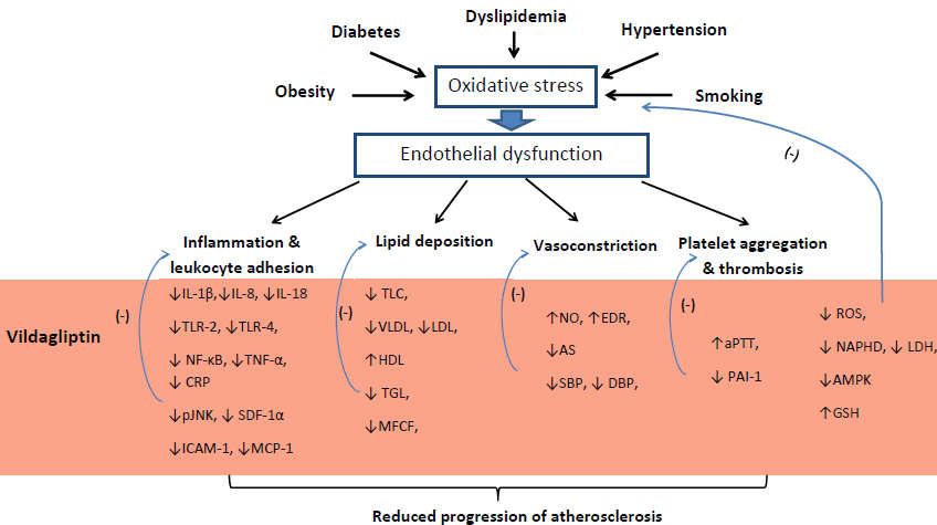
Int. J. Mol. Sci. 2020, 21, 2275 10 of 17
Figure
Figure 1.1.Proposed
Proposed influence
influenceofofvildagliptin on atherosclerosis
vildagliptin pathophysiology.
on atherosclerosis Note: ↓ = Note:
pathophysiology. reduction,
↓ = reduction, ↑
↑ = increase, (-) = inhibition, IL-1β = Interleukin 1 beta, IL-8 = Interleukin 8, IL-18 = Interleukin 18,
= increase, (-) = inhibition, IL-1β = Interleukin 1 beta, IL-8 = Interleukin 8, IL-18 = Interleukin 18, TLR2
TLR2 = Toll-like receptor 2, TLR4 = Toll-like receptor 4, NF-κB = nuclear factor kappa-light-chain-
= Toll-like receptor 2, TLR4 = Toll-like receptor 4, NF-κB = nuclear factor kappa-light-chain-enhancer of
enhancer of activated B cells, TNF-α = tumor necrosis factor α, CRP = C-reactive protein, pJNK =
activated B cells, TNF-α
phosphorylated = tumor necrosis
c-Jun N-terminal factor α,
kinase, SDF-1α = CRP = C-reactive
stromal cell-derivedprotein,
factor 1,pJNK = phosphorylated
ICAM-1 =
c-Jun N-terminal
intercellular kinase,
adhesion SDF-1α
molecule = stromal
1, MCP-1 cell-derived
= Monocyte chemoattractant protein-1, =TLC
factor 1, ICAM-1 intercellular
= total adhesion
molecule 1, MCP-1 = Monocyte chemoattractant protein-1, TLC = total cholesterol level, VLDL
cholesterol level, VLDL = very low density lipoprotein, LDL = low density lipoprotein, HDL = high
density
= very lowlipoprotein,
density TGC = triglycerides,
lipoprotein, LDL MFCF
= low= density
macrophages foam cellsHDL
lipoprotein, formation,
= highNOdensity
= nitric lipoprotein,
oxide, EDR = endothelial-dependent relaxation, AS – arterial stiffness, SBP = systolic blood pressure,
TGC = triglycerides, MFCF = macrophages foam cells formation, NO = nitric oxide, EDR =
DBP = diastolic blood pressure, aPTT = activated partial thromboplastin time, PAI-1 = plasminogen
endothelial-dependent
activator inhibitor-1, ROSrelaxation, AS –species,
= reactive oxygen arterial stiffness,
NADPH SBP = systolic
= Nicotinamide adenineblood pressure, DBP =
dinucleotide
diastolic blood pressure, aPTT = activated partial thromboplastin time,
phosphate, LDH = lactate dehydrogenase, AMPK = 5’AMP-activated protein kinase, GSH = PAI-1 = plasminogen activator
inhibitor-1,
glutathione.ROS = reactive oxygen species, NADPH = Nicotinamide adenine dinucleotide phosphate,
LDH = lactate dehydrogenase, AMPK = 5’AMP-activated protein kinase, GSH = glutathione.
Author Contributions: M.W. (Michał Wiciński): supervision and funding acquisition; M.W. (Michał Wiciński),
K.G., E.W.: formal analysis, resources, and writing—original draft preparation; M.W. (Maciej Walczak):
writing—review
Author and editing,
Contributions: visualization,
M.W. (Michał and critical
Wiciński): revision
supervision of funding
and the article; B.M.: participation
acquisition; in data
M.W. (Michał Wiciński), K.G.,
collection;
E.W.: formal M.N.: critical
analysis, revision of
resources, thewriting—original
and article. All authors have read
draft and agreed M.W.
preparation; to the published version of the
(Maciej Walczak): writing—review
andmanuscript.
editing, visualization, and critical revision of the article; B.M.: participation in data collection; M.N.: critical
revision of the
Funding: Thearticle.
present All
workauthors have read
was supported and
by the agreed toofthe
Department published version
Pharmacology of the manuscript.
and Therapeutics, Faculty of
Medicine,
Funding: TheCollegium
presentMedicum
work wasin Bydgoszcz,
supported Nicolaus
by theCopernicus University,
Department Toruń, Poland.
of Pharmacology and Therapeutics, Faculty of
Medicine, Collegium
Conflicts of Interest: Medicum
The authorsin Bydgoszcz,
declare Nicolaus
no conflict Copernicus University, Toruń, Poland.
of interest.
Conflicts of Interest: The authors declare no conflict of interest.
References
1. World Health Organization Diabetes. Available online: http://www.who.int (accessed on 30 October 2018).
2. The Emerging Risk Factors Collaboration. Diabetes mellitus, fasting blood glucose concentration, and risk
of vascular disease: A collaborative meta-analysis of 102 prospective studies. Lancet 2010, 375, 2215–2222.
[CrossRef]
3. Croxtall, J.D.; Keam, S.J. Vildagliptin: A Review of its Use in the Management of Type 2 Diabetes Mellitus.
Drugs 2008, 68, 2387–2409. [CrossRef] [PubMed]
4. Thornberry, N.A.; Gallwitz, B. Mechanism of action of inhibitors of dipeptidyl-peptidase-4 (DPP-4). Best Pract.
Res. Clin. Endocrinol. Metab. 2009, 23, 479–486. [CrossRef] [PubMed]
5. Näslund, E.; Gutniak, M.; Skogar, S.; Rössner, S.; Hellström, P.M. Glucagon-like peptide 1 increases the
period of postprandial satiety and slows gastric emptying in obese men. Am. J. Clin. Nutr. 1998, 68, 525–530.
[CrossRef] [PubMed]Int. J. Mol. Sci. 2020, 21, 2275 11 of 17
6. Vella, A. Mechanism of Action of DPP-4 Inhibitors—New Insights. J. Clin. Endocrinol. Metab. 2012, 97,
2626–2628. [CrossRef] [PubMed]
7. Choe, E.Y.; Cho, Y.; Choi, Y.; Yun, Y.; Wang, H.J.; Kwon, O.; Lee, B.-W.; Ahn, C.W.; Cha, B.S.; Lee, H.C.; et al.
The Effect of DPP-4 Inhibitors on Metabolic Parameters in Patients with Type 2 Diabetes. Diabetes Metab. J.
2014, 38, 211. [CrossRef]
8. He, Y.-L.; Sadler, B.M.; Sabo, R.; Balez, S.; Wang, Y.; Campestrini, J.; Laurent, A.; Ligueros-Saylan, M.;
Howard, D. The Absolute Oral Bioavailability and??Population-Based Pharmacokinetic Modelling of a Novel
Dipeptidylpeptidase-IV Inhibitor, Vildagliptin, in Healthy Volunteers. Clin. Pharmacokinet. 2007, 46, 787–802.
[CrossRef]
9. He, Y.-L. Clinical Pharmacokinetics and Pharmacodynamics of Vildagliptin. Clin. Pharmacokinet. 2012, 51,
147–162. [CrossRef]
10. Dejager, S.; Razac, S.; Foley, J.; Schweizer, A. Vildagliptin in Drug-naïve Patients with Type 2 Diabetes:
A 24-Week, Double-blind, Randomized, Placebo-controlled, Multiple-dose Study. Horm Metab Res. 2007, 39,
218–223. [CrossRef]
11. He, H.; Tran, P.; Yin, H.; Smith, H.; Batard, Y.; Wang, L.; Einolf, H.; Gu, H.; Mangold, J.B.; Fischer, V.; et al.
Absorption, Metabolism, and Excretion of [14 C]Vildagliptin, a Novel Dipeptidyl Peptidase 4 Inhibitor,
in Humans. Drug Metab Dispos. 2009, 37, 536–544. [CrossRef]
12. Trevisan, R. The Role of Vildagliptin in the Therapy of Type 2 Diabetic Patients with Renal Dysfunction.
Diabetes 2017, 8, 1215–1226. [CrossRef] [PubMed]
13. Lee, D.-S.; Lee, E.-S.; Alam, M.M.; Jang, J.-H.; Lee, H.-S.; Oh, H.; Kim, Y.-C.; Manzoor, Z.; Koh, Y.-S.;
Kang, D.-G.; et al. Soluble DPP-4 up-regulates toll-like receptors and augments inflammatory reactions,
which are ameliorated by vildagliptin or mannose-6-phosphate. Metabolism 2016, 65, 89–101. [CrossRef]
[PubMed]
14. Dei Cas, A.; Spigoni, V.; Cito, M.; Aldigeri, R.; Ridolfi, V.; Marchesi, E.; Marina, M.; Derlindati, E.; Aloe, R.;
Bonadonna, R.C.; et al. Vildagliptin, but not glibenclamide, increases circulating endothelial progenitor cell
number: A 12-month randomized controlled trial in patients with type 2 diabetes. Cardiovasc. Diabetol. 2017,
16, 27. [CrossRef] [PubMed]
15. Zhang, M.; Jin, X.; Zhang, Z.; Li, B.; Yang, G. Vildagliptin protects endothelial cells against high
glucose-induced damage. Biomed. Pharmacother. 2018, 108, 1790–1796. [CrossRef]
16. Qi, Y.; Du, X.; Yao, X.; Zhao, Y. Vildagliptin inhibits high free fatty acid (FFA)-induced NLRP3 inflammasome
activation in endothelial cells. Artif. CellsNanomed. Biotechnol. 2019, 47, 1067–1074. [CrossRef]
17. Liu, H.; Xiang, H.; Zhao, S.; Sang, H.; Lv, F.; Chen, R.; Shu, Z.; Chen, A.F.; Chen, S.; Lu, H. Vildagliptin
improves high glucose-induced endothelial mitochondrial dysfunction via inhibiting mitochondrial fission.
J. Cell Mol. Med. 2019, 23, 798–810. [CrossRef]
18. Seo, M.S.; Li, H.; An, J.R.; Jung, I.D.; Jung, W.-K.; Ha, K.-S.; Han, E.-T.; Hong, S.-H.; Choi, I.-W.; Park, W.S.
Vildagliptin, an Anti-diabetic Drug of the DPP-4 Inhibitor, Induces Vasodilation via Kv Channel and SERCA
Pump Activation in Aortic Smooth Muscle. Cardiovasc. Toxicol. 2019, 19, 244–254. [CrossRef]
19. Oeseburg, H.; de Boer, R.A.; Buikema, H.; van der Harst, P.; van Gilst, W.H.; Silljé, H.H.W. Glucagon-Like
Peptide 1 Prevents Reactive Oxygen Species–Induced Endothelial Cell Senescence Through the Activation of
Protein Kinase A. Arter. Thromb Vasc Biol. 2010, 30, 1407–1414. [CrossRef] [PubMed]
20. Terasaki, M.; Nagashima, M.; Watanabe, T.; Nohtomi, K.; Mori, Y.; Miyazaki, A.; Hirano, T. Effects
of PKF275-055, a dipeptidyl peptidase–4 inhibitor, on the development of atherosclerotic lesions in
apolipoprotein E–null mice. Metabolism 2012, 61, 974–977. [CrossRef] [PubMed]
21. Terasaki, M.; Nagashima, M.; Nohtomi, K.; Kohashi, K.; Tomoyasu, M.; Sinmura, K.; Nogi, Y.; Katayama, Y.;
Sato, K.; Itoh, F.; et al. Preventive Effect of Dipeptidyl Peptidase-4 Inhibitor on Atherosclerosis Is Mainly
Attributable to Incretin’s Actions in Nondiabetic and Diabetic Apolipoprotein E-Null Mice. PLoS ONE 2013,
8, e70933. [CrossRef] [PubMed]
22. Khan, S.; Khan, S.; Panda, B.P.; Akhtar, M.; Najmi, A.K. Potential effects of vildagliptin on biomarkers
associated with prothrombosis in diabetes mellitus. Expert Opin. Ther. Targets 2015, 19, 1607–1616. [CrossRef]
[PubMed]
23. Jain, S.; Sharma, B. Neuroprotective effect of selective DPP-4 inhibitor in experimental vascular dementia.
Physiol. Behav. 2015, 152, 182–193. [CrossRef] [PubMed]Int. J. Mol. Sci. 2020, 21, 2275 12 of 17
24. Koyama, A.; Komori, K.; Otsuka, R.; Kajikuri, J.; Itoh, T. Dipeptidyl peptidase 4 inhibitor reduces intimal
hyperplasia in rabbit autologous jugular vein graft under poor distal runoff. J. Vasc. Surg. 2016, 63, 1360–1370.
[CrossRef]
25. Zhang, Q.; Xiao, X.; Zheng, J.; Li, M.; Yu, M.; Ping, F.; Wang, T.; Wang, X. A Possible Mechanism: Vildagliptin
Prevents Aortic Dysfunction through Paraoxonase and Angiopoietin-Like 3. BioMed Res. Int. 2018, 2018,
1–14. [CrossRef] [PubMed]
26. Ji, Y.; Ge, Y.; Xu, X.; Ye, S.; Fan, Y.; Zhang, J.; Mei, L.; Zhang, X.; Ying, L.; Yang, T.; et al. Vildagliptin Reduces
Stenosis of Injured Carotid Artery in Diabetic Mouse Through Inhibiting Vascular Smooth Muscle Cell
Proliferation via ER Stress/NF-κB Pathway. Front. Pharm. 2019, 10, 142. [CrossRef] [PubMed]
27. van Poppel, P.C.M.; Netea, M.G.; Smits, P.; Tack, C.J. Vildagliptin Improves Endothelium-Dependent
Vasodilatation in Type 2 Diabetes. Dia Care 2011, 34, 2072–2077. [CrossRef] [PubMed]
28. Noguchi, K.; Hirota, M.; Miyoshi, T.; Tani, Y.; Noda, Y.; Ito, H.; Nanba, S. Single administration of vildagliptin
attenuates postprandial hypertriglyceridemia and endothelial dysfunction in normoglycemic individuals.
Exp. Ther. Med. 2015, 9, 84–88. [CrossRef]
29. Tani, S.; Takahashi, A.; Nagao, K.; Hirayama, A. Effect of Dipeptidyl Peptidase-4 Inhibitor, Vildagliptin on
Plasminogen Activator Inhibitor-1 in Patients with Diabetes Mellitus. Am. J. Cardiol. 2015, 115, 454–460.
[CrossRef]
30. Foley, J.E.; Evans, M.; Schweizer, A. Blood pressure and fasting lipid changes after 24 weeks’ treatment
with vildagliptin: A pooled analysis in >2,000 previously drug-naïve patients with type 2 diabetes mellitus.
VHRM 2016, 12, 337–340. [CrossRef]
31. Duvnjak, L.; Blaslov, K. Dipeptidyl peptidase-4 inhibitors improve arterial stiffness, blood pressure, lipid
profile and inflammation parameters in patients with type 2 diabetes mellitus. Diabetol. Metab. Syndr. 2016,
8, 26. [CrossRef]
32. Park, K.S.; Kwak, S.; Cho, Y.M.; Park, K.S.; Jang, H.C.; Kim, S.Y.; Jung, H.S. Vildagliptin reduces plasma
stromal cell-derived factor-1α in patients with type 2 diabetes compared with glimepiride. J. Diabetes Investig.
2017, 8, 218–226. [CrossRef] [PubMed]
33. Younis, A.; Eskenazi, D.; Goldkorn, R.; Leor, J.; Naftali-Shani, N.; Fisman, E.Z.; Tenenbaum, A.; Goldenberg, I.;
Klempfner, R. The addition of vildagliptin to metformin prevents the elevation of interleukin 1ß in patients
with type 2 diabetes and coronary artery disease: A prospective, randomized, open-label study. Cardiovasc.
Diabetol. 2017, 16, 69. [CrossRef]
34. El-Naggar, A.R.; Zaafar, D.; Elyamany, M.; Hassanin, S.; Bassyouni, A.; Abdel-Latif, H. The Role of Vildagliptin
in Treating Hypertension Through Modulating Serum VEGF in Diabetic Hypertensive Patients. J. Cardiovasc.
Pharm. 2019, 24, 254–261. [CrossRef] [PubMed]
35. Rajendran, P.; Rengarajan, T.; Thangavel, J.; Nishigaki, Y.; Sakthisekaran, D.; Sethi, G.; Nishigaki, I. The
Vascular Endothelium and Human Diseases. Int. J. Biol. Sci. 2013, 9, 1057–1069. [CrossRef] [PubMed]
36. Madden, J.A. Role of the vascular endothelium and plaque in acute ischemic stroke. Neurology 2012, 79,
S58–S62. [CrossRef]
37. Di Meo, S.; Reed, T.T.; Venditti, P.; Victor, V.M. Harmful and Beneficial Role of ROS. Oxidative Med. Cell.
Longev. 2016, 2016, 1–3. [CrossRef]
38. Dey, S.; DeMazumder, D.; Sidor, A.; Foster, D.B.; O’Rourke, B. Mitochondrial ROS Drive Sudden Cardiac
Death and Chronic Proteome Remodeling in Heart Failure. Circ. Res. 2018, 123, 356–371. [CrossRef]
39. Gimbrone, M.A.; García-Cardeña, G. Endothelial Cell Dysfunction and the Pathobiology of Atherosclerosis.
Circ. Res. 2016, 118, 620–636. [CrossRef]
40. Su, J.B. Vascular endothelial dysfunction and pharmacological treatment. World J. Cardiol. 2015, 7, 719.
[CrossRef]
41. Park, K.-H.; Park, W.J. Endothelial Dysfunction: Clinical Implications in Cardiovascular Disease and
Therapeutic Approaches. J. Korean Med. Sci. 2015, 30, 1213. [CrossRef]
42. Sharp, W.W.; Fang, Y.H.; Han, M.; Zhang, H.J.; Hong, Z.; Banathy, A.; Morrow, E.; Ryan, J.J.; Archer, S.L.
Dynamin-related protein 1 (Drp1)-mediated diastolic dysfunction in myocardial ischemia-reperfusion injury:
therapeutic benefits of Drp1 inhibition to reduce mitochondrial fission. Faseb J. 2014, 28, 316–326. [CrossRef]
[PubMed]Int. J. Mol. Sci. 2020, 21, 2275 13 of 17
43. Hausenloy, D.J.; Whittington, H.J.; Wynne, A.M.; Begum, S.S.; Theodorou, L.; Riksen, N.; Mocanu, M.M.;
Yellon, D.M. Dipeptidyl peptidase-4 inhibitors and GLP-1 reduce myocardial infarct size in a glucose-
dependent manner. Cardiovasc. Diabetol. 2013, 12, 154. [CrossRef] [PubMed]
44. Chinda, K.; Sanit, J.; Chattipakorn, S.; Chattipakorn, N. Dipeptidyl peptidase-4 inhibitor reduces infarct size
and preserves cardiac function via mitochondrial protection in ischaemia–reperfusion rat heart. Diabetes
Vasc. Dis. Res. 2014, 11, 75–83. [CrossRef]
45. Bayrami, G.; Karimi, P.; Agha-Hosseini, F.; Feyzizadeh, S.; Badalzadeh, R. Effect of Ischemic Postconditioning
on Myocardial Function and Infarct Size Following Reperfusion Injury in Diabetic Rats Pretreated With
Vildagliptin. J. Cardiovasc. Pharm. 2018, 23, 174–183. [CrossRef] [PubMed]
46. McMurray, J.J.V.; Ponikowski, P.; Bolli, G.B.; Lukashevich, V.; Kozlovski, P.; Kothny, W.; Lewsey, J.D.; Krum, H.
Effects of Vildagliptin on Ventricular Function in Patients with Type 2 Diabetes Mellitus and Heart Failure.
JACC Heart Fail. 2018, 6, 8–17. [CrossRef]
47. Loboda, A.; Damulewicz, M.; Pyza, E.; Jozkowicz, A.; Dulak, J. Role of Nrf2/HO-1 system in development,
oxidative stress response and diseases: An evolutionarily conserved mechanism. Cell. Mol. Life Sci. 2016, 73,
3221–3247. [CrossRef]
48. Werner, N.; Kosiol, S.; Schiegl, T.; Ahlers, P.; Walenta, K.; Link, A.; Böhm, M.; Nickenig, G. Circulating
Endothelial Progenitor Cells and Cardiovascular Outcomes. N. Engl. J. Med. 2005, 353, 999–1007. [CrossRef]
49. Wei, D.; Wang, G.; Tang, C.; Qiu, J.; Zhao, J.; Gregersen, H.; Deng, L. Upregulation of SDF-1 is Associated
with Atherosclerosis Lesions Induced by LDL Concentration Polarization. Ann. Biomed. Eng. 2012, 40,
1018–1027. [CrossRef]
50. Zhong, J.; Rajagopalan, S. Dipeptidyl Peptidase-4 Regulation of SDF-1/CXCR4 Axis: Implications for
Cardiovascular Disease. Front. Immunol. 2015, 6, 477. [CrossRef]
51. Deshane, J.; Chen, S.; Caballero, S.; Grochot-Przeczek, A.; Was, H.; Li Calzi, S.; Lach, R.; Hock, T.D.; Chen, B.;
Hill-Kapturczak, N.; et al. Stromal cell–derived factor 1 promotes angiogenesis via a heme oxygenase
1–dependent mechanism. J. Exp. Med. 2007, 204, 605–618. [CrossRef]
52. Hristov, M.; Zernecke, A.; Bidzhekov, K.; Liehn, E.A.; Shagdarsuren, E.; Ludwig, A.; Weber, C. Importance of
CXC Chemokine Receptor 2 in the Homing of Human Peripheral Blood Endothelial Progenitor Cells to Sites
of Arterial Injury. Circ. Res. 2007, 100, 590–597. [CrossRef]
53. Li, M.; Yu, J.; Li, Y.; Li, D.; Yan, D.; Qu, Z.; Ruan, Q. CXCR4 positive bone mesenchymal stem cells migrate to
human endothelial cell stimulated by ox-LDL via SDF-1α/CXCR4 signaling axis. Exp. Mol. Pathol. 2010, 88,
250–255. [CrossRef] [PubMed]
54. Abi-Younes, S.; Sauty, A.; Mach, F.; Sukhova, G.K.; Libby, P.; Luster, A.D. The Stromal Cell–Derived Factor-1
Chemokine Is a Potent Platelet Agonist Highly Expressed in Atherosclerotic Plaques. Circ. Res. 2000, 86,
131–138. [CrossRef] [PubMed]
55. Brenner, C.; Franz, W.M.; Kühlenthal, S.; Kuschnerus, K.; Remm, F.; Gross, L.; Theiss, H.D.; Landmesser, U.;
Kränkel, N. DPP-4 inhibition ameliorates atherosclerosis by priming monocytes into M2 macrophages. Int. J.
Cardiol. 2015, 199, 163–169. [CrossRef] [PubMed]
56. Subramanian, S.; Liu, C.; Aviv, A.; Ho, J.E.; Courchesne, P.; Muntendam, P.; Larson, M.G.; Cheng, S.; Wang, T.J.;
Mehta, N.N.; et al. Stromal Cell–Derived Factor 1 as a Biomarker of Heart Failure and Mortality Risk. Arter.
Thromb. Vasc. Biol. 2014, 34, 2100–2105. [CrossRef]
57. Donath, M.Y. Multiple benefits of targeting inflammation in the treatment of type 2 diabetes. Diabetologia
2016, 59, 679–682. [CrossRef]
58. Assar, M.E.; Angulo, J.; Rodríguez-Mañas, L. Diabetes and ageing-induced vascular inflammation. J. Physiol.
2016, 594, 2125–2146. [CrossRef]
59. Teague, H.L.; Ahlman, M.A.; Alavi, A.; Wagner, D.D.; Lichtman, A.H.; Nahrendorf, M.; Swirski, F.K.;
Nestle, F.; Gelfand, J.M.; Kaplan, M.J.; et al. Unraveling Vascular Inflammation. J. Am. Coll. Cardiol. 2017, 70,
1403–1412. [CrossRef]
60. Gordon, S.; Plüddemann, A. Tissue macrophages: heterogeneity and functions. BMC Biol. 2017, 15, 53.
[CrossRef]
61. Chen, L.; Deng, H.; Cui, H.; Fang, J.; Zuo, Z.; Deng, J.; Li, Y.; Wang, X.; Zhao, L. Inflammatory responses and
inflammation-associated diseases in organs. Oncotarget 2018, 9. [CrossRef]
62. Virdis, A.; Schiffrin, E.L. Vascular inflammation: A role in vascular disease in hypertension? Curr. Opin.
Nephrol. Hypertens. 2003, 12, 181–187. [CrossRef] [PubMed]You can also read


