ADAM-Mediated Signalling Pathways in Gastrointestinal Cancer Formation
←
→
Page content transcription
If your browser does not render page correctly, please read the page content below
International Journal of
            Molecular Sciences

Review
ADAM-Mediated Signalling Pathways in
Gastrointestinal Cancer Formation
Neele Schumacher, Stefan Rose-John                    and Dirk Schmidt-Arras *
 Institute of Biochemistry, Christian-Albrechts-University, 24118 Kiel, Germany;
 nschumacher@biochem.uni-kiel.de (N.S.); rosejohn@biochem.uni-kiel.de (S.R.-J.)
 * Correspondence: darras@biochem.uni-kiel.de; Tel.: +49-431-880-7112

 Received: 27 June 2020; Accepted: 15 July 2020; Published: 20 July 2020

 Abstract: Tumour growth is not solely driven by tumour cell-intrinsic mechanisms, but also depends
 on paracrine signals provided by the tumour micro-environment. These signals comprise cytokines
 and growth factors that are synthesized as trans-membrane proteins and need to be liberated by
 limited proteolysis also termed ectodomain shedding. Members of the family of A disintegrin and
 metalloproteases (ADAM) are major mediators of ectodomain shedding and therefore initiators of
 paracrine signal transduction. In this review, we summarize the current knowledge on how ADAM
 proteases on tumour cells but also on cells of the tumour micro-environment contribute to the formation
 of gastrointestinal tumours, and discuss how these processes can be exploited pharmacologically.

 Keywords: ADAM; protease; EGFR; tumour micro-environment; Notch; IL-6

1. Introduction
      Gastrointestinal organs are composed of highly complex tissue and most of these tissues
are constantly replenished by the proliferation and differentiation of multi-potent tissue stem
cells. These processes are regulated by paracrine signals provided by cells of the stem cell niche.
Moreover, signals emitted from inflammatory cells recruited to damaged or infected tissue contribute
to the mobilisation of tissue stem cells, regeneration and remodelling of the damaged tissue.
Several paracrine signal proteins are synthesized as membrane-bound proteins and need to be
proteolytically processed to give rise to soluble factors that then can act in a paracrine or even an
endocrine fashion. Additionally, receptor molecules and proteins of the extracellular matrix are
subject to proteolytic remodelling. Members of the A disintegrin and metalloprotease (ADAM)
family are involved in all these processes. Aberrant activation of paracrine signal transduction is
key to the development of gastrointestinal tumours and it is therefore not surprising that ADAM
proteases play a decisive role in gastrointestinal tumorigenesis. Here, we give an overview of ADAM
protease biology and the current knowledge regarding their tumour-promoting role in gastrointestinal
malignancies. We further discuss how ADAM proteases can be therapeutically targeted for the
treatment of gastrointestinal cancers.

2. ADAM Proteases

2.1. Overview
     Limited ectodomain proteolysis is a regulatory mechanism mediated by proteases that converts
membrane-bound proteins irreversibly into their soluble isoforms. The family of A disintegrin and
metalloproteases (ADAMs) consists of 21 members (Table 1) with 13 of them being proteolytically
active [1]. ADAM proteases belong to the superfamily of zinc-proteases that is characterised by
the presence of an invariant HEXXHXXGXXH zinc-binding motif with the catalytic domain [2,3].

Int. J. Mol. Sci. 2020, 21, 5133; doi:10.3390/ijms21145133                         www.mdpi.com/journal/ijms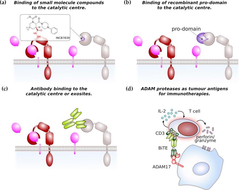
Int. J. Mol. Sci. 2020, 21, 5133                                                                                          2 of 19

ADAM proteases are expressed as inactive zymogens, consisting of an N-terminal signal sequence,
a pro-domain, a catalytic metalloproteinase domain, a disintegrin domain and a cysteine-rich,
EGF-like domain followed by a transmembrane part and cytoplasmic tail (Figure 1a). In place
of the EGF-like, cysteine-rich domain, ADAM17 and its close relative ADAM10 are characterized
by a membrane-proximal domain in their extracellular region, which has also been reported to be
involved in substrate recognition [4,5]. The prodomain has chaperone-like functions, inhibits enzymatic
activity and is removed during maturation by the intracellular proprotein-convertase furin (Figure 1b)
with additional proprotein convertase cleavage sites recently identified [6–8].

                                   Table 1. Classification of the human ADAM protease family.

                                             testis specific                                         ADAM 20,21,30
      Proteolytically Active                                           haematopoietic                  ADAM 8,28
                                           not testis specific
       (HEXXHXXGXXH)                                                                                     ADAM
                                                                    non haematopoietic
                                                                                                9,10,12,15,17,19,33,DEC1
                                             testis specific                                        ADAM 2,7, 18,29
     Proteolytically Inactive
                                           not testis specific                                      ADAM 11,22,23,32
 Int. J. Mol. Sci. 2020, 21, x FOR PEER REVIEW                                                                       4 of 20

      Figure 1. ADAMs and ADAM-mediated signalling pathways. (a) General domain structure of ADAM
        Figure 1. ADAMs
      proteases.            and ADAM-mediated
                   Pro, pro-domain;   Cat;catalyticsignalling
                                                     domain;pathways.       (a) General
                                                               Dis, disintegrin          domain
                                                                                    domain;   CR,structure  of ADAM
                                                                                                   cysteine-rich  domain;
        proteases.  Pro, pro-domain;  Cat;catalytic domain;  Dis,  disintegrin   domain;   CR,
      EGF, EGF-like domain; TM, transmembrane domain; CD, cytoplasmic domain (b) Maturation of cysteine-rich  domain;
        EGF, EGF-like
      ADAM      protease,domain;
                           notablyTM,  transmembrane
                                   ADAM10      and ADAM17 domain;    CD, cytoplasmic
                                                                is facilitated            domain (Tspans)
                                                                                 by tetraspanins   (b) Maturation    of
                                                                                                             and inactive
        ADAM     protease, notably ADAM10     and  ADAM17     is facilitated  by  tetraspanins
      rhomboid proteases (iRhom), respectively. Activation of ADAM proteases, notably ADAM17,   (Tspans) and  inactive
        rhomboid
      via           proteases
            C-terminal        (iRhom), respectively.
                         phosphorylation              Activation
                                            is mediated            of ADAM
                                                           by protein     kinaseproteases,
                                                                                    C (PKC) notably
                                                                                              or theADAM17,      via C-
                                                                                                      mitogen-activated
        terminal
      kinase      phosphorylation
               p38.                is mediated
                     (c) ADAM proteases    haveby   proteinsubstrates
                                                  diverse   kinase C (PKC)
                                                                         and areor the mitogen-activated
                                                                                   involved               kinase p38.
                                                                                              in cytokine/growth     factor
        (c) ADAM      proteases have diverse   substrates and   are  involved    in  cytokine/growth   factor
      release, shedding of receptor molecules and the induction of intracellular signal transduction via       release,
        shedding
      limited      of receptor
                proteolysis,    molecules
                             followed      and the induction
                                       by regulated              of intracellular
                                                      intramembrane       proteolysissignal transduction via limited
                                                                                        (RIP).
        proteolysis, followed by regulated intramembrane proteolysis (RIP).

 3. ADAM-Mediated Pathways in Gastrointestinal Tumour Formation

 3.1. ADAM Proteases in Gastric Cancer
     Gastric cancer is the fourth most frequently occurring but the second most deadly cancer
 worldwide. One major risk for the development of gastric cancer is infection by Helicobacter pylori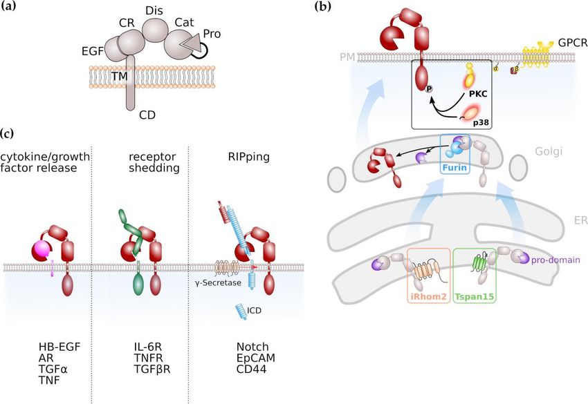
Int. J. Mol. Sci. 2020, 21, 5133                                                                     3 of 19

2.2. Regulation of ADAM Protease Activity
      Proteolytic activity of ADAM proteases can either be constitutive or activated by signal transduction
pathways. Regulation of ADAM protease activity is best described for the family members ADAM10
and ADAM17. Members of the membrane multi-pass families inactive rhomboids (iRhoms) [9,10] and
tetraspanins (Tspans) [11,12] are involved in maturation of ADAMs throughout the secretory pathway
(Figure 1b).
      The rhomboid family comprises several transmembrane proteins that are evolutionarily conserved.
The inactive rhomboid members lack the amino-acid residues that are indispensable for catalytic activity.
      ADAM17 needs to be located at the plasma membrane in order to exert its proteolytic activity.
An indispensable factor for ADAM17 activity is the inactive rhomboid protease (iRhom) 2, encoded by
the rhomboid family member (RHBDF) 2 gene. iRhom2 is localized in the ER and shuttles ADAM17
from the ER to the Golgi apparatus [9]. Importantly, it is specific for ADAM17, since the transport
of ADAM10 to the cell membrane is not affected by the absence of iRhom2 [10]. Whereas iRhom2 is
proposed to be a myeloid-specific regulator of ADAM17 activation, its closest homologue iRhom1 is
more broadly expressed [13]. The cleavage of ADAM17 substrates is seriously impaired in embryonic
fibroblasts derived from iRhom2-deficient mice. However, not all ADAM17 substrates are affected by an
iRhom2 deficiency, indicating a role of iRhom2 in ADAM17 substrate selection and discrimination [14].
      As the name indicates, tetraspanins span the membrane four times, resulting in the formation of
a short and a large extracellular loop. Tspans have the capacity to form large interacting networks,
the so-called tetraspanin-enrichted microdomains (TEMs) which regulate signal transduction pathways
similar to lipid rafts [15,16]. ADAM10 was previously identified to interact with members of the
TspanC8 family within the ER which promoted maturation of ADAM10 to the plasma membrane [11,12].
Therefore, Tspans might emerge in general as regulators and facilitators of ADAM protease activity.
      The intracellular region of ADAM17 contains numerous phosphorylation sites with
phosphorylation of threonine 735 leading to rapid translocation of ADAM17 to the cell surface,
resulting in increased ADAM17 activity [17,18]. It is described that the mitogen-activated protein (MAP)
kinases ERK and p38 are involved in ADAM17 phosphorylation thus regulating its activity [19,20],
as well as family members of the protein kinase C (PKC) family (Figure 1b). Activation of PKCs can
result from engagement of G-protein coupled receptors (GPCRs) that are coupled to an αq/11 subunit
resulting in phospholipase C (PLC)-mediated increase in second messengers diacylglycerol (DAG),
inositol triphosphate (IP3 ) and Ca2+ [21,22].
      Proteolytic activity of ADAM10 and ADAM17 is also triggered by exposure to negatively charged
phosphatidylserine [23,24]. Phosphatidylserine in the outer leaflet of the cell membrane usually
indicates cells which undergo apoptosis and cell death was indeed identified as a potent activator of
ADAM proteases [25]. Moreover, ADAM protease activity can be regulated by limited proteolysis
and ADAM proteases were suggested to be cleaved by other ADAM family members and the astacin
family member meprin β [26–29].
      The regulation of other ADAM family members is less well understood. The key to the activation
of all ADAM family members is the removal of the pro-domain by proprotein convertases, as described
above. However, the pro-domain of the family member ADAM8 is removed in an autocatalytic
process [30]. Expression of ADAM8 itself is regulated by inflammatory cytokines such as TNFα, IL-1β,
IL-4 and IL-13 [30]. ADAM12 is stored as active protease intracellularly and substrate exposure occurs
via trafficking to the plasma membrane [31].

2.3. Signalling Pathways Regulated by ADAM Proteases
    ADAM proteases play a decisive role in inflammation and cancer due to their variety of substrates.
Among these substrates are cytokines, growth factors and cell surface receptors (Figure 1c). The most
prominent substrate for ADAM17 is Tumor Necrosis Factor α (TNFα), a cytokine which binds to
TNFα receptors (TNFRs) 1 and 2 which both are ADAM17 substrates as well [32]. The resulting
soluble TNFR ectodomains can still bind TNFα thus act as antagonistic decoy receptors which
Int. J. Mol. Sci. 2020, 21, 5133 4 of 19
has been used therapeutically. In contrast to the antagonistic activity of soluble TNFRs in TNFα
signalling, the proteolytically cleaved receptor for the pleiotropic cytokine Interleukin-6 (IL-6) acts in an
agonistic fashion. IL-6 signals via a membrane-bound alpha-receptor (IL-6R) and a homodimer of the
signal-transducing beta-receptor subunit glycoprotein 130 (gp130), which is ubiquitously expressed,
while expression of IL-6R is limited to selected cell types, e.g., hepatocytes and leukocytes. This process
is termed IL-6 classic signalling. However, cells that do not express the membrane-bound IL-6R are
still responsive to IL-6 via IL-6 trans-signalling. In this process the complex of IL-6 and soluble IL-6R
(sIL-6R) which can be generated via limited proteolysis mediated by ADAM10 and ADAM17, is able
to engage gp130 homodimers on target cells [33–35].
ADAM protease activity was also demonstrated to increase bioavailability of epidermal growth
factor receptor (EGFR) ligands like transforming growth factor α (TGFα), heparin-bound EGF
(HB-EGF) and amphiregulin (AR) [36–38]. EGFR signalling is crucial for regenerative processes as it
was, e.g., demonstrated that both EGFR-hypomorphic mice (Waved-2) and ADAM17-hypomorphic
mice (ADAM17ex/ex ) display impaired intestinal epithelial regeneration [39,40]. The enhanced release
of EGFR ligands via ADAM17 was shown to drive cancer progression and drug resistance in cell lines
and experimental mouse models [41–43] (see also below).
Signal transduction via the Notch receptor is an evolutionary conserved pathway in which the
Notch receptor undergoes a series of proteolytic events. Notch signalling was demonstrated to be
essentially involved in stem cell maintenance and differentiation, but also in tumour formation [44,45].
In the majority of tissues analysed, ADAM10 was identified as the major extracellular protease
for Notch [46,47]. This cleavage is followed by regulated intramembrane proteolysis (RIP) by the
γ-secretase complex in order to release an intracellular domain (Figure 1c) that translocates to the
nucleus and induces expression of Notch target genes [44,45]. Next to ADAM10, ADAM17 is discussed
to promote Notch signalling [48,49].
Additionally, ADAM17 is described to be responsible for the proteolysis of Notch1 receptor,
which increases EGFR expression [49]. The Notch receptor undergoes a series of proteolytic events in
order to release an intracellular domain which is necessary for signal transduction. However, in this
process the involvement of both proteases, ADAM10 and ADAM17, is discussed and seems to be
context dependent [48].
3. ADAM-Mediated Pathways in Gastrointestinal Tumour Formation
3.1. ADAM Proteases in Gastric Cancer
Gastric cancer is the fourth most frequently occurring but the second most deadly cancer
worldwide. One major risk for the development of gastric cancer is infection by Helicobacter pylori
which is reflected by the fact that gastric cancer incidence is increased in areas with high H. pylori
prevalence. Transactivation of EGFR family members and chronic inflammation are potential
mechanisms how H. pylori infection contribute to the development of gastric cancer [50]. The vast
majority of gastric cancers are adenocarcinoma arising from glandular epithelial cells of the gastric
mucosa. Gastric atrophy, i.e., the loss of glandular tissue, including parietal cells, can progress to
metaplasia due to the absence of signals regulating stem cell proliferation and differentiation provided
by parietal cells [51]. Chronic inflammation of the gastric mucosa, e.g., triggered by bacterial infections
such as H.pylori often results in the formation of pre-malignant lesions. Accordingly, mice with
hyperactive gp130, the signal transducing subunit of the interleukin 6 (IL-6) receptor complex resulted
in an excessive inflammatory response of the gastric epithelium and the formation of gastric metaplasia
and spontaneous adenoma [52].
The most common genetic alterations in gastric cancer comprise among others, mutations in
genes encoding for the tumour suppressor p53, the small GTPase KRAS, phosphoinositide-3 kinase
(PI3K), but also members of the epidermal growth factor receptor (EGFR) family, including EGFR andInt. J. Mol. Sci. 2020, 21, 5133 5 of 19
ErbB2 [53,54]. KRAS, PI3K and EGFR proteins are connected within a common signal transduction
pathway, highlight the importance of the EGFR/MAPK pathway for the development of gastric cancer.
Several ADAM proteases including ADAM9, 10, 12, 15, 17 and 33 were shown to be overexpressed
in gastric cancer [55–60], while ADAM23 was shown to be epigenetically silenced by promoter CpG
methylation in gastric tumour tissue [61]. Expression of ADAM10 and ADAM17 was demonstrated
to be induced by H. pylori infection [55,62] and expression of ADAM17 correlated with the forkhead
transcription factor FoxM1 which is activated by PI3K/AKT [60]. ADAM17 activity is not solely
dependent on its transcription level but needs additional stimuli. It is therefore not surprising that the
enhanced release of EGFR ligands amphiregulin and HB-EGF upon H. pylori infection was dependent
on the phosphorylation of ADAM17 C-terminus [62].
As described above, ligands for the EGFR can be liberated by limited proteolysis mainly via
ADAM10 or ADAM17 activity. It is therefore plausible that ADAM proteases provide soluble ligands
for the activation of EGFR family members on gastric tumour cells. It was indeed demonstrated that
the inflammatory cytokines IL-1β and IL-8 [63], but TGFβ [64] also induced EGFR trans-activation
in gastric cancer cells, particularly via soluble amphiregulin and HB-EGF (Figure 2a). Depletion of
ADAM10 but not ADAM12 or ADAM17 abrogated the release of soluble EGFR ligands and hence
EGFR trans-activation [65]. Upon proteolytic release of HB-EGF ectodomain, a C-terminal fragment
(CTF) remains in the signal-sending cell and it was suggested that nuclear translocation of HB-EGF
CTF contributes to the pathogenicity of gastric cancer [63].
Mouse models with activating mutations in KRAS in gastric epithelial cells but not Helicobacter
infections resulted in an increase in AR and HB-EGF secretion, supporting the existence of a RTK/MAPK
feed-forward loop in gastric cancer. However, while aberrant KRAS activation alone induced
gastric metaplasia [66], the formation of adenoma was dependent on the additional activation of
gp130 [54], mainly through interleukin 11 (IL-11) [67]. While in vitro experiments showed that IL-6
trans-signalling can overcome the repressive effects of trefoil factor 1 (TFF1) on IL-6 classic-signalling [68],
gastric metaplasia induced by a hyperactive gp130 variant was not abrogated by a dimerised form of
soluble gp130 (sgp130Fc) [69]. These data suggest that in gastric cancer ADAM10 and ADAM17 rather
have a major role in EGFR trans-activation but not in boosting signal transduction via IL-6 family
cytokines as in other gastrointestinal tumours (see below).
ADAM17 was also shown to be overexpressed in gastrointestinal stroma tumours (GIST) where it
co-localised with EGF and EGFR [70]. Gastrointestinal stroma tumours are rare malignant tumours of
non-epithelial origin and the most common mesenchymal neoplasms of the gastrointestinal tract [71].
The majority of GISTs localise to the stomach and they are thought to originate from interstitial cells of
Cajal [71], a cell type that under physiological conditions controls gastric smooth muscle cell contraction.
GISTs are characterised by the occurrence of activating mutations in genes coding for the platelet
derived growth factor receptor alpha (PDGFRA) or the close homologue c-Kit [Schmidt-Arras and
Böhmer Trends Mol Med, in press]. PDGFRβ was previously shown to induce ADAM17 activation [72]
and it is likely that ADAM17 activity in cells is elevated by mutant PDGFRα or c-Kit, resulting in the
additional activation of EGFR. However, experimental evidence is still lacking.
The progression of gastric cancer correlates with the expression of ADAM10 and ADAM17 and
high ADAM10/ADAM17 expression levels were associated with patient’s poor prognosis [58,59] and
the establishment of lymph node metastasis [73]. A recent report demonstrated that inflammatory
cytokines IL-1α, IL-1β or TNFα secreted by diffuse-type gastric cancer cells induced upregulation
of rhomboid 5 homolog 2 (RHBDF2), also termed iRhom2 in cancer-associated fibroblasts (CAFs).
iRhom2 promoted ADAM17 activity and TGFβ receptor cleavage (Figure 2a) and as a consequence CAF
motility to invade extracellular matrix and lymphatic vessels [74]. Hence, progression of gastric cancer
is not only promoted by ADAM17 on tumour cells, but also on cells of the tumour micro-environment.Int. J. Mol. Sci. 2020, 21, 5133 6 of 19
Int. J. Mol. Sci. 2020, 21, x FOR PEER REVIEW 9 of 20
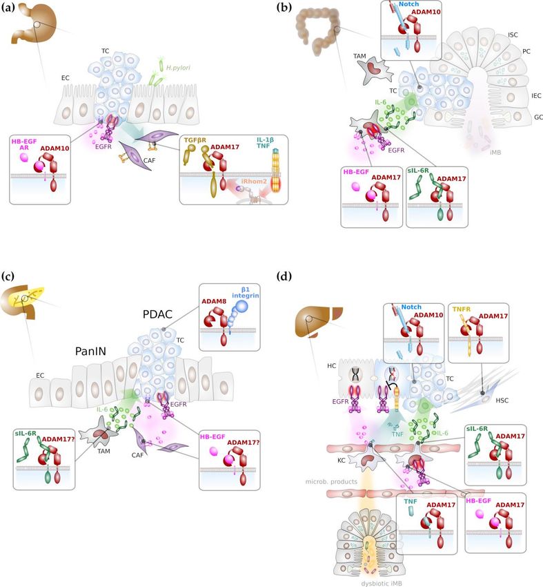
Figure 2. Major ADAM-mediated signalling pathways in gastrointestinal cancer formation. (a) ADAM
Figure 2. Major ADAM-mediated signalling pathways in gastrointestinal cancer formation. (a)
proteases in particular ADAM10 and ADAM17 promote gastric tumorigenesis mainly through activation
ADAM proteases in particular ADAM10 and ADAM17 promote gastric tumorigenesis mainly
of epidermal growth
through factor
activation (EGF) receptor
of epidermal (EGFR)
growth factor signalling
(EGF) on cancer
receptor (EGFR) cellson
signalling and through
cancer inhibition of
cells and
transforming growth
through factorof(TGF)
inhibition β receptor
transforming growth(TGFβR), thereby
factor (TGF) enhancing
β receptor (TGFβR), motility
thereby of cancer-associated
enhancing
fibroblastsmotility of cancer-associated
(in purple). (b) Colonicfibroblasts (in purple).
tumorigenesis (b) Colonic
depends on Notchtumorigenesis
signallingdepends
promoted on Notchby ADAM10.
signalling promoted by ADAM10. In addition, myeloid ADAM17 is supposed to promote
In addition, myeloid ADAM17 is supposed to promote inflammatory signalling via proteolytic
inflammatory signalling via proteolytic release of EGFR ligands, such as heparin-bound EGF (HB-
release ofEGF)EGFR ligands, such
and concomitant as heparin-bound
induction of interleukin (IL) 6 EGF
(green (HB-EGF) and with
circles). In concert concomitant
the soluble IL-6 induction of
interleukin (IL) 6(sIL-6R),
receptor (greenIL-6circles). In concert with
induces trans-signalling theepithelial
on colon solublecells.
IL-6(c)receptor (sIL-6R),
EGFR signalling IL-6 induces
and IL-6
trans-signalling
trans-signalling on colon promotes progression
epithelial cells.of pancreatic
(c) EGFRintraepithelial
signalling neoplasia
and IL-6(PanIN) to pancreatic promotes
trans-signalling
ductal adenocarcinoma (PDAC), presumably via ADAM17 activity. (d) DNA damage response (DDR)
progression of pancreatic intraepithelial neoplasia (PanIN) to pancreatic ductal adenocarcinoma
in hepatocytes is enhanced via EGFR signalling. Intestinal dysbiosis induces tumour necrosis factor
(PDAC), presumably via ADAM17 activity. (d) DNA damage response (DDR) in hepatocytes is
(TNF) secretion from Kupffer cells (KCs), the liver-resident macrophages that impairs the beneficial
enhancedeffects
via EGFR
of EGFR signalling Intestinal
signalling. on DDR. Both,dysbiosis induces
EGFR ligands andtumour
TNF are necrosis
presumably factor (TNF)
released by secretion
from Kupffer cells (KCs), the liver-resident macrophages that impairs the beneficial effects
ADAM17 from KCs. Liver fibrosis is reduced by ADAM17 via impaired TNF receptor signalling in of EGFR
hepatic stellate cells. Hepatocarcinogenesis is promoted by IL-6 trans-signalling
signalling on DDR. Both, EGFR ligands and TNF are presumably released by ADAM17 from KCs. that is presumably
Liver fibrosis is reduced by ADAM17 via impaired TNF receptor signalling in hepatic stellate cells.
Hepatocarcinogenesis is promoted by IL-6 trans-signalling that is presumably induced by ADAM17
on KCs. Notch signalling mediated by ADAM10 in hepatocytes promotes hepatic tumorigenesis.
Abbreviations: CAF: cancer associated fibroblast; EC: epithelial cell; GC: goblet cell; HSC: hepatic stellate
cell; IEC: intestinal epithelial cell; iMB: intestinal microbiome ISC: intestinal stem cell; KC: Kupffer cell;
PC: Paneth cell; TAM: tumour associated macrophage; TC: tumour cell.Int. J. Mol. Sci. 2020, 21, 5133 7 of 19
3.2. ADAM Proteases in Colorectal Cancer
The intestine is a complex organ comprising multiple types of differentiated and specialized
cell types. The intestinal lumen is lined by a single layer of columnar epithelial cels which are
constantly replenished by proliferative crypt cells. In the adult, Lgr5+ columnar base crypt (CBC)
stem cells locate to the crypt base and give rise to the multiple intestinal cell types. The stem cell
niche is formed and maintained by Paneth cells that intersperse the CBCs and provide Wnt ligands,
the Notch ligands delta like (Dll) 1 and 4 and EGF. Upon exiting the stem cell niche, CBCs give rise
to rapidly proliferating transit-amplifying (TA) progenitors and, subsequently, to either absorptive
progenitors or secretory progenitors, dependent on Notch receptor activity. High Notch activity results
in absorptive cells while low Notch activity induces secretory differentiation [75]. As outlined above,
ADAM10 is the major sheddase for the Notch receptor in multiples tissues and genetic deficiency
of ADAM10 in villin-expressing intestinal cells demonstrated its importance for intestinal stem
cell fitness [47]. In contrast, while complete loss of ADAM10 in Paneth cells did not alter crypt
homeostasis [76], overexpression of a catalytic inactive ADAM10 variant resulted in Paneth cell
mislocalisation, partially phenocopying EphB3-/- mice [77].
Proliferation of CBCs and TA progenitor cells is driven by EGFR activation [78].
Surprisingly, albeit ADAM17 plays a major role for the release of EGFR ligands, loss of ADAM17
did not influence intestinal crypt homeostasis. However, under conditions of epithelial regeneration,
loss of ADAM17 in intestinal epithelial cells but not in myeloid cells impaired intestinal regeneration
which was associated with impaired EGFR activation [40,79].
Intestinal tumorigenesis follows a typical adenoma–carcinoma sequence. The persistent activation
of the Wnt signalling pathway is a major hallmark in intestinal cancer formation. Wnt ligands bind
to its receptor Frizzled which results in stabilisation and activation of β-catenin which translocates
to the nucleus to induce gene expression. In the unliganded state, β-catenin is bound in a protein
complex also containing the protein adenomatous polyposis coli (APC) which is essential to induce
phosphorylation of β-catenin by glycogen synthase kinase 3 (GSK3) β and subsequent proteasomal
degradation of β-catenin. Loss of APC occurs often, particularly in colon cancer, and results in
ligand-independent β-catenin activation. The loss of APC alleles in mice is commonly used as a model
for intestinal adenoma formation. Other frequently found and early arising mutations in colorectal
cancer are loss of the tumour suppressor p53 and aberrant KRAS activation [80].
Genetic deficiency of ADAM10 in intestinal stem cells drastically reduced colonic and small
intestinal adenoma formation in mice with biallelic loss of APC which was linked to the absence of
Notch signalling [75], indicating that ADAM10 is a potential attractive target for intestinal cancer
therapy (Figure 2b).
ADAM17 has a dual role during intestinal cancer formation. On one hand, ADAM17 releases EGFR
ligands on intestinal epithelial cells and contributes to autocrine EGFR trans-activation [81]. In addition,
there is evidence that the release of EGFR ligands from cancer associated macrophages depends on
ADAM17 and it was demonstrated that myeloid EGFR activation (Figure 2b) is a prerequisite for
the release of tumour-promoting IL-6 [82]. On the other hand, ADAM17 provides the soluble IL-6
receptor (sIL-6R) from myeloid cells that in consequence induces IL-6 trans-signalling on intestinal
epithelial cells (Figure 2b) that was demonstrated to be essential for intestinal tumorigenesis [43,83].
Furthermore, there is evidence that ADAM17 on intestinal tumour cells promotes tumour angiogenesis
through enhanced vascular endothelial growth factor (VEGF)-A secretion [84].
Although there is strong experimental evidence that ADAM proteases, in particular ADAM10
and ADAM17 are involved in intestinal tumorigenesis, further work is warranted to translate these
findings to the human situation.
3.3. ADAM Proteases in Pancreatic Cancer
The pancreas is an organ with exo- and endocrine functions which synthesizes digestive enzymes
such as tryptic proteases and hormones like insulin and glucagon. The most frequently occurringInt. J. Mol. Sci. 2020, 21, 5133 8 of 19
type of pancreatic cancer is the pancreatic adenocarcinoma within the exocrine tissue. Almost all of
these cancers arise from the pancreatic ductal epithelium and are therefore termed pancreatic ductal
adenocarcinoma (PDAC), while the second most common type arises from acinar cells. The head of
the pancreas, which lies close to the duodenum, is the most common localisation of pancreatic cancer,
and tumours in this region can result in obstructions of the pancreatic or biliary tract which is the
reason why patients at diagnosis present signs of jaundice.
Pancreatic cancer mainly occurs at patients aged over 40 years and common risk factors are
smoking, obesity, chronic pancreatitis, which is often linked to excessive alcohol consumption,
and genetic predisposition.
PDAC is thought to arise from pre-cancerous lesions, that, depending on their localisation, can be
termed pancreatic intraepithelial neoplasia (PanIN), intraductal papillary mucinous neoplasia (IPMN),
pancreatic mucinous cystic neoplasia (MCN) or intraductal tubulopapillary neoplasia. Although the
pre-cancerous lesions differ in their occurrence of mutations, most common mutations in pancreatic
cancer are activating mutations in KRAS, TP53-deficiency and mutations in SMAD4 [85].
Experimental models have demonstrated the importance of EGFR activation in KRAS-mutant
pancreatic cancer [86,87], indicating that RAS/MAPK pathway activation is not sufficient to drive
pancreatic tumorigenesis. This also suggests a potential critical role of ADAM17 on tumour or stroma
cells to provide EGFR ligands (Figure 2c). Progression of pancreatic cancer is indeed associated with
increased expression of ADAM17 [88], and ADAM17 expression on PDAC cells was shown to be
induced by deoxycholic acid, resulting in enhanced release of the EGFR ligands AR and transforming
growth factor (TGF) α [89]. Consequently, PDAC formation in a KRAS and TP53-driven mouse model
of pancreatic cancer was reduced by the use of an ADAM17-directed antibody [90].
Furthermore, ADAM17 might be additionally involved in the generation of inflammatory
signalling, notably IL-6 trans-signalling through the generation of sIL-6R (Figure 2c).
IL-6 trans-signalling was demonstrated to promote pancreatitis-associated lung injury [91] and
the progression of pancreatic intraepithelial neoplasms [92]. However, direct experimental evidence
for ADAM17 in an early inflammatory stage of pancreatic cancer is still lacking.
Poor prognosis of PDAC also correlates with high expression levels of ADAM8 and ADAM9 [93,94].
While enhanced expression of ADAM9 in PDAC cell lines facilitated anchorage-independent growth
and was associated with increased vascularisation in a xenograft model, growth of tumour tissue was
unaltered [94]. In contrast, a peptidomimetic inhibitor of ADAM8 reduced tumour growth of pancreatic
tumour cells in a xenograft model but was also able to impair tumour growth in a KRAS-driven
pancreas cancer model and significantly prolonged overall survival [95]. The pro-tumorigenic effect
of ADAM8 was linked to its association with β1-integrin and the resulting increase in tumour cell
motility, invasiveness and activation of the MAP kinases ERK1/2 [95]. Hence, ADAM8 represents a
novel target for the treatment of pancreatic cancer [30].
3.4. ADAM Proteases in Hepatic Cancer
The liver is an organ with major functions in metabolism, innate immunity and detoxification.
It serves as a first line defence against intestinal pathogens. It is therefore not surprising that the
liver has a tremendous potential to regenerate. The liver consists of different resident cell types,
including hepatocytes, cholangiocytes, Kupffer cells, hepatic stellate cells (HSCs) and liver sinusoidal
endothelial cell. Hepatocytes, the hepatic epithelial cells make up the majority of cells in the liver.
Upon acute hepatic damage, hepatocytes have the capacity to restore the lost liver tissue via proliferation
and hypertrophy [96]. However, during chronic damage, hepatocytes undergo senescence and are
unable to proliferate. Under these conditions, liver stem and progenitor cells (LPCs) proliferate and
differentiate either into hepatocytes or cholangiocytes, the biliary epithelial cells [97]. ADAM10 was
demonstrated to be essential for hepatocellular homeostasis via regulation of bile acid transporters [98].
Interestingly, in the liver ADAM10 is dispensable for Notch processing and ADAM10-deficient mice
display normal formation of the biliary tree [98] which was linked to Notch2 activity [99].Int. J. Mol. Sci. 2020, 21, 5133 9 of 19
Hepatocellular carcinoma (HCC) is one of the most frequently occurring tumour entities
worldwide. Fibrotic livers can progress to liver cirrhosis which predisposes to HCC formation.
The differentiation of HSCs into collagen-secreting myofibroblasts is key to fibrosis and liver cirrhosis
development [100,101]. There are several indications that ADAM-induced signalling pathways
are involved in both the induction and the prevention of liver fibrosis. This has been recently
reviewed in depth elsewhere [102,103]. The release of EGFR ligands by HSCs can have pro- as
well as anti-fibrotic activities, depending on the ligand, while the release of TNFα promotes liver
fibrosis [103]. Consistent with a pro-fibrotic role of ADAM17, substrates of ADAM17 are elevated
in the serum of patients suffering from liver cirrhosis [104]. In contrast, ADAM17 seems also to
prevent the exacerbation of pro-fibrotic signalling (Figure 2d). HSCs lacking Rhbdf2 that is required
for ADAM17 maturation, displayed reduced TNFR1 and 2 shedding and as a consequence of
enhanced TNF signalling, Rhbdf2-/- mice displayed enhanced bile duct obstruction-induced liver
fibrosis [104]. Increased expression of ADAM12 was detected in activated HSCs and in liver tissue
sections from patients suffering from liver cirrhosis or HCC [105,106], suggesting that ADAM12 has a
tumour-promoting role in the liver by actively remodelling the extracellular matrix.
ADAM10 was shown to be overexpressed in HCC and associated with a poor prognosis [107].
miRNAs targeting ADAM10 reduced the invasive potential of HCC cell lines [108,109]. While direct
experimental evidence is still lacking, these findings suggest that ADAM10 has a tumour promoting
role in the liver, potentially via induction of Notch signalling (Figure 2d), which was suggested to
promote cholangiocarcinoma [110].
EGFR signalling has a dual role in the liver. EGFR signalling on hepatocytes was demonstrated
to enhance intrinsic DNA damage repair and chronic inflammatory signalling in obese mice via
IL-1β and TNFα abrogates this tumour suppressing effect of EGFR (Figure 2d) [111]. In contrast,
similar to its role in intestinal tumour formation, autocrine activation of EGFR on Kupffer cells was
demonstrated to promote IL-6 secretion (Figure 2d) [112]. The development of HCC in humans was
associated with elevated serum levels of IL-6 [113,114], and murine models have demonstrated that
HCC formation is blunted in the absence of IL-6 [115]. Interestingly, HCC formation seems to be
completely dependent on IL-6 trans-signalling (Figure 2d) and beside IL-6, Kupffer cells also provide
sIL-6R [116]. Although experimental evidence is still lacking, it is tempting to speculate that ADAM17
on Kupffer cells is essential to (i) release EGFR ligands to induce IL-6 release and (ii) generate sIL-6R in
order to promote tumorigenesis via IL-6 trans-signalling.
4. ADAM-Directed Therapeutic Approaches
Due to its major role in TNF processing, ADAM17 has previously gained interest as therapeutic
target for the treatment of chronic inflammatory diseases and has led to the development of a series
of small molecular inhibitors. The majority of these inhibitors is hydroxamate-based (Figure 3a) and
relatively specific for ADAM17 or dual inhibitors for ADAM10 and ADAM17. These inhibitors bind to
the zinc ion in the catalytic centre and to the S1’ pocket of ADAM17 [117–119]. However, none of the
earlier inhibitors reached beyond phase 3 in clinical trial due to musculoskeletal and liver toxicities [118].
This led to the development of more specific inhibitors such as INCB7839, a dual inhibitor of ADAM10
and ADAM17 which has been used in clinical trials for the treatment of HER2+ breast cancer, large B-cell
non-Hodgkin lymphoma [118] and is currently in clinical testing for the treatment of pediatric glioma.
These data will also show if severe side effects encountered by use of the earlier inhibitors is linked
to their unspecificity or to the fact that ADAM10/17 have a broad substrate spectrum with proteins
involved in different biological processes.Int. J. Mol. Sci. 2020, 21, 5133 10 of 19
Int. J. Mol. Sci. 2020, 21, x FOR PEER REVIEW 12 of 20
Figure 3. ADAM proteases as potential therapeutic target in cancer therapy. (a) Most of small molecule
Figure targeting
compounds 3. ADAMADAM proteases as potential
protease therapeutic
catalytic domain target in cancer
are based therapy. (a) Most
on a hydroxamate of small
structure (in red).
moleculeofcompounds
The structure targeting ADAM
the ADM17-specific protease
inhibitor catalytic
INCB7839 domainasare
is shown anbased on a (b)
example. hydroxamate
Recombinant
structure
pro-domain (in red).
binds Thecatalytic
to the structure centre
of the ADM17-specific
of its cognate inhibitor
ADAM INCB7839 is shown limiting
protease thereby as an example.
access to
(b) Recombinant pro-domain binds to the catalytic centre of its cognate ADAM protease thereby
the catalytic domain. Inhibition by pro-domains is considered to be highly specific. (c) Monoclonal
limiting access to the catalytic domain. Inhibition by pro-domains is considered to be highly specific.
antibodies targeting the catalytic site or regulatory exosites of ADAM proteases impair their catalytic
(c) Monoclonal antibodies targeting the catalytic site or regulatory exosites of ADAM proteases impair
activity. (d) ADAM protease can be target of immunotherapies. As an example, a bispecific T-cell
their catalytic activity. (d) ADAM protease can be target of immunotherapies. As an example, a
engager (BiTE) single-chain
bispecific T-cell engagerantibody against CD3
(BiTE) single-chain and ADAM17
antibody against CD3induces the T-cell-mediated
and ADAM17 killing of
induces the T-cell-
pancreatic cancer
mediated cells in
killing of vitro. Arrows
pancreatic indicate
cancer cellssecretion
in vitro.of Arrows
IL-2 or perforin/granzyme.
indicate secretion of IL-2 or
perforin/granzyme.
Another approach to target ADAM activity is the development of antibodies targeting the catalytic
site and the use of recombinant pro-domains of ADAM proteases (Figure 3b,c). Two monoclonal
5. Conclusions
antibodiesThere
D1 (A12)
is now[90,120,121] and of
a growing body MEDI3622 [122]
evidence that directed
members ofagainst
the ADAM ADAM17
proteasewere
familydemonstrated
are over-
to be efficient
expressedinon pre-clinical
tumour cellscancer
and models. The monoclonal
are correlated with tumourantibody
formation 8C7andwasthedirected against
progression of an
activegastrointestinal
conformation tumours
of ADAM10 and wasin
(summarized shown
Table to
2).inhibit
Some oflymphoma growth
the tumorigenic in a xenograft
signalling model
pathways
and gastrointestinal
initiated by ADAM tumour growth
proteases haveinbeen
a genetic mouse
deciphered. model [123].
However, unbiased proteomics approaches are
needed to identify ADAM substrates and ADAM protease-activated
The pro-domains of ADAM proteases share only a low level of signalling pathways
similarity and arethat might be
thought
represent novel targets for tumour therapy.
highly specific for their cognate protease. The use of recombinant pro-domains has therefore been
consideredGiven
as anthe fact that ADAM
alternative proteases
approach generate paracrine
to specifically signals,
target the the role
catalytic site of
of ADAM
ADAMproteases
proteasesin and
the tumour microenvironment needs more attention. Cell type-specific gene targeting in mouse
the inhibitory selectivity was demonstrated for pro-domains of ADAM10, 12 and 17 [7,117,124,125].
models of cancer combined with unbiased proteomic substrate identification will improve our
ADAM17 recombinant pro-domain has been shown to specifically inhibit ADAM17 in different mouse
understanding of how ADAM proteases shape the tumour microenvironment and promote tumour
models [8,126–128]
metastasis. Thisand
willmight enter
also help into clinical
to design development
tissue-specific ADAMfor the treatment
protease inhibitorsoforinflammatory
to target ADAM- bowel
disease [118]. novel tumour cell vulnerabilities, with the aim to reduce the severe side effects encountered
mediated
Peptidomimetics
with current ADAM were shown
protease to be useful to inhibit ADAM8 multimerisation and hence
inhibitors.
autoactivation in pancreatic cancer [95]. This approach might also be applicable to other ADAM family
members, where the association with integrins enhances cancer cell motility.Int. J. Mol. Sci. 2020, 21, 5133 11 of 19
Overexpression of ADAM family members on tumour cells make them an attractive
target for immunotherapies. An ADAM17/CD3 bispecific T-cell enganger (BiTE) antibody was
able to induce the T-cell mediated lysis of pancreatic cancer cells in vitro (Figure 3d) [129].
Furthermore, peptides derived from ADAM17 were found to be present on major histocompatibility
complex (MHC) I molecules on prostate cancer cells and therefore represent an immunotherapeutic
target [130]. However, ADAM protease-directed immunotherapy is only beginning to evolve and
definitely warrants further investigation.
5. Conclusions
There is now a growing body of evidence that members of the ADAM protease family are
over-expressed on tumour cells and are correlated with tumour formation and the progression of
gastrointestinal tumours (summarized in Table 2). Some of the tumorigenic signalling pathways
initiated by ADAM proteases have been deciphered. However, unbiased proteomics approaches are
needed to identify ADAM substrates and ADAM protease-activated signalling pathways that might
represent novel targets for tumour therapy.
Table 2. Overview of ADAM protease activity in gastroenterological tumours.
Signals
ADAM Biological
UpStream of Pathways Activated by ADAMs
ProTeases Effect
ADAMs
ADAM9, IL-1β, IL-8,
10,12,15,17,33↑ TGFβ TC AR, HB-EGF/EGFR↑ tumour growth
Gastric
ADAM17 PDGFR/c-Kit
Cancer
IL-1β, CAF, TC
ADAM17 CAF TGFβR↓
TNF/iRhom2 motility
block in
differentiation,
ADAM10 n.i. TC Notch signalling↑
adenoma
Colorectal formation
Cancer IL-6/gp130
n.i. TC tumour growth
ADAM17 trans-signalling↑
n.i. EC VEGF/VEGFR↑ vascularisation
tumour growth,
ADAM8 n.i. TC β1 integrin/ERK1/2
TC motility
integrins,
ADAM9 n.i. TC vascularisation
Pancreatic HB-EGF/EGFR
Cancer deoxycholic PDAC
TC, SC AR, TGFα/EGFR↑
ADAM17 acid formation
IL-6/gp130 PaNIN
n.i. TAM/TC
trans-signalling↑ progression
tumour
ADAM10 n.i. TC Notch signalling
progression
reduced DNA
Hepatic ADAM17 dysbiotic iMB KC/HC TNF/TNFR1/2↑
damage repair
Cancer
IL-6/gp130
ADAM17 n.i. KC/TC HCC formation
trans-signalling↑
ADAM17 iRhom2 HSC TNF/TNFR1/2↓ limiting fibrosis
Abbreviations used: CAF: cancer associated fibroblast, EC: endothelial cells, HC: hepatocyte, HSC: hepatic stellate
cell, KC: Kupffer cell, TC: tumour cell, n.i.: not identified, all other abbreviations are spelled out in the main text.
↑ indicates up-regulated; ↓ indicates down-regulated.
Given the fact that ADAM proteases generate paracrine signals, the role of ADAM proteases in the
tumour microenvironment needs more attention. Cell type-specific gene targeting in mouse models of
cancer combined with unbiased proteomic substrate identification will improve our understanding ofInt. J. Mol. Sci. 2020, 21, 5133 12 of 19
how ADAM proteases shape the tumour microenvironment and promote tumour metastasis. This will
also help to design tissue-specific ADAM protease inhibitors or to target ADAM-mediated novel
tumour cell vulnerabilities, with the aim to reduce the severe side effects encountered with current
ADAM protease inhibitors.
Author Contributions: Conceptualization, D.S.-A.; writing—original draft preparation, D.S.-A. and N.S.;
writing—review and editing, D.S.-A., S.R.-J. and N.S.; visualization, D.S.-A.; funding acquisition, D.S.-A. and
S.R.-J. All authors have read and agreed to the published version of the manuscript.
Funding: This work was supported by the Deutsche Forschungsgemeinschaft (DFG), Bonn, (grant number SFB841,
“Liver inflammation: Infection, immune regulation and consequences”, to D.S.-A., S.R.-J.; grant number SFB877 to
S.R.-J.), and the Cluster of Excellence ‘Precision Medicine’ to S.R.-J.
Acknowledgments: In this section you can acknowledge any support given which is not covered by the author
contribution or funding sections. This may include administrative and technical support, or donations in kind
(e.g., materials used for experiments).
Conflicts of Interest: S.R.-J. is an inventor of patents owned by the CONARIS Research Institute, which develops
the sgp130Fc protein together with Ferring Pharmaceuticals, and he has stock ownership in CONARIS. No conflict
of interest, financial or otherwise, are declared by N.S. and D.S.-A.
References
1. Edwards, D.R.; Handsley, M.M.; Pennington, C.J. The ADAM metalloproteinases. Mol. Asp. Med. 2008, 29, 258–289.
[CrossRef] [PubMed]
2. Huxley-Jones, J.; Clarke, T.-K.; Beck, C.; Toubaris, G.; Robertson, D.L.; Boot-Handford, R.P. The evolution
of the vertebrate metzincins; insights from Ciona intestinalis and Danio rerio. BMC Evol. Biol. 2007, 7, 63.
[CrossRef] [PubMed]
3. Gomis-Rüth, F.X. Catalytic domain architecture of metzincin metalloproteases. J. Biol. Chem. 2009, 284, 15353–15357.
[CrossRef] [PubMed]
4. Lorenzen, I.; Lokau, J.; Düsterhöft, S.; Trad, A.; Garbers, C.; Scheller, J.; Rose-John, S.; Grötzinger, J.
The membrane-proximal domain of A Disintegrin and Metalloprotease 17 (ADAM17) is responsible for
recognition of the interleukin-6 receptor and interleukin-1 receptor II. FEBS Lett. 2012, 586, 1093–1100.
[CrossRef]
5. Düsterhöft, S.; Michalek, M.; Kordowski, F.; Oldefest, M.; Sommer, A.; Röseler, J.; Reiss, K.; Grötzinger, J.;
Lorenzen, I. Extracellular Juxtamembrane Segment of ADAM17 Interacts with Membranes and Is Essential
for Its Shedding Activity. Biochemistry (Mosc.) 2015, 54, 5791–5801. [CrossRef]
6. Endres, K.; Anders, A.; Kojro, E.; Gilbert, S.; Fahrenholz, F.; Postina, R. Tumor necrosis factor-alpha converting
enzyme is processed by proprotein-convertases to its mature form which is degraded upon phorbol ester
stimulation. Eur. J. Biochem. 2003, 270, 2386–2393. [CrossRef]
7. Gonzales, P.E.; Solomon, A.; Miller, A.B.; Leesnitzer, M.A.; Sagi, I.; Milla, M.E. Inhibition of the tumor necrosis
factor-alpha-converting enzyme by its pro domain. J. Biol. Chem. 2004, 279, 31638–31645. [CrossRef]
8. Wong, E.; Cohen, T.; Romi, E.; Levin, M.; Peleg, Y.; Arad, U.; Yaron, A.; Milla, M.E.; Sagi, I. Harnessing the
natural inhibitory domain to control TNFα Converting Enzyme (TACE) activity in vivo. Sci. Rep. 2016, 6, 35598.
[CrossRef]
9. Adrain, C.; Zettl, M.; Christova, Y.; Taylor, N.; Freeman, M. Tumor necrosis factor signaling requires iRhom2
to promote trafficking and activation of TACE. Science 2012, 335, 225–228. [CrossRef]
10. McIlwain, D.R.; Lang, P.A.; Maretzky, T.; Hamada, K.; Ohishi, K.; Maney, S.K.; Berger, T.; Murthy, A.;
Duncan, G.; Xu, H.C.; et al. iRhom2 regulation of TACE controls TNF-mediated protection against Listeria
and responses to LPS. Science 2012, 335, 229–232. [CrossRef]
11. Dornier, E.; Coumailleau, F.; Ottavi, J.-F.; Moretti, J.; Boucheix, C.; Mauduit, P.; Schweisguth, F.; Rubinstein, E.
TspanC8 tetraspanins regulate ADAM10/Kuzbanian trafficking and promote Notch activation in flies and
mammals. J. Cell Biol. 2012, 199, 481–496. [CrossRef] [PubMed]
12. Prox, J.; Willenbrock, M.; Weber, S.; Lehmann, T.; Schmidt-Arras, D.; Schwanbeck, R.; Saftig, P.;
Schwake, M. Tetraspanin15 regulates cellular trafficking and activity of the ectodomain sheddase ADAM10.
Cell. Mol. Life Sci. Cmls 2012, 69, 2919–2932. [CrossRef] [PubMed]Int. J. Mol. Sci. 2020, 21, 5133 13 of 19
13. Christova, Y.; Adrain, C.; Bambrough, P.; Ibrahim, A.; Freeman, M. Mammalian iRhoms have distinct
physiological functions including an essential role in TACE regulation. EMBO Rep. 2013, 14, 884–890.
[CrossRef] [PubMed]
14. Maretzky, T.; McIlwain, D.R.; Issuree, P.D.A.; Li, X.; Malapeira, J.; Amin, S.; Lang, P.A.; Mak, T.W.;
Blobel, C.P. iRhom2 controls the substrate selectivity of stimulated ADAM17-dependent ectodomain
shedding. Proc. Natl. Acad. Sci. USA 2013, 110, 11433–11438. [CrossRef]
15. Charrin, S.; Jouannet, S.; Boucheix, C.; Rubinstein, E. Tetraspanins at a glance. J. Cell Sci. 2014, 127, 3641–3648.
[CrossRef]
16. Termini, C.M.; Gillette, J.M. Tetraspanins Function as Regulators of Cellular Signaling. Front. Cell Dev. Biol.
2017, 5, 34. [CrossRef]
17. Díaz-Rodríguez, E.; Montero, J.C.; Esparís-Ogando, A.; Yuste, L.; Pandiella, A. Extracellular signal-regulated
kinase phosphorylates tumor necrosis factor alpha-converting enzyme at threonine 735, a potential role in
regulated shedding. Mol. Biol. Cell 2002, 13, 2031–2044. [CrossRef]
18. Soond, S.M.; Everson, B.; Riches, D.W.H.; Murphy, G. ERK-mediated phosphorylation of Thr735 in
TNFalpha-converting enzyme and its potential role in TACE protein trafficking. J. Cell Sci. 2005, 118, 2371–2380.
[CrossRef]
19. Xu, P.; Derynck, R. Direct activation of TACE-mediated ectodomain shedding by p38 MAP kinase regulates
EGF receptor-dependent cell proliferation. Mol. Cell 2010, 37, 551–566. [CrossRef]
20. Xu, P.; Liu, J.; Sakaki-Yumoto, M.; Derynck, R. TACE activation by MAPK-mediated regulation of cell surface
dimerization and TIMP3 association. Sci. Signal. 2012, 5, ra34. [CrossRef]
21. Inoue, A.; Ishiguro, J.; Kitamura, H.; Arima, N.; Okutani, M.; Shuto, A.; Higashiyama, S.; Ohwada, T.;
Arai, H.; Makide, K.; et al. TGFα shedding assay: An accurate and versatile method for detecting GPCR
activation. Nat. Methods 2012, 9, 1021–1029. [CrossRef] [PubMed]
22. Newton, A.C. Protein kinase C: Perfectly balanced. Crit. Rev. Biochem. Mol. Biol. 2018, 53, 208–230.
[CrossRef] [PubMed]
23. Sommer, A.; Kordowski, F.; Büch, J.; Maretzky, T.; Evers, A.; Andrä, J.; Düsterhöft, S.; Michalek, M.;
Lorenzen, I.; Somasundaram, P.; et al. Phosphatidylserine exposure is required for ADAM17 sheddase
function. Nat. Commun. 2016, 7, 11523. [CrossRef] [PubMed]
24. Bleibaum, F.; Sommer, A.; Veit, M.; Rabe, B.; Andrä, J.; Kunzelmann, K.; Nehls, C.; Correa, W.; Gutsmann, T.;
Grötzinger, J.; et al. ADAM10 sheddase activation is controlled by cell membrane asymmetry. J. Mol. Cell. Biol.
2019, 11, 979–993. [CrossRef] [PubMed]
25. Chalaris, A.; Rabe, B.; Paliga, K.; Lange, H.; Laskay, T.; Fielding, C.A.; Jones, S.A.; Rose-John, S.; Scheller, J.
Apoptosis is a natural stimulus of IL6R shedding and contributes to the proinflammatory trans-signaling
function of neutrophils. Blood 2007, 110, 1748–1755. [CrossRef] [PubMed]
26. Tousseyn, T.; Thathiah, A.; Jorissen, E.; Raemaekers, T.; Konietzko, U.; Reiss, K.; Maes, E.; Snellinx, A.;
Serneels, L.; Nyabi, O.; et al. ADAM10, the rate-limiting protease of regulated intramembrane proteolysis of
Notch and other proteins, is processed by ADAMS-9, ADAMS-15, and the gamma-secretase. J. Biol. Chem.
2009, 284, 11738–11747. [CrossRef]
27. Moss, M.L.; Powell, G.; Miller, M.A.; Edwards, L.; Qi, B.; Sang, Q.-X.A.; De Strooper, B.; Tesseur, I.;
Lichtenthaler, S.F.; Taverna, M.; et al. ADAM9 inhibition increases membrane activity of ADAM10 and
controls α-secretase processing of amyloid precursor protein. J. Biol. Chem. 2011, 286, 40443–40451. [CrossRef]
28. Wichert, R.; Scharfenberg, F.; Colmorgen, C.; Koudelka, T.; Schwarz, J.; Wetzel, S.; Potempa, B.; Potempa, J.;
Bartsch, J.W.; Sagi, I.; et al. Meprin β induces activities of A disintegrin and metalloproteinases 9, 10, and 17
by specific prodomain cleavage. FASEB J. Off. Publ. Fed. Am. Soc. Exp. Biol. 2019, 33, 11925–11940. [CrossRef]
29. Scharfenberg, F.; Helbig, A.; Sammel, M.; Benzel, J.; Schlomann, U.; Peters, F.; Wichert, R.; Bettendorff, M.;
Schmidt-Arras, D.; Rose-John, S.; et al. Degradome of soluble ADAM10 and ADAM17 metalloproteases.
Cell. Mol. Life Sci. Cmls 2020, 77, 331–350. [CrossRef]
30. Conrad, C.; Benzel, J.; Dorzweiler, K.; Cook, L.; Schlomann, U.; Zarbock, A.; Slater, E.P.; Nimsky, C.;
Bartsch, J.W. ADAM8 in invasive cancers: Links to tumor progression, metastasis, and chemoresistance.
Clin. Sci. (Lond. Engl. 1979) 2019, 133, 83–99. [CrossRef]
31. Sundberg, C.; Thodeti, C.K.; Kveiborg, M.; Larsson, C.; Parker, P.; Albrechtsen, R.; Wewer, U.M. Regulation
of ADAM12 cell-surface expression by protein kinase C epsilon. J. Biol. Chem. 2004, 279, 51601–51611.
[CrossRef] [PubMed]Int. J. Mol. Sci. 2020, 21, 5133 14 of 19
32. Zunke, F.; Rose-John, S. The shedding protease ADAM17, Physiology and pathophysiology. Biochimica et
biophysica acta. Mol. Cell Res. 2017, 1864, 2059–2070. [CrossRef]
33. Müllberg, J.; Schooltink, H.; Stoyan, T.; Günther, M.; Graeve, L.; Buse, G.; Mackiewicz, A.; Heinrich, P.C.;
Rose-John, S. The soluble interleukin-6 receptor is generated by shedding. Eur. J. Immunol. 1993, 23, 473–480.
[CrossRef] [PubMed]
34. Scheller, J.; Chalaris, A.; Schmidt-Arras, D.; Rose-John, S. The pro- and anti-inflammatory properties of the
cytokine interleukin-6. Biochim. Biophys. Acta 2011, 1813, 878–888. [CrossRef] [PubMed]
35. Yan, I.; Schwarz, J.; Lücke, K.; Schumacher, N.; Schumacher, V.; Schmidt, S.; Rabe, B.; Saftig, P.; Donners, M.;
Rose-John, S.; et al. ADAM17 controls IL-6 signaling by cleavage of the murine IL-6Rα from the cell surface
of leukocytes during inflammatory responses. J. Leukoc. Biol. 2016, 99, 749–760. [CrossRef]
36. Borrell-Pagès, M.; Rojo, F.; Albanell, J.; Baselga, J.; Arribas, J. TACE is required for the activation of the EGFR
by TGF-alpha in tumors. Embo J. 2003, 22, 1114–1124. [CrossRef]
37. Gschwind, A.; Hart, S.; Fischer, O.M.; Ullrich, A. TACE cleavage of proamphiregulin regulates GPCR-induced
proliferation and motility of cancer cells. Embo J. 2003, 22, 2411–2421. [CrossRef]
38. Lee, D.C.; Sunnarborg, S.W.; Hinkle, C.L.; Myers, T.J.; Stevenson, M.Y.; Russell, W.E.; Castner, B.J.; Gerhart, M.J.;
Paxton, R.J.; Black, R.A.; et al. TACE/ADAM17 processing of EGFR ligands indicates a role as a physiological
convertase. Ann. N. Y. Acad. Sci. 2003, 995, 22–38. [CrossRef]
39. Egger, B.; Büchler, M.W.; Lakshmanan, J.; Moore, P.; Eysselein, V.E. Mice harboring a defective epidermal
growth factor receptor (waved-2) have an increased susceptibility to acute dextran sulfate-induced colitis.
Scand. J. Gastroenterol. 2000, 35, 1181–1187. [CrossRef]
40. Chalaris, A.; Adam, N.; Sina, C.; Rosenstiel, P.; Lehmann-Koch, J.; Schirmacher, P.; Hartmann, D.; Cichy, J.;
Gavrilova, O.; Schreiber, S.; et al. Critical role of the disintegrin metalloprotease ADAM17 for intestinal
inflammation and regeneration in mice. J. Exp. Med. 2010, 207, 1617–1624. [CrossRef]
41. Gijsen, M.; King, P.; Perera, T.; Parker, P.J.; Harris, A.L.; Larijani, B.; Kong, A. HER2 phosphorylation is
maintained by a PKB negative feedback loop in response to anti-HER2 herceptin in breast cancer. PLoS Biol.
2010, 8, e1000563. [CrossRef]
42. Kyula, J.N.; Van Schaeybroeck, S.; Doherty, J.; Fenning, C.S.; Longley, D.B.; Johnston, P.G.
Chemotherapy-induced activation of ADAM-17, a novel mechanism of drug resistance in colorectal
cancer. Clin. Cancer Res. Off. J. Am. Assoc. Cancer Res. 2010, 16, 3378–3389. [CrossRef] [PubMed]
43. Schmidt, S.; Schumacher, N.; Schwarz, J.; Tangermann, S.; Kenner, L.; Schlederer, M.; Sibilia, M.; Linder, M.;
Altendorf-Hofmann, A.; Knösel, T.; et al. ADAM17 is required for EGF-R-induced intestinal tumors via IL-6
trans-signaling. J. Exp. Med. 2018, 215, 1205–1225. [CrossRef] [PubMed]
44. Koch, U.; Lehal, R.; Radtke, F. Stem cells living with a Notch. Development (Camb. Engl.) 2013, 140, 689–704.
[CrossRef]
45. Bigas, A.; Espinosa, L. The multiple usages of Notch signaling in development, cell differentiation and cancer.
Curr. Opin. Cell Biol. 2018, 55, 1–7. [CrossRef] [PubMed]
46. Hartmann, D.; de Strooper, B.; Serneels, L.; Craessaerts, K.; Herreman, A.; Annaert, W.; Umans, L.; Lübke, T.;
Lena Illert, A.; von Figura, K.; et al. The disintegrin/metalloprotease ADAM 10 is essential for Notch
signalling but not for alpha-secretase activity in fibroblasts. Hum. Mol. Genet. 2002, 11, 2615–2624. [CrossRef]
47. Tsai, Y.-H.; VanDussen, K.L.; Sawey, E.T.; Wade, A.W.; Kasper, C.; Rakshit, S.; Bhatt, R.G.; Stoeck, A.;
Maillard, I.; Crawford, H.C.; et al. ADAM10 regulates Notch function in intestinal stem cells of mice.
Gastroenterology 2014, 147, 822–834.e13. [CrossRef]
48. Bozkulak, E.C.; Weinmaster, G. Selective use of ADAM10 and ADAM17 in activation of Notch1 signaling.
Mol. Cell. Biol. 2009, 29, 5679–5695. [CrossRef]
49. Baumgart, A.; Seidl, S.; Vlachou, P.; Michel, L.; Mitova, N.; Schatz, N.; Specht, K.; Koch, I.; Schuster, T.;
Grundler, R.; et al. ADAM17 regulates epidermal growth factor receptor expression through the activation
of Notch1 in non-small cell lung cancer. Cancer Res. 2010, 70, 5368–5378. [CrossRef]
50. Wallasch, C.; Crabtree, J.E.; Bevec, D.; Robinson, P.A.; Wagner, H.; Ullrich, A. Helicobacter pylori-stimulated
EGF receptor transactivation requires metalloprotease cleavage of HB-EGF. Biochem. Biophys. Res. Commun.
2002, 295, 695–701. [CrossRef]
51. Fox, J.G.; Wang, T.C. Inflammation, atrophy, and gastric cancer. J. Clin. Investig. 2007, 117, 60–69. [CrossRef]
[PubMed]You can also read


