Clinical Practice Guidelines for Cervical Screening in New Zealand 2020
←
→
Page content transcription
If your browser does not render page correctly, please read the page content below
Clinical Practice
                Guidelines for
                Cervical Screening in
                New Zealand 2020

Released 2020                           health.govt.nz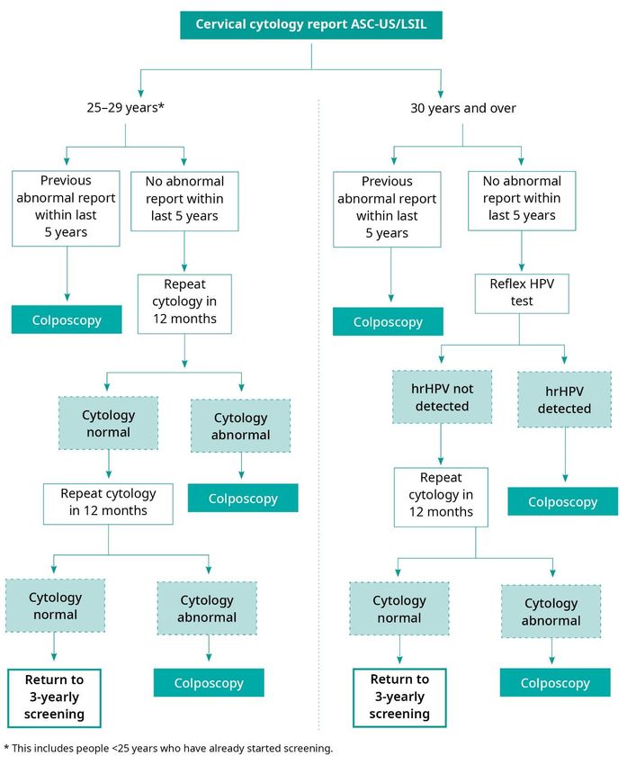
Published in 2020 by the National Screening Unit, Ministry of Health
           PO Box 5013, Wellington 6140, New Zealand

                  ISBN 978-1-98-859774-4 (print)

                 ISBN 978-1-98-859773-7 (online)

     These guidelines can be downloaded or ordered from the
        National Screening Unit website: www.nsu.govt.nz

                             HP 7348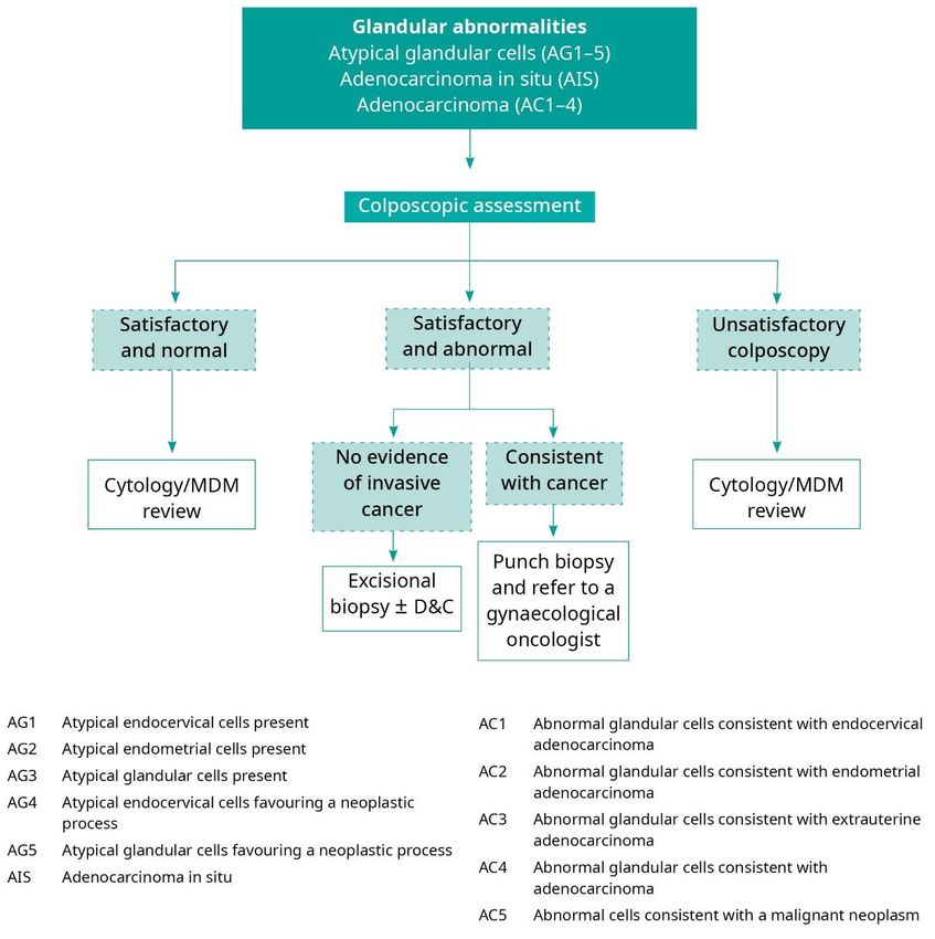
Contents
Foreword                                                                                 1

PART A: INTRODUCTION                                                                     2
Introduction                                                                             3
Background                                                                               3
Development of these guidelines                                                          4
Overview of cervical screening in New Zealand                                            5
Coverage                                                                                 7
Cervical cancer incidence and mortality                                                  7
When to screen and how often                                                             11
Age to start screening                                                                   11
Age to stop screening                                                                    12

PART B: THE GUIDELINES                                                                   13
Using the guidelines                                                                     14
Glossary                                                                                 15
Management of normal cervical cytology tests                                             18
Management of unsatisfactory cervical cytology tests                                     18
Management of abnormal cervical cytology tests                                           19
  Low-grade squamous abnormalities: ASC-US and LSIL                                      19
  Colposcopic assessment of ASC-US/LSIL                                                  23
  Management of histologically confirmed LSIL (HPV/CIN 1)                                24
  High-grade squamous abnormalities: ASC-H / HSIL (CIN 2/3)                              25
  Colposcopic assessment of ASC-H/HSIL                                                   26
  Management of histologically confirmed HSIL (CIN 2 or 3)                               28
  Follow-up of people treated for HSIL (CIN 2 or 3)                                      31
  Management of suspected invasion or SCC                                                33
  Cervical glandular abnormalities: AGC/AIS/AC                                           34
  Cytology report of cervical glandular abnormalities                                    34
  Colposcopic assessment and treatment of glandular abnormalities                        35
  Follow-up of people with AIS                                                           37
Special clinical circumstances                                                           38
  Pregnancy                                                                              38
  People under 25 years                                                                  39

               Clinical Practice Guidelines for Cervical Screening in New Zealand 2020        iii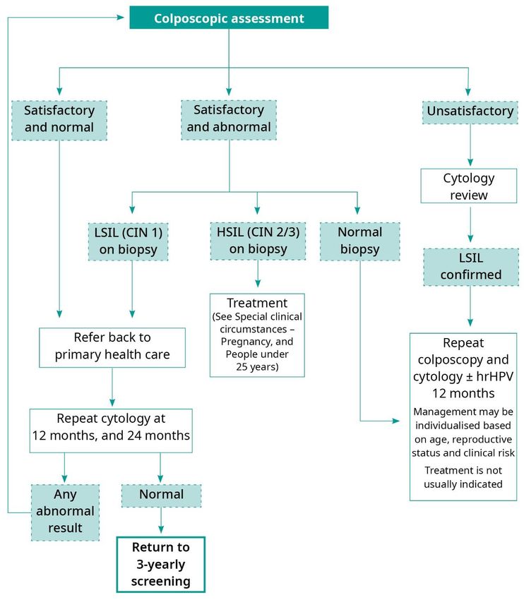
Abnormal vaginal bleeding in people under 25 years                                   41
     Assessment and management of people with persistent abnormal vaginal bleeding        41
     People over 40 years with normal endometrial cells                                   45
     Immune deficiency                                                                    46
     Hysterectomy                                                                         48
     Exposure in utero to diethylstilboestrol                                             50
Summary of indications for cytological review                                             50
References                                                                                52

PART C: GUIDANCE ON HPV TESTING                                                           57
Introduction                                                                              58
Indications for HPV testing                                                               58
HPV testing for indications outside of the NCSP Guidelines                                59
Summary of indications for HPV testing                                                    60
Bibliography for Part C: Guidance on HPV testing                                          61

APPENDICES                                                                                64
Appendix 1: Advisory and working group members                                            65
Appendix 2: AGREE tool                                                                    67

iv              Clinical Practice Guidelines for Cervical Screening in New Zealand 2020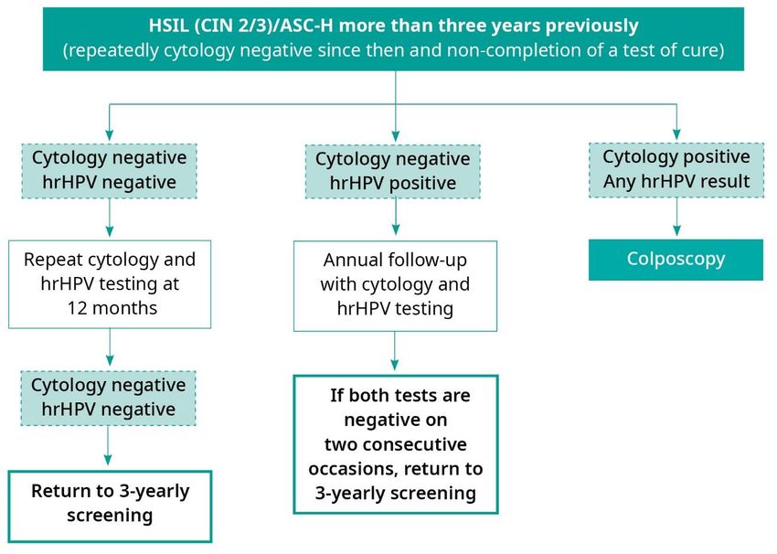
Foreword
The Clinical Practice Guidelines for Cervical Screening in New Zealand 2020 have been
developed for practitioners providing health services across the cervical screening
pathway, including nurses, general practitioners, gynaecologists, cytologists and
pathologists. The guidelines aim to assist providers to achieve best-practice outcomes
when delivering cervical screening and colposcopy services.

These guidelines replace the Guidelines for Cervical Screening in New Zealand published
in 2008.

While the guidelines are evidence-based where possible, they are a guide to best clinical
practice. Clinicians should continue to exercise professional judgement and make
decisions that reflect individual circumstances, in consultation with their patients.

 Dr Jane O’Hallahan              Dr Howard Clentworth             Dr Margaret Sage
 Clinical director, National     Clinical leader, colposcopy,     Clinical leader, pathology,
 Screening Unit                  NCSP                             NCSP

               Clinical Practice Guidelines for Cervical Screening in New Zealand 2020          1
PART A: INTRODUCTION

2   Clinical Practice Guidelines for Cervical Screening in New Zealand 2020
Introduction
Four key changes in this document are as follows:
1. In November 2019 the National Cervical Screening Programme (NCSP) raised the
   recommended commencement age for screening to 25 years for any person with a
   cervix or vagina who has ever been sexually active. People aged 20–25 years who
   have already commenced screening, including those with abnormal cytology, will be
   recalled and managed according to these guidelines.
2. A new section on abnormal bleeding does not wholly relate to the cervical screening
   pathway but has been included to assist medical practitioners in primary care with
   assessment, management and referral decisions. The most important message from
   this section is that symptomatic people need to be examined.
3. A new recommendation that people aged 70 years and older who were unscreened or
   under-screened prior to age 70 have two consecutive normal cytology samples (taken
   12 months apart) before ceasing cytology screening. Unscreened and under-screened
   people in this age group are at increased risk of cervical cancer because of potential
   undetected cervical lesions.
4. A change to the recommendation for follow-up after successful treatment for high-
   grade squamous disease is discharge from colposcopy to primary care for a test of
   cure. Cytology and hrHPV testing should be performed 6 months post-treatment, with
   a repeat co-test (cytology and hrHPV testing) at a further 12 months to complete a test
   of cure. Where there are clinical concerns, colposcopy with hrHPV and cytology
   testing at 6 months post-treatment is recommended.
Other areas have been updated where further evidence and clinical experience have
suggested that changes are required.
The Ministry of Health will update these guidelines when HPV primary screening is
introduced.

Background
Cervical screening is important for any person with a cervix or vagina who has ever been
sexually active, regardless of their sexual orientation, including people who are
transgender or non-binary.

There is overwhelming evidence that the primary underlying cause of cervical cancer is
persistent infection with high-risk types of human papillomaviruses (hrHPV), and that
these viruses are primarily transmitted sexually. Most HPV infections resolve
spontaneously, but those that persist can lead to the development of precancerous
abnormalities, which, if untreated, may progress to cancer.

Cervical cancer has a long latency period, taking on average 10 to 20 years to develop.
This means that screening for the detection of precursor (precancerous) lesions can be
very effective for people who participate regularly in cervical screening.

New technologies introduced to cervical screening, such as liquid-based cytology (LBC)
and HPV testing are increasingly important. Widespread vaccination will reduce the
incidence of cervical cancer, but cervical abnormalities which still occur will be more

              Clinical Practice Guidelines for Cervical Screening in New Zealand 2020      3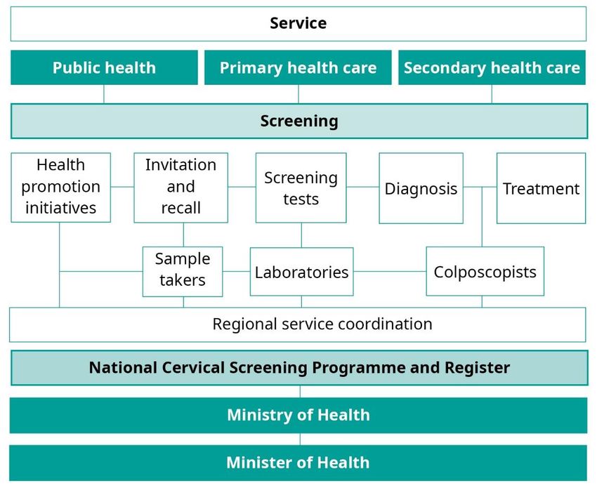
difficult to detect when they are less prevalent in the population, unless there are
improvements in the sensitivity of screening tests.

HPV immunisation with Gardasil®4, which was used in New Zealand between 2008 and
2016, provides effective immunisation against hrHPV types 16 and 18, which cause 70
percent of cervical cancers. The 9-valent vaccine, Gardasil®9 has been used in New
Zealand since 2017 and protects against nine types of HPV – seven that cause HPV-
related cancers and two that cause genital warts. Ninety-two percent of cancers
attributable to HPV can be prevented by Gardasil®9 (CDC, 2019).

Over time, HPV immunisation will have a marked effect on the incidence of and mortality
from cervical cancer, will reduce the volumes of abnormal cytology and colposcopy
assessments and will result in further changes to these guidelines.

Development of these guidelines
In 2005, the NCSP established a multidisciplinary team to update Guidelines for the
Management of Women with Abnormal Cervical Smears, published in 1999 (Appendix 1).
This work resulted in the Guidelines for Cervical Screening in New Zealand, published in
2008.

The team adopted an evidence-based methodology for this process recommended by the
New Zealand Guidelines Group (NZGG). This involved an extensive review of the cervical
screening literature, the development of clinical questions and group decisions on the
content of the guidelines. In addition, they undertook a comprehensive search of
international guidelines relating to the management of people with abnormal cervical
cytology results, and critically appraised this information using the AGREE tool (see
Appendix 2).

However, at that time, there was insufficient or inconsistent external evidence to provide
direct answers to many relevant clinical questions. In these cases, the team developed
recommendations by discussion, by using ‘considered judgement’ and by seeking the
consensus of the entire group. The team graded its recommendations based on the
strength of the evidence using the NZGG’s grading system (see ‘Using the guidelines’ in
Part B).

The team used the Australian guidelines Screening to Prevent Cervical Cancer:
Guidelines for the management of asymptomatic people with screen detected
abnormalities (National Health and Medical Research Council (Australia), 2005) as a key
resource.

In April 2010, the Ministry of Health added an update to the 2008 Guidelines, entitled
Guidance on HPV Testing Update 1: April 2010. This set out clinical guidelines for the use
of HPV testing in New Zealand in three clinical situations:
•   as a triage test after low-grade cytology for people 30 years of age and over with no
    abnormalities in the previous five years
•   as a test of cure after treatment (or follow-up) of high-grade squamous lesions

4            Clinical Practice Guidelines for Cervical Screening in New Zealand 2020
•   for use in clinical assessment in colposcopy, particularly for people with discordant
    results.

The current 2020 guidelines incorporate an increase to the starting age for screening to
25 years, and update other areas where further evidence and clinical experience have
suggested that changes are required.

The Ministry of Health will update the guidelines when HPV primary screening is
introduced.

Overview of cervical screening in New
Zealand
In New Zealand, approximately 160 people are diagnosed with cervical cancer every year,
and 60 die from this largely preventable disease, despite the availability of an organised
screening programme, the NCSP. Research has identified that over 85 percent of people
who develop cervical cancer in New Zealand either have never been screened or have
been screened infrequently (Sykes, 2019).

The NCSP was established in 1990 to reduce the number of people who develop cervical
cancer and the number who die from it. Through routine screening at regular intervals, the
programme aims to detect precancerous squamous cell changes that, if not treated, may
lead to cancer.

The programme has a number of separate components (see Figure 1).

               Clinical Practice Guidelines for Cervical Screening in New Zealand 2020      5
Figure 1: The screening pathway 6 Clinical Practice Guidelines for Cervical Screening in New Zealand 2020
Successful cervical screening requires a high standard of quality at each step in the
screening pathway, from invitation and recall, through to cervical screening, laboratory
testing, colposcopy and the management and information systems that support these
processes. The Health (National Cervical Screening Programme) Amendment Act, which
came into effect in 2004, underpins the NCSP’s operations to ensure the co-ordination of
a high-quality cervical screening programme in New Zealand.

Coverage
New Zealand is in the top five of OECD countries in terms of overall high cervical
screening population coverage rates (Organization for Economic Co-operation and
Development, 2019).

In June 2019, overall programme coverage in New Zealand was 71.4 percent for the total
population. However, there are significant inequalities in coverage by ethnicity: as at June
2019 coverage was 66.8 percent for Māori, 66.6 percent for Pacific and 60.9 percent for
Asian people. Coverage is lower among people living in the most deprived areas.

Cervical cancer incidence and mortality
Cervical cancer mortality began to decline many years before the introduction of the
NCSP, probably reflecting opportunistic screening and improvements in treatment.

Since the introduction of the NCSP in 1990, the age-standardised incidence rate of
invasive cervical cancer in women over 25 years of age has decreased substantially.
Relative reductions have been similar in both Māori and non-Māori populations (Smith,
Edwards, Canfell, 2017). However, between 1990 and 2014 invasive cervical cancer
incidence did not decline in people aged 20–24 despite 25 years of cytology-based
screening (see Figure 2, Figure 3, and Figure 4).

              Clinical Practice Guidelines for Cervical Screening in New Zealand 2020      7
Figure 2: Five-year average cervical cancer incidence by age, 1987–2016
Source: Ministry of Health data from the NCSP Register
35
30
25
Incidence per 100,000 (age–standardised)
20
15
10
5
0
1993
1987
1988
1989
1990
1991
1992
1994
1995
1996
1997
1998
1999
2000
2001
2002
2003
2004
2005
2006
2007
2008
2009
2010
2011
2012
2013
2014
2015
2016
20–24 25–49
Year 50–69 70+
Figure 3: Māori five-year average cervical cancer incidence by age, 2000–2016
Source: Ministry of Health data from the NCSP Register
8 Clinical Practice Guidelines for Cervical Screening in New Zealand 2020Figure 4: Non-Māori five-year average cervical cancer incidence by age, 2000–2016
Source: Ministry of Health data from the NCSP Register
Figure 5: Age-standardised cervical cancer incidence by ethnicity, 2012–2016
Source: Ministry of Health, 2016
Vertical bars represent 95 percent confidence intervals.
Clinical Practice Guidelines for Cervical Screening in New Zealand 2020 9Figure 6: Age-standardised cervical cancer mortality rates by ethnicity, 2011–2015
Source: Ministry of Health, 2016
Vertical bars represent 95 percent confidence intervals.
Note: no deaths were recorded for Asian people in 2011.
10 Clinical Practice Guidelines for Cervical Screening in New Zealand 2020When to screen and how often
The NCSP policy on the screening age and interval is as follows.
Screening age and interval
Anyone with a cervix or vagina who has ever been sexually active should be offered
three-yearly cervical screening from age 25 to age 69.
If this is the ‘first ever’ cervical cytology test, or more than five years have elapsed
since the previous test, a second cytology test is recommended one year after the
first, with three-yearly screening thereafter if both results are normal.
Cervical screening over age 70 years is recommended in people who are unscreened
or have a lapsed screening history prior to age 70.
Clinical management of people under 25 years who have started screening
People under 25 who have already been screened (including those with normal
cytology) will continue to be recalled for screening and referred and managed in the
same way as people aged 25 to 69 years, according to the Clinical Practice
Guidelines for Cervical Screening in New Zealand 2020.
Age to start screening
In 2004, the World Health Organization (WHO)’s International Agency for Research on
Cancer (IARC) concluded that there was minimal benefit and potential treatment harm
associated with cervical screening below age 25 years; it was recommended that
organised screening programmes should not start cervical screening before 25 years of
age (International Agency for Research on Cancer, 2005). Although treatment has a low
complication rate, it is now recognised that the consequences of treatment complications
are greater for younger people who have not completed their family than they are for older
people (Kyrgiou, Koliopoulos, Martin-Hirsch et al, 2006; Sadler, Saftlas, Wang et al, 2004).
In New Zealand, invasive squamous or adenocarcinoma of the cervix is rarely diagnosed
in people under 25 years of age (Ministry of Health, 2019).
Australia, England, Wales, Scotland, Ireland, France, Belgium, Italy and Norway start
screening at 25 years of age. Many other European countries, such as the Netherlands
and Finland, start screening at age 30 years.
In line with international evidence and practice in other countries, in November 2019, the
Ministry of Health raised the recommended commencement age for cervical screening in
New Zealand from 20 to 25 years of age.
Clinical Practice Guidelines for Cervical Screening in New Zealand 2020 11A summary of the evidence supporting the change is available on the National Screening
Unit website: https://www.nsu.govt.nz/health-professionals/national-cervical-screening-
programme/age-range-change-cervical-screening.
Age to stop screening
Many countries do not screen people aged over 60 or 65 years. People aged 65 and over
who have had many normal cervical cytology tests, particularly those who have had three
normal tests in the previous 10 years, are at low risk of developing cervical cancer. The
current policy in New Zealand is to continue regular screening up to age 69 years. The
Ministry of Health will review the exit age for screening when HPV primary screening is
implemented.
People over 70 years of age who are unscreened or under-screened remain at risk of
cervical cancer (Landy, 2015; Lynge, Lonnberg, Tornberg, 2017). It is therefore important
to have adequate screening prior to ceasing screening at age 69. The NCSP’s policy in
this regard is as follows.
• People who have been regularly screened and who have had at least two consecutive
normal cytology samples between 62 and 69 years can cease screening at age 69
years.
• People who have not been adequately screened at a younger age and who have not
had two normal cytology samples reported between 62 and 69 years of age should
have two cervical cytology samples taken 12 months apart, and can cease screening if
both are negative.
• People aged 70 years and older who are unscreened should have two consecutive
normal cytology samples taken 12 months apart before ceasing cytology screening.
• People with abnormal results at any age should follow the recommended NCSP
guidelines for follow-up and management.
Note - The results of women over 70 years will be recorded on the NCSP Register, but
health providers need to take responsibility for adequate follow-up in this group, as the
NCSP Register may not provide recall back-up.
12 Clinical Practice Guidelines for Cervical Screening in New Zealand 2020PART B: THE GUIDELINES Clinical Practice Guidelines for Cervical Screening in New Zealand 2020 13
Using the guidelines
The following guidelines are intended as an aid to clinical practice, not as a substitute for
clinical judgement. Clinicians should continue to manage patients on the basis of personal
and family medical history and clinical signs and symptoms. The results of all cervical
cytology and histology samples taken in New Zealand are recorded on the NCSP Register
(a legislative requirement) regardless of whether such samples are taken in accordance
with these guidelines or not.

These guidelines use technical terminology that will be familiar to many health
professionals but may be foreign to those outside the health system. The glossary that
follows explains the key terms and abbreviations.

The guidelines are presented as shown in the example below.

Example of a guideline

 Assessment/
                         Guideline                                                     Evidence
 Report

 Histologically          Treatment is not recommended because such
 confirmed low-grade     lesions are considered to be an expression of a
                                                                                       Grade C
 squamous                productive HPV infection.
 abnormalities.

In the table above, the first column gives the result of the cervical screening test or
assessment, the second column provides guidelines for management and the third
column gives a grading of the level of evidence on which the guideline is based. Potential
grades are A, B, C, I and ✓, as given in the table below.

 Grade                                          Details

     A    The recommendation is supported by good evidence. The evidence is
          based on a number of studies that are valid, consistent, applicable and
          clinically relevant.
     B    The recommendation is supported by fair evidence. This is based on
          relevant studies that are valid, but there are some concerns about the volume,
          consistency, applicability and/or clinical relevance of the evidence; however,
          the studies are not likely to be overturned by other evidence.
     C    The recommendation is supported by expert opinion only. The evidence
          may be published or unpublished (eg, consensus guidelines).
     I    No recommendation can be made. The evidence is lacking, of poor quality or
          conflicting, and the balance of benefits and harms cannot be determined.
     ✓    Good practice point: No external evidence is available. In this case, best
          practice recommendations are made by consensus, based on the experience
          of the Guideline Development Team, or feedback from consultation within New
          Zealand.

14           Clinical Practice Guidelines for Cervical Screening in New Zealand 2020
Glossary
AC Adenocarcinoma. Cervical cancer arising from the glandular cells
lining the endocervical canal rather than the squamous cells that
cover the outer surface of the cervix
AGC Atypical glandular cells (replaces the previously used term
‘AGUS’)
AIS Adenocarcinoma in situ. High-grade precancerous change in the
glandular (endocervical) cells of the cervix
ASC-US Atypical squamous cells of undetermined significance
ASC-H Atypical squamous cells – cannot exclude a high-grade
squamous lesion
Biopsy A sample of tissue taken during a colposcopy
CIN Cervical intra-epithelial neoplasia. Abnormal squamous cell
changes in the surface epithelial layers of the cervix. These
changes are not invasive cancer, but a small proportion of cases
would develop into cancer if not treated. CIN is graded as low-
grade CIN 1 or high-grade CIN 2 or 3: CIN 3 is the most severe
Colposcopist A health professional with expertise in colposcopy
Colposcopy Examination using a colposcope. This magnifies the cervix and
vagina so that a clinician can detect abnormal areas
Coverage The proportion of people aged 25–69 years who have had a
screening result recorded on the NCSP Register
Cytology test Microscopic examination of cells from an LBC sample
Cytology review A review of cytology and histology slides by a
pathologist/cytologist. This may be undertaken during
multidisciplinary case review by health professionals (eg, a
pathologist, colposcopist, cytologist and colposcopy nurse)
D&C Dilatation and curettage
DHB District Health Board
Dysplasia Older terminology referring to all grades of precancerous lesions:
mild (CIN 1), moderate (CIN 2) or severe (CIN 3)
Clinical Practice Guidelines for Cervical Screening in New Zealand 2020 15Ectocervix The outer surface of the cervix, usually covered by squamous
cells
Endocervix The lining of the canal in the centre of the cervix, usually lined by
endocervical glandular cells
Endometrium The tissue lining the uterus
Histology Microscopic examination of a sample of tissue
HPV Human papillomavirus
hrHPV High-risk human papillomavirus
HSIL High-grade squamous intra-epithelial lesion (equivalent to CIN
2/3)
LBC Liquid-based cytology. The type of collection system specimen
used for both cytology and HPV testing. The sampled cells are
put into a liquid preserving solution in a small plastic vial
Low-grade Encompasses possible LSIL (ASC-US) and definite LSIL in
abnormality cytology samples. In histology samples, ‘low-grade’ encompasses
HPV infection and CIN 1
LSIL Low-grade squamous intra-epithelial lesion involving mild
changes encompassing HPV effect and CIN 1
MDM Multidisciplinary meeting
NCSP National Cervical Screening Programme
RANZCOG Royal Australian and New Zealand College of Obstetrics and
Gynaecology
SCC Squamous cell carcinoma. A type of cervical cancer arising from
squamous cells
Test of cure HPV testing and cytology (co-testing) on two occasions 12
months apart. The person can return to three-yearly screening if
HPV testing and cytology are negative on two occasions 12
months apart (ie, successful completion of the test of cure)
Transformation The region of the cervix where the glandular (columnar) precursor
zone cells have changed or are changing to squamous cells (a normal
physiological process)
16 Clinical Practice Guidelines for Cervical Screening in New Zealand 2020Triage The clinical process of assigning people into follow-up or treatment
pathways based on their clinical risk
Unsatisfactory An inadequate test that cannot be assessed by the laboratory.
cervical screening
test
Type 1, 2 or 3 Depending on the type of transformation zone and the length of
excision the endocervix removed, an excision can be of type 1, type 2 or
type 3. A type 1 excision is adequate for a purely ectocervical
lesion, whereas a type 3 excision is required if the endocervical
extent of the lesion is not visible
Vault sample A sample taken from the top of the vagina in people who have
had their cervix removed as a result of a hysterectomy
Clinical Practice Guidelines for Cervical Screening in New Zealand 2020 17Management of normal cervical cytology tests
The cervical screening test is a screening test of asymptomatic people to detect and treat
pre-invasive abnormalities of the cervix. If the first ever result is negative, a follow-up test
is recommended in 12 months to reduce the risk of non-detection of a significant lesion
due to a false negative result. If this second cervical screening test is also negative, recall
should be every three years.
Guideline 1: Negative (normal) cervical cytology test
Cervical Cytology Result Guideline Evidence
Negative for a squamous or Recall in 3 years for cervical cytology Grade B
glandular intra-epithelial unless the result falls into one of the
lesion or malignancy following two categories.
Negative for a squamous or Recall in 12 months for cervical Grade C
glandular intra-epithelial cytology.
lesion or malignancy, but
this is the first test, or more
than 5 years have elapsed
since the previous test
Negative for a squamous or Recall according to the relevant
glandular intra-epithelial guideline in this document.
lesion or malignancy, but
with a previous abnormality
Management of unsatisfactory cervical
cytology tests
An unsatisfactory cervical cytology test is inadequate for some reason, and therefore the
laboratory cannot report it. The adequacy of the sample is based on the number of well-
visualised, well-preserved squamous cells that have been sampled. Laboratories reading
cervical cytology samples have a standardised procedure for assessing the adequacy of
the sample. The presence or absence of cells from the endocervical canal/transformation
zone is recorded in the report, but does not affect the adequacy of the test or the report
recommendation.
Three main factors cause unsatisfactory samples:
• sample taking – inadequate numbers of cells sampled, contact bleeding or
contaminants such as lubricant
• clinical factors eg, bleeding, inflammation or cytolysis
• laboratory technical processing issues.
An unsatisfactory cytology sample is recorded as a non-result on the NCSP Register.
After three consecutive unsatisfactory samples colposcopy is recommended to exclude a
high-grade lesion as the person is inadequately screened.
18 Clinical Practice Guidelines for Cervical Screening in New Zealand 2020Guideline 2: Unsatisfactory cervical cytology tests
Cervical cytology result Guideline Evidence
Unsatisfactory Repeat the test after 4–6 weeks and before 3 Grade C
months.
Refer for colposcopy after 3 consecutive
unsatisfactory cytology reports.
In people who are post-menopausal, postnatal
or breastfeeding give a course of vaginal
oestrogen cream nightly for 2–3 weeks prior to
repeating the cytology test.
Management of abnormal cervical cytology
tests
With the introduction of the LAST terminology (Darragh, Colgan, Cox et al, 2012) to New
Zealand in 2020, squamous lesions in cervical biopsies are reported in the form of ‘LSIL
(CIN 1)’ or ‘HSIL (CIN 2/3)’.
Abnormal results reported outside of New Zealand including hysterectomy information can
usually be added to the NCSP Register if a copy of the cytology or histology result or a
specialist letter that documents the result is provided to NCSP Register staff. Refer to 3.2
Recall Process, Overseas Test Results in Section 3 of the National Policy and Quality
Standards – Cervical Screening https://www.nsu.govt.nz/health-professionals/national-
cervical-screening-programme/policies-and-standards.
Low-grade squamous abnormalities: ASC-US and LSIL
Cervical cancer is a rare outcome after a low-grade abnormality (Woodman, Collins &
Young, 2007; Mitchell, 2005). A diagnosis of cancer after a low-grade cytology result can
occur for any of the following reasons: sampling error (the abnormal cells were not picked
up by the sampling device, or not transferred to the sample vial, or not selected for
examination by sample processing), non-detection or misinterpretation at cytology
reporting, or true progression over time from a low-grade intra-epithelial abnormality to
cancer.
Studies indicate that people with ASC-US who are also hrHPV positive are at similar risk
of HSIL (CIN 2/3) as people with LSIL (Cox, Schiffman, Solomon, 2003). These groups
show similar high regression rates and are managed similarly.
Low-grade cytology is a manifestation of a viral infection that will resolve spontaneously in
the majority of people (Moscicki, Schiffman, Kjaer et al, 2006; Schiffman, Castle, Jeronimo
et al, 2007). For people under 30 years of age, the recall timeframe is 12 months, given
the evidence that the median time for clearance of HPV infection is 6–18 months
(Plummer, Schiffman, Castle et al, 2007) - see Guideline 3. HPV testing is not used in this
age group because the positivity rate is too high for the test to be a good way of
identifying people who need referral for colposcopy.
Clinical Practice Guidelines for Cervical Screening in New Zealand 2020 19People aged 30 years and over with a hrHPV infection are at increased risk of developing
a high-grade lesion, because the infection is more likely to be persistent (Castle,
Schiffman, Herrero et al, 2005). HPV triage for people in this age group with a first ASC-
US/LSIL cytology result is therefore of greater benefit than repeated cytology to assess
the underlying risk of HSIL (CIN 2/3) (Arbyn, Sasieni, Meijer et al, 2006; Ronco, Cuzick,
Segnan et al, 2007). See Flowchart 1.
Clinicians should advise all people of the significance of their low-grade cytology results
and the low risk of harboring or developing cancer. If a person is unduly anxious, or
specifically requests specialist reassurance, referral for colposcopic assessment may
alleviate their anxiety, bearing in mind that this is not a complete safeguard against a
diagnosis of underlying HSIL (CIN 2/3) or cervical cancer.
Where a clinician finds symptoms suspicious of cervical cancer or is concerned about the
clinical appearance of the cervix, the person must be investigated appropriately with
colposcopy irrespective of the cytology result.
Guideline 3: Cervical cytology report ASC-US/LSIL (see Flowchart 1)
Cervical
Guideline Evidence
cytology result
ASC-US/LSIL People aged 25–69 years with ASC-US/LSIL who
have had an abnormal cytology or histology
Grade C
report within the last 5 years
Refer for colposcopy.
People aged 25−29 years with ASC-US/LSIL who
have had no abnormal cytology or histology
Grade C
reports within the last 5 years
Refer to colposcopy if there has been a prior high-
grade abnormality more than 5 years previously. If
there has been no previous high-grade abnormality,
repeat the test in 12 months.
Grade B
If the 12-month repeat cytology test is reported as:
• negative – repeat the test in 12 months (ie, 24
months after the index cytology test)
• ASC-US/LSIL – refer for colposcopy
• HSIL or ASC-H– refer for colposcopy.
If the next 12-month repeat cytology test (ie, 24
months after the index result of ASC-US/LSIL) is
Grade B
reported as:
• normal – return to 3-yearly screening
• abnormal – refer for colposcopy.
20 Clinical Practice Guidelines for Cervical Screening in New Zealand 2020Cervical
Guideline Evidence
cytology result
People aged 30 years and over with ASC-
US/LSIL and no abnormal cytology or histology
Grade C
reports within the last 5 years have a reflex
hrHPV test added on by the laboratory
If the reflex hrHPV test is:
• negative – repeat cytology in 12 months. If the
repeat cytology is:
− normal – return to normal 3-yearly screening
− abnormal – refer for colposcopy
• positive – refer for colposcopy.
Clinical Practice Guidelines for Cervical Screening in New Zealand 2020 21Flowchart 1: Management of low-grade abnormalities: ASC-US or LSIL 22 Clinical Practice Guidelines for Cervical Screening in New Zealand 2020
Colposcopic assessment of ASC-US/LSIL
Colposcopy assessment and management of people with a cytology result of ASC-
US/LSIL should comply with the guidelines published by RANZCOG and the Australian
Society of Colposcopy and Cervical Pathology (ASCCP) (RANZCOG, 2001).

A fluctuating status between low-grade change and negative cytology is not uncommon,
but the significance of this is unclear. It could reflect a transition from active HPV infection
to resolution followed by re-infection, or there could be an underlying persistent lesion that
is not being consistently sampled or detected by cytology. HrHPV testing may help with
further management.

Guideline 4: Colposcopic assessment of ASC-US/LSIL (see Flowchart 2)

 Colposcopic
                                Guideline                                           Evidence
 assessment
 Satisfactory and normal        Refer back to the sample taker for 2 annual         Grade C
                                cytology tests after discharge from
                                colposcopy.
                                If either test is abnormal, refer for repeat
                                colposcopy.
                                If both tests are normal, resume regular 3-
                                yearly screening.
 Satisfactory and abnormal      Perform a target biopsy to make a diagnosis.        Grade C
 Unsatisfactory                 Cytology review is recommended.                     ✓
                                If low-grade cytology is confirmed on review,
                                undertake repeat colposcopy, cytology and           Grade C
                                hrHPV testing, as appropriate, in 12 months.
                                Management may be individualised, based on
                                age, reproductive status and clinical risk.
                                Treatment is not usually indicated.

         Clinical Practice Guidelines for Cervical Screening in New Zealand 2020        23
Management of histologically confirmed LSIL (HPV/CIN 1)
Guideline 5: Histologically confirmed LSIL (HPV/CIN 1) (see Flowchart 2)
Histology Result Guideline Evidence
Histologically Treatment is not recommended, because such Grade C
confirmed low-grade lesions are considered to be an expression of a
squamous productive HPV infection.
abnormalities
Refer back to the sample taker for repeat
cytology at 12 and 24 months. A return to regular Grade C
3-yearly screening is recommended if both tests
are negative.
Refer back to colposcopy if either repeat test
shows ASC-US/LSIL or a higher degree of
abnormality (ie, ASC-H or HSIL, AGC or AIS, or
possible/definite invasive malignancy).
24 Clinical Practice Guidelines for Cervical Screening in New Zealand 2020Flowchart 2: Colposcopic management of low-grade cytology (ASC-US/LSIL)
Note: Colposcopists may vary these guidelines on the basis of hrHPV status.
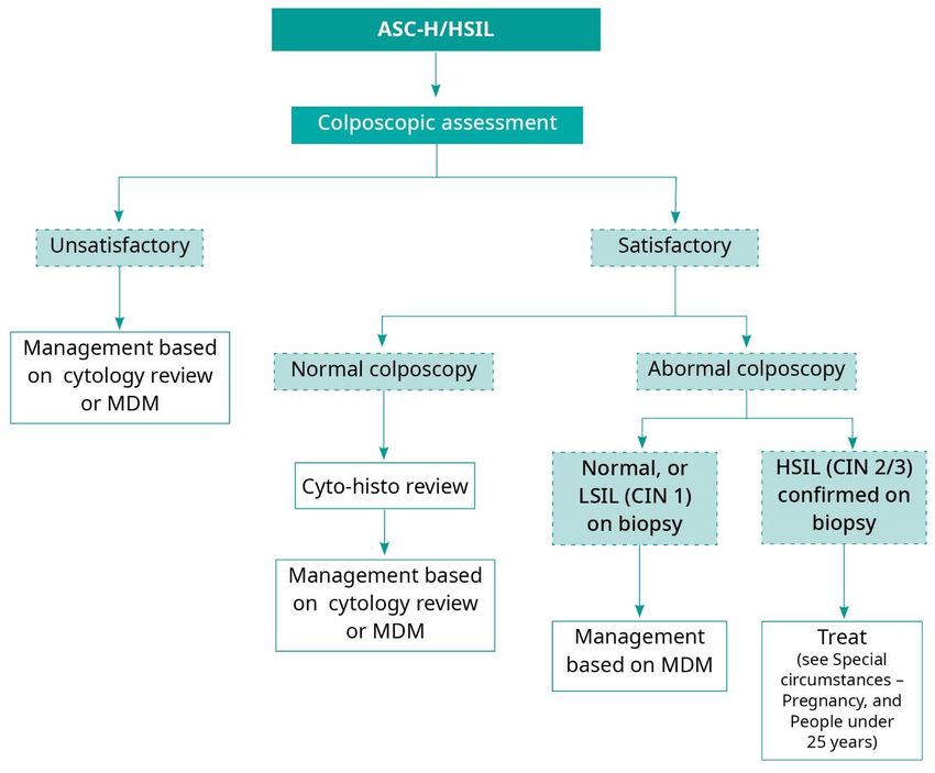
High-grade squamous abnormalities: ASC-H / HSIL (CIN 2/3)
This category encompasses cases with a definite prediction of HSIL (CIN 2/3) as well as
cases that are suspicious of HSIL (CIN 2/3), without being definite (ASC-H - Atypical
squamous cells, cannot exclude HSIL). The finding of a high-grade result on cytology
carries a high risk of significant cervical disease.
The main objective of the NCSP is to detect high-grade abnormalities in order to treat
these effectively and prevent cervical cancer. People with untreated HSIL (CIN 3) lesions
are at high risk of cervical cancer (McCredie, Sharples, Paul et al, 2008; Moscicki,
Clinical Practice Guidelines for Cervical Screening in New Zealand 2020 25Schiffman, Kjaer, et al, 2006). HSIL (CIN 2) lesions are more heterogeneous and variable
in cancer potential than HSIL (CIN 3) (Schiffman, Castle, Jeronimo et al, 2007; Moscicki,
Schiffman, Kjaer et al, 2006).
Using the Bethesda reporting system terminology in cytology reports, high-grade in-situ
squamous lesions in cytology samples are usually reported as ‘HSIL. The features are
consistent with CIN 2 or CIN 3’. Laboratories may elect to subcategorise HSIL into HSIL
(CIN 2) and HSIL (CIN 3) in cytology reports if they wish to do so.
In histology reports, HSIL (CIN 2 and CIN 3) are usually reported as separate diagnoses,
although it is recognised that this distinction is subjective and not reliable to permit clear
stratification of risk (Carreon, Sherman, Guillen et al, 2007). HSIL (CIN 2) is generally the
threshold for treatment. Exceptions include people under 25 years of age with histologic
HSIL (CIN 2), who (after MDM review) are often managed conservatively because of high
regression rates. In pregnancy, treatment for HSIL (CIN 2) and/or HSIL (CIN 3) is usually
deferred until the post-partum period.
Guideline 6: Cervical cytology report ASC-H or HSIL
Cervical Cytology
Guideline Evidence
Result
ASC-H or HSIL Refer for colposcopy and a targeted biopsy, Grade B
where indicated.
Colposcopic assessment of ASC-H/HSIL
A significant number of lesions can be missed on colposcopic impression (Gage, Hanson,
Abbey et al, 2006; Jeronimo, Schiffman, 2007). Where cytology is ASC-H or HSIL but
colposcopic examination of the cervix shows no sign of any abnormality, there should be
careful clinical inspection and colposcopy of the entire lower genital tract, and a review
should be undertaken of possible sites of origin for neoplastic cells in the upper genital
tract (National Cervical Screening Programme,1999).
26 Clinical Practice Guidelines for Cervical Screening in New Zealand 2020Guideline 7: Colposcopic assessment of ASC-H/HSIL (see Flowchart 3)
Colposcopic
Guideline
Assessment
Satisfactory Undertake a targeted biopsy for histology. Grade B
and abnormal
Note: For ‘See and treat’ see Guideline 8.
colposcopy
Where the biopsy confirms CIN 1, manage based on
MDM review.
Satisfactory Cytology review is recommended. Grade C
and normal
If the review confirms high-grade abnormalities, repeat
colposcopy or
colposcopy and cytology within 3 months.
negative
biopsy If colposcopy and cytology are normal at 3 months,
✓
repeat cytology in 12 months.
If colposcopy or cytology is LSIL at 3 months,
individualise management based on an MDM review.
If colposcopy or cytology is HSIL (CIN 2/3) at 3 months,
treatment is indicated (refer to Guideline 8).
As Part C: Guidance on HPV testing indicates, HPV
testing should be used in colposcopy to assist with the
management of people with discordant results.
Unsatisfactory Cytology review is recommended. Grade C
colposcopy
If the review confirms ASC-H/HSIL, a type 3 excision is
recommended.
✓
If the review confirms normal or ASC-US or LSIL,
manage based on an MDM review.
As Part C: Guidance on HPV testing indicates, HPV
testing can be used at colposcopy to assist with the
management of people with an unsatisfactory
colposcopy.
Clinical Practice Guidelines for Cervical Screening in New Zealand 2020 27Flowchart 3: Management of high-grade abnormalities: ASC-H or HSIL Management of histologically confirmed HSIL (CIN 2 or 3) No substantial differences have been found between the different treatment modalities in terms of reducing cancer risk (Martin-Hirsch, Paraskevaidis, Kitchener, 1999; Kalliala, Nieminen, Dyba, 2007; Nuovo, Melnikow, Willan et al, 2000). Evidence suggests that all excisional treatment methods are associated with a small but real increase in long-term adverse obstetric outcomes, including pre-term delivery, low birth weight and premature rupture of membranes (Kyrgiou, Koliopoulos, Martin-Hirsch et al, 2006; Sadler, Saftlas, Wang et al, 2004; Crane, 2003). The available data indicates a significantly increased risk if the excision depth is more than 10mm (Kyrgiou, Koliopoulos, Martin-Hirsch et al, 2006). This evidence reinforces the need for caution when treating young people with mild cervical abnormalities and supports management by surveillance. Follow-up after treatment serves to identify both complications of treatment and recurrent disease, which may be the result of inadequately treated disease, persistent disease or new infection. People treated for HSIL (CIN 2/3) are at increased risk of developing further high-grade disease and invasive cancer (Soutter, Sasieni, Panoskaltsis, 2006; Mitchell & Hocking, 2002). Persistence and recurrence rates are greatest in the initial two years following treatment, but the risk has been found to persist for at least 10 years after initial 28 Clinical Practice Guidelines for Cervical Screening in New Zealand 2020
treatment (Soutter, Sasieni, Panoskaltsis, 2006; Kalliala, Anttila, Pukkala et al, 2005;
Flannelly, Bolger, Fawzi et al, 2001; Arbyn, Sasieni, Meijer et al, 2006).

Treatment failure rates have been reported to average around 10 percent (Arbyn, Sasieni,
Meijer et al 2006). Involved excision margins after an excision biopsy are a risk factor for
treatment failure (Flannelly, Bolger, Fawzi et al, 2001). The risk of further high-grade
disease and invasive cervical cancer increases with age (Mitchell & Hocking, 2002;
Flannelly, Bolger, Fawzi et al, 2001).

Guideline 8: Management of people with histologically confirmed HSIL (CIN 2 or CIN
3)

 Histology Result     Guideline                                                    Evidence

 HSIL (CIN 2 or 3)    Treat in order to reduce the risk of developing              Grade A
                      invasive cervical carcinoma.

 Treatment            Guideline

 Ablative therapy     Ablative therapy may be considered if:                       Grade C
                      •   colposcopic assessment is satisfactory
                      •   a targeted biopsy has confirmed the diagnosis
                      •   there is no evidence of invasive cancer on
                          cytology, colposcopic assessment or biopsy
                      •   there is no evidence of a glandular lesion on
                          cytology, biopsy or colposcopy
                      •   the entire lesion can be visualised.
 Cryotherapy          Cryotherapy is not recommended.                              Grade B

 Type 1, 2 or 3       Loop excision                                                Grade C
 excision
                      Avoid excess diathermy artefact when using
                      diathermy loops to allow comprehensive pathological
                      examination, including margin status.

                      Cone biopsy                                                  Grade C
                      A type 3 excision may be necessary to treat people
                      with high-grade squamous lesions. Indications
                      include:
                      •   failure to visualise the upper limit of the cervical
                          transformation zone in a person with a high-grade
                          squamous abnormality on the referral cervical
                          cytology test (ie, unsatisfactory colposcopy)
                      •   suspicion of an early invasive cancer on cytology,
                          biopsy or colposcopic assessment

         Clinical Practice Guidelines for Cervical Screening in New Zealand 2020       29
• the suspected presence of an additional glandular
abnormality (eg, AIS) on cytology or biopsy (ie, a
mixed lesion).
• Pay careful attention to tailoring treatment to the
individual, taking into account the size, extent,
situation and severity of the lesion.
Hysterectomy Hysterectomy is not generally indicated for the Grade B
management of HSIL (CIN 2 or 3) alone. If
performed for concurrent clinical indications, the
following conditions must be met:
• colposcopic assessment is satisfactory
• a targeted biopsy has confirmed the diagnosis
• there is no evidence of invasive cancer on
cytology, colposcopic assessment or biopsy
• there is no evidence of a glandular lesion on
cytology or biopsy or colposcopy
• the entire lesion can be visualised.
See and treat Consider ‘see and treat’ for high grade lesions, if this Grade C
seems to be the only opportunity to undertake
treatment and the following apply:
• circumstances are appropriate or immediate
treatment is necessary
• the colposcopic examination is consistent with the
referral
• the limits of the lesion are visible
• the whole abnormality can be excised
• there is no suspicion of invasion
• there is an excisional specimen available for
histological examination (ie, no ablative therapy).
People who plan Local ablative or excisional treatments should Grade B
to have children destroy or remove abnormal tissue to a depth of at
least 7 mm. There is no clearly superior method of
fertility-sparing treatment for HSIL (CIN 2 and 3).
30 Clinical Practice Guidelines for Cervical Screening in New Zealand 2020Follow-up of people treated for HSIL (CIN 2 or 3)
HrHPV testing has a high sensitivity for detecting persistent HSIL (CIN 2/3) post-treatment
(Arbyn, Sasieni, Meijer et al, 2006; Paraskevaidis, Arbyn, Sotiriadis et al, 2004; Zielinski,
Bais, Helmerhorst et al, 2004), and when used as a test of cure allows a safe pathway for
women successfully treated for HSIL (CIN 2/3) to return to three yearly cytology
screening.
All people who have been treated for a high-grade squamous lesion should have hrHPV
testing as part of their follow-up. Follow-up after successful treatment of high-grade
squamous disease is discharge from colposcopy to primary care for a test of cure.
Cytology and hrHPV testing should be performed 6 months post-treatment, with a repeat
co-test (cytology and hrHPV testing) at a further 12 months to complete a test of cure.
Where there are clinical concerns, colposcopy with hrHPV and cytology testing at 6
months post-treatment is recommended.
If the HPV test is positive 6 or 18 months after treatment, they should be re-referred to
colposcopy to ensure that treatment has been complete. If the colposcopic evaluation is
negative, they should have annual HPV and cytology co-testing until they have two
consecutive negative co-tests a year apart (ie, two normal cytology and two negative
hrHPV test results). Following successful completion of a test of cure, they can return to 3-
yearly screening.
Some people remain hrHPV-positive with negative cytology. The risk of completely treated
people with negative cytology but persistent hrHPV having high-grade abnormalities
declines with time, but never returns to the same level of risk as for hrHPV-negative
people.
People treated for high-grade squamous lesions before introduction of the hrHPV test of
cure as a regular part of post-treatment follow-up should be offered a test of cure. If they
have two normal cytology tests and two negative hrHPV tests 12 months apart, they can
return to 3-yearly screening.
People treated for HSIL (CIN2/3) who have been cytologically negative repeatedly for over
3 years and are found to be hrHPV positive should be followed up annually with cytology
and hrHPV testing.
Refer to Part C: Guidance on HPV testing and Flowchart 4 and Flowchart 5 in this section.
Guideline 9: Follow-up of people treated for HSIL (CIN 2/3)
Follow-Up Guideline Evidence
Routine follow-up Ensure a person treated for HSIL (CIN 2 or 3): Grade B
• has HPV and cytology co-testing at 6 and 18
months post-treatment as part of their follow-up.
• If HPV testing and cytology (co-testing) are
negative on two occasions 12 months apart (ie,
successful completion of the test of cure), they
can return to 3-yearly screening.
Any symptoms should be appropriately managed.
Clinical Practice Guidelines for Cervical Screening in New Zealand 2020 31Flowchart 4: HPV testing after treatment for HSIL (CIN 2/3) in the previous three years 32 Clinical Practice Guidelines for Cervical Screening in New Zealand 2020
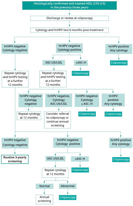
Flowchart 5: HPV testing after HSIL (CIN 2/3)/ASC-H more than three years previously, with
subsequent negative cytology and non-completion of a test of cure

Management of suspected invasion or SCC
Guideline 10: HSIL with suspected invasion or SCC

 Cervical Cytology
                       Guideline                                                  Evidence
 Result

 HSIL with             Refer for urgent assessment to a colposcopist or           Grade B
 suspected             oncologist.
 invasion or SCC

        Clinical Practice Guidelines for Cervical Screening in New Zealand 2020      33
Cervical glandular abnormalities: AGC/AIS/AC
In New Zealand, and internationally, glandular lesions are now estimated to represent
15−20 percent of invasive cervical cancers (Lewis, Almendral, Neal et al, 2008; Bulk,
Visser, Rozendaal et al, 2005; Pak, Martens, Bekkers et al, 2007).
Cervical screening is less effective at preventing cervical AC compared to SCC because
of the limitations of the cervical cytology test (Azodi, Chambers, Rutherford et al, 1999;
Krane, Granter, Trask et al, 2001).
Infection with hrHPV types is associated with cervical AC and AIS in approximately
90 percent of cases (Castellsague, Diaz, de Sanjose et al, 2006; El-Ghobashy, Shaaban,
Herod et al, 2005).
Detecting and reporting abnormal glandular abnormalities by cytology is a difficult task. A
significant number of glandular abnormalities reported by cytology are high-grade
squamous lesions on histology. It is also relatively common for squamous and glandular
lesions to co-exist, and a significant number of cytology-detected glandular abnormalities
result in either a squamous or co-existing squamous/glandular lesion (Rabelo-Santos,
Derchain, Westin et al, 2008; Irvin, Evans, Andersen et al, 2005; Saqi, Gupta, Erroll et al,
2005).
Further, AGC in a cervical cytology sample may be associated with a neoplastic condition,
including AC of the cervix, endometrium, ovary or fallopian tube (Sharpless, Schnatz,
Mandavilli et al, 2005; DeSimone, Day, Tovar et al, 2006; Derchain, Rabelo-Santos,
Sarian et al, 2004; Dias-Montes, Farinola, Zahurak et al, 2006)..
Due to these complexities, anyone with glandular abnormalities should be referred to
colposcopy or a gynaecological oncologist for assessment.
Because of the high incidence of neoplasia and poor sensitivity of testing methods, once
atypical glandular cells are detected then diagnostic excisional procedures may be
necessary (National Health and Medical Research Council, 2005; Wright, Massad, Dunton
et al, 2007).
A sample for hrHPV testing should be taken in the colposcopy clinic prior to treatment for
definite or suspected AIS, if HPV testing has not already been performed in the previous 6
months. HrHPV testing is a useful adjunct in the management of cases at colposcopy in
which a lesion is suspected by cytology but not confirmed by colposcopy or histology
(Dias-Montes, Farinola, Zahurak et al, 2006; Saqi, Gupta, Erroll et al, 2005; Wright,
Massad, Dunton et al, 2007).
Cytology report of cervical glandular abnormalities
Guideline 11: Cervical cytology report of AGC, AIS or AC
Cervical Cytology
Guideline Evidence
Result
AGC, AIS or AC Refer to a colposcopist. Grade B
34 Clinical Practice Guidelines for Cervical Screening in New Zealand 2020Colposcopic assessment and treatment of glandular
abnormalities
Guideline 12: Colposcopic assessment and treatment of glandular abnormalities
(see Flowchart 6)
Situation Guideline Evidence
Assessment Undertake colposcopy assessment if cervical Grade B
cytology suggests glandular abnormalities (AGC
or AIS).
• If the colposcopy is satisfactory and normal, it
is recommended that the cytology be reviewed.
• If abnormal glandular cytology is confirmed on
review, a type 3 excision as a single specimen,
and dilatation and curettage (D&C) are
recommended.
• If abnormal glandular cytology is not confirmed
on review, management should be based on
an MDM decision.
1. If the colposcopy is satisfactory and
abnormal, and consistent with cancer, a
biopsy should be taken and then an urgent
referral made to a gynaecological
oncologist.
2. If colposcopy is satisfactory and abnormal,
and suspicious of a pre-invasive neoplastic
process, a type 3 excision and D&C are
recommended.
3. If colposcopy is unsatisfactory, it is
recommended that the cytology be
reviewed.
• If abnormal glandular cytology is confirmed as
favouring a neoplastic process, a type 3
excision and D&C are recommended.
• If abnormal glandular cytology is not confirmed
on review, management should be based on
an MDM decision.
A sample for hrHPV testing should be taken in the
colposcopy clinic prior to treatment for definite or
suspected AIS, if HPV testing has not already
been performed in the previous 6 months.
Treatment Undertake a type 3 excision. Grade B
Clinical Practice Guidelines for Cervical Screening in New Zealand 2020 35Referral for people Refer to a gynaecological oncologist or an Grade B
with AC on type 3 oncology unit for subsequent management.
excision or punch
biopsy
Management of If invasive carcinoma is not identified at Grade C
people with a colposcopic assessment, a type 3 excision should
cytology report of be undertaken.
AIS
Hysterectomy should not be undertaken without a
prior type 3 excision to exclude invasive
carcinoma.
Management of Management will depend on age and fertility Grade B
people with a type 3 expectations and the status of the excision
excision report of margins.
AIS
AIS treatment (with 1. If the type 3 excision has positive margins on Grade B
a type 3 excision) histology, further treatment should be
follow-up considered.
2. If the margins are clear, follow-up colposcopy
and cytology should be undertaken, including
an endocervical brush sample 6 months after
treatment.
3. Repeat cytology at 12 months, then annually if
both tests and examinations are normal.
4. Early follow-up of symptoms is recommended.
HPV testing may aid follow up in colposcopy
where complete excision of glandular disease has
occurred: see below.
Glandular People in this category can cease cervical Grade B
abnormalities in screening.
people who have
had a total
hysterectomy, with
no evidence of a
squamous high-
grade lesion
36 Clinical Practice Guidelines for Cervical Screening in New Zealand 2020Follow-up of people with AIS
There is a lack of randomised studies of people with AIS. Under these circumstances, the
recommendations within these guidelines are conservative. They are as follows.
• For people who wish to retain their fertility, the treatment goal is to have clear
histological margins.
• Even when the margins are clear, the risk of recurrence can reach approximately 20
percent.
• HPV testing is more sensitive than cytology, and both are more sensitive than
colposcopy. If HPV testing is undertaken and the results are negative, where there are
clear histological margins there is a positive predictive value of no identifiable disease
of 90 percent after one year and 100 percent after two years (Dillner et al, 2008;
Koliopoulos, Nyaga, Santesso et al, 2017).
Flowchart 6: Colposcopic assessment and treatment of glandular abnormalities
Clinical Practice Guidelines for Cervical Screening in New Zealand 2020 37Special clinical circumstances
Pregnancy
Colposcopy is safe for both baby and mother in the prenatal period; if indicated, a referral
to colposcopy should be made during pregnancy. It is unlikely that a biopsy or treatment
would be undertaken during pregnancy, but colposcopic assessment can exclude the
presence of invasive cervical cancer and provide reassurance.
The risk of progression of HSIL to invasive cancer during pregnancy is low (National
Health and Medical Research Council, 2005; Hunter, Bradley, Monk et al, 2008).
However, some studies have found a high probability that a high-grade lesion will persist
during pregnancy (Kaplan, Dainty, Dolinsky et al, 2004; Palle, Bangsboll, Andreasson,
2000), pointing to the need for continued colposcopic and cytological surveillance during
the pregnancy (at about 20−30 weeks) and postpartum period (after 6 weeks) (National
Health and Medical Research Council, 2005; Wright, Massad, Dunton et al, 2007). Other
studies show a high rate of regression (Yost, Santoso, McIntire et al, 1999).
In the prenatal period colposcopy should be undertaken by a colposcopist experienced in
assessing the pregnant cervix (National Health and Medical Research Council, 2005).
Treatment of HSIL during pregnancy has been associated with complications and a high
rate of recurrence or persistence (National Health and Medical Research Council, 2005;
Wright, Massad, Dunton et al, 2007; Hunter, Bradley, Monk et al, 2008). Therefore, the
only indication for treatment in pregnancy is suspicion of invasive cancer.
Guideline 13: Management during pregnancy
Situation Guideline Evidence
Cervical screening Take cytology tests according to these guidelines. Grade B
during pregnancy
A cervical sample can be taken at any time during
pregnancy, particularly if the person has never been
screened, is overdue for a test, has an abnormal
screening history and is due for a test, or if there
have been specific indications or recommendations
for a follow-up test. If the person has a normal
screening history, a decision may be made to delay
screening until 3 months postpartum.
After delivery, it is recommended that cervical
screening is delayed until 3 months postpartum, to
allow the changes associated with pregnancy to
resolve.
If a person is screened when postnatal and/or
breastfeeding, a course of vaginal oestrogen cream
nightly for 2–3 weeks is recommended prior to the
test.
38 Clinical Practice Guidelines for Cervical Screening in New Zealand 2020You can also read