Ubiquitination in the ERAD Process - Review - MDPI
←
→
Page content transcription
If your browser does not render page correctly, please read the page content below
International Journal of
            Molecular Sciences

Review
Ubiquitination in the ERAD Process
Anna Lopata , Andreas Kniss, Frank Löhr , Vladimir V. Rogov               and Volker Dötsch *
 Institute of Biophysical Chemistry and Center for Biomolecular Magnetic Resonance, Goethe University,
 Max-von-Laue Str. 9, 60438 Frankfurt am Main, Germany; lopata@bpc.uni-frankfurt.de (A.L.);
 andikniss@aol.com (A.K.); murph@bpc.uni-frankfurt.de (F.L.); rogov@bpc.uni-frankfurt.de (V.V.R.)
 * Correspondence: vdoetsch@em.uni-frankfurt.de

 Received: 29 May 2020; Accepted: 27 July 2020; Published: 28 July 2020

 Abstract: In this review, we focus on the ubiquitination process within the endoplasmic reticulum
 associated protein degradation (ERAD) pathway. Approximately one third of all synthesized proteins
 in a cell are channeled into the endoplasmic reticulum (ER) lumen or are incorporated into the
 ER membrane. Since all newly synthesized proteins enter the ER in an unfolded manner, folding
 must occur within the ER lumen or co-translationally, rendering misfolding events a serious threat.
 To prevent the accumulation of misfolded protein in the ER, proteins that fail the quality control
 undergo retrotranslocation into the cytosol where they proceed with ubiquitination and degradation.
 The wide variety of misfolded targets requires on the one hand a promiscuity of the ubiquitination
 process and on the other hand a fast and highly processive mechanism. We present the various ERAD
 components involved in the ubiquitination process including the different E2 conjugating enzymes,
 E3 ligases, and E4 factors. The resulting K48-linked and K11-linked ubiquitin chains do not only
 represent a signal for degradation by the proteasome but are also recognized by the AAA+ ATPase
 Cdc48 and get in the process of retrotranslocation modified by enzymes bound to Cdc48. Lastly we
 discuss the conformations adopted in particular by K48-linked ubiquitin chains and their importance
 for degradation.

 Keywords: ERAD; ubiquitination; CUE domain; ubiquitin chain conformation

1. Introduction
      Quality-control processes are essential for every aspect of cellular function. Genetic quality-control
systems ensure that the genetic information is copied with high fidelity and is efficiently repaired when
damage is detected. On the protein level additional quality-control systems monitor the state of the
proteome ensuring that misfolded proteins are tagged and degraded efficiently. This tight surveillance
of the folding state of the proteome is critical as misfolded proteins can expose regions with high
aggregation propensity which not only inactivates the affected protein but can cause the breakdown of
crucial cellular functions via co-aggregation with other cellular factors [1]. The formation of prion-like
fibrils is the most dramatic, as they catalyze their further growth by forcing proteins to adopt the
prion-conformation. As prions are infectious and have been shown to be able to spread to uninfected
cells this mechanism poses a serious threat beyond the level of a single cell but affecting tissues and the
entire organism [2]. This is the most studied molecular mechanism underlying a neurodegenerative
disease [3], and the mechanism could be extended to other proteins as well. As basically all proteins
can transform into an amyloid state and also non-fibrils forming aggregates are dangerous, cells use
several surveillance systems to monitor the integrity of their proteome [4]. These systems include
a whole array of different chaperones as well as the ubiquitin-proteasome system (UPS) and the
autophagy-lysosomal pathway (ALP) as the main degradation routes for proteins that do not meet
quality criteria [5]. In addition to the general UPS and ALP systems, cells have developed specific

Int. J. Mol. Sci. 2020, 21, 5369; doi:10.3390/ijms21155369                         www.mdpi.com/journal/ijms
Int. J. Mol. Sci. 2020, 21, 5369                                                                   2 of 21

systems for certain organelles that feed directly into the cellular degradation pathways but are also
coupled to cell death signaling in case the stress cannot be resolved [6–8]. The endoplasmic reticulum
(ER) plays an important role as both newly synthesized membrane proteins and soluble proteins of the
secretory pathway pass through a special quality-control process [9,10] in the lumen of the ER and those
that do not fold properly are removed in a process called endoplasmic reticulum associated protein
degradation (ERAD) [11,12]. Approximately one third of all expressed proteins of a cell get channeled
through the ER. As E3 ligases are responsible for target selection and, therefore, bind specifically
to certain proteins, the vast number of different targets requires them to have a high promiscuity.
To prevent aggregation of the misfolded proteins at the ER membrane the E2 enzymes involved in
ERAD must be efficient and highly processive. These characteristics of the ERAD E3 and E2 enzymes
in combination with the retrotranslocation of target proteins from the ER lumen/membrane to the
cytosol makes the ERAD process unique within the UPS. There are several excellent reviews focusing
on the entire ERAD process including the retrotranslocation mechanism [11–16]. Here, we provide
a general overview of the machinery but focus on the ubiquitination process itself and discuss the
various E2 and E3 enzymes involved as well as interaction with their cofactors. The result of this entire
process is the attachment of a K48-linked ubiquitin chain to the target protein which functions as a
specific degradation signal. We describe the various factors involved in processing of the resulting
ubiquitin chain and discuss how its conformation influences its interaction with binding partners.
Although the focus of this review is on the well investigated yeast system, we provide links to the
mammalian counterpart as well.

2. Recognition of Target Proteins
     Modification of newly synthesized polypeptides in the ER lumen with asparagine-linked
oligosaccharide structures serves as a marker and timer for the folding process in order to distinguish
folding intermediates from terminally misfolded proteins [17,18]. The folding process is accompanied
by a successive trimming of this oligosaccharide structure to finally yield a (Man)8 GlcN(Ac)2 moiety.
Most glycoproteins are labelled with this glycan by the time they leave the ER and after passing the
quality control system. For proteins that do not pass this quality-control check-point further trimming
of the oligosaccharide occurs. Processing by Htm1/Mnl1 in yeast creates a terminal α-1,6 mannose
on the C-branch [19,20] of a (Man)7 GlcN(Ac)2 construct. This glycan structure gets recognized by
the mannose 6-phoshate receptor homology (MRH) domain of the lectin Yos9 (yeast homologue
of amplified in osteosarcoma 9 protein) [21–23]. The mannosidase Htm1 is tightly associated with
the protein-oxidoreductase Pdi1 and preferentially processes glycoproteins that display a prolonged
interaction with Pdi1 based on their abnormal conformation [24,25]. The substrate selection process
is further supported by Hrd3 (HMG-CoA reductase degradation 3), which is associated with Yos9,
and supposed to have additional interactions with surface-exposed unstructured regions on proteins.
The misfolded protein is subsequently handed over to transmembrane components of the Hrd complex.

3. Retrotranslocation of the Substrate
      The recent cryo-electron microscopy studies of the Hrd complex have shown for the first time with
high resolution the molecular architecture of the central component of the ERAD system and suggested
a model for the retrotranslocation of misfolded proteins from the ER lumen back to the cytosol and
its handover to the ubiquitination machinery [26,27]. The Hrd complex consists of the following five
proteins: Hrd1, Hrd3, Der1, Usa1 and Yos9 [28–30] that are required for the degradation of proteins from
the ER lumen (ERAD-L pathway, Figure 1A). For the degradation of ER resident membrane proteins
(ERAD-M pathway, Figure 1B) only Hrd1, Hrd3, and Usa1 with the addition of the Der1 paralog, Dfm1,
are required [31], while a third pathway, ERAD-C (Figure 1C) that degrades proteins with cytosolic
components uses a different ER membrane embedded E3 ligase, Doa10 (degradation of alpha 10).
In ERAD-L, of its five components Hrd1 is sufficient for retrotranslocation when overexpressed or
in vitro when incorporated into proteoliposomes [32,33]. The cryo-electron microscopy structure shows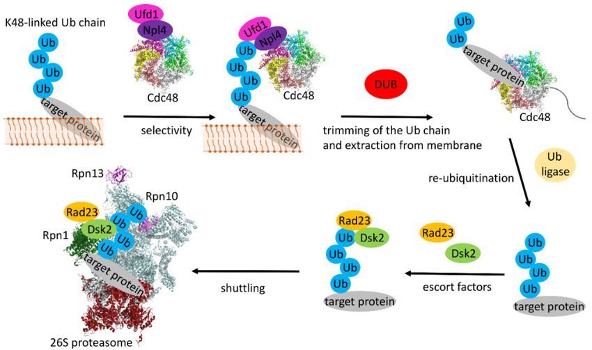
Int. J. Mol. Sci. 2020, 21, 5369 3 of 21
that
Int.the membrane
J. Mol. Sci. 2020, 21,protein partREVIEW
x FOR PEER of this multispanning E3 Really Interesting New Gene (RING)-finger 3 of 21
ligase forms half of a translocation channel with the other half provided by Der1 [26]. Both half
other half
channels markprovided
a site byof aDer1 [26]. Both
thinned half channels
membrane regionmark
that afacilitates
site of a thinned membrane regioninto
the retrotranslocation thatthe
facilitates the retrotranslocation into the cellular cytosol. Recognition of the to-be-degraded
cellular cytosol. Recognition of the to-be-degraded substrate is achieved by the combination of the substrate
MRHis achieved
domain by the combination
of Yos9 of the
that recognizes theMRH domain ofpolysaccharide
α-1,6-mannose Yos9 that recognizes theofα-1,6-mannose
part and a groove of the
luminal domain of Hrd3 that probably binds to the polypeptide segment downstreambinds
polysaccharide part and of a groove of the luminal domain of Hrd3 that probably of thetoglycan
the
polypeptide segment downstream of the glycan attachment site. Retrotranslocation is initiated by a
attachment site. Retrotranslocation is initiated by a polypeptide hairpin that is moved into the cytosol
polypeptide hairpin that is moved into the cytosol where it gets ubiquitinated followed by complete
where it gets ubiquitinated followed by complete extraction of the ubiquitinated polypeptide from the
extraction of the ubiquitinated polypeptide from the ER into the cytosol by the Cdc48 adenosine
ER into the cytosol by the Cdc48 adenosine triphosphatase (ATPase) and its cofactors Ufd1-Npl4 and
triphosphatase (ATPase) and its cofactors Ufd1-Npl4 and final degradation by the proteasome (see
final degradation by the proteasome (see below).
below).
Figure 1. Schematic representation of major components of the ERAD machinery that are involved in the
Figure 1. Schematic representation of major components of the ERAD machinery that are involved in
three
the main
three ERAD subtypes
main ERAD ERAD-L,
subtypes ERAD-M,
ERAD-L, and ERAD-C.
ERAD-M, (A) The
and ERAD-C. (A)ERAD-L pathway.
The ERAD-L PanelPanel
pathway. shows
theshows
components
the components of the Hrd1 complex that are required for degradation of targets localizedthe
of the Hrd1 complex that are required for degradation of targets localized within
ERwithin
lumentheandERadditional
lumen andimportant
additionalfactors suchfactors
important as thesuch
AAA+ ATPase
as the AAA+Cdc48
ATPaseand its two
Cdc48 andassociated
its two
interaction partners Npl4 and Ufd1; (B) The ERAD-M pathway. Panel shows the
associated interaction partners Npl4 and Ufd1; (B) The ERAD-M pathway. Panel shows thecentral components
central
and associated factors
components requiredfactors
and associated for degradation of intra-membrane
required for substrates. This substrates.
degradation of intra-membrane pathway does Thisnot
involve Yos9does
pathway andnot
Der1 is exchanged
involve Yos9 andwith Dfm1;
Der1 (C) The ERAD-C
is exchanged pathway.
with Dfm1; (C) ThePanel shows
ERAD-C the components
pathway. Panel
of shows
the Doa10 complex required
the components of the for degradation
Doa10 of proteins
complex required forwith misfolded
degradation of cytoplasmic
proteins withdomains.
misfolded
cytoplasmic domains.
4. Components of the Ubiquitination Machinery
4. Components
Targeting the of the Ubiquitination
retrotranslocated Machinery
polypeptide for degradation is essential to prevent the accumulation
of misfolded proteins
Targeting at the membrane
the retrotranslocated of the ER with
polypeptide potential detrimental
for degradation is essentialeffects.
to preventIn general,
the
ubiquitin conjugating
accumulation E2 enzymes
of misfolded proteins specify
at the the ubiquitin
membrane chain
of the ERlinkage type and
with potential also the processivity
detrimental effects. In
of general,
ubiquitination
ubiquitin(i.e., the number
conjugating of ubiquitin
E2 enzymes specifymolecules attached
the ubiquitin chaintolinkage
the growing
type and chain
alsointhe
one
processivity
round of ubiquitination
of association) (i.e., the number
[34]. Originally, only of ubiquitin molecules
K48-linked ubiquitinattached
chains wereto the found
growingtochainact as
in one round
degradation of association)
signals [35,36]. Later[34].itOriginally, only K48-linked
was demonstrated that alsoubiquitin
K11-linkedchains weremixed
chains, found chains
to act as
and
degradation signalsproteins
mono-ubiquitinated [35,36]. [37,38]
Later itcanwasbe demonstrated
degraded bythat the also K11-linked
proteasome. In chains, mixed
yeast the chains and
E2 enzymes Ubc7
andmono-ubiquitinated
Ubc6 (and to a lesser proteins
extent[37,38]
Ubc1)can arebe degraded
involved in by
thethe
ERADproteasome. In yeast
process [39]. the E2 enzymes
In mammalian cells a
larger number of E2 enzymes have been identified with two homologues for each In
Ubc7 and Ubc6 (and to a lesser extent Ubc1) are involved in the ERAD process [39]. of mammalian
the two yeast
cells a larger number
proteins-Ube2g1 and Ube2g2of E2 (Ubc7)
enzymes andhave beenand
Ube2j1 identified
Ube2j2 with
(Ubc6) two homologues
[40–45]. for each of
For a summary ofthe two
yeast and
yeast proteins-Ube2g1 and Ube2g2 (Ubc7) and Ube2j1 and Ube2j2 (Ubc6)
mammalian ERAD components see Table 1. Ubc6 and its mammalian homologues have hydrophobic [40–45]. For a summary of
yeast and mammalian ERAD components see Table 1. Ubc6 and its mammalian homologues have
C-termini that get tethered to the ER membrane via posttranslational modifications. In contrast, Ubc7
hydrophobic C-termini that get tethered to the ER membrane via posttranslational modifications. In
and its homologues are soluble proteins that are recruited to the membrane by interaction with Cue1,
contrast, Ubc7 and its homologues are soluble proteins that are recruited to the membrane by
another component of the ERAD machinery (see below). Ubc7 and Ubc6 are involved in different
interaction with Cue1, another component of the ERAD machinery (see below). Ubc7 and Ubc6 are
aspects of the ERAD process. Although Ubc7 is the main E2 enzyme that cooperates with the Hrd1 E3
involved in different aspects of the ERAD process. Although Ubc7 is the main E2 enzyme that
ligase, the central component of the Hrd complex, Ubc6 interacts with another E3 ligase complex, the
cooperates with the Hrd1 E3 ligase, the central component of the Hrd complex, Ubc6 interacts with
Doa10 complex. Doa10
another E3 ligase consists
complex, theofDoa10
14 transmembrane
complex. Doa10 helices as well
consists of 14astransmembrane
N- and C-terminal helicescytoplasmic
as well
domains (Figure 1C) [46]. It is not only expressed in the ER membrane but
as N- and C-terminal cytoplasmic domains (Figure 1C) [46]. It is not only expressed in the also in the nuclear envelope.
ER
In membrane
contrast to but
the Hrd
also complex that isenvelope.
in the nuclear involved In in degrading
contrast to proteins
the Hrd in the ER that
complex lumen and in theinER
is involved
membrane
degrading(ERAD-L
proteins andin theERAD-M
ER lumen pathways),
and in the Doa10 is involved
ER membrane in the ubiquitination
(ERAD-L and ERAD-M of membrane
pathways),
proteins with cytoplasmic domains (both ER and nuclear envelope) and
Doa10 is involved in the ubiquitination of membrane proteins with cytoplasmic domains (bothsoluble proteins in the cytosol
ER
and nuclear envelope) and soluble proteins in the cytosol (ERAD-C pathway). Recently, a third E3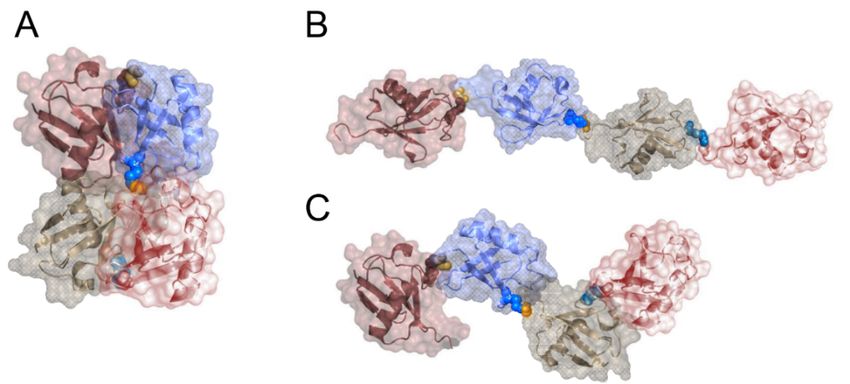
Int. J. Mol. Sci. 2020, 21, 5369                                                                    4 of 21

(ERAD-C pathway). Recently, a third E3 ligase, the Asi complex, was identified in yeast that functions
exclusively at the inner membrane of the nuclear envelope [47,48] where it cooperates with the E2
enzymes Ubc7, Ubc6 and to a lesser extent with Ubc4 [48]. In the mammalian system four major
E3 ligases (Hrd1, TEB4, gp78, and carboxy-terminus of Hsc70 interacting protein (CHIP)) have been
identified in the ERAD process but up to 19 additional E3 ligases have been linked to the degradation
of specific ER-associated targets [14,15]. Of the four major E3 ligases Hrd1 and gp78 are homologues of
yeast Hrd1 and both of them bind to the Ubc7 homologue Ube2g2 [42,49,50]. In addition to its RING
domain, gp78 contains a CUE domain and a G2BR domain for recruiting Ube2g2 (see below) as well.
The mammalian Doa10 homologue E3 ligase is TEB4 that was found to adopt a membrane topology
similar to Doa10 [46,51] and is predicted to bind to Ube2g2, Ube2j1 and Ubxd8 E2 enzymes [52] while
CHIP is a U-box E3 ligase that also acts as a co-chaperone [53].

                     Table 1. Comparison of ERAD components from yeast and mammalian cells.

                                   Yeast                 Human Homologue           Reference
               Target recognition
                             Htm1/Mnl1                        EDEM1-3                 [54]
                                Yos9                            OS-9                  [55]
                                Pdi1                            Pdi                   [56]
                                Hrd3                           Sel1L                  [57]
               Retrotranslocation
                               Der1                            Derlin1                [58]
                               Dfm1                            Derlin1                [58]
                               Usa1                            HERP                   [59]
                               Cdc48                            p97                   [60]
                               Ufd1                             Ufd1                  [61]
                               Npl4                             Npl4                  [62]
                               Ubx2                            Ubxd8                  [63]
               E2 ubiquitin conjugating enzymes
                                Ubc1                           Ube2k                  [64]
                                Ubc4                           Ube2d1                 [65]
                                Ubc6                          Ube2j1-2                [43]
                                Ubc7                          Ube2g1-2                [66]
               E3 ubiquitin ligase enzymes
                                Hrd1                            Hrd1                  [57]
                                Doa10                           TEB4                  [51]
                                 Asi
                                                                gp78                  [49]
                                                               CHIP                   [67]
                                                               RMA1                   [68]
               E4 ubiquitin chain elongation factors
                                Ufd2                            Ufd2                  [69]
                                Doa1                            Ufd3                  [70]
                                Cue1
                                                                AUP1                  [71]
               Deubiquitinase
                                   Otu1                         Yod1                  [72]
               Escort factors
                                   Rad23                      Hr23a-b                 [73]
                                   Dsk2                      Ubiquilin-1              [74]

     In yeast, the two E3 enzymes were also shown to cooperate with Ubc6 that is implicated in priming
of the ubiquitin chain build-up by attaching a single ubiquitin moiety [75]. Interestingly, it seems to be
able to attach ubiquitin not only to lysine residues but also to serines and possibly to threonines [75].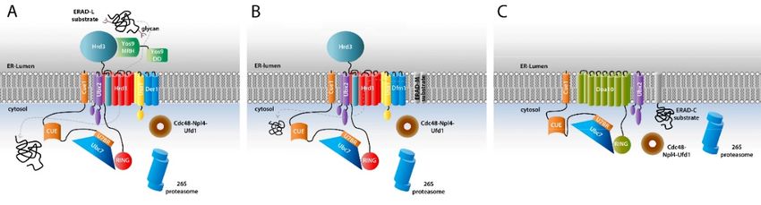
Int. J. Mol. Sci. 2020, 21, 5369                                                                    5 of 21

This ability to modify amino acids with hydroxyl group bearing side chains was also reported
for the mammalian homologue of Ubc6, Ube2j2 [76]. The purpose of this relatively promiscuous
mono-ubiquitination reaction could be to have many potential sites for attaching ubiquitin chains.
Once a site is marked with mono-ubiquitin Ubc7 takes over which is better suited for (K48-linked)
chain elongation but less for priming. In support of this model, attachment of a non-cleavable ubiquitin
monomer to a substrate resulted in fast ubiquitination mediated only by Ubc7 [75]. Interestingly,
it was reported that also mammalian Hrd1 can ubiquitinate non-lysine amino acids [77]. Investigation
of the ubiquitination of the ERAD-L (i.e., Hrd1 dependent) substrate NS-1 κ LC (non-secreted-1
immunoglobulin κ light chain) showed that ubiquitination could still be detected when all its lysine
residues were mutated to arginine. Ubiquitination by Hrd1 was only suppressed when also all serine
and threonine residues were mutated as well which also resulted in inhibition of the degradation of
NS-1 κ LC [77]. Likewise, it was shown that the T-cell antigen receptor α-chain becomes ubiquitinated
on its cytoplasmic tail upon failure to correctly assemble the complete T-cell receptor complex. As the
cytoplasmic tail of this protein does not contain any lysine residues (RLWSS), it was proposed that
serines get modified which was validated by mutational analysis [78].

5. High Processivity in ERAD

5.1. The Special Role of the E2 Conjugating Enzymes
      Within the UPS system the E3 ligase is responsible for target specificity by connecting the E2
enzyme with the to-be-ubiquitinated protein. The three ERAD specific E3 ligases in yeast (including the
Asi complex of the nuclear membrane), however, have to interact with a large number of diverse targets
as approximately one third of all expressed proteins of a cell get channeled through the ER. As the
translocation of the newly synthesized proteins into the ER lumen or into the ER membrane occurs in an
unfolded form, a high risk of misfolding of a wide variety of proteins in the ER exists [79]. The required
high promiscuity of the ERAD E3 ligases makes a highly efficient and processive ubiquitination
reaction necessary. This is achieved by several features of the E2 conjugating enzymes. First, Ubc6
and Ubc7 have the ability to pre-assemble longer ubiquitin chains on their active site cysteine that are
then transferred en bloc to the target protein mediated by the E3 ligases [39,80–82]. Second, efficient
elongation of the growing ubiquitin chain is also achieved by the interaction of Ubc7 with various
domains of another protein, Cue1. This protein is anchored in the ER membrane with a single
transmembrane helix and has additional domains in the cytosol. One of these domains is a peptide
sequence that interacts directly with Ubc7. This region is named U7BR (Ubc7 binding region) that
binds to a site of the E2 enzyme opposite from the active site cysteine. The binding event recruits
Ubc7 to the ER membrane, increases its local concentration in the vicinity of the ERAD E3 ligases and
stabilizes Ubc7 (by preventing its degradation, see below). In addition, detailed biochemical studies
have shown that binding enhances the ubiquitination reaction by an allosteric mechanism. Ubc7 bound
to the isolated U7BR showed strongly enhanced ubiquitination activity, independently of the presence
of E3 ligases [83]. Another study further showed that the U7BR region is the only required domain of
Cue1 for ERAD if Ubc7 is tethered to the ER membrane [84]. Structure determination of a complex
of Ubc7 with the U7BR region of Cue1 has revealed the molecular mechanism of interaction [85].
The U7BR domain consists of three helices that bind to the “backside” of the E2 enzyme (Figure 2A,C,D).
Comparison of the apo Ubc7 structure with the structure of the Ubc7/U7BR complex did not show any
major structural alterations but mainly different orientations and flexibility of the loops surrounding
the active site Cys89. The observed changes in the crystal structures were further analyzed by nuclear
magnetic resonance (NMR) experiments that confirmed a higher flexibility of the β4α2 loop in the
complex. Consequently, charging Ubc7/U7BR with ubiquitin by an E1 enzyme is more efficient.
Likewise the transfer of the thioester bound ubiquitin to another ubiquitin is accelerated and the
affinity of binding to the Hrd1 RING-finger domain is increased [85]. A similar “backside” interaction
is seen between the mammalian ERAD E2 enzyme Ube2g2 and the RING-finger E3 ligase gp78 [86].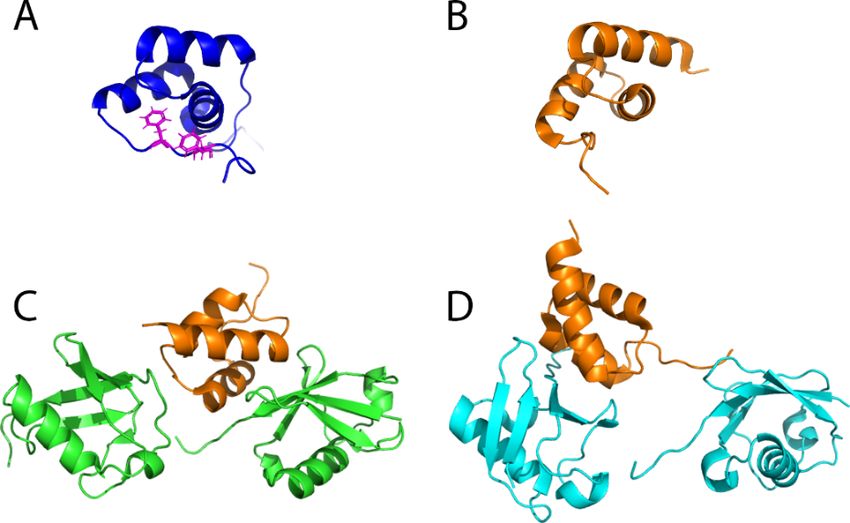
Int. J. Mol. Sci. 2020, 21, 5369 6 of 21
Differences are, however, also evident with the Ube2g2-binding region (G2BR) of gp78 comprising
only a single helix (Figure 2B–D). Structurally, the binding of the G2BR to Ube2g2 has an impact on
Int. J. Mol. Sci. 2020, 21, x FOR PEER REVIEW 6 of 21
the β4α2 and α2α3 loops as well, however, leading to a partial occlusion of the active site cysteine
which
of also shows
the active a different
site cysteine orientation
which of the
also shows side chain
a different in the complex.
orientation Model
of the side building
chain in thesuggested
complex.
Model building suggested that this conformation makes charging with ubiquitin bywhich
that this conformation makes charging with ubiquitin by an E1 enzyme more difficult an E1 was also
enzyme
confirmed
more experimentally.
difficult which was also At confirmed
the same time, however, binding
experimentally. At theof the G2BR
same to Ube2g2binding
time, however, enhances the
of the
affinity towards the gp78 RING-finger domain, which in this case is located in cis
G2BR to Ube2g2 enhances the affinity towards the gp78 RING-finger domain, which in this casethe with respect to is
G2BR peptide
located [86].respect to the G2BR peptide [86].
in cis with
Figure 2.
2. Crystal
Crystalstructures ofof
structures E2E2
enzymes
enzymes in complex
in complexwithwith
the U7BR/G2BR
the U7BR/G2BR domains. (A) Yeast
domains. Ubc7
(A) Yeast
in complex
Ubc7 with with
in complex U7BRU7BR
(PDB(PDB
ID: 4JQU). TheThe
ID: 4JQU). green and
green orange
and orange colors
colorsrepresent
representUbc7
Ubc7and
and U7BR,
U7BR,
respectively; (B)
(B)Mammalian
MammalianUbe2g2
Ube2g2inin
complex
complexwith G2BR
with (PDB
G2BR ID: 3H8K).
(PDB The cyan
ID: 3H8K). The and
cyanyellow colors
and yellow
represent Ube2g2 and G2BR, respectively; (C) Superimposed structures of (A,B); (D)
colors represent Ube2g2 and G2BR, respectively; (C) Superimposed structures of (A) and (B); (D) Superimposed
structures of (A,B), rotatedof ◦ along the vertical axis compared to (C). In both cases the binding domain
90(A)
Superimposed structures and (B), rotated 90° along the vertical axis compared to (C). In both
interacts
cases the with the backside
binding of the E2with
domain interacts enzyme, opposite of
the backside from
thethe
E2 catalytic
enzyme,cysteine.
opposite from the catalytic
cysteine.
5.2. CUE Domains in the ERAD Process
5.2. CUE Domainstointhe
In addition theU7BR
ERAD ProcessCue1 contains a CUE domain. CUE domains were first identified
domain
and Innamed after yeast Cue1
addition to the U7BR domain [87] for ‘coupling of ubiquitin
Cue1 contains a CUE to domain.
ER degradation’.
CUE domains They were
consist of
first
approximately
identified 40 amino
and named acid
after residues
yeast Cue1and [87]are
forsemi-conserved in a range
‘coupling of ubiquitin of eukaryotic
to ER degradation’.proteins. In line
They consist
with the origin of its name, the CUE domain containing proteins Cue1 [88],
of approximately 40 amino acid residues and are semi-conserved in a range of eukaryotic proteins. gp78 [40] and AUP1 [89]
are indeed involved in the ERAD process. However, other CUE domain
In line with the origin of its name, the CUE domain containing proteins Cue1 [88], gp78 [40] andcontaining proteins such as
Vps9 [90]
AUP1 and
[89] Tollip
are [91] have
indeed otherin
involved functions.
the ERAD Originally,
process.investigations
However, other showed CUEthatdomain
the CUEcontaining
domain of
Cue1 is dispensable for ubiquitination within the ERAD process. Later studies,
proteins such as Vps9 [90] and Tollip [91] have other functions. Originally, investigations showed however, demonstrated
that in
that thetheCUEpresence
domain of Hrd1
of Cue1the full length Cue1for
is dispensable protein stimulatedwithin
ubiquitination ubiquitination
the ERAD stronger
process.than the
Later
isolated U7BR and this effect was traced to the presence of the CUE domain [92].
studies, however, demonstrated that in the presence of Hrd1 the full length Cue1 protein stimulated The same stimulation
was observed when
ubiquitination Doa10
stronger was
than used
the as E3 ligase
isolated U7BR in these
and thisexperiments showing
effect was traced thatpresence
to the the CUE domain
of the CUEhas
a similar function
domain [92]. Theforsame
both the Hrd1/Ubc7
stimulation andobserved
was Doa10/Ubc7-based
when Doa10 ubiquitination
was usedreactions [92]. However,
as E3 ligase in these
investigation of different substrate classes—soluble proteins and membrane-bound
experiments showing that the CUE domain has a similar function for both the Hrd1/Ubc7 and proteins—revealed
that degradation of only
Doa10/Ubc7-based the membrane-bound
ubiquitination reactions [92].substrates
However,was affected by either
investigation ofdeletion of the
different entire
substrate
CUE domain or destabilizing mutations in this domain in vivo [92].
classes—soluble proteins and membrane-bound proteins—revealed that degradation of only the
membrane-bound substrates was affected by either deletion of the entire CUE domain or
destabilizing mutations in this domain in vivo [92].
CUE domains have been shown to be ubiquitin binding domains (UBDs) that bind to both mono-
ubiquitin as well as to poly-ubiquitin chains [87,90,93–95]. They consist of three α-helices, similar to
the ubiquitin-associated (UBA) fold. The CUE domain of yeast Cue1 differs from canonical CUEInt. J. Mol. Sci. 2020, 21, 5369 7 of 21
CUE domains have been shown to be ubiquitin binding domains (UBDs) that bind to both
mono-ubiquitin as well as to poly-ubiquitin chains [87,90,93–95]. They consist of three α-helices,
similar to the ubiquitin-associated (UBA) fold. The CUE domain of yeast Cue1 differs from canonical
CUE domains by requiring a C-terminal extension containing two phenylalanine residues that are
crucial for stabilization of the structure (Figure 3A) [96]. All structural investigations of different
CUE domains have revealed that they recognize the hydrophobic patch of ubiquitin around residues
Int. J. Mol. Sci. 2020, 21, x FOR PEER REVIEW 7 of 21
L8, I44 and V70 with dissociation constants for mono-ubiquitin in the ~10 µM range. In contrast
to mostdomains
CUE domains, by requiring thea dissociation constant
C-terminal extension of the two
containing CUE domain of residues
phenylalanine Cue1 for ubiquitin
that are crucialbinding
is high and was determined
for stabilization to be ~150
of the structure µM 3A)
(Figure [96][96].
most Alllikely due to
structural the replacement
investigations of anCUE
of different otherwise
domains
invariable have revealed
Met-Phe-Pro triple that they recognize
amino acid stretch the hydrophobic patch of ubiquitin
with the sequence Leu-Ala-Proaround residues
within theL8,ubiquitin
I44 and V70 with dissociation constants for mono-ubiquitin in the ~10 μM
binding interface. Measuring the binding affinity of different ubiquitin chains revealed that the affinityrange. In contrast to most
CUE domains, the dissociation constant of the CUE domain of Cue1 for ubiquitin binding is high and
significantly increased with increasing chain length and that K48-linked chains are preferred over
was determined to be ~150 μM [96] most likely due to the replacement of an otherwise invariable
K63-linked or linear
Met-Phe-Pro chains
triple amino (~90
acidµM for tetra
stretch with theK48 Ub andLeu-Ala-Pro
sequence 110 µM for tetrathe
within K63 Ub) [96].
ubiquitin This result
binding
is also consistent with the the
interface. Measuring observation that Ubc7
binding affinity exclusively
of different assembles
ubiquitin K48-linked
chains revealed ubiquitin
that the affinity chains
on substrates in vivo
significantly [80,97]with
increased andincreasing
unanchored chain ubiquitin chains
length and that in vitrochains
K48-linked [92]. are
In preferred
general, overelongation
kinetics decrease with increasing chain length, an effect attributed to the growing distance isbetween
K63-linked or linear chains (~90 μM for tetra K48 Ub and 110 μM for tetra K63 Ub) [96]. This result
also consistent with the observation that Ubc7 exclusively assembles K48-linked ubiquitin chains on
the distal end of the chain and the active center of the involved E2-E3 ligase complex which has for
substrates in vivo [80,97] and unanchored ubiquitin chains in vitro [92]. In general, elongation kinetics
exampledecrease
been observed for the anaphase promoting complex (APC/C) [98,99]. In the presence of the
with increasing chain length, an effect attributed to the growing distance between the distal
CUE domain
end of the of Cue1,
chain and however,
the activeacceleration
center of thewas observed
involved with increasing
E2-E3 ligase complex which chain
has length.
for example The slight
preference
beenfor K48-linked
observed for thechains
anaphaseover K63-linked
promoting ones(APC/C)
complex could be traced
[98,99]. In back to additional
the presence of the CUEinteraction
with thedomain
C-terminus of Cue1, however,
of the distal acceleration was observed
ubiquitin molecule within with
theincreasing
chain which chainalso
length. The slight
increases the affinity
preference for K48-linked chains over K63-linked ones could be traced
to the proximal moiety (over the distal ubiquitin unit) approximately two-fold. These studies further back to additional interaction
with the C-terminus of the distal ubiquitin molecule within the chain which also increases the affinity
revealed that K48 itself is not part of the interaction interface of the Cue1 CUE domain, in contrast
to the proximal moiety (over the distal ubiquitin unit) approximately two-fold. These studies further
for example
revealed to the
thatCUE domain
K48 itself is notofpart
Cue2 [94].
of the Furtherinterface
interaction kinetic ofstudies
the Cue1withCUEubiquitin
domain, chains harboring
in contrast
ubiquitinformolecules
example to with the CUE mutated
domainCUE of Cue2domain binding
[94]. Further interfaces
kinetic at various
studies with ubiquitinpositions with the chain
chains harboring
suggested a model
ubiquitin in which
molecules with the CUECUE
mutated domain
domain binds preferentially
binding to thepositions
interfaces at various penultimate
with theubiquitin
chain in a
suggested a model in which the CUE domain binds preferentially to
chain [96]. The binding most likely orients Ubc7 relative to the distal end of the growing chain andthe penultimate ubiquitin in a
chain [96]. The binding most likely orients Ubc7 relative to the distal end of the growing chain and
thus accelerates the transfer of the next ubiquitin unit from the E2 enzyme to the acceptor lysine of the
thus accelerates the transfer of the next ubiquitin unit from the E2 enzyme to the acceptor lysine of
distal moiety (Figure 4).
the distal moiety (Figure 4).
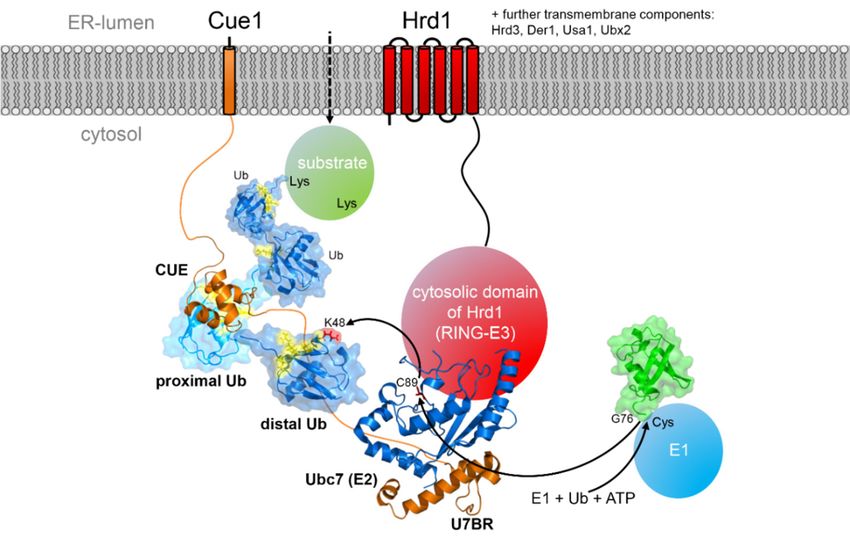
Figure
Figure 3. 3. Structural
Structural comparison
comparison of CUE
of CUE domains and
domains and their
theirbinding
bindingto di-ubiquitin. (A) NMR
to di-ubiquitin. (A) structure
NMR structure
of the CUE domain of Cue1 (PDB ID: 2MYX). Phenylalanine residues that are crucial for the stability
of the CUE domain of Cue1 (PDB ID: 2MYX). Phenylalanine residues that are crucial for the stability
are shown with side chains; (B) NMR structure of the CUE domain of gp78 (PDB ID: 2LVN); (C) NMR
are shown with side chains; (B) NMR structure of the CUE domain of gp78 (PDB ID: 2LVN); (C) NMR
structure of the CUE domain of gp78 bound to the distal moiety in a K48-linked di-ubiquitin (PDB
structure of the CUE
ID: 2LVP). domain
Orange of gp78
and green bound
colors to the
represent thedistal moiety and
CUE domain in adi-ubiquitin,
K48-linkedrespectively;
di-ubiquitin (PDB ID:
(D)
2LVP). Orange and green
NMR structure colors
of the CUE represent
domain the
of gp78 CUEtodomain
bound andmoiety
the proximal di-ubiquitin, respectively;
in a K48-linked (D) NMR
di-ubiquitin
(PDB
structure of ID:
the2LVQ). Orange and
CUE domain of cyan
gp78colors represent
bound to thethe CUE domain
proximal and di-ubiquitin,
moiety respectively.
in a K48-linked di-ubiquitin
(PDB ID:The structures
2LVQ). involving
Orange and the
cyan gp78 CUErepresent
colors domain and thedi-ubiquitin
CUE domain showand the di-ubiquitin,
dynamic interaction
respectively.
between both molecules.
The structures involving the gp78 CUE domain and di-ubiquitin show the dynamic interaction between
both molecules.Int. J. Mol. Sci. 2020, 21, 5369 8 of 21
Int. J. Mol. Sci. 2020, 21, x FOR PEER REVIEW 8 of 21
Figure 4.
Figure Schematic representation
4. Schematic representation ofof the
the effect
effect of
of the
the CUE
CUE domain
domain ofof Cue1
Cue1 onon the
the Hrd1
Hrd1 dependent
dependent
ubiquitination of a retrotranslocated substrate. The CUE domain preferentially
ubiquitination of a retrotranslocated substrate. The CUE domain preferentially binds to binds to the
the proximal
proximal
ubiquitin in the growing ubiquitin chain, thereby positioning Ubc7 that is also tethered
ubiquitin in the growing ubiquitin chain, thereby positioning Ubc7 that is also tethered to Cue1 to Cue1 via via
the
U7BR
the domain
U7BR relative
domain to the
relative substrate
to the protein.
substrate Except
protein. for Hrd1
Except andand
for Hrd1 Cue1 all other
Cue1 components
all other componentsof the
of
Hrd1 complex are omitted for clarity.
the Hrd1 complex are omitted for clarity.
In mammalian cells the ERAD E3 ligase gp78 contains in addition to the G2BR domain that binds
In mammalian cells the ERAD E3 ligase gp78 contains in addition to the G2BR domain that binds
to the E2 enzyme Ube2g2 also a CUE domain that recognizes the hydrophobic patch of ubiquitin
to the E2 enzyme Ube2g2 also a CUE domain that recognizes the hydrophobic patch of ubiquitin
similar to other CUE domains. Its interaction with di-ubiquitin is not well-defined, but dynamic
similar to other CUE domains. Its interaction with di-ubiquitin is not well-defined, but dynamic and
and enables multiple conformations of di-ubiquitin [95], supporting growth of the ubiquitin chain
enables multiple conformations of di-ubiquitin [95], supporting growth of the ubiquitin chain and
and thus processivity of ubiquitination [100] (Figure 3B–D). Using NMR titrations and isothermal
thus processivity of ubiquitination [100] (Figure 3B–D). Using NMR titrations and isothermal titration
titration calorimetry (ITC) the binding of mono-ubiquitin and both K63- and K48-linked di-ubiquitin
calorimetry (ITC) the binding of mono-ubiquitin and both K63- and K48-linked di-ubiquitin was
was measured and found to have dissociation constants in the 10–25 µM range and thus a higher
measured and found to have dissociation constants in the 10–25 μM range and thus a higher affinity
affinity than the yeast Cue1 CUE domain [95]. A recent study suggested that the CUE domain of gp78
than the yeast Cue1 CUE domain [95]. A recent study suggested that the CUE domain of gp78 is
is responsible for proofreading the growing poly-ubiquitin chain to ensure K48-linkage specificity by
responsible for proofreading the growing poly-ubiquitin chain to ensure K48-linkage specificity by
restricting its activity for non-K48-linked chain assembly when bound to K48-linked chain [101].
restricting its activity for non-K48-linked chain assembly when bound to K48-linked chain [101].
5.3. Role of E4 Enzymes and Different Chain Linkages
5.3. Role of E4 Enzymes and Different Chain Linkages
The effects of the various Cue1 domains show how the concerted action of a domain that activates
The effects of the various Cue1 domains show how the concerted action of a domain that
the E2 enzyme by “backside binding” (U7BR) and by positioning the distal end of the growing chain
activates the E2 enzyme by “backside binding” (U7BR) and by positioning the distal end of the
in an E4-like manner (CUE) facilitates ubiquitination to be highly processive. For other enzymes
growing chain in an E4-like manner (CUE) facilitates ubiquitination to be highly processive. For other
of the ubiquitin system it has been shown as well that processive poly-ubiquitin chain formation
enzymes of the ubiquitin system it has been shown as well that processive poly-ubiquitin chain
can be promoted by noncovalent interactions with ubiquitin. Examples are activation by “backside
formation can be promoted by noncovalent interactions with ubiquitin. Examples are activation by
binding” to certain E2 enzymes [102,103] as well as by RING domains showing ubiquitin binding
“backside binding” to certain E2 enzymes [102,103] as well as by RING domains showing ubiquitin
activity [104,105]. E4 enzymes that participate in ubiquitination reactions in yeast have been described
binding activity [104,105]. E4 enzymes that participate in ubiquitination reactions in yeast have been
as well and shown to be necessary for highly processive chain elongation but not in the initial steps [106].
described as well and shown to be necessary for highly processive chain elongation but not in the
In in vitro ubiquitination reactions, using the E1 enzyme Uba1, the E2 enzyme Ubc4, and the E3 ligase
initial steps [106]. In in vitro ubiquitination reactions, using the E1 enzyme Uba1, the E2 enzyme Ubc4,
Ufd4 only resulted in relatively short ubiquitin chains of two to three moieties. Adding the ubiquitin
and the E3 ligase Ufd4 only resulted in relatively short ubiquitin chains of two to three moieties.
binding protein Ufd2 stimulated the ubiquitination reaction, resulting in significantly longer ubiquitin
Adding the ubiquitin binding protein Ufd2 stimulated the ubiquitination reaction, resulting in
chains [106]. A recent study, however, has not found an effect on ubiquitin chain elongation. Instead
significantly longer ubiquitin chains [106]. A recent study, however, has not found an effect on
these investigations showed that Ufd2 adds a single ubiquitin moiety onto proximal ubiquitin molecules
ubiquitin chain elongation. Instead these investigations showed that Ufd2 adds a single ubiquitin
to form K48-ubiquitin branches [107], in particular creating branched K29/K48 ubiquitin chains on
moiety onto proximal ubiquitin molecules to form K48-ubiquitin branches [107], in particular
ERAD substrates. These branching reactions are necessary to target the originally K29 decorated
creating branched K29/K48 ubiquitin chains on ERAD substrates. These branching reactions are
necessary to target the originally K29 decorated substrates for proteasomal degradation. Ufd2 alsoInt. J. Mol. Sci. 2020, 21, 5369 9 of 21
substrates for proteasomal degradation. Ufd2 also interacts with the AAA+ ATPase Cdc48 connected
to the extraction of ubiquitinated substrates from the ER [70]. The exact role of Ufd2, however, remains
to be investigated.
In addition to the canonical K48-linked and branched K29/K48 chains, other chain types have
also been detected on ERAD substrates. Although Ubc7 is committed to the formation of K48-linked
ubiquitin chains quantitative proteomics studies have revealed that Ubc6 can be decorated with
K11-linked chains in an auto-ubiquitination process and that other targets can be modified with
K11-linked chains by Ubc6 as well [39]. Treating yeast with dithiothreitol (DTT) that prevents the
formation of disulfide bonds in the ER or tunicamycin (inhibits N-linked glycosylation) to induce ER
stress selectively increased the level of K11-linked ubiquitin chains while the level of all other linkage
types remained constant. This suggested that K11-linked chains play a role via the Ubc6/Doa10 E2/E3
complex in the ERAD process [39]. Similarly, inhibiting the proteasome in mammalian cells did not
only increase the abundance of ubiquitin chains with a K48-linkage, but also other chain types, in
particular K11-linked ones [108].
5.4. Degradation of the Ubiquitination Machinery
The high and necessary promiscuity of the ERAD system for target proteins makes it not only
efficient to prevent the accumulation of misfolded proteins in and at the ER but poses also great risks.
This is demonstrated by overexpression experiments of Hrd1 in a Hrd3 deletion mutant strain which
targets also stable ER resident proteins for degradation [29]. This observation probably also explains
the growth retardation effect seen with overexpression of Hrd1 in yeast [29]. Although overexpression
of Hrd1 circumvents the need for the other ERAD components by forming oligomers that are able to
retrotranslocate substrate proteins [32], complex formation with Usa1 and in particular Hrd3 stabilize
Hrd1 and prevent its poly-ubiquitination, extraction and degradation [109–113] and it has been
speculated that Hrd3 regulates auto-ubiquitination of Hrd1. One effective way of down-regulating the
activity of the ERAD machinery is to remove the associated E2 enzymes [32]. Ubc6 itself is an ERAD
target and its degradation requires both the membrane-bound tail domain as well as the catalytic
cysteine [114]. Ubiquitination of Ubc6 depends on its own catalytic function and on the activity of Ubc7,
in a Doa10-dependent manner [115], consistent with the involvement of Doa10 in the degradation
of ER membrane proteins with domains in the cytosol (ERAD-C pathway). Similarly, the human
homologues of Ubc6, Ube2j1, and Ube2j2, were found to be targeted for proteasomal degradation and
both their membrane-anchored domains and catalytic cysteines were crucial in that process [116,117].
Ubc7 also gets degraded and it was found to be down-regulated in the absence of its binding partner,
Cue1 [88]. Later it was discovered that the degradation of Ubc7 involves auto-ubiquitination at the
catalytic cysteine supported by Ufd4, a HECT-domain E3 ligase [80].
6. The Conformation of the Synthesized Chains
6.1. Binding and Modification of Ubiquitin Chains by Cdc48 and Associated Factors
The ubiquitin chain attached to the substrate does not only constitute a signal for proteasomal
degradation [118] but is also required to extract the ubiquitin decorated protein completely into
the cytosol (in the ERAD-L pathway) [119]. There is also increasing experimental evidence that
the chain is subject to extensive modifications during the extraction process (Figure 5). The final
retrotranslocation of the substrate is dependent on the homohexameric AAA+ ATPase Cdc48 in yeast (or
p97/valosin-containing protein (VCP) in mammalian cells) and its two cofactors Ufd1 and Npl4 [120,121].
The complex gets recruited to the ER membrane by interaction with different receptor domains located
on Ubx2 (being part of both the Hrd1 and Doa10 complexes), Dfm1 and Hrd1 [31,122–124]. The two
cofactors, Ufd1 and Npl4, contain UBDs that interact with the ubiquitinated target proteins [125–127]
and recruit them to Cdc48 by also binding to the N-terminal domain of the AAA+ ATPase. In addition,
Cdc48 interacts with several ubiquitin chain modifying enzymes and is therefore a hub for chainInt. J. Mol. Sci. 2020, 21, 5369 10 of 21
elongation
Int. J. Mol. Sci.and
2020,trimming.
21, x FOR PEER These modifying factors include Ufd2 [106,128,129] as well as the protein
REVIEW 10 of 21
Ufd3 which does not have a chain modifying activity itself but acts as an inhibitor of Ufd2 [130].
It was
acts as also shown that
an inhibitor of Ufd2the [130].
presence of Cdc48
It was inhibits
also shown thatthe
theformation
presence ofofCdc48
very long ubiquitin
inhibits chains
the formation
and
of very restricts the average
long ubiquitin sizeand
chains to three to six
restricts the moieties [131]
average size to by recruiting
three de-ubiquitinating
to six moieties (DUB)
[131] by recruiting
enzymes such as Otu1 [132]. In yeast, the expression of an inactive
de-ubiquitinating (DUB) enzymes such as Otu1 [132]. In yeast, the expression of an inactive Otu1Otu1 mutant (Otu1p C120S)
inhibited
mutant (Otu1p the efficient
C120S)degradation
inhibited theofefficient
sCPY*-DHFR and deletion
degradation of the Cdc48
of sCPY*-DHFR andbinding
deletionUBX of the domain
Cdc48
from
binding theUBXmutant DUBfrom
domain counter-acted
the mutantthisDUB effect [123]. In vitro
counter-acted thisexperiments with
effect [123]. In poly-ubiquitinated
vitro experiments with
Hrd1 suggested thatHrd1
poly-ubiquitinated in general Cdc48
suggested thatacts before Otu1-mediated
in general Cdc48 acts before trimming of the ubiquitin
Otu1-mediated trimmingchainsof the
occurs
ubiquitin [123]. These
chains investigations
occurs [123]. Thesealso suggested that
investigations also Ubx2
suggestedprevents premature
that Ubx2 de-ubiquitination
prevents premature de-
before substratebefore
ubiquitination extraction by competing
substrate extraction with Otu1 for with
by competing bindingOtu1 tofor
Cdc48 [123].
binding to In the mammalian
Cdc48 [123]. In the
system
mammalian it wassystem
shown it that
wastheshown
p97-associated
that the DUBs Yod1 andDUBs
p97-associated Usp13Yod1 de-ubiquitinate
and Usp13substrates before
de-ubiquitinate
they are channeled
substrates before they through the narrow through
are channeled pore of thetheAAA+
narrow ATPase
pore ofp97the
[72]. Subsequently,
AAA+ ATPase p97 they[72].
get
re-ubiquitinated involving the p97 bound cofactors. Interestingly, recent
Subsequently, they get re-ubiquitinated involving the p97 bound cofactors. Interestingly, recentstudies suggest an alternative
model
studiesinsuggest
which an Cdc48 not only
alternative unfolds
model the extracted
in which Cdc48 not substrate in an ATP-dependent
only unfolds mannerinbut
the extracted substrate an
that trimmed ubiquitin
ATP-dependent mannerchainsbut thatpass through
trimmed the central
ubiquitin chainspore
passofthrough
the AAA+ ATPase
the central in an
pore unfolded
of the AAA+
state
ATPase as well
in an[123,127,132].
unfolded state Inas
this model,
well poly-ubiquitination
[123,127,132]. In this model, is required to bind to the
poly-ubiquitination Ufd1/Npl4
is required to
bind to the
cofactors Ufd1/Npl4
of Cdc48. Aftercofactors
unfolding of and
Cdc48. After unfolding
translocation through andthetranslocation through
pore of the AAA+ the pore
ATPase of the
trimming
AAA+
of ATPase trimming
the poly-ubiquitin chainoftothean poly-ubiquitin
oligo-ubiquitinchain chaintois an oligo-ubiquitin
necessary chainrelease,
for substrate is necessary
followedfor
substrate
by release,
translocation of followed
this trimmed by translocation
chain [132] and ofsubsequent
this trimmed chain chain [132] to
extension and subsequent
create chain
a proteasomal
extension to tag
degradation create a proteasomal
(Figure 5). degradation tag (Figure 5).
Figure 5.
Figure 5. Cartoon showing the main main components
components involved in target target protein
protein extraction
extraction and
and ubiquitin
ubiquitin
chain processing.
chain processing. For clarity, all components of the Hrd1 or Doa10 complexes are omitted. The AAA+
clarity, all components of the Hrd1 or Doa10 complexes are omitted. The AAA+
ATPase
ATPaseCdc48
Cdc48(PDB(PDB ID:ID:
6OPC)
6OPC)withwith
its two
its associated factorsfactors
two associated Ufd1 and
Ufd1Npl4andinteracts with the ubiquitin
Npl4 interacts with the
chain of the
ubiquitin target
chain of protein
the targetwhich leads
protein to itsleads
which extraction
to its from the membrane.
extraction The ubiquitin
from the membrane. Thechain gets
ubiquitin
processed
chain gets first by trimming
processed first byto an oligo-chain.
trimming After translocation
to an oligo-chain. through the
After translocation centralthe
through pore of Cdc48
central pore
the chain the
of Cdc48 is again
chainelongated, then bound
is again elongated, thentobound
the escort
to thefactors
escort Rad23
factorsand
Rad23Dsk2andand
Dsk2recruited to the
and recruited
proteasome for final
to the proteasome fordegradation. Interaction
final degradation. with the
Interaction proteasome
with is initiated
the proteasome by binding
is initiated of the escort
by binding of the
factors to Rpn1toand
escort factors of the
Rpn1 andubiquitin chain to Rpn10
of the ubiquitin chain and Rpn13and
to Rpn10 on the lid structure
Rpn13 on the lid of structure
the proteasome
of the
(PDB ID: 4CR2).
proteasome (PDB ID: 4CR2).
6.2. Interaction with the Proteasome and the Conformational Space of K48-linked Chains
6.2. Interaction with the Proteasome and the Conformational Space of K48-linked Chains
Once processed to the optimal chain length, ubiquitinated proteins are escorted to the proteasome
Once processed to the optimal chain length, ubiquitinated proteins are escorted to the
via the soluble escort factors, Rad23p and Dsk2p [133,134] and interact with the proteasomal receptor
proteasome via the soluble escort factors, Rad23p and Dsk2p [133,134] and interact with the
proteasomal receptor Rpn1 [135] located on the “cap” of the proteasome (19S particle). This
interaction gets further strengthened by binding of the poly-ubiquitin chain to the two ubiquitin
receptors Rpn10 [136] and Rpn13 [137,138]. In all these cases the optimal length for interaction was
determined to be four to six ubiquitin moieties [139–141]. In general, the question how differentInt. J. Mol. Sci. 2020, 21, 5369 11 of 21 Rpn1 [135] located on the “cap” of the proteasome (19S particle). This interaction gets further strengthened by binding of the poly-ubiquitin chain to the two ubiquitin receptors Rpn10 [136] and Rpn13 [137,138]. In all these cases the optimal length for interaction was determined to be four to six ubiquitin moieties [139–141]. In general, the question how different ubiquitin linkage types are recognized by interaction partners and which conformations different chain types can adopt are of central importance for understanding how the various ubiquitination patterns govern so many different cellular processes [142,143]. It was initially found that a K48-linked tetra-ubiquitin chain is the minimal length for efficient degradation [141] and it was demonstrated that the proteasomal ubiquitin receptor Rpn13 stoichiometrically binds K48-linked di-ubiquitin [137,138]. Initial studies investigated the structure of K48-linked di-ubiquitin and tetra-ubiquitin by applying various NMR techniques [144,145] and X-ray crystallography [146]. Open and closed conformations of di-ubiquitin were identified in solution and the transition was found to be dependent on the pH. Although mobility was observed at the interdomain interface at neutral pH, the structure was mainly reported to be in a closed conformation with the hydrophobic patches sequestered at the interdomain interfaces [144,145]. This was further supported by the closed and compact conformation seen in the crystal structure of K48-linked tetra-ubiquitin that was interpreted as a degradation signal [147] (Figure 6A) and by single molecule fluorescence energy transfer experiments in combination with two color coincidence detection [148]. In contrast, in a later study di-ubiquitin was observed in an open conformation, both in solution and in the crystal phase [149]. For other linkage types modelling suggested closed conformations for K6-, K11-, and K27-linked chains and extended conformations for K29-, K33- and K63-linked ones [150]. These predictions were supported for K63-linked chains of different length by X-ray crystallography [151–153] (Figure 6B) and solution studies [154,155]. For K11-linked chains, different conformations were seen in crystal structures showing compact structures in which the hydrophobic patches centered around I44 are solvent exposed but adopt different orientations and are located either on the same face of the dimer [156] or pointing into different directions [157]. Yet, another conformation was identified in solution studies of K11-linked di-ubiquitin in which the hydrophobic patch of the distal moiety is involved in the inter-ubiquitin interface and the hydrophobic patch of the proximal moiety is exposed [158]. Increasing salt concentrations results in more compact conformation, while pH changes have virtually no influence. Whether tetra-ubiquitin (K48-, K11-linked) adopts a closed conformation that serves as a degradation signal also poses the question how the receptors on the proteasome as well as other interaction partners should be able to bind to this compact conformation [159]. Furthermore, results showing that mono-ubiquitination on one or multiple sites constitutes a sufficient degradation signal for the proteasome [37,160] supporting the interpretation that a closed tetra-ubiquitin conformation does not represent a degradation signal. Further underlining this hypothesis more recent studies concluded that K48-linked ubiquitin chains do not adopt a predominantly closed conformation. Using NMR techniques Cook and colleagues showed that K48-linked di-ubiquitin attached to the active site of the E2 enzyme Ube2k adopts an extended conformation [161]. A mix of closed and extended conformations was also seen in crystal structures of tetra-ubiquitin [162]. Kniss and colleagues studied the entire conformational space of K48-linked di-ubiquitin and tetra-ubiquitin with pulsed electron paramagnetic resonance (EPR) and found high flexibility in both structures [163] suggesting that K48-linked ubiquitin chains adopt a wide range of conformations including completely open and closed ones (Figure 6C). Interaction studies with the CUE domain of Cue1 revealed that this conformational space gets narrower by a conformation-selection process. In contrast, interaction with an inactive form of the K48-linkage-specific Otubain protease family member OTUB1 showed a remodeling of the conformational space with conformations adopted that are not seen in free di-ubiquitin. A recent modelling study also concluded that the structure of di-ubiquitin samples a wide conformational space, with the experimentally observed conformations accounting for only 24% of all possible structures [164]. Berg and colleagues performed coarse grained simulations on differently linked di-ubiquitin and tri-ubiquitin and applied neural network-based dimensionality reduction technique to obtain a two-dimensional representation of the conformational
You can also read