COVID-19 The right clinical information, right where it's needed - Last updated: Mar 12, 2020 - SNLG-ISS
←
→
Page content transcription
If your browser does not render page correctly, please read the page content below

COVID-19

The right clinical information, right where it's needed

                                                  Last updated: Mar 12, 2020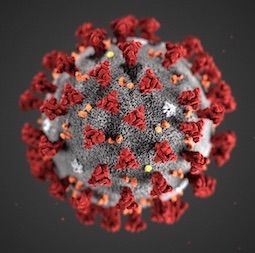
Table of Contents
Summary                                               3

Basics                                                4
    Definition                                        4
    Epidemiology                                      4
    Aetiology                                         5
    Pathophysiology                                   6

Prevention                                            8
    Primary prevention                               8
    Screening                                         9
    Secondary prevention                             9

Diagnosis                                            10
    Case history                                     10
    Step-by-step diagnostic approach                 10
    Risk factors                                     15
    History & examination factors                    15
    Diagnostic tests                                 18
    Differential diagnosis                           21
    Diagnostic criteria                              23

Treatment                                            25
    Step-by-step treatment approach                  25
    Treatment details overview                       28
    Treatment options                                29
    Emerging                                         36

Follow up                                            37
    Recommendations                                  37
    Complications                                    38
    Prognosis                                        39

Guidelines                                           40
    Diagnostic guidelines                            40
    Treatment guidelines                             41

Online resources                                     43

References                                           44

Images                                               52

Disclaimer                                           53
Summary ◊ The World Health Organization has declared the COVID-19 outbreak a pandemic and rates the global risk assessment as very high. ◊ The situation is evolving rapidly with global case counts and deaths increasing each day. The number of cases being reported outside of China each day is now more than the number of cases being reported from China each day. ◊ Person-to-person spread has been confirmed, but it is uncertain how easily the virus spreads between people. ◊ Clinical trials and investigations to learn more about the virus, its origin, and how it affects humans are ongoing.
COVID-19                                                                                                               Basics

         Definition
         Coronavirus disease 2019 (COVID-19) is a potentially severe acute respiratory infection caused by severe
         acute respiratory syndrome coronavirus 2 (SARS-CoV-2). The virus was identified as the cause of an
BASICS

         outbreak of pneumonia of unknown cause in Wuhan City, Hubei Province, China, in December 2019.[1] The
         clinical presentation is that of a respiratory infection with a symptom severity ranging from a mild common
         cold-like illness, to a severe viral pneumonia leading to acute respiratory distress syndrome that is potentially
         fatal.

         The International Committee on Taxonomy of Viruses has confirmed SARS-CoV-2 as the name of the virus
         owing to the virus's genetic similarity to the SARS-CoV virus, but taking into account that there may be
         differences in disease spectrum and transmission.[2] [3] The World Health Organization has confirmed
         COVID-19 (a shortened version of coronavirus disease 2019) as the name of the disease that SARS-CoV-2
         infection causes.[4] Prior to this, the virus and/or disease was known by various names including novel
         coronavirus (2019-nCoV), 2019-nCoV, or variations on this.

         Epidemiology
         The World Health Organization (WHO) was informed of 44 cases of pneumonia of unknown microbial
         aetiology associated with Wuhan City, Hubei Province, China on 31 December 2019. Most of the patients
         in the outbreak reported a link to a large seafood and live animal market (Huanan South China Seafood
         Market).[14] The WHO announced that a novel coronavirus had been detected in samples taken from these
         patients. Laboratory tests ruled out severe acute respiratory syndrome coronavirus (SARS-CoV), Middle East
         respiratory syndrome (MERS)-CoV, influenza, avian influenza, and other common respiratory pathogens.[15]

         Since then, the outbreak has escalated rapidly, with the WHO first declaring a public health emergency of
         international concern on 30 January 2020 and then formally declaring it a pandemic on 11 March 2020. The
         outbreak spread rapidly from a single city in China to the entire country in only 30 days.[13] The numbers
         of cases and deaths have surpassed the toll from the 2002-2003 outbreak of severe acute respiratory
         syndrome (SARS).

         Globally, 118,326 cases have been reported as of 11 March 2020.

         [WHO: novel coronavirus (COVID-19) situation dashboard]

         Cases in China

             • 80,955 cases and 3162 deaths have been reported in China (as of 11 March 2020). The majority
               of cases are in Hubei Province. The number of cases reported each day in China has decreased
               dramatically.[16] [17]
         Cases outside of China

             • 37,371 cases and 1130 deaths have been reported in 113 countries/territories/areas outside of China
               (as of 11 March 2020). Local transmission has been reported in many countries. Italy, Iran, and South
               Korea have the most number of cases outside of China.[16]
         These case counts are correct at the time of publication; however, they are increasing daily, and you should
         consult the case count resources below for updated information if necessary:

         4           This PDF of the BMJ Best Practice topic is based on the web version that was last updated: Mar 12, 2020.
                                    BMJ Best Practice topics are regularly updated and the most recent version
                                     of the topics can be found on bestpractice.bmj.com . Use of this content is
                                  subject to our disclaimer. © BMJ Publishing Group Ltd 2020. All rights reserved.
COVID-19 Basics
• [WHO: coronavirus disease (COVID-2019) situation reports]
• [CDC: coronavirus disease 2019 (COVID-19) in the US]
• [CDC: locations with confirmed COVID-19 cases, by WHO region]
BASICS
• [National Health Committee of the People’s Republic of China: outbreak report]
The Chinese Center for Disease Control and Prevention recently published data from the largest case series
to date (72,314 cases from 31 December 2019 to 11 February 2020). The majority of confirmed cases (87%)
were aged 30 to 79 years, 1% were aged 9 years or younger, 1% were aged 10 to 19 years, and 3% were
aged 80 years or older. Approximately 51% of patients were male and 49% were female. Nearly 4% of cases
were in health care workers.[13]
Infection in children is being reported much less commonly than among adults, and all cases so far have
been in family clusters or in children who have a history of close contact with an infected patient.[10] [11]
Aetiology
Severe acute respiratory syndrome coronavirus 2 (SARS-CoV-2) is a previously unknown betacoronavirus
that was discovered in bronchoalveolar lavage samples taken from clusters of patients who presented with
pneumonia of unknown cause in Wuhan City, Hubei Province, China, in December 2019.[1]
SARS-CoV-2 belongs to the Sarbecovirus subgenus of the Coronaviridae family, and is the seventh
coronavirus known to infect humans. The virus has been found to be similar to severe acute respiratory
syndrome (SARS)-like coronaviruses from bats, but it is distinct from SARS-CoV and Middle East respiratory
syndrome (MERS)-CoV.[18] [19] The full genome has been determined and published in GenBank.
[GenBank] A preliminary study suggests that there are two major types (or strains) of the SARS-CoV-2
virus in China, designated L and S. The L type was found to be more prevalent during the early stages of
the outbreak in Wuhan City and may be more aggressive (although this is speculative), but its frequency
decreased after early January. The relevance of this finding is unknown at this stage and further research is
required.[20]
[Fig-1]
Coronaviruses are a large family of enveloped RNA viruses, some of which cause illness in people (e.g.,
common cold, SARS, MERS), and others that circulate among mammals (e.g., bats, camels) and birds.
Rarely, animal coronaviruses can spread to humans and subsequently spread between people, as was the
case with SARS and MERS.
A majority of patients in the initial stages of this outbreak reported a link to the Huanan South China Seafood
Market, a live animal or ‘wet’ market, suggesting a zoonotic origin of the virus.[5] [6] [21] While the potential
animal reservoir and intermediary host(s) are unknown at this point, studies suggest they may derive from a
recombinant virus between the bat coronavirus and an origin-unknown coronavirus; however, this is yet to be
confirmed.[18] [19] [22] [23]
Transmission dynamics of the virus are currently unknown and the situation is evolving. Person-to-person
spread has been confirmed in community and healthcare settings in China and other countries.[16] An
initial assessment of the transmission dynamics in the first 425 confirmed cases found that 55% of cases
before 1 January 2020 were linked to the Huanan South China Seafood Market, whereas only 8.6% of cases
after this date were linked to the market. This confirms that person-to-person spread occurred among close
This PDF of the BMJ Best Practice topic is based on the web version that was last updated: Mar 12, 2020.
BMJ Best Practice topics are regularly updated and the most recent version
5
of the topics can be found on bestpractice.bmj.com . Use of this content is
subject to our disclaimer. © BMJ Publishing Group Ltd 2020. All rights reserved.COVID-19 Basics
contacts since the middle of December 2019, including infections in healthcare workers. One study of a
family cluster of five patients in Shenzhen who had a history of travel to Wuhan City (with one other family
member who did not travel to Wuhan City) found that person-to-person spread is possible in both hospital
and family settings.[21] Nosocomial transmission in healthcare workers and patients has been reported in
BASICS
41% of patients in one case series.[7]
It is uncertain how easily the virus spreads between people, but transmission in chains involving several links
is increasingly recognised. Similar to SARS and MERS, it is thought that human transmission occurs via
respiratory droplets produced when a person sneezes or coughs.[24] The contribution to transmission by the
presence of the virus in other body fluids is unknown; however, the virus has been detected in blood, saliva,
tears, and conjunctival secretions, and faecal transmission may also be possible.[25] [26] [27] [28]
An early report of of transmission from an asymptomatic contact in Germany has been criticised.[29] [30]
However, there is mounting evidence that spread from asymptomatic contacts can occur and has been
observed in endemic areas.[31]
Anecdotal reports suggest that some people can act as superspreaders early in the course of their infection.
These individuals can pass the infection on to large numbers of contacts, including healthcare workers. This
phenomenon is well documented for infections such as SARS and Ebola virus infection, and more recently
with MERS.[32] [33] Some of these individuals are also supershedders of virus, but the reasons underlying
superspreader events are often more complex than just excess virus shedding and can include a variety of
behavioural and environmental factors.[32]
It is unknown whether perinatal transmission or transmission via breastfeeding is possible; however
perinatal transmission has been suspected in one case.[34] Retrospective reviews of pregnant women
with COVID-19 found that there is no evidence for intrauterine infection caused by vertical transmission in
women who develop the infection late in pregnancy. However, there is currently a lack of data about the risk
of transmission to the newborn during vaginal delivery.[35] [36]
Pathophysiology
Current estimates of the incubation period range from 1 to 14 days, according to the World Health
Organization and the US Centers for Disease Control and Prevention.[37] [38] The median incubation period
has been estimated to be 5 days.[21] Transmission may be possible during the incubation period.[39]
Preliminary reports suggest that the reproductive number (R₀), the number of people who acquire the
infection from an infected person, is approximately 2.2.[21] [40] However, as the situation is still evolving, the
R₀ may actually be higher or lower. The secondary attack rate for SARS-CoV-2 is estimated to be 0.45% for
close contacts of US patients.[41]
While the pathophysiology of this condition is currently unknown, it is thought that the virus binds to
the angiotensin-converting enzyme-2 (ACE2) receptor in humans, which suggests that it may have a
similar pathogenesis to SARS.[19] [42] However, a unique structural feature of the spike glycoprotein
receptor binding domain of SARS-CoV-2 (which is responsible for the entry of the virus into host cells)
confers potentially higher binding affinity for ACE2 on host cells compared to SARS-CoV.[43] A furin-like
cleavage site has been identified in the spike protein of the virus; this does not exist in other SARS-like
coronaviruses.[44]
6 This PDF of the BMJ Best Practice topic is based on the web version that was last updated: Mar 12, 2020.
BMJ Best Practice topics are regularly updated and the most recent version
of the topics can be found on bestpractice.bmj.com . Use of this content is
subject to our disclaimer. © BMJ Publishing Group Ltd 2020. All rights reserved.COVID-19 Basics
High viral loads have been detected in nasal and throat swabs soon after symptom onset, and it is thought
that the viral shedding pattern may be similar to that of patients with influenza. An asymptomatic patient was
found to have a similar viral load compared with symptomatic patients.[45]
BASICS
This PDF of the BMJ Best Practice topic is based on the web version that was last updated: Mar 12, 2020.
BMJ Best Practice topics are regularly updated and the most recent version
7
of the topics can be found on bestpractice.bmj.com . Use of this content is
subject to our disclaimer. © BMJ Publishing Group Ltd 2020. All rights reserved.COVID-19 Prevention
Primary prevention
General prevention measures
• The only way to prevent infection is to avoid exposure to the virus and people should be advised to:[48]
[49]
• Wash hands often with soap and water or an alcohol-based hand sanitiser and avoid touching
the eyes, nose, and mouth with unwashed hands
• Avoid close contact with people (i.e., maintain a distance of at least 1 metre [3 feet]), particularly
those who have a fever or are coughing or sneezing
• Practice respiratory hygiene (i.e., cover mouth and nose when coughing or sneezing, discard
tissue immediately in a closed bin, and wash hands)
• Seek medical care early if they have a fever, cough, and difficulty breathing, and share their
previous travel and contact history with their healthcare provider
PREVENTION
• Avoid direct unprotected contact with live animals and surfaces in contact with live animals when
visiting live markets in affected areas
• Avoid the consumption of raw or undercooked animal products, and handle raw meat, milk, or
animal organs with care as per usual good food safety practices.
• [WHO: coronavirus disease (COVID-19) advice for the public]
Medical masks
• The World Health Organization (WHO) does not recommend that people wear a medical mask in
community settings if they do not have respiratory symptoms as there is no evidence available on
its usefulness to protect people who are not ill. However, masks may be worn in some countries
according to local cultural habits. Individuals with fever and/or respiratory symptoms are advised to
wear a mask, particularly in endemic areas.[50]
• It is mandatory to wear a medical mask in public in certain areas of China, and local guidance should
be consulted for more information.
• It is important to wash your hands with soap and water (or an alcohol-based sanitiser) prior to putting
on a face mask.[51]
• [BMJ: facemasks for the prevention of infection in healthcare and community settings]
Screening and quarantine
• People travelling from areas with a high risk of infection may be screened using questionnaires about
their travel, contact with ill persons, symptoms of infection, and/or measurement of their temperature.
Combined screening of airline passengers on exit from an affected area and on arrival elsewhere has
been relatively ineffective when used for other infections such as Ebola virus infection, and has been
modelled to miss up to 50% of cases of COVID-19, particularly those with no symptoms during an
incubation period, which may exceed 10 days.[52] Symptom-based screening processes have been
reported to be ineffective in detecting SARS-CoV-2 infection in a small number of patients who were
later found to have evidence of SARS-CoV-2 in a throat swab.[53]
• Enforced quarantine has been used in some countries to isolate easily identifiable cohorts of people at
potential risk of recent exposure (e.g., groups evacuated by aeroplane from affected areas, or groups
on cruise ships with infected people on board). The psychosocial effects of enforced quarantine may
have long-lasting repercussions.[54] [55]
8 This PDF of the BMJ Best Practice topic is based on the web version that was last updated: Mar 12, 2020.
BMJ Best Practice topics are regularly updated and the most recent version
of the topics can be found on bestpractice.bmj.com . Use of this content is
subject to our disclaimer. © BMJ Publishing Group Ltd 2020. All rights reserved.COVID-19 Prevention
Vaccine
• There is currently no vaccine available. Vaccines are in development, but it may take up to 12 months
before a vaccine is available.[56] An mRNA vaccine (mRNA-1273) has been shipped to the National
Institute of Allergy and Infectious Diseases for phase 1 clinical trials in the US, with an estimated start
date of 19 March 2020.[57]
Screening
Management of contacts
People who may have been exposed to individuals with suspected COVID-19 (including healthcare workers)
should be advised to monitor their health for 14 days from the last day of possible contact, and seek
immediate medical attention if they develop any symptoms, particularly fever, respiratory symptoms such as
coughing or shortness of breath, or diarrhoea.[78] Some people may be put into voluntary or compulsory
quarantine depending on the guidance from local health authorities.
Screening of travellers
PREVENTION
Exit and entry screening may be recommended in some countries, particulary when repatriating nationals
from affected areas. Travellers returning from affected areas should self-monitor for symptoms for 14 days
and follow local protocols of the receiving country. Some countries may require returning travellers to
enter quarantine. Travellers who develop symptoms are advised to contact their local health care provider,
preferably by phone.[79]
Secondary prevention
Early recognition of new cases is the cornerstone of prevention of transmission. Immediately isolate all
suspected and confirmed cases and implement recommended infection prevention and control procedures
according to local protocols, including standard precautions at all times, and contact, droplet, and airborne
precautions while the patient is symptomatic.[58] COVID-19 is a notifiable disease; report all suspected and
confirmed cases to your local health authorities.
Detailed guidance on infection prevention and control measures are available from the World Health
Organization and the Centers for Disease Control and Prevention:
• [WHO: infection prevention and control during health care when novel coronavirus (nCoV) infection is
suspected]
• [CDC: interim infection prevention and control recommendations for patients with confirmed
coronavirus disease 2019 (COVID-19) or persons under investigation for COVID-19 in healthcare
settings]
This PDF of the BMJ Best Practice topic is based on the web version that was last updated: Mar 12, 2020.
BMJ Best Practice topics are regularly updated and the most recent version
9
of the topics can be found on bestpractice.bmj.com . Use of this content is
subject to our disclaimer. © BMJ Publishing Group Ltd 2020. All rights reserved.COVID-19 Diagnosis
Case history
Case history #1
A 30-year-old man presents to his general practitioner on 14 January 2020 with a bad cough. He has
had the cough for 4 days and now feels a little short of breath. He also has a headache and reports that
his muscles ache. On examimation, his pulse is 100 bpm and his temperature is 38.5°C (101.3°F). The
patient reports that he returned from a business trip in mainland China 6 days ago.
Case history #2
A 61-year-old man presents to hospital on 3 February 2020 with fever, cough, and difficulty breathing. He
also reports feeling very tired and unwell. He has a history of congestive heart failure, which is controlled
with medication. On examination, his pulse is 120 bpm and his temperature is 38.7°C (101.6°F). Chest x-
ray shows bilateral lung infiltrates. He is admitted to hospital in an isolation room and is started on oxygen,
intravenous fluids, empirical antibiotics, and paracetamol. Later that day, he tests positive for severe
acute respiratory syndrome coronavirus 2 (SARS-CoV-2) on real-time reverse transcriptase polymerase
chain reaction testing. The patient develops respiratory distress 7 days after admission and is started on
mechanical ventilation.
Other presentations
Other non-specific mild symptoms may include anorexia, confusion, dizziness, sore throat, rhinorrhoea,
and sputum production. Some patients may present with chest pain or haemoptysis. Gastrointestinal
symptoms such as diarrhoea, nausea, vomiting, and abdominal pain have been reported in 1% to 10%
of patients in case series, although this may be underestimated.[5] [6] [7] [8] One case series reported
gastrointestinal symptoms in nearly 40% of patients.[9] Some patients may be minimally symptomatic or
asymptomatic, especially children.[10] [11] [12]
DIAGNOSIS
Approximately 80% of patients present with mild illness, 14% present with severe illness, and 5% present
with critical illness.[13] Patients with severe illness may have signs and symptoms of viral pneumonia, or
complications including acute distress syndrome, acute cardiac injury, arrhythmias, acute kidney injury,
secondary infection, sepsis, or shock.[5] [6] [7]
Atypical presentations may occur as the full spectrum of clinical illness is yet to be characterised.
Step-by-step diagnostic approach
Early recognition and rapid diagnosis are essential to prevent transmission and provide supportive care
in a timely manner. Have a high index of clinical suspicion for COVID-19 in all patients who present with
fever and/or respiratory symptoms and who report a travel history to an affected area or close contact with
a suspected or confirmed case in the 14 days prior to symptom onset. Evaluation should be performed
according to pneumonia severity indexes and sepsis guidelines (if sepsis is suspected) in all patients with
severe illness.
10 This PDF of the BMJ Best Practice topic is based on the web version that was last updated: Mar 12, 2020.
BMJ Best Practice topics are regularly updated and the most recent version
of the topics can be found on bestpractice.bmj.com . Use of this content is
subject to our disclaimer. © BMJ Publishing Group Ltd 2020. All rights reserved.COVID-19 Diagnosis
There is limited information available to characterise the spectrum of clinical illness. Much of the information
in this section is based on early evidence, analysis of case series and reports, and data from previous
betacoronavirus infections such as severe acute respiratory syndrome (SARS) and Middle East respiratory
syndrome (MERS). You should consult local guidance for further detailed information as the situation is
evolving rapidly.
Infection prevention and control
Triage all patients on admission and immediately isolate all suspected and confirmed cases in an
area separate from other patients. Implement appropriate infection prevention and control procedures.
Screening questionnaires may be helpful. COVID-19 is a notifiable disease; report all suspected and
confirmed cases to your local health authorities.
The World Health Organization (WHO) recommends the following basic principles:[58]
• Immediately isolate all suspected cases in an area that is separate from other patients
• Implement standard precautions at all times:
• Practice hand and respiratory hygiene
• Offer a medical mask to patients who can tolerate one
• Wear personal protective equipment
• Prevent needlestick and sharps injury
• Practice safe waste management, environmental cleaning, and sterilisation of patient care
equipment and linen
• Implement additional contact and droplet precautions until the patient is asymptomatic:
• Place patients in adequately ventilated single rooms; when single rooms are not available,
DIAGNOSIS
place all suspected cases together in the same ward
• Wear a medical mask, gloves, an appropriate gown, and eye/facial protection (e.g., goggles
or a face shield)
• Use single-use or disposable equipment
• Consider limiting the number of healthcare workers, family members, and visitors in contact
with the patient, ensuring optimal patient care and psychosocial support for the patient
• Consider placing patients in negative pressure rooms, if available
• Implement airborne precautions when performing aerosol-generating procedures
• All specimens collected for laboratory investigations should be regarded as potentially infectious.
It is important to disinfect inanimate surfaces in the surgery or hospital as patients may touch and
contaminate surfaces such as door handles and desktops.[59]
Detailed guidance on infection prevention and control procedures are available from the WHO and the
Centers for Disease Control and Prevention (CDC):
This PDF of the BMJ Best Practice topic is based on the web version that was last updated: Mar 12, 2020.
BMJ Best Practice topics are regularly updated and the most recent version
11
of the topics can be found on bestpractice.bmj.com . Use of this content is
subject to our disclaimer. © BMJ Publishing Group Ltd 2020. All rights reserved.COVID-19 Diagnosis
• [WHO: infection prevention and control during health care when novel coronavirus (nCoV) infection
is suspected]
• [CDC: interim infection prevention and control recommendations for patients with confirmed
coronavirus disease 2019 (COVID-19) or persons under investigation for COVID-19 in healthcare
settings]
History
Take a detailed history to ascertain the level of risk for COVID-19 and assess the possibility of other
causes. Travel history is key; it is crucial for timely diagnosis and to prevent further transmission.
The diagnosis should be suspected in patients with fever and/or signs/symptoms of lower respiratory
illness (e.g., cough, dyspnoea) who reside in or have travelled to a country/area or territory reporting local
transmission of COVID-19 or who report close contact with a confirmed or probable case of COVID-19 in
the 14 days prior to symptom onset.[60] [61]
Clinical presentation
The clinical presentation resembles viral pneumonia, and the severity of illness ranges from mild to
severe. Approximately 80% of patients present with mild illness, 14% present with severe illness, and 5%
present with critical illness. Early reports suggest that illness severity is associated with older age and the
presence of underlying health conditions.[13]
Some patients may be minimally symptomatic or asymptomatic. Large-scale screening in non-endemic
areas may pick up more of these types of patients. A milder clinical course has been reported in cases
identified outside of China, with most patients being healthy adults.[62]
Based on an early analysis of case series, the most common symptoms are:[5] [6] [7]
• Fever
• Cough
DIAGNOSIS
• Dyspnoea
• Myalgia
• Fatigue.
Less common symptoms include:
• Anorexia
• Sputum production
• Sore throat
• Confusion
• Dizziness
• Headache
• Rhinorrhoea
12 This PDF of the BMJ Best Practice topic is based on the web version that was last updated: Mar 12, 2020.
BMJ Best Practice topics are regularly updated and the most recent version
of the topics can be found on bestpractice.bmj.com . Use of this content is
subject to our disclaimer. © BMJ Publishing Group Ltd 2020. All rights reserved.COVID-19 Diagnosis
• Chest pain
• Haemoptysis
• Diarrhoea
• Nausea/vomiting
• Abdominal pain
• Conjunctival congestion.
Approximately 90% of patients present with more than one symptom, and 15% of patients present with
fever, cough, and dyspnoea.[6] It appears that fewer patients have prominent upper respiratory tract or
gastrointestinal symptoms compared with SARS, MERS, or influenza.[5] [6] Patients may present with
nausea or diarrhoea 1 to 2 days prior to onset of fever and breathing difficulties.[7] Most children present
with mild symptoms, without fever or pneumonia. However, they may have signs of pneumonia on chest
imaging despite having minimal or no symptoms.[10] [11] [12] Retrospective reviews of pregnant women
with COVID-19 found that the clinical characteristics in pregnant women were similar to those reported
for non-pregnant adults.[35] [36] A retrospective case series of 62 patients in Zhejiang province found
that the clinical features were less severe than those of the primary infected patients from Wuhan City,
indicating that second-generation infection may result in milder infection. This phenomenon was also
reported with MERS.[63]
Perform a physical examination. Patients may be febrile (with or without chills/rigors) and have obvious
cough and/or difficulty breathing. Auscultation of the chest may reveal inspiratory crackles, rales, and/or
bronchial breathing in patients with pneumonia or respiratory distress. Patients with respiratory distress
may have tachycardia, tachypnoea, or cyanosis accompanying hypoxia.
Initial investigations
Order the following investigations in all patients with severe illness:
• Pulse oximetry
DIAGNOSIS
• ABG (as indicated to detect hypercarbia or acidosis)
• FBC
• Comprehensive metabolic panel
• Coagulation screen
• Inflammatory markers (serum procalcitonin and C-reactive protein)
• Serum troponin
• Serum lactate dehydrogenase
• Serum creatine kinase.
The most common laboratory abnormalities in patients hospitalised with pneumonia include leukopenia,
lymphopenia, leukocytosis, and elevated liver transaminases. Other abnormalities include neutrophilia,
thrombocytopenia, decreased haemoglobin, decreased albumin, and renal impairment.[5] [6] [7]
Pulse oximetry may reveal low oxygen saturation (SpO₂COVID-19 Diagnosis
[VIDEO: Radial artery puncture animated demonstration ]
Blood and sputum cultures
Collect blood and sputum specimens for culture in all patients to rule out other causes of lower respiratory
tract infection, especially patients with an atypical epidemiological history.
Specimens should be collected prior to starting empirical antimicrobials if possible.
Molecular testing
Molecular testing is required to confirm the diagnosis. Diagnostic tests should be performed according
to guidance issued by local health authorities and should adhere to appropriate biosafety practices. If
testing is not available nationally, specimens should be shipped to an appropriate reference laboratory.
Specimens for testing should be collected under appropriate infection prevention and control procedures.
Perform a nucleic acid amplification test, such as real-time reverse-transcription polymerase chain
reaction (RT-PCR), for severe acute respiratory syndrome coronavirus 2 (SARS-CoV-2) in all patients with
suspected infection, with confirmation by nucleic acid sequencing when necessary.[64]
• Collect upper respiratory specimens (nasopharyngeal and oropharyngeal swab or wash) in
ambulatory patients and/or lower respiratory specimens (sputum and/or endotracheal aspirate or
bronchoalveolar lavage) in patients with more severe respiratory disease. Also consider collecting
additional clinical specimens (e.g., blood, stool, urine).
One or more negative results do not rule out the possibility of infection. If a negative result is obtained
from a patient with a high index of suspicion for COVID-19, additional specimens should be collected and
tested, especially if only upper respiratory tract specimens were collected initially.[64]
Also rule out infection with other respiratory pathogens (e.g., influenza, atypical pathogens) according to
local guidance. Collect nasopharyngeal swabs for testing. It is important to note that co-infections can
occur.
DIAGNOSIS
Serological testing is not available as yet, but assays are in development.[65] Serum samples can be
stored to retrospectively define cases when validated serology tests become available.
Imaging
All imaging procedures should be performed according to local infection prevention and control
procedures to prevent transmission.
Chest x-ray
• Order a chest x-ray in all patients with suspected pneumonia. Unilateral lung infiltrates are found in
25% of patients, and bilateral lung infiltrates are found in 75% of patients.[5] [6] [66]
Computed tomography (CT) chest
• Consider ordering a CT scan of the chest. Abnormal chest CT findings have been reported in up to
97% of patients in one meta-analysis of 50,466 hospitalised patients.[67] CT is the primary imaging
modality in China.[68]
• CT imaging generally shows bilateral multiple lobular and subsegmental areas of ground-glass
opacity or consolidation in most patients.[5] [59] [63] Other features include interlobular or septal
14 This PDF of the BMJ Best Practice topic is based on the web version that was last updated: Mar 12, 2020.
BMJ Best Practice topics are regularly updated and the most recent version
of the topics can be found on bestpractice.bmj.com . Use of this content is
subject to our disclaimer. © BMJ Publishing Group Ltd 2020. All rights reserved.COVID-19 Diagnosis
thickening (smooth or irregular), thickening of the adjacent pleura, and air bronchograms. Some
patients may present with pleural effusion, lymphadenopathy, and round cystic changes. None of
these findings appear to be specific or diagnostic for COVID-19. Abnormalities can rapidly evolve
from focal unilateral to diffuse bilateral ground-glass opacities that progress to, or co-exist with,
consolidations within 1 to 3 weeks.[69]
• Small nodular ground-glass opacities are the most common finding in children.[70] Consolidation
with surrounding halo signs is a typical finding in children.[71]
• Evidence of viral pneumonia on CT may precede a positive RT-PCR result for SARS-CoV-2
in some patients.[65] However, CT imaging abnormalities may be present in asymptomatic
patients.[69] Some patients may present with a normal chest finding despite a positive RT-PCR.[72]
• In a cohort of over 1000 patients in a hyperendemic area in China, chest CT had a higher
sensitivity for diagnosis of COVID-19 compared with initial RT-PCR from swab samples (88%
versus 59%). Improvement of abnormal CT findings also preceded change from RT-PCR positivity
to negativity in this cohort during recovery. The sensitivity of chest CT was 97% in patients who
ultimately had positive RT-PCR results. However, in this setting, 75% of patients with negative RT-
PCR results also had positive chest CT findings. Of these patients, 48% were considered highly
likely cases, while 33% were considered probable cases.[73]
Risk factors
Strong
residence in/travel to affected area 14 days prior to symptom onset
• Diagnosis should be suspected in patients with fever and/or signs/symptoms of lower respiratory
illness (e.g., cough, dyspnoea) who reside in, or have travelled to a country/area or territory reporting
local transmission of COVID-19 in the 14 days prior to symptom onset.[46] [47]
• [WHO: novel coronavirus (COVID-19) situation dashboard]
• [CDC: locations with confirmed 2019-nCoV cases]
DIAGNOSIS
close contact with infected individual
• Person-to-person spread has been confirmed in community and healthcare settings in China and other
countries.[16] Diagnosis should be suspected in patients with fever and/or signs/symptoms of lower
respiratory illness (e.g., cough, dyspnoea) who report close contact with a confirmed or probable case
of COVID-19 in the 14 days prior to symptom onset.[46] [47]
History & examination factors
Key diagnostic factors
fever (common)
• Reported in 83% to 98% of patients in case series.[5] [6] [7] [67] In one case series, 44% of patients
had a fever on presentation, but it developed in 89% of patients after hospitalisation.[8]
• Children may not present with fever.[10]
• Patients may present with chills/rigors.
• The course of fever is not fully understood yet.
This PDF of the BMJ Best Practice topic is based on the web version that was last updated: Mar 12, 2020.
BMJ Best Practice topics are regularly updated and the most recent version
15
of the topics can be found on bestpractice.bmj.com . Use of this content is
subject to our disclaimer. © BMJ Publishing Group Ltd 2020. All rights reserved.COVID-19 Diagnosis
cough (common)
• Reported in 59% to 82% of patients in case series.[5] [6] [7] [8] [67]
• Cough is usually dry.
dyspnoea (common)
• Reported in 18% to 55% of patients in case series.[5] [6] [7] [8]
• Median time from onset of symptoms to development of dyspnoea is 5 to 8 days.[5] [6] [7]
Other diagnostic factors
fatigue (common)
• Reported in 38% to 69% of patients in case series.[5] [7] [8]
• Patients may also report malaise.
myalgia (common)
• Reported in 11% to 44% of patients in case series.[5] [6] [7] [8] [67]
anorexia (common)
• Reported in 40% of patients in case series.[7]
sputum production (common)
• Reported in 26% to 33% of patients in case series.[5] [7] [8]
sore throat (common)
• Reported in 5% to 17% of patients in case series, and usually presents early in the clinical course.[6]
[7] [8]
confusion (uncommon)
• Reported in 9% of patients in case series.[6]
DIAGNOSIS
dizziness (uncommon)
• Reported in 9% of patients in case series.[7]
headache (uncommon)
• Reported in 6% to 14% of patients in case series.[5] [6] [7] [8]
haemoptysis (uncommon)
• Reported in 1% to 5% of patients in case series.[5] [8]
rhinorrhoea (uncommon)
• Reported in 4% to 5% of patients in case series.[6] [8]
chest pain (uncommon)
• Reported in 2% to 5% of patients in case series.[5] [6]
• May indicate pneumonia.
gastrointestinal symptoms (uncommon)
16 This PDF of the BMJ Best Practice topic is based on the web version that was last updated: Mar 12, 2020.
BMJ Best Practice topics are regularly updated and the most recent version
of the topics can be found on bestpractice.bmj.com . Use of this content is
subject to our disclaimer. © BMJ Publishing Group Ltd 2020. All rights reserved.COVID-19 Diagnosis
• Nausea, vomiting, and diarrhoea have been reported in 1% to 10% of patients in case series, although
this may be underestimated.[5] [6] [7] [8] One case series reported gastrointestinal symptoms in nearly
40% of patients.[9]
• Abdominal pain has been reported in 2% of patients in case series.[7]
• Patients may present with nausea or diarrhoea 1 to 2 days prior to onset of fever and breathing
difficulties.[7]
conjunctival congestion (uncommon)
• Reported inCOVID-19 Diagnosis
Diagnostic tests
1st test to order
Test Result
pulse oximetry may show low ox ygen
saturation (SpO₂COVID-19 Diagnosis
Test Result
• Order in patients with severe illness.
• May be elevated in patients with cardiac injury.[5]
blood and sputum cultures negative for bacterial
infection
• Collect blood and sputum specimens for culture in all patients to
rule out other causes of lower respiratory tract infection, especially
patients with an atypical epidemiological history.
• Specimens should be collected prior to starting empirical
antimicrobials if possible.
real-time reverse transcription polymerase chain reaction (RT- positive for severe acute
PCR) respiratory syndrome
coronavirus 2 (SARS-
• Molecular testing is required to confirm the diagnosis. Nucleic acid
CoV-2) viral RNA; negative
sequencing may be required to confirm the diagnosis.[64]
• Collect upper respiratory specimens (nasopharyngeal and for influenza A and
B viruses and other
oropharyngeal swab or wash) in ambulatory patients and/or lower
respiratory pathogens
respiratory specimens (sputum and/or endotracheal aspirate or
bronchoalveolar lavage) in patients with more severe respiratory
disease. Also consider collecting additional clinical specimens (e.g.,
blood, stool, urine). Specimens should be collected under appropriate
infection prevention and control procedures.[64]
• If a negative result is obtained from a patient with a high index of
suspicion for COVID-19, additional specimens should be collected
and tested, especially if only upper respiratory tract specimens were
collected initially.[64]
• The US Food and Drug Administration has issued an emergency-use
authorisation to enable emergency use of the US Center for Disease
Control and Prevention (CDC)’s RT-PCR diagnostic panel, which
allows testing at any CDC-qualified laboratory in the US.[75] This test
is also available in many laboratories worldwide and testing should be
done according to instructions from local health authorities.
• Collect nasopharyngeal swabs to rule out influenza and other
respiratory infections according to local guidance. It is important to
note that co-infections can occur.
DIAGNOSIS
chest x-ray unilateral or bilateral lung
infiltrates
• Order in all patients with suspected pneumonia.
• Unilateral lung infiltrates are found in 25% of patients, and bilateral
lung infiltrates are found in 75% of patients.[5] [6] [66]
This PDF of the BMJ Best Practice topic is based on the web version that was last updated: Mar 12, 2020.
BMJ Best Practice topics are regularly updated and the most recent version
19
of the topics can be found on bestpractice.bmj.com . Use of this content is
subject to our disclaimer. © BMJ Publishing Group Ltd 2020. All rights reserved.COVID-19 Diagnosis
Other tests to consider
Test Result
computed tomography (CT) chest bilateral ground-glass
opacity or consolidation
• Consider a CT scan of the chest. Abnormal chest CT findings have
been reported in up to 97% of patients in one meta-analysis of
50,466 hospitalised patients.[67] CT is the primary imaging modality
in China.[68]
• CT imaging generally shows bilateral multiple lobular and
subsegmental areas of ground-glass opacity or consolidation in most
patients.[5] [59] [63] Other features include interlobular or septal
thickening (smooth or irregular), thickening of the adjacent pleura,
and air bronchograms. Some patients may present with pleural
effusion, lymphadenopathy, and round cystic changes. None of
these findings appear to be specific or diagnostic for COVID-19.
Abnormalities can rapidly evolve from focal unilateral to diffuse
bilateral ground-glass opacities that progress to, or co-exist with,
consolidations within 1 to 3 weeks.[69]
• Small nodular ground-glass opacities are the most common finding
in children.[70] Consolidation with surrounding halo signs is a typical
finding in children.[71]
• Evidence of viral pneumonia on CT may precede a positive RT-PCR
result for SARS-CoV-2 in some patients.[65] However, CT imaging
abnormalities may be present in asymptomatic patients.[69]
• In a cohort of over 1000 patients in a hyperendemic area in China,
chest CT had a higher sensitivity for diagnosis of COVID-19
compared with initial RT-PCR from swab samples (88% versus
59%). Improvement of abnormal CT findings also preceded change
from RT-PCR positivity to negativity in this cohort during recovery.
The sensitivity of chest CT was 97% in patients who ultimately had
positive RT-PCR results. However, in this setting, 75% of patients
with negative RT-PCR results also had positive chest CT findings. Of
these patients, 48% were considered highly likely cases, while 33%
were considered probable cases.[73]
DIAGNOSIS
Emerging tests
Test Result
serology positive for SARS-CoV-2
virus antibodies
• Serological testing is not available as yet, but assays are in
development.[65] Serum samples can be stored to retrospectively
define cases when validated serology tests become available.
20 This PDF of the BMJ Best Practice topic is based on the web version that was last updated: Mar 12, 2020.
BMJ Best Practice topics are regularly updated and the most recent version
of the topics can be found on bestpractice.bmj.com . Use of this content is
subject to our disclaimer. © BMJ Publishing Group Ltd 2020. All rights reserved.COVID-19 Diagnosis
Differential diagnosis
Condition Differentiating signs / Differentiating tests
symptoms
Middle East respiratory • Lack of travel history to • Reverse-transcriptase
syndrome (MERS) mainland China or other polymerase chain reaction
affected areas, or of close (RT-PCR): positive for
contact with an infected MERS-CoV viral RNA.
person in the 14 days prior to
symptom onset.
• Initial reports suggest
that the clinical course of
COVID-19 is less severe
and the case fatality rate is
lower compared with MERS
(approximately 2% to 3%
for COVID-19 versus 37%
for MERS); however, there
are no data to confirm this
and the situation is rapidly
evolving.[76]
• Gastrointestinal symptoms
and upper respiratory tract
symptoms appear to be less
common in COVID-19 based
on early data.[76] [77]
Severe acute respiratory • There have been no cases of • RT-PCR: positive for SARS-
syndrome (SARS) SARS reported since 2004. CoV viral RNA.
• Lack of travel history to
mainland China or other
affected areas, or of close
contact with an infected
person in the 14 days prior to
DIAGNOSIS
symptom onset.
• Initial reports suggest
that the clinical course of
COVID-19 is less severe
and the case fatality rate is
lower compared with SARS
(approximately 2% to 3%
for COVID-19 versus 10%
for SARS); however, there
are no data to confirm this
and the situation is rapidly
evolving.[76]
• Gastrointestinal symptoms
and upper respiratory tract
symptoms appear to be less
common in COVID-19 based
on early data.[76] [77]
Community-acquired • Lack of travel history to • Blood or sputum culture or
pneumonia mainland China or other molecular testing: positive for
affected areas, or of close causative organism.
contact with an infected
This PDF of the BMJ Best Practice topic is based on the web version that was last updated: Mar 12, 2020.
BMJ Best Practice topics are regularly updated and the most recent version
21
of the topics can be found on bestpractice.bmj.com . Use of this content is
subject to our disclaimer. © BMJ Publishing Group Ltd 2020. All rights reserved.COVID-19 Diagnosis
Condition Differentiating signs / Differentiating tests
symptoms
person in the 14 days prior to
symptom onset.
• Differentiating COVID-19
from community-acquired
respiratory tract infections is
not possible from signs and
symptoms.
Influenza infection • Lack of travel history to • RT-PCR: positive for
mainland China or other influenza A or B viral RNA.
affected areas, or of close
contact with an infected
person in the 14 days prior to
symptom onset.
• Differentiating COVID-19
from community-acquired
respiratory tract infections
is not possible from signs
and symptoms. However,
early reports suggest that
sore throat is less common
in COVID-19.[77]
Common cold • Lack of travel history to • RT-PCR: positive for
mainland China or other causative organism, or
affected areas, or of close negative for SARS-CoV-2
contact with an infected viral RNA.
person in the 14 days prior to
symptom onset.
• Differentiating COVID-19
from community-acquired
respiratory tract infections is
not possible from signs and
DIAGNOSIS
symptoms. However, early
reports suggest that coryza
and sore throat are less
common in COVID-19.[77]
Avian influenza A (H7N9) • May be difficult to • RT-PCR: positive for H7-
virus infection differentiate based on specific viral RNA.
epidemiological history as
avian influenza H7N9 is
endemic in China.
• Close contact with infected
birds (e.g., farmer or visitor
to a live market in endemic
areas), or living in an area
when avian influenza is
endemic.
• Early reports suggest that
sore throat is less common
in COVID-19.[77]
Avian influenza A (H5N1) • Lack of travel history to • RT-PCR: positive for H5N1
virus infection mainland China or other viral RNA.
affected areas, or of close
22 This PDF of the BMJ Best Practice topic is based on the web version that was last updated: Mar 12, 2020.
BMJ Best Practice topics are regularly updated and the most recent version
of the topics can be found on bestpractice.bmj.com . Use of this content is
subject to our disclaimer. © BMJ Publishing Group Ltd 2020. All rights reserved.COVID-19 Diagnosis
Condition Differentiating signs / Differentiating tests
symptoms
contact with an infected
person in the 14 days prior to
symptom onset.
• Close contact with infected
birds (e.g., farmer or visitor
to a live market in endemic
areas), or living in an area
when avian influenza is
endemic.
• Early reports suggest that
sore throat is less common
in COVID-19.[77]
Other viral or bacterial • Lack of travel history to • Blood or sputum culture of
respiratory infections mainland China or other molecular testing: positive for
affected areas, or of close causative organism.
contact with an infected
person in the 14 days prior to
symptom onset.
• Differentiating COVID-19
from community-acquired
respiratory tract infections is
not possible from signs and
symptoms.
• Adenovirus and
Mycoplasma should be
considered in clusters
of pneumonia patients,
especially in closed settings
such as military camps and
schools.
Pulmonary tuberculosis • Consider diagnosis in • Chest x-ray: fibronodular
DIAGNOSIS
endemic areas, especially opacities in upper lobes with
in patients who are or without cavitation; atypical
immunocompromised. pattern includes opacities
• History of symptoms is in middle or lower lobes,
usually longer. or hilar or paratracheal
• Presence of night sweats lymphadenopathy, and/or
and weight loss may help to pleural effusion.
differentiate. • Sputum acid-fast bacilli
smear and sputum culture:
positive.
• Molecular testing: positive for
Mycoplasma tuberculosis .
Diagnostic criteria
World Health Organization: case definitions for surveillance[61]
Suspect case
This PDF of the BMJ Best Practice topic is based on the web version that was last updated: Mar 12, 2020.
BMJ Best Practice topics are regularly updated and the most recent version
23
of the topics can be found on bestpractice.bmj.com . Use of this content is
subject to our disclaimer. © BMJ Publishing Group Ltd 2020. All rights reserved.COVID-19 Diagnosis
• A. Patients with acute respiratory illness (i.e., fever and at least one sign/symptom of respiratory
disease such as cough or shortness of breath) AND with no other aetiology that fully explains the
clinical presentation AND a history of travel to or residence in a country/area or territory reporting local
transmission of COVID-19 disease during the 14 days prior to symptom onset; OR
• B. Patients with any acute respiratory illness AND having been in contact with a confirmed or probable
COVID-19 case in the last 14 days prior to onset of symptoms; OR
• C. Patients with severe acute respiratory infection (i.e., fever and at least one sign/symptom of
respiratory disease such as cough or shortness of breath) AND requiring hospitalisation AND with no
other aetiology that fully explains the clinical presentation.
Probable case
• A suspect case for whom testing is inconclusive.
Confirmed case
• A person with laboratory confirmation of infection, irrespective of signs and symptoms.
[WHO: global surveillance for COVID-19 disease caused by human infection with novel coronavirus
(COVID-19)]
Centers for Disease Control and Prevention: criteria to guide
evaluation of patients under investigation (PUI) for COVID-19[60]
Clinicians should use their judgement to determine whether a patient has signs and symptoms compatible
with COVID-19 and whether the patient should be tested. Decisions on which patients receive testing should
be based on the local epidemiology of COVID-19, as well as the clinical course of illness.
Most patients with confirmed COVID-19 have developed fever and/or symptoms of acute respiratory illness
(e.g., cough, difficulty breathing). Epidemiological factors that may help guide decisions on whether to test
include: any persons, including healthcare workers, who have had close contact with a laboratory-confirmed
DIAGNOSIS
COVID-19 patient within 14 days of symptom onset, or a history of travel from affected geographical
areas (international areas with sustained/ongoing transmission) within 14 days of symptom onset. [CDC:
coronavirus disease 2019 information for travel]
Clinicians are strongly encouraged to test for other causes of respiratory illness, including infections such as
influenza.
[CDC: criteria to guide evaluation of persons under investigation (PUI) for COVID-19]
24 This PDF of the BMJ Best Practice topic is based on the web version that was last updated: Mar 12, 2020.
BMJ Best Practice topics are regularly updated and the most recent version
of the topics can be found on bestpractice.bmj.com . Use of this content is
subject to our disclaimer. © BMJ Publishing Group Ltd 2020. All rights reserved.COVID-19 Treatment
Step-by-step treatment approach
No specific treatments are known to be effective for COVID-19 yet; therefore, the mainstay of management is
optimised supportive care to relieve symptoms and to support organ function in more severe illness. Patients
should be managed in a hospital setting where possible; however, home care may be suitable for selected
patients with mild illness. Much of the information in this section is based on early evidence, analysis of
case series and reports, and data from previous betacoronavirus infections such as severe acute respiratory
syndrome (SARS) and Middle East respiratory syndrome (MERS). You should consult local guidance for
further detailed information as the situation is evolving rapidly.
Infection prevention and control
Immediately isolate all suspected or confirmed cases in an area separate from other patients. Implement
appropriate infection prevention and control procedures. COVID-19 is a notifiable disease; report all
suspected and confirmed cases to your local health authorities.
Detailed guidance on infection prevention and control procedures are available from the World Health
Organization (WHO) and the US Centers for Disease Control and Prevention (CDC):
• [WHO: infection prevention and control during health care when novel coronavirus (nCoV) infection
is suspected]
• [CDC: interim infection prevention and control recommendations for patients with confirmed
coronavirus disease 2019 (COVID-19) or persons under investigation for COVID-19 in healthcare
settings]
Management of patients with pneumonia or comorbidities
Promptly admit patients with pneumonia or respiratory distress to an appropriate healthcare facility and
start supportive care depending on the clinical presentation. The median time from onset of symptoms to
hospital admission is reported to be approximately 7 days.[5] [7] Patients with impending or established
respiratory failure should be admitted to an intensive care unit. Between 23% to 32% of hospitalised
patients require intensive care for respiratory support.[5] [6] [7] However, this estimate may be lower
based on current case counts. Symptomatic patients who no longer require hospitalisation may be
considered for home care if suitable (see below).
Supportive therapies
• Oxygen: give supplemental oxygen at a rate of 5 L/minute to patients with severe acute respiratory
infection and respiratory distress, hypoxaemia, or shock. Titrate flow rates to reach a target SpO₂
≥90%.[80]
• Fluids: manage fluids conservatively in patients with severe acute respiratory infection when there
is no evidence of shock as aggressive fluid resuscitation may worsen oxygenation.[80]
• Symptom relief: give an antipyretic/analgesic for the relief of fever and pain.[80]
TREATMENT
• Antimicrobials: consider starting empirical antimicrobials in patients with suspected infection
to cover other potential bacterial pathogens that may cause respiratory infection according
to local protocols. Give within 1 hour of initial patient assessment for patients with suspected
sepsis. Choice of empirical antimicrobials should be based on the clinical diagnosis, and
This PDF of the BMJ Best Practice topic is based on the web version that was last updated: Mar 12, 2020.
BMJ Best Practice topics are regularly updated and the most recent version
25
of the topics can be found on bestpractice.bmj.com . Use of this content is
subject to our disclaimer. © BMJ Publishing Group Ltd 2020. All rights reserved.COVID-19 Treatment
local epidemiology and susceptibility data. Consider treatment with a neuraminidase inhibitor
until influenza is ruled out. De-escalate empirical therapy based on test results and clinical
judgement.[80] Some patients with severe illness may require continued antimicrobial therapy once
COVID-19 has been confirmed depending on the clinical circumstances.
Monitoring
• Monitor patients closely for signs of clinical deterioration, such as rapidly progressive respiratory
failure and sepsis, and start general supportive care interventions as indicated (e.g., haemodialysis,
vasopressor therapy, fluid resuscitation, ventilation, antimicrobials) as appropriate.[80]
Mechanical ventilation
• It is important to follow local infection prevention and control procedures to prevent transmission to
healthcare workers. Endotracheal intubation should be performed by an experienced provider using
airborne precautions.
• Intubation and mechanical ventilation are recommended in patients who are deteriorating and
cannot maintain an SpO₂ ≥90% with oxygen therapy.[80] Some patients may develop severe
hypoxic respiratory failure, requiring a high fraction of inspired oxygen, and high air flow rates to
match inspiratory flow demand. Patients may also have increased work of breathing, demanding
positive pressure breathing assistance.
• High-flow nasal oxygen and non-invasive ventilation are recommended in select patients.
Mechanically ventilated patients with acute respiratory distress syndrome should receive a lung-
protective, low tidal volume/low inspiratory pressure ventilation strategy. Those with persistent
severe hypoxic failure should be considered for prone ventilation.[80]
• The risk of treatment failure is high in patients with non-acutely reversible conditions, and there is
also concern about nosocomial transmission with open ventilation systems and suboptimal non-
invasive face mask or nasal pillow seals. More research to define the balance of benefits and risks
to patients and health workers is needed.
• Some patients may require extracorporeal membrane oxygenation (ECMO) according to availability
and expertise.[80]
Management of patients without pneumonia or comorbidities
Although treatment in a hospital setting is preferred, sometimes inpatient care may not be available or
may be considered unsafe, or the patient refuses to be hospitalised. Home care may be considered on a
case-by-case basis.[78] The location of home care may depend on guidance from local health authorities
as forced quarantine orders are being used in some countries.
Patients suitable for home care
• Mild symptoms only (e.g., low-grade fever, cough, fatigue, rhinorrhoea, sore throat).
TREATMENT
• No warning signs (e.g., shortness of breath or difficulty breathing, haemoptysis, increased sputum
production, gastrointestinal symptoms, mental status changes).
• No underlying health conditions.
Home infection prevention and control measures
26 This PDF of the BMJ Best Practice topic is based on the web version that was last updated: Mar 12, 2020.
BMJ Best Practice topics are regularly updated and the most recent version
of the topics can be found on bestpractice.bmj.com . Use of this content is
subject to our disclaimer. © BMJ Publishing Group Ltd 2020. All rights reserved.You can also read