Folic Acid and Vitamin B12 Administration in CKD, Why Not? - MDPI
←
→
Page content transcription
If your browser does not render page correctly, please read the page content below
nutrients
Review
Folic Acid and Vitamin B12 Administration in CKD,
Why Not?
Irene Capelli, Giuseppe Cianciolo, Lorenzo Gasperoni, Fulvia Zappulo, Francesco Tondolo,
Maria Cappuccilli and Gaetano La Manna *
 Department of Experimental Diagnostic and Specialty Medicine (DIMES), Nephrology, Dialysis and Renal
 Transplant Unit, S. Orsola Hospital, University of Bologna, 40138 Bologna, Italy; irene.capelli@gmail.com (I.C.);
 giuseppe.cianciolo@aosp.bo.it (G.C.); lorenzo.gasperoni3@gmail.com (L.G.);
 fulvia.zappulo@studio.unibo.it (F.Z.); francesco.tondolo@studio.unibo.it (F.T.); maria.cappuccilli@unibo.it (M.C.)
 * Correspondence: gaetano.lamanna@unibo.it

 Received: 29 January 2019; Accepted: 11 February 2019; Published: 13 February 2019

 Abstract: Patients affected by chronic kidney disease (CKD) or end-stage renal disease (ESRD)
 experience a huge cardiovascular risk and cardiovascular events represent the leading causes of
 death. Since traditional risk factors cannot fully explain such increased cardiovascular risk, interest in
 non-traditional risk factors, such as hyperhomocysteinemia and folic acid and vitamin B12 metabolism
 impairment, is growing. Although elevated homocysteine blood levels are often seen in patients with
 CKD and ESRD, whether hyperhomocysteinemia represents a reliable cardiovascular and mortality
 risk marker or a therapeutic target in this population is still unclear. In addition, folic acid and
 vitamin B12 could not only be mere cofactors in the homocysteine metabolism; they may have a
 direct action in determining tissue damage and cardiovascular risk. The purpose of this review was
 to highlight homocysteine, folic acid and vitamin B12 metabolism impairment in CKD and ESRD
 and to summarize available evidences on hyperhomocysteinemia, folic acid and vitamin B12 as
 cardiovascular risk markers, therapeutic target and risk factors for CKD progression.

 Keywords:   cardiovascular disease; chronic kidney disease;                       end-stage renal disease;
 hyperhomocysteinemia; folic acid; vitamin B12

1. Introduction
      Patients affected by chronic kidney disease (CKD) or end-stage renal disease (ESRD) have a
shorter life expectancy than those with normal renal function, primarily due to the dramatic increase in
cardiovascular mortality [1]. Chronic hemodialysis treatment is associated with a 10 to 50-fold higher
risk of premature death than in the general population, and cardiovascular disease (CVD) represents
the leading cause of death in hemodialysis patients [2,3]. Nevertheless, such increased cardiovascular
risk is present since earlier stages of CKD [4].
      In randomized clinical trials (RCTs), the traditional Framingham factors, such as hypertension,
dyslipidemia and diabetes mellitus have been proven to be poor predictors of cardiovascular risk in this
population. Therefore, there has been growing attention on non-traditional cardiovascular risk factors,
in particular oxidative stress, endothelial dysfunction, chronic inflammation, vascular calcification
Chronic Kidney Disease—Mineral and Bone Disorder (CKD-MBD) and hyperhomocysteinemia [5].
      The “homocysteine hypothesis” arises from the observation that subjects with very high
homocysteine blood levels due to congenital homocysteine metabolism impairment are more
susceptible to develop a severe form of progressing atherosclerosis. Thus, over the years, research
has been conducted into the possible link between an even moderate rise in homocysteine levels and
cardiovascular risk and mortality, with conflicting results [6,7].

Nutrients 2019, 11, 383; doi:10.3390/nu11020383                                   www.mdpi.com/journal/nutrients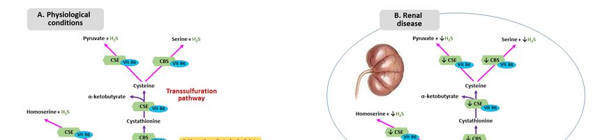
Nutrients 2019, 11, 383                                                                             2 of 20

     Although patients with CKD and ESRD display elevated homocysteine levels, the role of
hyperhomocysteinemia as a cardiovascular and mortality risk factor in this population is still to
be fully elucidated and deserves further investigation [8–12].
     Furthermore, the high prevalence of hyperhomocysteinemia in patients with CKD has increased
interest in speculating the role for hyperhomocysteinemia as a risk factor for the progression of
CKD [13,14].
     The role of folic acid and vitamin B12 role is well recognized, as they are not only essential
cofactors for homocysteine metabolism, but their homeostasis disruption may be related directly to
cardiovascular risk and CKD progression [11,15].
     The aim of this review was to summarize folic acid, vitamin B12 and homocysteine metabolism
in CKD patients and to analyze the published evidences on folic acid and vitamin B12 deficiency as
cardiovascular risk markers and therapeutic targets in CKD and ESRD patients.

2. B Vitamins—Homocysteine Pathway
     B vitamins, including vitamin B9 (folate) and vitamin B12 (cobalamin) are water-soluble vitamins
involved in several normal cellular functions: they are providers of carbon residues for purine and
pyrimidine synthesis, nucleoprotein synthesis and maintenance in erythropoiesis [16].
     Folic acid is derived from polyglutamates that are converted into monoglutamates in the bowel,
and then transported across mucosal epithelia by a specific carrier. The circulating form of folic acid is
5-methyltetrahydrofolate (5-MTHF) [17].
     Vitamin B12, ingested with nutrients such as cobalamin, complexes with salivary haptocorrin,
and is released abruptly from cobalamin by pancreatic proteases in the duodenum. Then, cobalamin,
binds to an intrinsic factor secreted from the parietal cells of the stomach: when this complex arrives at
the distal ileum, it is endocytosed from the enterocytes through cubilin. Then, cobalamin is carried into
the plasma by a plasma transport protein named transcobalamin [16]. B12 is filtered by the glomerulus;
however urine excretion is minimal due to reabsorption in the proximal tubule.
     In target tissues, cobalamin is metabolized into two active forms: adenosylcobalamin in the
mitochondria and methylcobalamin in the cytosol. Methylcobalamin is a methyl-transferring cofactor
to the enzyme methionine synthase allowing homocysteine remethylation to methionine [17].
     Homocysteine is a thiol-containing amino acid, not involved in protein synthesis, deriving from
methionine metabolism. Plasma levels of homocysteine depend on several factors, such as genetic
alteration of methionine metabolism enzymes or deficiency of vitamin B12, vitamin B6 or folic acid [18].
     Methionine is transformed into S-adenosylmethionine (SAM) and then converted in
S-adenosylhomocysteine (SAH) through a reaction catalyzed by methionine synthase reductase
(MTRR). SAM, one of the most important methyl group donors, is formed within mitochondria
and is a cofactor for a mutase known as methylmalonyl-CoA-mutase. This enzyme converts
methylmalonyl-CoA into succinyl CoA, representing a crucial step in the catabolism of various amino
acids and fatty acids. These processes also require pyridoxine (vitamin B6) as a cofactor [18].
     Homocysteine is the final product derived from hydrolysis of SAH to homocysteine and adenosine.
Metabolism of homocysteine includes two different pathways: remethylation and transsulfuration
(Figure 1A). In the remethylation pathway, methionine is regenerated through a reaction catalyzed
by the enzyme methionine synthase (MTS), requiring folate and vitamin B12 as cofactors. Given
that folate is not biologically active, it necessitates transformation into tetrahydrofolate that is then
converted into methylenetetrahydrofolate (MTHF) by the enzyme methylenetetrahydrofolate reductase
(MTHFR) [19].
     The other pathway responsible for the homocysteine metabolism is transsulfuration. First,
homocysteine combines with serine forming cystathionine by cystathionine beta synthase (CBS),
then, cystathionine is hydrolyzed into cysteine and α-ketobutyrate by cystathionine γ-lyase (CTH)
Human CBS is expressed in the liver, kidneys, muscle brain and ovary, and during early embryogenesis
in the neural and cardiac systems [20].
Nutrients 2019, 11, x FOR PEER REVIEW 3 of 21
Nutrients 2019, 11, 383 3 of 20
Human CBS is expressed in the liver, kidneys, muscle brain and ovary, and during early
embryogenesis in the neural and cardiac systems [20].
The sulfur
The sulfur atom, atom,
in the in the of
form form of sulfane
sulfane sulfur sulfur or hydrogen
or hydrogen sulfidesulfide
(H2S), (H 2 S),
can becan be involved
involved in in
vitamin B12-dependent methyl group transfer [21,22]. Alterations in
vitamin B12-dependent methyl group transfer [21,22]. Alterations in methylation pathway, which methylation pathway, which
causescauses a reduction
a reduction of proteins
of proteins and DNA and methylation,
DNA methylation,resultsresults in abnormal
in abnormal vascularvascular
smoothsmooth
musclemuscle
cell proliferation
cell proliferation and increased
and increased lipid peroxidation
lipid peroxidation [23]. isSulfur
[23]. Sulfur a sideisproduct
a side ofproduct of conversion
conversion of
homocysteine to cysteine by the enzymes CBS and cystathionine gamma-lyase (CSE). H2S is anH2 S is
of homocysteine to cysteine by the enzymes CBS and cystathionine gamma-lyase (CSE).
an angiogenic
angiogenic agent with agent with antioxidant
antioxidant and vasorelaxing
and vasorelaxing properties.
properties. Moreover, Moreover, H2 S represents
H2S represents an an
endogenous gaseous mediator, similarly to nitric oxide (NO) and carbon
endogenous gaseous mediator, similarly to nitric oxide (NO) and carbon monoxide [24], which playsmonoxide [24], which plays
a role ainrole in several
several physiological
physiological processes,
processes, namelynamely
vascularvascular
smoothsmooth
musclemuscle relaxation,
relaxation, inhibition
inhibition of of
vascular
vascular smoothsmooth
musclemuscle cell proliferation
cell proliferation and bloodandpressure
blood pressure
loweringlowering
[25]. Li et[25]. Li et al.
al. proved thatproved
H2S that
H2 S metabolism
metabolism impairment impairment might contribute
might contribute to the development
to the development of uremia-associated
of uremia-associated accelerated
accelerated
atherosclerosis
atherosclerosis in CKDinpatients
CKD patients with diabetic
with diabetic nephropathy
nephropathy [26]. Patients
[26]. Patients with CKDwithandCKD and show
ESRD ESRD show
lower lower
H2S plasmaH2 S plasma
levels, levels,
which which can from
can result result downregulation
from downregulation of CBSofand CBSCSE,
and mediated
CSE, mediated
by by
hyperhomocysteinemia (Figure 1B). Whether this phenomenon
hyperhomocysteinemia (Figure 1B). Whether this phenomenon can be attributed can be attributed to additional
additional factors
factorsisisstill
stillunclear
unclear[21].
[21].
Figure 1. Homocysteine Metabolism in physiological condition (A) and in renal disease (B). CSE:
Figure 1. Homocysteine Metabolism in physiological condition (A) and in renal disease (B). CSE:
cystathionine gamma-lyase; CBS: cystathionine beta synthase.
cystathionine gamma-lyase; CBS: cystathionine beta synthase.
Homocysteine can be found in reduced and oxidized form in the bloodstream: more than 90%
Homocysteine can behomocysteine
of the total plasma found in reduced and oxidized
is oxidized and boundform to
in proteins,
the bloodstream:
while the more than 90%
remaining oxidized
of the homocysteine
total plasma homocysteine is oxidized and bound to proteins, while the remaining
exists as a disulfide form. Only 2% of the total homocysteine in plasma is present as a oxidized
homocysteine existsform
free reduced as a [27].
disulfide form. Only 2% of the total homocysteine in plasma is present as a
free reduced form [27].
Normal homocysteine plasmatic level is 10; however, levels
Normal homocysteine
100 mmol/L
is minimally [28]. by the kidney, since in physiological conditions, only
eliminated
Homocysteine is minimally eliminated
non-protein bound homocysteine is subjected by the kidney, since in physiological
to glomerular filtration, and conditions,
then for only
most part
non-protein bound homocysteine is subjected to glomerular filtration, and
reabsorbed in the tubuli and oxidized to carbon dioxide and sulfate in the kidney cells then for most part
[25].
reabsorbed Moreover,
in the tubuliin and oxidized
the kidney, to carbon dioxide
homocysteine is aboveandall
sulfate in the kidney
transsulfurated and cells [25].
deficiency of this renal
Moreover, in the kidney, homocysteine is above all transsulfurated
transsulfuration contributes to the elevation of plasma homocysteine [18]. and deficiency of this renal
transsulfuration contributes to the elevation of plasma homocysteine [18].
3. Metabolism of Homocysteine, Folic Acid and Vitamin B12 in CKD
3. Metabolism of Homocysteine, Folic Acid and Vitamin B12 in CKD
Patients with CKD and ESRD have been shown to have higher homocysteine blood levels
Patients
compared withtoCKD and ESRD
the general have been
population shown
[8,29]. It hastobeen
havehypothesized
higher homocysteine blood levels
that hyperhomocysteinemia
compared
in these patients may be induced by the abnormality of homocysteine metabolism in the in
to the general population [8,29]. It has been hypothesized that hyperhomocysteinemia kidneys
these patients mayby
rather than be reduced
induced glomerular
by the abnormality
filtrationofrate.
homocysteine metabolism
In fact, although in the kidneys rather
free homocysteine can pass theNutrients 2019, 11, 383 4 of 20
ultrafiltration barrier due to its low molecular weight, it circulates in the bloodstream mostly (about
90%) in the protein-bound form [27]. In particular, transsulfuration and remethylation pathways
occurring in the kidney may be affected by renal disease. Stable isotope studies in nondiabetic and
diabetic patients with CKD have shown impaired metabolic clearance of homocysteine determined by
dysfunction in both pathways [30].
In both CKD and ESRD patients, several metabolic alterations, including acidosis, systemic
inflammation and hormonal dysregulation, together with comorbidities and multidrug therapies,
can lead to malnutrition with subsequent folic acid and vitamin B12 deficiency. In addition,
anorexia, gastroparesis, slow intestinal transit or diarrhea, increased gut mucosal permeability and gut
microbiota impairment may represent worsening factors [31,32].
Folic acid metabolism is impaired in uremic patients. Organic and inorganic anions, whose
clearance is reduced in CKD, inhibit the membrane transport of 5-MTHF, thus compromising the
incorporation into nucleic acids and proteins. Data suggest that transport of folates is slower in uremia
and this implicated that, even with normal plasmatic folate levels, the uptake rate of folates into tissues
may be altered [33]. In fact, serum folate concentration does not represent a reliable measure of tissue
folate stores, but rather reflects recent dietary intake of the vitamin. Erythrocyte folate concentration is
a better indicator of whole folate status. In a population of 112 dialysis patients, Bamonti et al. found
serum folate levels normal in only 37% of cases, despite over 80% of red blood cells folate levels within
the normal range [34].
Regarding vitamin B12, several studies have shown a correlation between low serum vitamin
B12 concentrations and high BMI, insulin resistance, type 2 diabetes, dyslipidemia and CVD [35].
Vitamin B12 in the blood is primarily protein-bound. Approximately 20% of circulating B12 is bound
to transcobalamin: this is the biologically active form that can be taken up into cells. Although CKD
patients display increased transcobalamin levels, they show an impaired vitamin tissue uptake of
B12 [36]. Moreover, in uremic patients a functional vitamin B12 deficiency can be observed because of
increased transcobalamin losses in the urine and reduced absorption in the proximal tubule. This can
lead to a “paradoxical” increase in cellular homocysteine levels despite normal total B12 [37].
On the other hand, potentially overdosage-related vitamin B12 toxicity could result exacerbated
in individuals with CKD. Cyanocobalamin, the most commonly used form of B12 supplementation
therapy, is indeed metabolized to active methylcobalamin, releasing small amounts of cyanide whose
clearance is reduced in CKD [34]. Under normal conditions, methylcobalamin is required to remove
cyanide from the circulation through conversion to cyanocobalamin. However, in CKD patients,
the reduced cyanide clearance prevents conversion of cyanocobalamin to the active form and therefore
supplementation is less effective [38].
The appropriate range of B12 levels in CKD remains to be defined adequately. Downstream
metabolites, such as methylmalonic acid and homocysteine, may more accurately reflect functional
B12 status in uremic patients [35].
4. Homocysteine-Mediated Tissue Damage
The pathogenic role of hyperhomocysteinemia on cardiovascular system in CKD and ESRD is
related to atherosclerosis progression in the context of an already enhanced risk of vascular damage
determined by uremic syndrome. One possible mechanism is the induction of local oxidative stress,
generating Reactive Oxygen Species (ROS) because of the thiol group, which rapidly undergoes
autoxidation in the presence of oxygen and metal ion. Besides, hyperhomocysteinemia promotes
Nicotinamide Adenine Dinucleotide Phosphate (NADPH) oxidase activity with further increase in
ROS generation. Hyperhomocysteinemia also determines Nitric Oxide (NO) metabolism impairment
in endothelial cells (including Nitric Oxide Synthase expression, localization, activation, and activity)
leading, together with ROS-induced local microinflammation, to endothelial dysfunction [39].
In cultured endothelial cells, hyperhomocysteinemia has been shown to upregulate monocyte
chemotactic protein 1 (MCP-1) and interleukin-8 (IL-8) production, resulting in monocyte adhesionNutrients 2019, 11, 383 5 of 20
to the endothelium [40]. The link between homocysteine and inflammatory factors seems to be
the activated transcription factor NF-κB (nuclear factor kappa-light-chain-enhancer of activated B
cells) [41].
Additionally, hyperhomocysteinemia induces vascular smooth muscle cells (VSMC) proliferation
by promoting the expression of adhesion molecules, chemokine and VSMC mitogen leading to several
interactions with platelets, clotting factors and lipids [42], and might contribute to the scavenger
receptor-mediated uptake of oxidized- Low Density Lipoprotein (LDL) by macrophages, triggering
foam cell formation in atherosclerotic plaque [43–46]. Hyperhomocysteinemia also determines a
vascular remodeling process that involves activation of metalloproteinase and induction of collagen
synthesis, with subsequent reduction of vascular elasticity [47].
Likewise, elevated blood levels of homocysteine can cause endothelial reticulum stress with
increase endothelial apoptosis and inflammation through a process mediated by ROS production
and NF-κB activation [48–50]. Endothelial cells are known to be particularly vulnerable to
hyperhomocysteinemia, since they do not express CBS, the first enzyme of the hepatic reverse
transsulfuration pathway, or betaine-homocysteine methyltransferase (BHMT), which catalyzes the
alternate remethylation pathway in the liver using betaine as a substrate [51].
Lastly, N-homocysteinylation of proteins is one process responsible for homocysteine toxicity,
since it causes structural and functional loss. For LDL, homocysteinylation produces aggregation,
accumulation of cholesterol and formation of foam-cells. Fibronectin is also involved in
N-homocysteinylation: this reaction contributes to extracellular matrix remodeling, promoting the
development of sclerotic processes [52].
Some peculiar effects of hyperhomocysteinemia on renal tissue have been described.
Homocysteine can act directly on glomerular cells inducing sclerosis, and it can initiate renal injury
by reducing plasma and tissue level of adenosine. Decreased plasma adenosine leads to enhanced
proliferation of VSMC, accelerating sclerotic process in arteries and glomeruli. In a rat model of
hyperhomocysteinemia induced by a folate-free diet, glomerular sclerosis, mesangial expansion,
podocyte dysfunction and fibrosis occurred due to enhanced local oxidative stress. After treatment
of the animals with apocynin, a NADPH oxidase inhibitor, glomerular injury was significantly
attenuated [53].
5. Folic Acid and Vitamin B12 Impairment and Tissue Injury
Both folic acid and vitamin B12 have shown a potential direct relationship with cardiovascular
outcomes with mechanism unrelated to homocysteine levels, although not clearly understood [54].
Folic acid improves endothelial function without lowering homocysteine, suggesting an
alternative explanation for its effect on endothelial function that is possibly related to its
anti-inflammatory, anti-oxidative and anti-apoptotic properties [55–57]. Experimental models revealed
that folic acid can reduce endothelial dysfunction through the limitation of oxidative stress generation
and the increasing of NO half-life [17]. 5-MTHF, the circulating form of folic acid, acutely improves
NO-mediated endothelial function and decreases superoxide production. Moreover, 5-MTHF prevents
oxidation of BH4 increasing enzymatic coupling of eNOS, enhancing NO production. Because 5-MTHF
is a reduced form of folic acid that does not require conversion by dihydrofolate reductase, some direct
effects may be attributable to redox mechanisms that are not seen when oral folic acid is used to
increase plasma folate levels [58,59].
Doshi et al. investigated the direct effects of folic acid on endothelial function in patients with
coronary artery disease (CAD) through Flow Mediated Dilatation (FMD) measurement before and
after folic acid intake. FMD improved at 2 h in parallel with folic acid blood concentration, while
homocysteine blood level did not change significantly. These data suggest that folic acid improves
endothelial function in CAD acutely by a mechanism largely independent of homocysteine [60].
Other authors demonstrated that high-dose folic acid (5 mg/day) improves endothelial function in
CAD patients with an action not related to homocysteine level [60–63]. We have previously reportedNutrients 2019, 11, 383 6 of 20
Nutrients 2019, 11, x FOR PEER REVIEW 6 of 21
that supplementation
homocysteine with 5-MTHF
levels [11]. Pan et al. versus folic acid
recently improved
showed that survival
folic acid ratetreatment
without differences
can inhibit in
homocysteine levels [11]. Pan et al. recently showed that folic acid treatment
atherosclerosis progression through the reduction of VSMC dedifferentiation in high-fat-fed LDL can inhibit atherosclerosis
progression through
receptor-deficient mice the[64].
reduction of VSMC dedifferentiation in high-fat-fed LDL receptor-deficient
miceFrom
[64]. the vitamin B12 side, patients with chronic inflammation, such as the hemodialysis
From the
population, vitamin
display B12 side,production
decreased patients with chronic inflammation,
of transcobalamin II, duesuch as the hemodialysis
to impaired uptake of
population, display decreased production of transcobalamin II, due
circulating B12 by peripheral tissues. This can determine increased synthesis of transcobalaminsto impaired uptake of circulating I
B12 by peripheral tissues. This can determine increased synthesis
and III that brings to further accumulation of B12 in blood [65–68]. Therefore, in the context of transcobalamins I and III that
of
brings to further
inflammatory accumulation
syndromes, despiteofhigh
B12 vitamin
in bloodB12 [65–68].
blood Therefore,
levels, thereinisthe contextB12
a vitamin of inflammatory
deficiency in
target tissues, potentially leading to hyperhomocysteinemia and increased cardiovascular risktissues,
syndromes, despite high vitamin B12 blood levels, there is a vitamin B12 deficiency in target [69].
potentially leading
Concerning to hyperhomocysteinemia
anemia, unless CKD and and ESRD increased
patients cardiovascular
show significantrisk [69].
folate depletion,
Concerning
additional anemia, unless
supplementation CKD
of folic acidand ESRD
does notpatients
appear to showhave significant
a beneficialfolate depletion,
effect additional
on erythropoiesis
supplementation of folic acid does not appear to have a
or on responsiveness to Recombinant Human Erythropoietin (rHuEPO) therapy. However,beneficial effect on erythropoiesis or ona
responsiveness to Recombinant Human Erythropoietin (rHuEPO) therapy.
diagnosis of folate deficiency should be considered in such patients when significant elevation in However, a diagnosis of
folate deficiency
mean cell volume should be considered in such
or hypersegmented patients when significant
polymorphonuclear leucocytes elevation in mean
are found, cell volume
especially in
or hypersegmented polymorphonuclear leucocytes are found, especially
subjects with malnutrition, history of alcohol abuse, or in patients hyporesponsive to rHuEPO. in subjects with malnutrition,
history of alcohol
Measurements of abuse, or in patients
circulating serum folatehyporesponsive to rHuEPO.
do not necessarily Measurements
mirror of circulating
tissue folate stores, and serum
red
folate do not necessarily mirror tissue folate stores, and red blood cell folate
blood cell folate measures provide a more accurate picture. Low red blood cells folate concentrations measures provide a more
accurate
in picture. suggest
these patients Low redthe blood
need cells
for folate
folate concentrations
supplementation in these
[70]. patients suggest the need for folate
supplementation
In patients with [70].CKD, folate and vitamin B12 deficiency may represent an influential factor in
In patients with CKD, folate and vitamin
renal anemia and hyporesponsiveness B12 deficiency
to rHuEPO therapy.may As represent
such, theanpossibility
influential and
factorthein
renal anemiaof
requirement and hyporesponsiveness
a regular supplementation to rHuEPO
is still atherapy.
matter of Asdebate
such, the possibility and the requirement
[71].
of a regular supplementation is still a matter of debate [71].
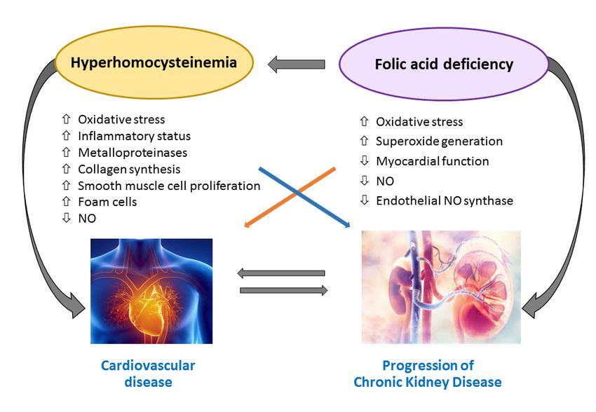
Figure 2 illustrates the pathways involved in the amplification of atherosclerosis and
Figure 2 illustrates
inflammation triggered the pathways involved in theinamplification
by hyperhomocysteinemia CKD patients. of atherosclerosis and inflammation
triggered by hyperhomocysteinemia in CKD patients.
Figure 2. Hyperhomocysteinemia-induced amplification of atherosclerosis and inflammation in chronic
Figure
kidney 2. Hyperhomocysteinemia-induced
disease amplification
(CKD) patients. Abbreviations: of atherosclerosis and inflammation in
NO, Nitric Oxide.
chronic kidney disease (CKD) patients. Abbreviations: NO, Nitric Oxide.
6. MTHFR Gene Polymorphisms
6. MTHFR Gene
MTHFR Polymorphisms
is an enzyme that plays a fundamental role in folate and homocysteine metabolism by
catalyzing
MTHFR theisconversion
an enzymeofthat
5,10-methenyltetrahydrofolate
plays a fundamental role ininto 5-MTHF,
folate the main circulating
and homocysteine formby
metabolism of
folate [72]. Several MTHFR gene polymorphisms have been described, and some of them
catalyzing the conversion of 5,10-methenyltetrahydrofolate into 5-MTHF, the main circulating formseem to affect
the
of individual
folate susceptibility
[72]. Several MTHFR to a number
gene of pathological
polymorphisms conditions
have been associated
described, and somewith
of homocysteine
them seem to
disorders,
affect the like myocardial
individual infarction, stroke,
susceptibility to a neurodegenerative diseases, conditions
number of pathological autoimmuneassociated
diseases, cancer,
with
diabetes, birth defects and kidney disease [73].
homocysteine disorders, like myocardial infarction, stroke, neurodegenerative diseases,
autoimmune diseases, cancer, diabetes, birth defects and kidney disease [73].Nutrients 2019, 11, 383 7 of 20
The most characterized are four functional single nucleotide polymorphisms at position 677
(MTHFR 677 C > T), at position 1298 (MTHFR 1298 A > C), at position 1317 (MTHFR 1317 T > C) and
at position 1793 (MTHFR 1793 G > A) [74].
Although some studies excluded an association between MTHFR 677 C > T genotype and
long-term kidney outcomes [75], MTHFR 677 C > T polymorphism has been shown to contribute to
increase cardiovascular risk in ESRD patients [76]. A study of 2015 by Trovato et al. on 630 Italian
Caucasian subjects found a lower frequency of MTHFR 677 C > T and A1298 A > C polymorphisms
among ESRD patients requiring hemodialysis, suggesting a protective role of these gene variants on
renal function [77].
Despite the fact that the main function of the MTHFR enzyme is to regulate the availability of
5-MTHF for homocysteine remethylation, the pathological consequences of functional variants of
MTHFR gene cannot only be attributed to the increase in homocysteine levels. While the homocysteine
lowering effect of routine folate supplementation in general population has been proven, patients
with ESRD seem to display a folate resistance even to higher doses of folate [78]. Folate and vitamin
B12 supplementation effects on hemodialysis patients are controversial, and possibly dependent on
MTHFR polymorphisms [79].
Anchour et al. recently evaluated folic acid response in terms of homocysteine lowering with
respect to MTHFR polymorphism carrier status in a prospective cohort of 132 hemodialysis patients.
The authors found that 677 C > T MTHFR genotype influences vitamin B supplementation response,
as reported in previous studies [79–86]. In particular, simultaneous supplementation of vitamin B12
and folate was useful only for the homozygous for the C allele, and the homocysteine reduction was
significantly higher in carriers of TT genotype than in other genotypes [84].
Other authors reported that after B12 supplementation, homocysteine reduction in CC carriers
was higher than in CT or TT carriers [82]. A renal substudy of the China Stroke Primary Prevention
Trial (CSPPT) evaluated the effects of the combination of Angiotensin Converting Enzyme (ACE)
inhibitors and folic acid with ACE inhibitors alone in reducing the risk of renal function decline in a
hypertensive population without folic acid fortification. In 7545 patients treated with 10 mg enalapril
and 0.8 mg folic acid, out of 15,104 participants, the greatest drop in serum homocysteine was in TT
homozygotes of MTHFR 677 C > T polymorphism compared to other genotypes (CC/CT) [87].
In summary, the majority of available evidences suggest that MTHFR polymorphisms may
influence folic acid and vitamin B12 treatment response in terms of homocysteine lowering and
cardiovascular risk reduction in patients with CKD and ESRD on dialysis although indication of
routine testing is matter of debate [88].
7. Role of Folic Acid, Vitamin B12 and Homocysteine as Cardiovascular Risk Markers
Although hyperhomocysteinemia has been accepted for years as a cardiovascular risk factor,
its association with CVD and mortality has been recently questioned and literature data are
controversial [7,89,90]. Epidemiologic and case-control studies generally support an association
of elevated plasma homocysteine levels with an increased incidence of CVD and stroke, whereas
prospective, randomized, placebo-controlled studies do not [7].
Moreover, a discrepancy still exists about the indication of routine screening for
hyperhomocysteinemia and its treatment in the general population [7].
For CKD and ESRD patients, in spite of the increased homocysteine levels (average homocysteine
level in the general population about 10–15 mmol/L versus 25–35 mmol/L in uremic patients), the role
of homocysteine as a cardiovascular and mortality risk factor is still uncertain and many retrospective
and interventional studies resulted in conflicting evidences [8–12].
A meta-analysis including retrospective studies, prospective observational studies and
interventional trials (total population 5123 patients) showed that elevated homocysteine blood levels
represent a risk factor for both CVD and mortality in patients with ESRD not treated with folic acid
supplementation [10].Nutrients 2019, 11, 383 8 of 20
The prospective studies included in the meta-analysis showed that in unsupplemented patients
with ESRD, an increase of 5 mmol/L in homocysteine concentration is associated with an increase
of 7% in the risk of total mortality and an increase of 9% in the risk of cardiovascular events [10].
Conversely, in a prospective cohort of 341 hemodialysis patients, we previously failed to demonstrate
a relationship between baseline homocysteine as well as MTHFR polymorphisms and mortality [11].
At the origin of these divergences, several possible factors may be hypothesized, such as
non-homogeneous populations selection, temporal discrepancies between competitive risk factors and
influence of common complication including inflammation and protein-energy wasting (PEW) that
could influence circulating homocysteine and that are associated with poorer outcomes [9].
An inverse correlation between homocysteine levels and cardiovascular outcomes in advanced
CKD and in hemodialysis patients has also been documented, configuring the phenomenon known as
“reverse epidemiology” that also involves other cardiovascular risk factors, including Body Mass Index
(BMI), serum cholesterol and blood pressure [91]. Some evidence indeed suggests that the presence
of PEW and inflammation may justify the observed reverse association between homocysteine and
clinical outcome in CKD and ESRD patients [34–37]. Specifically, two studies showed that patients
with very low homocysteine plasma levels had worse outcomes, as confirmed by a higher incidence of
hospitalization and mortality [92,93].
These data call into question the reliability of homocysteine as a marker of cardiovascular risk
and mortality in patients with CKD and ESRD, raising the suspicion that other mechanisms beyond
elevated homocysteine levels might be implicated. Given that DNA methyltransferases are among the
main targets of hyperhomocysteinemia, it has been hypothesized that epigenetic alterations could play
a role in hyperhomocysteinemia-mediated tissue damage [12].
Sohoo et al. recently carried out a retrospective study on a large cohort of hemodialysis patients
investigating the association between baseline folic acid and vitamin B12 levels and all-cause mortality
after an observation period of 5 years (9517 patients for folic acid group and 12,968 patients for B12
group). The authors found that higher B12 concentrations (550 pg/mL) were associated with a higher
risk of mortality after adjusting for sociodemographic and laboratory variables, while only lower
serum folate concentrations (Nutrients 2019, 11, 383 9 of 20
Table 1. Cont.
Case
Study Design Participants, n Results
Definition/Outcome
HHcy predicted
Ducloux et al.,
Prospective observational 459 HD Mortality and fatal CVD outcome only in patients
2006 [92]
without CIMS
Nair et al., 2005 HHcy did not predict
Retrospective case-control 146 HD MI, heart surgery
[96] CVD risk
London et al., HHcy did not predict
Prospective observational 78 HD Mortality
2004 [97] outcome
Kalantar-Zadeh Lower Hcy levels
Prospective observational 367 HD Mortality
et al., [98] predicted mortality
Buccianti et al., HHcy predicted
Prospective observational 77 HD Fatal CVD
2004 [99] outcome
Bayès et al., 2003 HHcy did not predict
Prospective observational 94 HD Mortality, fatal CVD
[100] outcome
Mallamaci et al., HHcy predicted
Prospective observational 175 HD Mortality, fatal CVD
2002 [101] outcome
Ducloux et al., HHcy did not predict
Prospective observational 240 PD Fatal and nonfatal CVD
2002 [102] outcome
Haraki et al.,2001 Coronary, cerebral and
(retrospective Retrospective case-control 43 HD/PD peripheral vascular HHcy CVD risk factor
part) [103] disease
Haraki et al., 2001
HHcy predicted
(prospective part) Prospective observational 55 HD/PD Fatal and nonfatal CVD
outcome
[103]
Wrone et al., 2001 MI, stroke, TIA, carotid HHcy did not predict
Retrospective case-control 459 HD/PD
[104] endarterectomy. CVD risk
Dierkes et al., Mortality, fatal/nonfatal HHcy predicted
Prospective observational 102 HD
2000 [105] CVD outcome
Coronary, cerebral and
Suliman et al., HHcy did not predict
Retrospective case-control 117 HD peripheral vascular
2000 [106] CVD risk
disease
Coronary, cerebral and
Kunz et al., 1999 HHcy cardiovascular
Retrospective case-control 63 HD peripheral vascular
[107] risk factor
disease
Coronary, cerebral and
Manns et al., 1999 HHcy cardiovascular
Retrospective case-control 218 HD peripheral vascular
[108] risk factor only in males
disease
Sirrs et al., 1999 Mortality and CVD Lower Hcy levels
Prospective observational 88 HD
[109] events predicted mortality
Moustapha et al., Mortality and CVD HHcy predicted
Prospective observational 167 HD/PD
1998 [110] events outcome
Coronary, cerebral and
Vychytil et al., HHcy did not predict
Retrospective case-control 154 PD peripheral vascular
1998 [111] CVD risk
disease
Bostom et al., HHcy predicted
Prospective observational 73 HD/PD CVD events
1997 [112] outcome
Coronary, cerebral and
Robinson et al., HHcy cardiovascular
Retrospective case-control 176 HD/PD peripheral vascular
1996 [113] risk factor
disease
Coronary, cerebral and
Bachmann et al., HHcy cardiovascular
Retrospective case-control 45 HD peripheral vascular
1995 [114] risk factor
disease
Coronary, cerebral and
Bostom et al., HHcy did not predict
Retrospective case-control 24 HD/PD peripheral vascular
1995 [115] CVD risk
disease
Abbreviations: CVD, Cardiovascular Disease; MI, Myocardial Infarction; LVH, left ventricular hypertrophy; LVD,
left ventricular dysfunction; HHcy, hyperhomocysteinemia; HD, Hemodialysis; PD, peritoneal dialysis; CKD,
Chronic Kidney Disease; CIMS, chronic inflammation-malnutrition state.Nutrients 2019, 11, 383 10 of 20
8. Effect of Folic Acid and Vitamin B12 Supplementation on CVD and Mortality in CKD
and ESRD
Regarding folic acid and vitamin B12 supplementation, the role such vitamins administration
with the aim of reducing mortality and prevent progression to ESRD is still to be determined.
Moreover, effective folic acid and vitamin B12 supplementation dosages are not clearly established
in the category of patients that take dosages ranging from 2.5 to 5 mg of folic acid three times a week up
to more than 15 mg/day. Simultaneous administration of intravenous B complex vitamins is proven to
be more efficient in reducing homocysteine serum levels and restoring the remethylation pathway in
ESRD patients [116].
Righetti et al. in a one-year, placebo-controlled, non-blinded randomized control trial on a cohort
of 81 chronic hemodialysis patients, showed no survival benefit of treatment with folic acid compared
to placebo, and only 12% of patients on treatment reached normal homocysteine blood levels [117].
Wrone et al. found no difference in terms of mortality and cardiovascular events in a multicentre study
on 510 patients on chronic dialysis randomized to 1, 5, or 15 mg/day of folic acid [76].
In the ASFAST study (Cardiovascular Morbidity and Mortality in the Atherosclerosis and Folic
Acid Supplementation Trial), a double blinded, placebo controlled trail, a randomized cohort of 315
CKD dialysis patients (with eGFR < 25 mL/min) were treated with folic acid 15 mg/die or placebo.
After a median follow-up of 3.6 years, the results failed to demonstrate a benefit of folic acid therapy
regarding all-cause mortality, cardiovascular mortality and control of atheroma progression (carotid
intima-media thickness progression) [118].
The HOST trial (Homocysteinemia in Kidney and End Stage Renal Disease) is a double
blind, placebo-controlled trial in which 2056 patients with advanced CKD or ESRD requiring renal
replacement therapy and elevated homocysteine levels, were randomized to a combined therapy
with folic acid, vitamin B12 and piridoxin or placebo. After a median follow-up of 3.2 years, the
study showed a significant reduction in homocysteine levels, but failed to reach its primary end-point,
reduction of all-cause mortality, and its secondary end-point, reduction in cardiovascular death,
amputation and thrombosis of the vascular access. A possible explanation for these negative results
may be ascribed to the high cardiovascular comorbidity burden and the suboptimal compliance to
therapy. Moreover, the study considered CKD and ESRD population together and was underpowered
to evaluate the two populations separately. The disparity between these findings and the previously
reported epidemiologic data could reflect limitations of observational studies [119].
Recently, Heinz et al. designed a multicenter trial on 650 chronic hemodialysis patients
randomized to 5 mg folic acid, 50 µg vitamin B12 and 20 mg vitamin B6 versus placebo three times a
week (post-dialysis) for 2 years. No differences were observed between the two groups in terms of all
cause-mortality and fatal and non-fatal cardiovascular events. On the other side, post-hoc analysis
revealed a significant reduction in unstable angina pectoris and fewer vascularization procedures [120].
In a meta-analysis by Heinz et al. involving five intervention trials for a total of 1642 dialysis
patients treated with folic acid, vitamin B12 and vitamin B6, a significant CVD risk reduction but not
mortality risk reduction was demonstrated [10]. Another meta-analysis including 3886 patients with
ESRD or advanced CKD (creatinine clearance < 30 mL/min) assessed the relationship between folic
acid therapy (with or without vitamin B6 and B12) and CVD. Folic acid reduced cardiovascular risk by
15% in ESRD patients with greater benefit in those treated for longer than 24 months and in those from
areas with no or partial grain fortification [121].
Ji et al. performed a large meta-analysis including 14 RCTs (54,913 participants) that demonstrated
overall stroke events reduction resulting from homocysteine lowering following folic acid, vitamin
B12 and vitamin B6 supplementation. Beneficial effects in reducing stroke events were observed in
the subgroup with CKD [122]. A meta-analysis of 10 studies concluded that homocysteine-lowering
therapy is not associated with a significant decrease in the risks for CVD events, stroke, and all-cause
mortality among patients with CKD. It has to be pointed out that, respect to previous meta-analysis
a high number of participants with diabetes were included and that RCTs were performed in grainNutrients 2019, 11, 383 11 of 20
fortification areas [123]. More recently, a meta-analysis took into account six studies (comprising the
abovementioned ones) for 2452 patients on chronic hemodialysis, finding no significant differences in
mortality and in the incidence of cardiovascular events in patients treated with homocysteine lowering
therapy [124].
Finally, many published post-hoc analyses have shown that several factors including age, baseline
homocysteine levels, folic acid fortification of grains, B12 status, renal function, comorbidities and
medications could be modifiers of folic acid and vitamin B12 effects on cardiovascular risk [12].
Table 2 summarizes the interventional studies investigating the effects of folic acid and vitamin
B12 administration on CVD risk, mortality and CKD progression.
Table 2. Interventional trials on the effects of folic acid and vitamin B12 administration and CVD risk,
mortality and CKD progression.
Follow-up,
Study Design/Intervention Participants, n End Point Results
Years
Double blind RCT: enalapril 15,104 (eGFR ≥ 30 Enalapril plus folic
Xu et al., 2016 CKD
10 mg versus enalapril mL/min). No folic 4.4 acid delayed CKD
[87] progression
10 mg plus folic acid acid fortification progression
Double blind RCT: folic acid 238 (diabetic Greater GFR
House et al., 2.5 mg + Vitamin B6 25 mg + nephropathy with CKD decrease and more
2.6
2010 [125] Vitamin B12 1 mg versus eGFR > 30 mL/min). progression CVD events in
placebo Folic acid fortification treatment group
Double blind RCT: folic acid All-cause
Heinz et al., 5 mg, vitamin B12 50 µg, 650 hemodialysis mortality,
2 No differences
2010 [120] vitamin B6 20 mg versus patients cardiovascular
placebo 3 times a week events
Double blind RCT: folic acid All-cause
Mann et al., 2.5 mg + vitamin B6 50 mg + 619 CKD (eGFRNutrients 2019, 11, 383 12 of 20
the incidence of cardiovascular events in patients with CKD and ESRD has not been demonstrated.
Moreover, the beneficial effect could depend on anti-inflammatory and vascular protective effects.
9. Folic Acid and Vitamin B12: Evidences on CKD Progression
Regarding the relationship between folic acid, vitamin B12 supplementation and CKD progression,
available interventional studies have demonstrated no clear benefit or even harmful effects on renal
outcomes [15], while observational studies showed a correlation between hyperhomocysteinemia and
risk of CKD development and progression [129].
The China Stroke Primary Prevention Trial (CSPPT) is a large RCT (20,702 patients) that enrolled
adults with hypertension without a history of stroke or myocardial infarction with determination of
MTHFR genotype and baseline folate level. The study aimed at evaluating the effect of treatment with
folic acid in an Asian population without folic acid fortification. Authors found that the ACE inhibitors
plus folic acid therapy, compared with ACE inhibitors alone, reduced stroke risk. Moreover, subjects
with the CC or CT MTHFR genotype had the highest risk of stroke and the greatest benefit of folic
acid supplementation while those with the TT genotype required a higher dosage of folic acid to reach
sufficient levels [80].
A renal sub-study of the China Stroke Primary Prevention Trial (CSPPT) compared the efficacy
of combination of enalapril and folic acid with enalapril alone in reducing the risk of renal function
decline in a large hypertensive population (15,104 patients). The study included patients with eGFR
greater than 30 mL/min and participants were randomized to receive enalapril 10 mg plus folic acid
0.8 mg or enalapril 10 mg alone. Compared with the enalapril group, the enalapril-folic acid group
showed reduced risk of CKD progression by 21% and a reduction of eGFR decline rate of 10% after
a four-year follow-up. Authors performed a subgroup analysis that compared patients with CKD
(defined as eGFR < 60 mL/min or presence of proteinuria) and without CKD at baseline. It resulted
that CKD at baseline was a strong modifier of the treatment effect [85]. Specifically, the greatest
decrease in serum homocysteine was in TT homozygotes of MTHFR 677 C > T polymorphism, while
the magnitude of the declines in those with CC/CT genotypes was smaller. Finally, an exploratory
subgroup analysis aimed to assess the treatment effect on the primary outcome in various subgroups
among CKD participants showed that CKD progression risk reduction was more represented in
diabetes subgroup [87]. This has been the first study showing renal protection from folic acid therapy
in a population without folic acid fortification. Previous trials have reported a null or harmful effect of
supplementation with folic acid and vitamin B12 [15,118].
In the abovementioned HOST study, treatment with high doses of folic acid, vitamin B6 and
vitamin B12 failed to delay the time to initiating dialysis in patients with advanced CKD [15].
Diabetic Intervention with Vitamins to Improve Nephropathy (DIVINe) was a double blind RCT
on a population of 238 patients with diabetic kidney disease randomized to receive 2.5 mg folic acid
plus 25 mg vitamin B6 plus 1 mg vitamin B12, or placebo (mean eGFR was 64 mL/min in treatment
group and 58 mL/min in the placebo group). Results showed that treatment with folic acid, vitamin B6
and vitamin B12 led to a greater decrease in GFR and to an increase in cardiovascular events compared
to placebo after a follow-up of 2.6 years [125]. The possible explanations for such conflicting results
may be multiple. First of all, baseline folic acid levels were different between studies (7.7 ng/mL for
CSPPT renal sub study, 15 and 16.5 ng/mL for DIVINe and HOST study respectively) corroborating
the hypothesis that beneficial effects of folic acid supplementation on renal outcomes could be stronger
in patients with low folic acid level at baseline. In addition, vitamin B doses may play a role, as
suggested by the elevated folate blood levels reached in the HOST study (2000 ng/mL) compared
to CSPPT (23 ng/mL). This allowed postulating a potential toxicity determined by unmetabolized
folic acid accumulated in the bloodstream [85]. Finally, CKD severity differed between the studies.
In fact, the HOST study population was composed by patients with advanced CKD and with high
comorbidity burden and this could have attenuated the study power with respect to renal outcomes.
The fact that the CSPPT renal sub study demonstrated a benefit of folic acid therapy on the progressionNutrients 2019, 11, 383 13 of 20
of renal damage could be related to the choice of a population with mild-moderate CKD. Furthermore,
only CSPPT trial selected a population without folic acid grain fortification, while the other two studies
were carried out in countries with folic acid fortification programs.
Besides, a recent study on 630 Italian Caucasian population found a lower frequency of MTHFR
677 C > T and A1298 A > C polymorphisms among dialysis patients compared to subjects without or
with slight-moderate renal impairment, suggesting a protective role of both polymorphisms on renal
function [39].
In conclusion, the available evidences regarding the effect of homocysteine lowering therapies on
CKD progression are controversial and further studies with CKD progression as primary end-point
and more homogeneous population selection are needed. While awaiting further evidences, it seems
reasonable to treat patients with folic acid deficiency in order to reduce the risk of CKD progression
avoiding accumulation phenomena that could lead to toxicity.
10. Folic Acid and Vitamin B12 in Kidney Transplant Recipients
In kidney transplant recipients several factors such as dialytic history, anemia, immunosuppression,
inflammatory state and dysmetabolic alterations may influence cardiovascular risk [130].
A decline in homocysteine blood levels after kidney transplantation is frequently observed;
nonetheless, hyperhomocysteinemia usually persists [131–133]. It has been documented that
homocysteine can be further lowered among stable transplant recipients through high-dose B-vitamin
therapy [134]. The effect of folic acid, vitamin B12 and vitamin B6 supplementation on cardiovascular
risk and mortality reduction has been investigated by the Folic Acid for Vascular Outcome Reduction in
Transplantation (FAVORIT) trial. Stable transplant recipients were randomized to daily multi-vitamin
drug containing high-doses of folate (5.0 mg), vitamin B12 (1.0 mg) and vitamin B6 (50 mg) or placebo.
The study was terminated early after an interim analysis because, despite effectively homocysteine
lowering action, the incidence of CVD, all-cause mortality and onset of dialysis-dependent kidney
failure did not differ between the treatment arms [135].
A longitudinal ancillary study of the FAVORIT trial recently showed that high-dose B-vitamin
supplementation determined modest cognitive benefit in patients with elevated baseline. It has to be
pointed out that almost all subjects had no folate or B12 deficiency; thus, the potential cognitive
benefits of folate and B12 supplementation in individuals with poor B-vitamin status remains
controversial [136].
11. Conclusions
At present, the available evidence does not provide full support to consider hyperhomocysteinemia,
folic acid and vitamin B12 alterations reliable cardiovascular disease and cardiovascular mortality risk
markers in CKD and ESRD populations. Furthermore, such factors do not represent a validated
therapeutic target regarding the reduction of cardiovascular risk and CKD progression.
While waiting for the results of confirmatory trials, it seems reasonable to consider folic acid with
or without vitamin B12 supplementation as appropriate adjunctive therapy in patients with CKD.
Concerning patients in early CKD stages for which potassium or phosphorus dietary intake
restriction is not indicated, folic acid could come in the form of a healthy diet rich in natural
sources of folate. For patients with advanced CKD and on dialysis, folic acid can be supplemented
pharmacologically after accurate folate status assessment.
Author Contributions: I.C., G.C. and G.L.M.: design of the work and manuscript revision. L.G., F.Z. and
F.T.: literature review, writing and manuscript preparation. M.C.: figure preparation and manuscript revision.
All authors approve the submitted version, agree to be personally accountable for the author’s own contributions
and for ensuring that questions related to the accuracy or integrity of any part of the work, even ones in which an
author was not personally involved, are appropriately investigated, resolved and documented in the literature.
Funding: The authors declare no funding.
Conflicts of Interest: The authors declare no conflict of interest.Nutrients 2019, 11, 383 14 of 20
References
1. Go, A.S.; Chertow, G.M.; Fan, D.; McCulloch, C.E.; Hsu, C.Y. Chronic kidney disease and the risks of death,
cardiovascular events, and hospitalization. N. Engl. J. Med. 2004, 351, 1296–1305. [CrossRef] [PubMed]
2. Foley, R.N.; Parfrey, P.S.; Sarnak, M.J. Epidemiology of cardiovascular disease in chronic renal disease. J. Am.
Soc. Nephrol. 1998, 9, S16–S23. [CrossRef] [PubMed]
3. Jha, V.; Garcia-Garcia, G.; Iseki, K.; Li, Z.; Naicker, S.; Plattner, B.; Saran, R.; Wang, A.Y.; Yang, C.W. Chronic
kidney disease: Global dimension and perspectives. Lancet 2013, 382, 260–272. [CrossRef]
4. McCullough, P.A.; Steigerwalt, S.; Tolia, K.; Chen, S.C.; Li, S.; Norris, K.C.; Whaley-Connell, A.; KEEP
Investigators. Cardiovascular disease in chronic kidney disease: Data from the Kidney Early Evaluation
Program (KEEP). Curr. Diab. Rep. 2011, 11, 47–55. [CrossRef]
5. Ekdahl, K.; Soveri, I.; Hilborn, J.; Fellström, B.; Nilsson, B. Cardiovascular disease in hemodialysis: Role of
the intravascular innate immune system. Nat. Rev. Nephrol. 2017, 13, 285–296. [CrossRef] [PubMed]
6. McCully, K.S. Homocysteine and vascular disease. Nat. Med. 1996, 2, 386–389. [CrossRef] [PubMed]
7. Chrysant, S.G.; Chrysant, G.S. The current status of homocysteine as a risk factor for cardiovascular disease:
A mini review. Expert. Rev. Cardiovasc. Ther. 2018, 16, 559–565. [CrossRef]
8. Robinson, K. Renal disease, homocysteine, and cardiovascular complications. Circulation 2004, 109, 294–295.
[CrossRef]
9. Suliman, M.E.; Lindholm, B.; Barany, P.; Qureshi, A.R.; Stenvinkel, P. Homocysteine-lowering is not a
primary target for cardiovascular disease prevention in chronic kidney disease patients. Semin. Dial. 2007,
20, 523–529. [CrossRef]
10. Heinz, J.; Kropf, S.; Luley, C.; Dierkes, J. Homocysteine as a risk factor for cardiovascular disease in patients
treated by dialysis: A meta-analysis. Am. J. Kidney Dis. 2009, 54, 478–489. [CrossRef]
11. Cianciolo, G.; La Manna, G.; Colì, L.; Donati, G.; D’Addio, F.; Persici, E.; Comai, G.; Wratten, M.; Dormi, A.;
Mantovani, V.; et al. 5-methyltetrahydrofolate administration is associated with prolonged survival and
reduced inflammation in ESRD patients. Am. J. Nephrol. 2008, 28, 941–948. [CrossRef] [PubMed]
12. Cianciolo, G.; De Pascalis, A.; Di Lullo, L.; Ronco, C.; Zannini, C.; La Manna, G. Folic Acid and Homocysteine
in Chronic Kidney Disease and Cardiovascular Disease Progression: Which Comes First? Cardiorenal. Med.
2017, 7, 255–266. [CrossRef] [PubMed]
13. Marti, F.; Vollenweider, P.; Marques-Vidal, P.M.; Mooser, V.; Waeber, G.; Paccaud, F.; Bochud, M.
Hyperhomocysteinemia is independently associated with albuminuria in the population-based CoLaus
study. BMC Public Health. 2011, 11. [CrossRef] [PubMed]
14. Ponte, B.; Pruijm, M.; Marques-Vidal, P.; Martin, P.Y.; Burnier, M.; Paccaud, F.; Waeber, G.; Vollenweider, P.;
Bochud, M. Determinants and burden of chronic kidney disease in the population-based CoLaus study:
A cross-sectional analysis. Nephrol. Dial. Transpl. 2013, 28, 2329–2339. [CrossRef] [PubMed]
15. Soohoo, M.; Ahmadi, S.F.; Qader, H.; Streja, E.; Obi, Y.; Moradi, H.; Rhee, C.M.; Kim, T.H.; Kovesdy, C.P.;
Kalantar-Zadeh, K. Association of serum vitamin B12 and folate with mortality in incident hemodialysis
patients. Nephrol. Dial. Transpl. 2017, 32, 1024–1032. [CrossRef] [PubMed]
16. Kang, S.S.; Wong, P.W.K.; Malinow, M.R. Hyperhomocyst(e)inemia as a risk factor for occlusive vascular
disease. Annu. Rev. Nutr. 1992, 12, 279–298. [CrossRef] [PubMed]
17. Randaccio, L.; Geremia, S.; Demitri, N.; Wuerges, J. Vitamin B12: Unique metalorganic compounds and the
most complex vitamins. Molecules 2010, 15, 3228–3259. [CrossRef]
18. Long, Y.; Nie, J. Homocysteine in Renal Injury. Kidney Dis. 2016, 2, 80–87. [CrossRef]
19. Van Guldener, C.; Stam, F.; Stehouwer, C.D. Hyperhomocysteinaemia in chronic kidney disease: Focus on
transmethylation. Clin. Chem. Lab. Med. 2005, 43, 1026–1031. [CrossRef]
20. Jacobsen, D.W. Homocysteine and vitamins in cardiovascular disease. Clin. Chem. 1998, 44, 1833–1843.
21. Cianciolo, G.; Cappuccilli, M.; La Manna, G. The Hydrogen Sulfide-Vitamin B12-Folic Acid Axis: An
Intriguing Issue in Chronic Kidney Disease. A Comment on Toohey JI: “Possible Involvement of
Hydrosulfide in B12-Dependent Methyl Group Transfer”. Molecules 2017, 22, 1216. [CrossRef] [PubMed]
22. Toohey, J.I. Possible Involvement of Hydrosulfide in B12-Dependent Methyl Group Transfer. Molecules 2017,
22, 582. [CrossRef] [PubMed]
23. Welch, G.N.; Loscalzo, J. Homocysteine and atherothrombosis. N. Engl. J. Med. 1998, 338, 1042–1050.
[CrossRef] [PubMed]You can also read


