E-cadherin Beyond Structure: A Signaling Hub in Colon Homeostasis and Disease - MDPI
←
→
Page content transcription
If your browser does not render page correctly, please read the page content below
International Journal of
            Molecular Sciences

Review
E-cadherin Beyond Structure: A Signaling Hub in
Colon Homeostasis and Disease
Amanda C. Daulagala , Mary Catherine Bridges and Antonis Kourtidis *
 Department of Regenerative Medicine and Cell Biology, Medical University of South Carolina,
 173 Ashley Avenue, Charleston, SC 29425, USA; gunarath@musc.edu (A.C.D.); bridgmar@musc.edu (M.C.B.)
 * Correspondence: kourtidi@musc.edu; Tel.: +1-843-792-9170

 Received: 1 May 2019; Accepted: 1 June 2019; Published: 5 June 2019

 Abstract: E-cadherin is the core component of epithelial adherens junctions, essential for tissue
 development, differentiation, and maintenance. It is also fundamental for tissue barrier formation, a
 critical function of epithelial tissues. The colon or large intestine is lined by an epithelial monolayer that
 encompasses an E-cadherin-dependent barrier, critical for the homeostasis of the organ. Compromised
 barriers of the colonic epithelium lead to inflammation, fibrosis, and are commonly observed in
 colorectal cancer. In addition to its architectural role, E-cadherin is also considered a tumor suppressor
 in the colon, primarily a result of its opposing function to Wnt signaling, the predominant driver of
 colon tumorigenesis. Beyond these well-established traditional roles, several studies have portrayed
 an evolving role of E-cadherin as a signaling epicenter that regulates cell behavior in response
 to intra- and extra-cellular cues. Intriguingly, these recent findings also reveal tumor-promoting
 functions of E-cadherin in colon tumorigenesis and new interacting partners, opening future avenues
 of investigation. In this Review, we focus on these emerging aspects of E-cadherin signaling, and we
 discuss their implications in colon biology and disease.

 Keywords: adherens junctions; CDH1; cadherin; catenin; colorectal cancer; inflammatory bowel
 disease; epithelial; colon crypt; microbiome

1. Introduction

1.1. The Adherens Junctions
      Cell-cell adhesion complexes are indispensable for tissue integrity and organ function; however,
their disruption can lead to numerous diseases, including inflammation and cancer. The Adherens
Junction (AJ) is a major cell-cell adhesion structure, key for maintaining tissue integrity and architecture
through its intimate tethering to the actin and microtubule cytoskeleton [1]. The core components of AJs
are the members of the classical cadherin superfamily, such as epithelial cadherin (E-cadherin), neural
cadherin (N-cadherin), placental cadherin (P-cadherin), as well as members of the catenin family of
proteins, namely p120 catenin (p120), α-catenin, and β-catenin [2]. Nectin is another important cell-cell
adhesion molecule present at the AJs, which binds intracellularly to Afadin via its C-terminus [3].
      The cadherin superfamily includes classical, desmosomal, protocadherins, and unconventional
types of cadherins [2,4,5]. In this review, we focus on the classical type I cadherin E-cadherin, which is
the predominant member of the family in epithelial tissues and is encoded by the CDH1 gene. Classical
mammalian cadherins have five extracellular domains, spanning EC1 to EC5, with calcium-binding
sites. Each of these sites contain negatively charged motifs that can bind to three Ca2+ molecules, thus
strengthening the interactions between the extracellular domains [4,6]. The homophilic binding of EC1
domains between cells is known as “trans” interactions; binding of the EC1 domain of one cadherin

Int. J. Mol. Sci. 2019, 20, 2756; doi:10.3390/ijms20112756                           www.mdpi.com/journal/ijms
Int. J. Mol. Sci. 2019, 20, 2756                                                                        2 of 17

molecule to the EC2 domain of another within the same cell is known as “cis” interactions. Both cis
and trans interactions are important for the formation of cadherin-based adhesions [6].
      Armadillo repeats are homologous tandem repeats of approximately 40 amino acids, a defining
characteristic of β-catenin and p120. The cytoplasmic carboxy terminal region of E-cadherin binds
with β-catenin, which, in turn, interacts with α-catenin [7]. The “PEST” sequence of type I cadherins is
subjected to rapid turnover via the action of ubiquitin ligases. However, this motif overlaps with the
β-catenin binding region, thus preventing cadherins from proteasomal degradation when bound to
β-catenin [7]. α-catenin binds to the 118–149 amino acid sequence of β-catenin. Further, it binds to
F-actin via its 697–906 amino acid sequence and to Afadin, another actin-associated protein, through
its 391–631 amino acid sequence in the M-domain [7,8]. In addition, α-catenin has a homologous
region to another actin-binding protein known as Vinculin [4]. p120 is also involved in cytoskeletal
dynamics through interaction with small GTPases [9]. Importantly, p120 is essential for the stability
of cadherin junctions. p120 binds to the juxtamembrane domain (JMD) of E-cadherin, which blocks
binding of the ubiquitin ligase Hakai, protecting E-cadherin from endocytosis and turnover [10–12].
p120 downregulation causes downregulation of E-cadherin and negatively affects morphology of
SW48 colorectal adenocarcinoma epithelial cells [13]. Restoration of p120 significantly enhances
epithelial morphology and E-cadherin levels [13]. A more recently identified protein named PLEKHA7
(Pleckstrin Homology domain-containing, family A member 7) binds to the N-terminus of p120 at the
AJs and to the minus ends of microtubules through a protein termed Nezha [14]. PLEKHA7 is also
critical in stabilizing the actin cytoskeleton and the overall integrity of the AJs, potentially through
interaction with several cytoskeletal components at the AJs, such as Actin, α-actinin (ACTN1), and
myosin light chain 6 (MYL6) [15,16].
      Although cadherin-based junctions form across lateral areas of cell-cell contact, mature adherens
junctions are found at the apical areas of cell-cell contact in polarized differentiated epithelial cells and
tissues, where they also tether to an apical circumferential actin ring, forming a structure called the
zonula adherens (ZA) [1]. The ZA is in close proximity and closely related to the tight junctions (TJ), the
cell-cell adhesion complex that is primarily responsible for the barrier function of epithelial tissues [17].
For example, several components of the ZA, such as PLEKHA7, associate with TJ components such
as ZO-1 and Cingulin, affecting barrier function [16,17]. In addition, the ZA and the TJs are tethered
through the actin circumferential ring [18,19]. Importantly, E-cadherin is required for TJs and tissue
barrier formation [20,21]. Therefore, E-cadherin is a quintessential molecule for enabling of the core
function of epithelial tissues, which is formation of a tissue barrier. This is well understood in the
context of intestinal tissues, such as the colon.

1.2. The Colonic Crypt
      The colon, or large intestine, is the part of the digestive system primarily responsible for the
absorption of water and electrolytes that remain after nutrient absorption in the small intestine, and
to passage stool. Anatomically, the colon continues from the small intestine to the segment called
the cecum, which is followed by the ascending colon, the transverse colon, the descending colon,
the sigmoid colon, and the rectum. The colonic wall is covered by a columnar epithelial monolayer
called the mucosa, which contains invaginations called crypts. The epithelial monolayer is supported
by a basement membrane and an underlying layer of connective tissue called lamina propria. The
existence of crypts is also a feature of the small intestine; however, colonic crypts do not extend into villi
structures, which specifically appear in the small intestinal tissue. The colonic crypt is a well-organized
and intriguing structure that contains a gradient of distinct subpopulations of different cell types: an
Lgr5+ stem cell niche that lies at the base of the crypt and produces adjacent progenitor cells, which, in
turn, progressively fully differentiate towards the apical part of the crypt to colonocytes (or absorptive
cells), to the mucus-secreting goblet cells, to the peptide hormone-secreting endocrine cells, and to the
Paneth cells that are occasionally found in the ascending colon [22]. This structure provides the colon
with a robust renewal mechanism: the intestinal epithelium has a turnover rate of four to five days,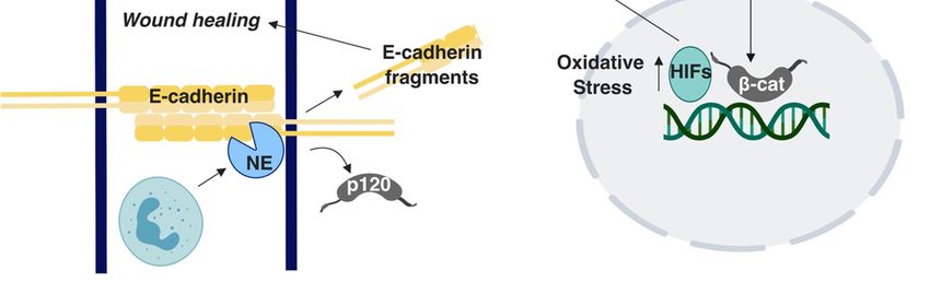
Int. J. Mol. Sci. 2019, 20, 2756                                                                     3 of 17

making it the tissue with the fastest turnover in the human body. This mechanism allows the colon to
maintain homeostasis under the harsh conditions of the intestinal lumen, which induces constant cell
shedding from the top of the crypt [23]. Accordingly, this cell gradient across the crypt is accompanied
by a signaling gradient. Two major signaling pathways that determine cell fate in the colonic crypt
are the Wnt and the BMP signaling pathways. The Wnt signaling is activated at the bottom and is
gradually suppressed towards the top part of the crypt; in contrast, BMP signaling is activated at the
top of the crypt [24–26]. This elegant balance allows for the maintenance of a stem cell niche at the
bottom of the crypt, giving rise to cells that proliferate and eventually fully differentiate at the top of
the crypt.
      E-cadherin is the main cadherin expressed in the colonic crypt epithelium. E-cadherin is vital
for the proper morphogenesis of the intestine [27]. E-cadherin expression across the developed crypt
is not uniform; E-cadherin levels are lower towards the base of the crypt but are strongly expressed
at the apical part of the crypt, which supports the formation of the intestinal barrier, an essential
function of the organ [26,28]. This E-cadherin expression gradient is consistent with the state of
differentiation of the crypt cells, which occurs at the top part of the crypt as well as with the activation
status of Wnt signaling. Indeed, β-catenin is nuclear and Wnt is active at the bottom of the crypt;
however, Wnt signaling becomes gradually inactive towards the top of the crypt due to increased
APC (Adenomatous Polyposis Coli) expression, resulting in β-catenin association with E-cadherin
and cell-cell junction stabilization at the differentiated cell compartment at the top of the crypt [26,28].
Nevertheless, there are heterogeneous crypts with clusters of cells towards the bottom of the crypt
that strongly express E-cadherin [28]. It is not clear why these cells express high E-cadherin and what
type of cells these are. It has been suggested that high E-cadherin expression may serve in stabilizing
contacts between stem cells and surrounding cells at the bottom of the crypt, and that E-cadherin
expression is suppressed in the proliferating cells to allow them to progress towards the top part of the
crypt [28]. Ephrin EphB - ADAM10 - mediated shedding of E-cadherin results in compartmentalization
of E-cadherin contacts, which is critical in fine-tuning cell migration and proper organization of cells
in the crypt [29]. Furthermore, E-cadherin stabilization promotes colony formation of colonic stem
cells [30]. Interestingly, a recent work showed that E-cadherin is required for Lgr5+ gastric stem cell
survival [31]. However, association of E-cadherin with the Lgr5+ colonic stem cells has yet to be
established. The role of E-cadherin in stem cell survival and its potential repercussions in cancer stem
cell survival and pro-tumorigenic transformation has been explored in a computational model [32];
however, this also has yet to be experimentally tested.

2. Colorectal Cancer and E-cadherin

2.1. E-cadherin is a Double-Faced Signaling Molecule in the Colon
     Although the fast renewal capacity and turnover of the colonic epithelium provides plasticity
and the ability to maintain homoeostasis, it also makes the colon susceptible to mutagenesis and
potentially tumorigenesis. Indeed, colorectal cancer (CRC) is the third most prevalent and second
deadliest form of the disease [33], and most cancers in the colon arise from the mucosal epithelial layer.
Preventative screening has led to a gradual decrease in CRC incidence in recent decades, especially
among older populations. However, studies conducted in cancer patients diagnosed from 1995 to 2014
in the USA showed a surprising increase in CRC incidence rates in young populations [34,35]. This
and other studies have suggested a link between increased obesity rates and colon cancer. In a recent
work, it was demonstrated that obesity and ensuing diabetes and hyperglycemia negatively impact
intestinal barrier function, which, in turn, results in microbial infection and inflammation, a common
precursor to CRC [36]. However, incidents in East Asia, where a lower obesity level has been observed
in comparison with the global numbers [37], the most prevalent cancer type remains CRC, suggesting
other contributing factors to the disease.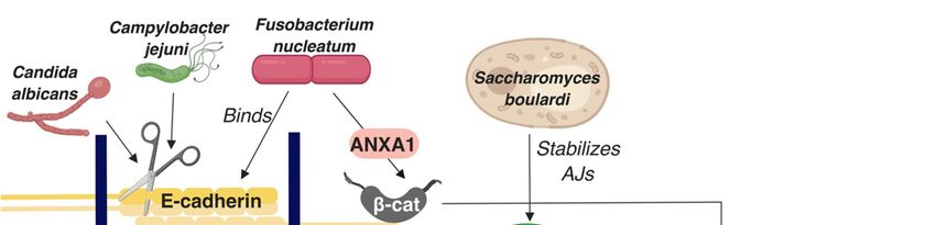
Int. J. Mol. Sci. 2019, 20, 2756 4 of 17
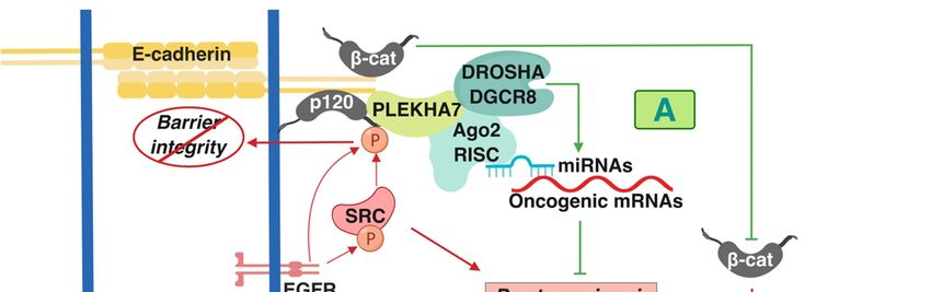
The status of E-cadherin has been extensively studied in CRC in the context of Wnt/β-catenin
signaling because dysregulation of this pathway is a predominant driver of tumorigenesis in the
colon [38–40]. Overall, E-cadherin has a predominantly tumor-suppressing role in this context. For
example, E-cadherin suppresses the pro-tumorigenic transformation that is promoted by β-catenin
activating mutations by keeping β-catenin at areas of cell-cell contact as opposed to allowing it to go
to the nucleus (Figure 1A) [41,42]. However, other functions of E-cadherin have recently emerged
with regards to its role in colon tumorigenesis. Interestingly, many of these studies point towards
signaling directly driven by the AJs and not indirectly in the nucleus, through the release of β-catenin.
One interaction that regulates this signaling is the cross-talk of cadherin complexes with EGFR and
Src. Src is an oncogenic non-receptor tyrosine kinase that is overexpressed and/or activated in colon
tumors and is one of the major drivers in colon tumorigenesis [43–45]. Both EGFR and Src can directly
phosphorylate p120 [46,47]; this interaction and overall Src activity disrupts strong adhesion, resulting
in compromised barrier function (Figure 1B) [48,49]. However, this interaction also has consequences
in promoting pro-tumorigenic cell behavior. Disruption of cadherin-mediated adhesion promotes
metastatic and
Int. J. Mol. Sci. invasive
2019, 20, x phenotypes (Figure 1B) [43,49]. 5 of 17
Figure1. 1.AAsummary
Figure summaryofofthe thepro-
pro-and
andanti-tumorigenic
anti-tumorigenicsignaling
signalingmediated
mediatedbybyE-cadherin-based
E-cadherin-based
Adherens Junctions AJ complexes that has been described in colon cells and tissues. (A)
Adherens Junctions AJ complexes that has been described in colon cells and tissues. (A)Shades
Shadesof
green/blue depict tumor-suppressing components and functions, whereas (B) shades
of green/blue depict tumor-suppressing components and functions, whereas (B) shades of red of red represent
the tumor-promoting
represent ones described
the tumor-promoting ones in the text. T-arrows
described represent
in the text. inhibition
T-arrows of molecules
represent or pro-
inhibition of
tumorigenic
molecules signaling processes;
or pro-tumorigenic straight
signaling arrows represent
processes; activation
straight arrows of molecules
represent or pro-tumorigenic
activation of molecules
orsignaling processes.
pro-tumorigenic β-cat: β-catenin;
signaling p120:
processes. p120β-catenin;
β-cat: catenin; RISC:
p120:RNA-induced
p120 catenin; silencing complex.
RISC: RNA-induced
silencing complex.
2.2. E-cadherin as a Colon Cancer Biomarker?
E-cadherin and p120 are required for Src-dependent, anchorage-independent growth and
E-cadherin has been proposed as an additional biomarker for CRC because of its downregulation
downstream suppression of RhoA signaling [50]. p120 acts as an obligatory haploinsufficient tumor
or loss in many cancers [59]. Currently, the Carcinoembryonic Antigen (CEA) is the most commonly
suppressor, whereby one allele of p120 is required for early stages of tumorigenesis in the intestine in
used CRC marker. Other markers in serum or plasma such as APC and KRAS mutations, DNA
Apc-mutated mouse models [51]. Findings also imply a similar role for E-cadherin. Another work has
integrity, histone and DNA methylation, and some microRNAs have also been suggested as CRC
shown that E-cadherin forms a complex together with the polarity component DLG1 and with the cell
biomarkers [60–62]. A meta-analysis reported that low or lost E-cadherin levels in CRC correlate with
death regulator FAS at areas of cell-cell contact (Figure 1B) [52]. This interaction suppresses apoptosis of
poor prognosis in Asian patients but not in European patients [63]. Signet ring cell carcinoma (SRCC)
the HCT15 colon cancer cells by inhibiting the formation of the pro-apoptotic, death-inducing signaling
is a rare adenocarcinoma that primarily occurs in the stomach and occasionally in the colon [64]. The
World Health Organization (WHO) defines SRCC as the cancer type where >50% of tumor cells have
intracytoplasmic mucin present [65]. A study that investigated 59 patients reported a statistically
significant higher survival for patients with E-cadherin positive SRCC when compared with lower
survival rates of patients with E-cadherin negative SRCC [64]. In addition, increased levels of soluble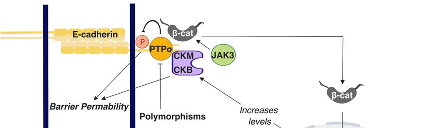
Int. J. Mol. Sci. 2019, 20, 2756                                                                     5 of 17

complex (DISC), which signifies a pro-survival role of E-cadherin in colon cancer cells (Figure 1B) [52].
E-cadherin-positive cells and tumors appear chemotherapy-resistant [53,54]. These studies challenge
the dogma of Epithelial-to-Mesenchymal Transition EMT-mediated cancer progression. One study
demonstrated that L1-induced metastasis of colon cancer cells is E-cadherin and EMT-independent [55],
whereas a more recent work has shown that Rab11 stabilizes E-cadherin levels and promotes collective
cell migration of colon cells (Figure 1B) [56,57]. These findings unravel a tumor-promoting role of
E-cadherin complexes, contrary to the prevailing notion that E-cadherin is a de facto tumor suppressor. In
attempts to reconcile these findings, it was demonstrated that there are distinct E-cadherin complexes at
the AJs of polarized monolayers of the well-differentiated colon epithelial Caco2 cells: an apical-specific
complex with tumor suppressing properties and a basolateral-specific that promotes pro-tumorigenic
behavior, dependent on Src activity and Src-mediated p120 phosphorylation [16]. This work led
to another revelation regarding cadherin-mediated signaling, demonstrating that E-cadherin-p120
complexes, though their interacting partner PLEKHA7, recruit the core and accessory components of
the RNA interference (RNAi) machinery, including DROSHA, DGCR8, Ago2, and the RNA-induced
silencing complex (RISC) at the apical AJs of the well-differentiated colon Caco2 cells. Cadherins can
regulate miRNA processing and activity to suppress expression of a series of pro-tumorigenic factors
and anchorage-independent growth (Figure 1A) [15,16,58]. In summary, the above studies have altered
our perception on the role of cadherin complexes in cancer by: a) demonstrating that E-cadherin-based
complexes can also act as tumor promoters; b) revealing that E-cadherin complexes are signaling hubs
and not merely structural components of cells. It would be of interest to examine the extent to which
these interactions occur in colon cells and tumors and how they contribute to the tumor suppressing or
tumor promoting functions of E-cadherin.

2.2. E-cadherin as a Colon Cancer Biomarker?
      E-cadherin has been proposed as an additional biomarker for CRC because of its downregulation
or loss in many cancers [59]. Currently, the Carcinoembryonic Antigen (CEA) is the most commonly
used CRC marker. Other markers in serum or plasma such as APC and KRAS mutations, DNA
integrity, histone and DNA methylation, and some microRNAs have also been suggested as CRC
biomarkers [60–62]. A meta-analysis reported that low or lost E-cadherin levels in CRC correlate with
poor prognosis in Asian patients but not in European patients [63]. Signet ring cell carcinoma (SRCC)
is a rare adenocarcinoma that primarily occurs in the stomach and occasionally in the colon [64]. The
World Health Organization (WHO) defines SRCC as the cancer type where >50% of tumor cells have
intracytoplasmic mucin present [65]. A study that investigated 59 patients reported a statistically
significant higher survival for patients with E-cadherin positive SRCC when compared with lower
survival rates of patients with E-cadherin negative SRCC [64]. In addition, increased levels of soluble
plasma E-cadherin, which would indicate E-cadherin cleavage and compromised cell-cell adhesion,
has been associated with advanced stage colorectal cancer and with familial adenomatous polyposis
(FAP), a rare condition in the colon that strongly predisposes for CRC [66]. However, in the same
study, plasma E-cadherin levels were unaltered in patients with inflammatory bowel disease (IBD)
or early stage colorectal tumors [66]. Similarly, although E-cadherin loss was found to strongly
predict lymph node-positive colorectal cancers [67], another study found no statistically significant
correlation of reduced E-cadherin expression with development of metastatic colon disease [68] and
loss of membranous expression of E-cadherin, which would indicate junction-bound cadherin was not
significantly correlated to Duke’s staging, tumor grade, sex, size, and site of tumor [69]. Furthermore,
E-cadherin is still expressed in several colon cancer cell lines [70]. HCT116 and HT-29 cells in 3D
cultures adopt an invasive phenotype without progressing through EMT while continuing to express
robust levels of E-cadherin [71]. Together, these data are in agreement with the recent conflicting
findings in E-cadherin signaling in colon tumorigenesis and further challenge the traditional view of
E-cadherin as a tumor suppressor. They also suggest that the use of additional markers that broadly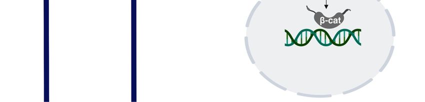
Int. J. Mol. Sci. 2019, 20, 2756                                                                     6 of 17

incorporate other members of cadherin complexes that modulate E-cadherin’s barrier and signaling
functions in the colon is required to provide a better association with disease initiation and progression.

2.3. The Role of Other Cadherins in Colon Tumorigenesis
      Although absent in normal colon tissues, P-cadherin is abnormally expressed early in colorectal
carcinogenesis, promoting colonic crypt fission and metastasis in the liver [72–74]. Similarly, N-cadherin
is upregulated in a cohort of colon tumors [75]. Interestingly, this expression coincides with E-cadherin
expression and is independent of expression of EMT promoters, such as SNAI1 and TWIST [75]. This,
together with other studies [16] that show retention of E-cadherin and simultaneous overexpression
of mesenchymal cadherins, such as N-cadherin or Cadherin-11, also demonstrates a deviation from
the classical model of EMT in tumorigenesis. Heterotypic cadherin interactions may drive tumor cell
migration and metastasis, as was recently shown in other epithelial cancer cell types [76]. It remains
to be shown whether this is the case in the colon. Nevertheless, a role of N-cadherin in promoting
colon myofibroblast migration and invasion upon TGF-β stimulation has been demonstrated [77].
Increased expression of Cadherin-11, which is another mesenchymal cadherin, has also been shown to
promote pro-tumorigenic signaling in Caco2 cells [16] or cell migration in HT-29 cells [78]. Interestingly,
Cadherin-11 expression is increased in patients with IBD, although the significance of this finding in
cell signaling and behavior has yet to be explored [79]. Overall, the status and role of cadherins other
than E-cadherin in the colon and in colon tumorigenesis is understudied in comparison with other
epithelial tumors, leaving this an open field of investigation.

3. E-cadherin in Inflammatory Bowel Disease
     A suggested culprit for the increased incidence of colon cancer in younger ages is increased cases
of Inflammatory Bowel Disease (IBD) [80], which is a general term for two conditions: Ulcerative
Colitis (UC), which is predominantly found in the large intestine, and Crohn’s Disease (CD), which
occurs in both the small and large intestine [81]. The reasons of increased IBD in the general population
are still not well understood. However, IBD patients carry a significantly increased risk for developing
CRC [82]. The intestinal barrier is compromised in IBD, allowing the flux of water and dissolved
solutes, ions, and nutritional molecules across the intestinal barrier [83,84]. Because E-cadherin is key
to barrier maintenance, its dysregulation could increase the risk of developing IBD and ultimately CRC.
Indeed, genome-wide association studies have shown the E-cadherin gene CDH1 as a susceptibility
locus in UC [85], along with HNF4 and LAMB1 [86]. N-terminal truncation of E-cadherin due to
polymorphisms in CDH1 results in cytoplasmic aggregation of E-cadherin in CD while indirectly
mis-localizing β-Catenin [87]. Mutations in other genes can also affect AJ- associated proteins.
Polymorphisms in the C1orf106 gene is a risk factor in UC [88]. A study conducted using C1orf106−/−
colonic organoid-derived epithelial cells noted decreased surface E-cadherin levels and increased
intracellular E-cadherin levels [88]. Furthermore, C1orf106−/− cellular monolayers exhibited increased
permeability in luciferase permeability and trans-epithelial electrical resistance (TEER) assays [88].
In the same study, C1orf106−/− mice demonstrated impaired recovery from DSS-induced colitis and
damaged colon crypts when Citrobacter rodentium was introduced in comparison with C1orf106+/+
mice [88]. Additionally, in a Chloride channel protein-2 (CIC-2; CLCN2) null mouse model, recovery
from DSS-induced colitis was impaired and the epithelial permeability was decreased [89]. Although
these results were firstly attributed to compromised TJ function [89], it was subsequently shown that
the AJs were also responsible [90]. E-cadherin and β-catenin distribution as well as the ultrastructural
tissue morphology were specifically altered in the colon while it was retained in small intestine [90].
These observations suggest that CIC-2 is associated with the AJs’ function specifically in the colon [90].
Polymorphisms in the receptor-type tyrosine-protein phosphatase-S gene (PTPRS) that encodes for
the PTPσ protein are associated with UC. Importantly, PTPσ has been demonstrated to localize at the
apical region. E-cadherin and β-catenin act as substrates for PTPσ in the brain and epithelial barrier is
perturbed due to tyrosine phosphorylation; accordingly, it has been suggested that polymorphisms in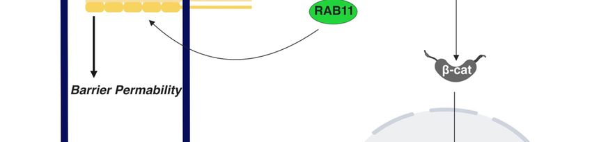
Int. J. Mol. Sci. 2019, 20, 2756                                                                                7 of 17

   Int. J. Mol. Sci. 2019, 20, x                                                                                  8 of 17
the PTPRS gene can cause disruption in the apical junctions in the colon, promoting UC [91] (Figure 2).
  studies
Earlier    demonstrate
        work has shown that   E-cadherin junctions
                          hypermethylation            are apromoter
                                             in the CDH1     central node  in and
                                                                      region   a variety  of mechanisms
                                                                                    CpG island            that
                                                                                                methylation
  promote   barrier  function  and  IBD  progression.   However,    what  is  still missing  is
of CDH1 in UC conditions [92,93]. Although epigenetic regulations of AJ proteins, especially of whether these
  observations
E-cadherin, havecan  provide
                   been       mechanistic
                         extensively       insights
                                     studied          into theofreasons
                                              in the context            for the
                                                                 CRC, there      increased
                                                                             is overall      CRCknowledge
                                                                                        limited  risk for IBD
  patients,
available onwhich  remains
             this topic     an unresolved
                        regarding  IBD.    conundrum. Given the extensive signaling roles of E-cadherin
  complexes mentioned throughout this paper, this is a fertile ground for future investigation.

         Figure
      Figure  2. A2.summary
                      A summary      of the E-cadherin-mediated
                             of the E-cadherin-mediated              signaling
                                                          signaling and           andinvolved
                                                                         interactions   interactions   involved in
                                                                                                in Inflammatory
      Bowel   Disease IBD.
         Inflammatory      T-arrows
                        Bowel        represent
                               Disease         inhibition
                                        IBD. T-arrows     of molecules
                                                        represent       or processes;
                                                                  inhibition          straight
                                                                              of molecules   orarrows   represent
                                                                                                processes;  straight
      activation  of molecules
         arrows represent      or processes.
                           activation        β-cat: β-catenin;
                                      of molecules             p120:
                                                    or processes.    p120
                                                                  β-cat:   catenin. p120: p120 catenin
                                                                         β-catenin;

      E-cadherin Interacts
   4. E-cadherin   regulateswith
                             colonthe
                                    homeostasis  also through interactions with immune cells. CD11c+
                                      Colon Microbiome
mononuclear phagocytes in an IBD mouse model have higher than usual number of adhesions to the
         Projects such as Human Microbiome Project have extended our understanding of the gut
epithelium due to upregulated E-cadherin expression, leading to inflammation [94]. Polymorphonuclear
   microbiome, which consists of trillions of microbes. Although the commensal microbe community
neutrophils (PMNs) are a type of white blood cells that have been shown to affect mucosal barrier
   positively affects the overall health of the host, disturbances in the healthy microbiome, known as
during the inflammation process by altering the localization patterns of E-cadherin and β-catenin,
   dysbiosis, have been shown to corelate with colon cancer occurrence [108]. The colonic epithelium
eventually leading to perturbation of AJs [95]. A recent study demonstrated that E-cadherin is
   acts as a barrier and blocks microorganisms from passing through. When microorganisms penetrate
enzymatically cleaved to several peptide fragments by neutrophil elastase (NE), a known inflammatory
   the epithelial barrier and enter into the inner layers, this can cause inflammation. Bacteroides fragilis
protease present in IBD (Figure 2). These peptide fragments were present in the patient tissues sample
   is one such microorganism that has a positive correlation with IBD patients, both in CD and UC [109].
analyzed in the study and could enter the cytosol of Caco2 cells in vitro by crossing the lipid bilayer.
   This bacterial species produces a metalloprotease known as Bacteroides fragilis toxin, which
Although these fragments did not alter proliferation rates, they improved wound healing in in vitro
   stimulates γ-secretase to cleave E-cadherin, resulting in AJs disruption and nuclear localization of β-
assays (Figure 2) [96]. Although E-cadherin fragmentation would seemingly impair barrier function
   catenin, ultimately promoting cell proliferation in HT29/C1 cells [110–112]. A study has shown that
and exacerbate IBD, the faster wound healing could instead be beneficial for IBD; the action of these
   CRC patients with tumors with bacterial biofilms, which are dense bacterial populations encased in
fragments implies downstream signaling, which warrants further investigation.
   a polymeric matrix, also exhibited biofilms in their normal colonic tissue, which resulted in decreased
      Although not extensively investigated as E-cadherin, studies have investigated the roles of other AJ
   E-cadherin expression, increased cell proliferation, and IL-6/STAT3 activation [113]. Changes in the
proteins in IBD, demonstrating that E-cadherin, p120, and α-catenin expression is downregulated in the
   colonic microbiome, e.g., in CRC, can affect colonic tissue homeostasis and the E-cadherin status in
colonic mucosa of IBD patients [97]. In contrast, another study showed focal increases of the E-cadherin
   distant places in the colon. Notably, no specific bacterial species, but the overall presence or absence
- β-catenin complex in the mucosa of IBD patients, suggesting a putative defensive response against
   of biofilms, was associated with this phenotype. Candida albicans is a yeast species that has been
inflammation [98]. An in vivo study reported that p120 loss caused inflammation due to increased
   shown to disturb the epithelial integrity of Caco2 colon epithelial cells by cleaving E-cadherin into an
association of neutrophils with the disturbed epithelial barrier [99]. Similarities were noted between
   extracellular fragment and an intracellular fragment that acts as a substrate for γ-secretase (Figure 3)
the p120-ablated phenotype and IBD, caused by overexpression of a dominant negative cadherin [99].
   [114]. In Caco2 cells, E-cadherin was shown to be displaced from AJs when infected with Escherichia
When Citrobacter rodentium-induced IBD mice were treated with the γ-secretase inhibitor Dibenzazepine
   coli in vitro [115]. In contrast, an in vitro study conducted using HCT-8/E11 human colonic
(DBZ) to block Notch signaling, the AJs were affected, as demonstrated by E-cadherin and β-catenin
   adenocarcinoma cells demonstrated that Saccharomyces boulardi strengthens AJs by improving E-
   cadherin transportation to the cell surface via regulation of recycling of Rab11-associated endosomes,
   (Figure 3) [97]. Fusobacterium nucleatum is a bacterium that directly binds E-cadherin through its
Int. J. Mol. Sci. 2019, 20, 2756 8 of 17
altered expression. These mice showed signs of altered mucous makeup and bacterial dysbiosis
that resulted in serious colitis and inflammation [100]. Numb is a regulatory protein that directs
epithelial cell transformation to goblet cells via inhibition of Notch signaling. Co-immunoprecipitation
studies conducted using Caco2 cells demonstrated that Numb interacts with E-cadherin while its
downregulation compromises the epithelial barrier in a Notch signaling-independent manner [101].
Lastly, in vitro and in vivo experiments revealed Janus kinase-3 (JAK3) as a potential regulator of IBD
due to its ability to control β-catenin localization at the apical junctions (Figure 2) [102].
Other conditions in the body can locally affect adherens junction integrity, leading to the
development of IBD. Creatine kinases (CKs) are enzymes regulated by hypoxia-inducible transcription
factors (HIFs), which fluctuate with oxygen concentrations. CKM and CKB have been shown to
localize at the apical junctions, suggesting a role in regulating epithelial permeability in IBD (Figure 2);
however, the exact mechanism for junction stabilization remains unclear [103]. In PIK3C3 mutant
zebrafish, induction of IBD is accompanied by cytoplasmic retention and decreased localization of
E-cadherin at the cell membrane of intestinal epithelial cells [104]. Although vitamin D deficiency is
an unexpected candidate, it has been shown to correlate with increased risk for IBD [105] (Figure 2).
An in vivo study demonstrated that vitamin D receptor null mice (VDR-/- ) exhibit severe colitis [106].
In the same study, in vitro cultures of VDR-depleted Caco2 cells showed lower TEER and reduced
E-cadherin levels by qRT-PCR [106]. Nevertheless, transmission electron microscope images of
VDR-/- mice colons did not display significant alteration in adherens junction morphology. However,
induction of 1,25-dihydroxy-vitamin D3 [1,25(OH)2 D3] increased the E-cadherin levels in SW480
colon adenocarcinoma cells [106]. Notably, genome-wide data analysis suggests an association among
UC, CD, and polymorphisms in VDR [107]. Taken together, these studies demonstrate that E-cadherin
junctions are a central node in a variety of mechanisms that promote barrier function and IBD
progression. However, what is still missing is whether these observations can provide mechanistic
insights into the reasons for the increased CRC risk for IBD patients, which remains an unresolved
conundrum. Given the extensive signaling roles of E-cadherin complexes mentioned throughout this
paper, this is a fertile ground for future investigation.
4. E-cadherin Interacts with the Colon Microbiome
Projects such as Human Microbiome Project have extended our understanding of the gut
microbiome, which consists of trillions of microbes. Although the commensal microbe community
positively affects the overall health of the host, disturbances in the healthy microbiome, known as
dysbiosis, have been shown to corelate with colon cancer occurrence [108]. The colonic epithelium acts
as a barrier and blocks microorganisms from passing through. When microorganisms penetrate the
epithelial barrier and enter into the inner layers, this can cause inflammation. Bacteroides fragilis is one
such microorganism that has a positive correlation with IBD patients, both in CD and UC [109]. This
bacterial species produces a metalloprotease known as Bacteroides fragilis toxin, which stimulates
γ-secretase to cleave E-cadherin, resulting in AJs disruption and nuclear localization of β-catenin,
ultimately promoting cell proliferation in HT29/C1 cells [110–112]. A study has shown that CRC
patients with tumors with bacterial biofilms, which are dense bacterial populations encased in a
polymeric matrix, also exhibited biofilms in their normal colonic tissue, which resulted in decreased
E-cadherin expression, increased cell proliferation, and IL-6/STAT3 activation [113]. Changes in the
colonic microbiome, e.g., in CRC, can affect colonic tissue homeostasis and the E-cadherin status in
distant places in the colon. Notably, no specific bacterial species, but the overall presence or absence of
biofilms, was associated with this phenotype. Candida albicans is a yeast species that has been shown to
disturb the epithelial integrity of Caco2 colon epithelial cells by cleaving E-cadherin into an extracellular
fragment and an intracellular fragment that acts as a substrate for γ-secretase (Figure 3) [114]. In Caco2
cells, E-cadherin was shown to be displaced from AJs when infected with Escherichia coli in vitro [115].
In contrast, an in vitro study conducted using HCT-8/E11 human colonic adenocarcinoma cells
demonstrated that Saccharomyces boulardi strengthens AJs by improving E-cadherin transportation toInt. J. Mol. Sci. 2019, 20, 2756 9 of 17
Int. J. Mol. Sci. 2019, 20, x 9 of 17
the cell surface via regulation of recycling of Rab11-associated endosomes, (Figure 3) [97]. Fusobacterium
nucleatum is a bacterium that directly binds E-cadherin through its Fusobacterium adhesin A (FadA)
Fusobacterium adhesin A (FadA) domain, promoting β-catenin signaling and stimulating
domain, promoting β-catenin signaling and stimulating proliferation in CRC cells, as confirmed by
proliferation in CRC cells, as confirmed by in vitro and in vivo studies [116]. Additional research has
in vitro and in vivo studies [116]. Additional research has demonstrated that it is through Annexin 1
demonstrated that it is through Annexin 1 (ANXA1) that Fusobacterium nucleatum can mediate β-
(ANXA1) that Fusobacterium nucleatum can mediate β-catenin signaling [117]. Work that investigated
catenin signaling [117]. Work that investigated the effects of four different Lactobacillus strains on
the effects of four different Lactobacillus strains on the adherens junctions of T84 colon adenocarcinoma
the adherens junctions of T84 colon adenocarcinoma cells noted differentially regulated E-cadherin
cells noted differentially regulated E-cadherin and elevated phosphorylated β-catenin levels by some of
and elevated phosphorylated β-catenin levels by some of the strains; it also noticed an overall
the strains; it also noticed an overall improvement in barrier function by gram positive lactobacilli [118].
improvement in barrier function by gram positive lactobacilli [118]. Another bacterial species,
Another bacterial species, Campylobacter jejuni, proteolytically cleaves E-cadherin through proteases
Campylobacter jejuni, proteolytically cleaves E-cadherin through proteases secreted in outer
secreted in outer membrane vesicles (Figure 3) [119]. Although Campylobacter jejuni can be associated
membrane vesicles (Figure 3) [119]. Although Campylobacter jejuni can be associated with
with inflammatory enteritis, its role in IBD is not clear. Overall, E-cadherin seems to be a critical node
inflammatory enteritis, its role in IBD is not clear. Overall, E-cadherin seems to be a critical node in
in the cross-talk between the intestinal epithelium and the microbiome, adding an important parameter
the cross-talk between the intestinal epithelium and the microbiome, adding an important parameter
to consider in E-cadherin’s broad role in colon homeostasis and disease.
to consider in E-cadherin’s broad role in colon homeostasis and disease.
FigureFigure 3. A schematic
3. A schematic summarizing
summarizing interactions
interactions of E-cadherin
of E-cadherin with
with the the colon
colon microbiome.
microbiome. Straight
Straight
arrowsarrows represent
represent activation
activation of molecules
of molecules or processes
or processes β-cat: β-cat: β-catenin;
β-catenin; p120: p120:
p120 p120 catenin
catenin.
5. E-cadherin as a Sensor
5. E-cadherin of Physical
as a Sensor Strain
of Physical in theinColon
Strain the Colon
Colon tissues
Colon fromfrom
tissues CRC CRC
and IBD
and patients exhibit
IBD patients extensive
exhibit fibrosis,
extensive characterized
fibrosis, by increased
characterized by increased
deposition and reorganization
deposition and reorganizationof theofextracellular matrix
the extracellular (ECM)
matrix [120].[120].
(ECM) Impaired barrier
Impaired integrity
barrier and and
integrity
permeability are both causes and consequences of fibrosis [84,121]. Changes
permeability are both causes and consequences of fibrosis [84,121]. Changes in the ECM promotein the ECM promote
physical cues and
physical cuesstrain
andthat can be
strain thattransmitted throughout cells
can be transmitted and tissues,
throughout cellsaltering their physiology.
and tissues, altering their
For example,
physiology. For example, different ECM components, such as collagen I, collagen IV, different
different ECM components, such as collagen I, collagen IV, and laminin, generated and laminin,
brushgenerated
border enzyme expression
different of Caco2
brush border cells, whereas
enzyme expressioncollagen I promoted
of Caco2 their proliferation
cells, whereas collagen I [122].
promoted
Similarly,
their when Caco2 cells
proliferation [122].are put under
Similarly, increased
when Caco2 strain,
cells areexpression
put under ofincreased
brush border enzymes
strain, is of
expression
altered [123]. Changes in the ECM also translate to changes in the stromal stiffness,
brush border enzymes is altered [123]. Changes in the ECM also translate to changes in the stromal which can affect
cellular morphology
stiffness, which andcan promote cancermorphology
affect cellular progression and [124]. It has been
promote proposed
cancer that collagen
progression [124]. Ithas
hasa been
role in this process
proposed thatascollagen
one the has
main components
a role of ECMas[125].
in this process Indeed,
one the mainHCT-8 colon cancer
components of ECM cells exhibit
[125]. Indeed,
HCT-8 colon cancer cells exhibit a more metastatic phenotype when cultured under low stiffness of
20–47 kPa; however, this phenotype was not observed under higher stiffness [126]. Interestingly,
decrease in E-cadherin levels was also observed in cells cultured on less stiff substrates, which is inInt. J. Mol. Sci. 2019, 20, 2756 10 of 17
a more metastatic phenotype when cultured under low stiffness of 20–47 kPa; however, this phenotype
was not observed under higher stiffness [126]. Interestingly, decrease in E-cadherin levels was also
observed in cells cultured on less stiff substrates, which is in agreement with the higher metastatic
potential of cells in these conditions [127]. Similarly, when colon samples from APC heterozygous
mice for truncated amino acid loci 1638 were harvested and put under mechanical strain, an increase
of nuclear β-catenin, MYC, and TWIST1 expression was observed [128]. A later study revealed that,
when mechanical pressure was magnetically induced, phosphorylated β-catenin levels in the colon
were elevated [129]. Indeed, it was shown that the Y654-β-catenin and D665-E-cadherin binding sites
are affected by mechanical stress, which eventually stimulates the β-catenin signaling pathway in
developing Drosophila melanogaster embryos [130]. The effects of mechanical stress and stiffness in the
overall tumor development are described in detail in Broders-Bondon et al. (2018) [131].
Cells adhere to the ECM through integrins, which connect to the cytoskeleton and are mediators of
extracellular signals. Integrins and the ECM have an intimate relationship with the AJs [132]. Integrins
interact with Focal Adhesion Kinase (FAK) and Src; together, they regulate RhoGTPase activity
and affect strong adhesion [133]. When Caco2 cells were put under cyclic strain, phosphorylation
of JNK2 and c-MYC affected localization of E-cadherin and β-catenin while increasing epithelial
permeability [134]. An in vitro study showed that TGF-β induced E-cadherin to mediate cellular
adhesions in a FAK-dependent manner during ECM remodeling [135]. Overall, the data demonstrate
that there is cross-talk among extracellular mechanical cues, stromal composition, and stiffness, with
the integrity of the adherens junctions in the colon. Driven by these findings, further research is
required to understand how the cells translate these mechanical cues to regulate junctional integrity
and to better understand fibrosis and mechanical stress in the context of colon tumorigenesis.
6. Conclusions
E-cadherin has long been considered a critical homeostatic component of the colonic epithelium,
primarily due to its central role in cellular architecture and barrier function. It has also been thought
to primarily act as a tumor suppressor in CRC. However, numerous emerging roles of E-cadherin in
intracellular signaling and cell behavior as well as its extensive cross-talk with the colonic epithelial
microenvironment reveal a broader and more complicated role. Furthermore, the identification of
new E-cadherin partners at the AJs add to the complexity, introducing new aspects and questions in
cadherin biology. These recent findings portray E-cadherin and of the AJs as not merely structural
components of cells and tissues but in new roles as signaling hubs, opening novel and exciting avenues
of investigation.
Funding: This work was supported by a South Carolina IDeA Networks of Biomedical Research Excellence (SC
INBRE; NIH P20 GM103499) Development Research Program Award and by the Concern Foundation (Conquer
Cancer Now Award) to AK; and a NIH TL1 TR001451/UL1 TR001450 Predoctoral Clinical & Translational Research
Training Program to MCB.
Acknowledgments: We apologize to our colleagues that their work was not cited due to space limitations.
Conflicts of Interest: The authors declare no conflict of interest.
Abbreviations
AJ Adherens Junction
CD Crohn’s Disease
CRC Colorectal Cancer
DISC death-inducing signaling complex
DSS Dextran Sulfate Sodium
ECM extracellular matrix
EMT Epithelial to Mesenchymal Transition
IBD Inflammatory Bowel Disease
NE neutrophil elastaseInt. J. Mol. Sci. 2019, 20, 2756 11 of 17
p120 p120 catenin
PMNs Polymorphonuclear neutrophils
qRT-PCR quantitative reverse transcription polymerase chain reaction
RNAi RNA interference
RISC RNA-induced silencing complex
TEER trans-epithelial electrical resistance
TJ tight junction
UC Ulcerative Colitis
ZA zonula adherens
References
1. Takeichi, M. Dynamic Contacts: Rearranging Adherens Junctions to Drive Epithelial Remodelling. Nat. Rev.
Mol. Cell Biol. 2014, 15, 397–410. [CrossRef] [PubMed]
2. Harris, T.J.C.; Tepass, U. Adherens Junctions: From Molecules to Morphogenesis. Nat. Rev. Mol. Cell Biol.
2010, 11, 502–514. [CrossRef] [PubMed]
3. Ogita, H.; Rikitake, Y.; Miyoshi, J.; Takai, Y. Cell Adhesion Molecules Nectins and Associating Proteins:
Implications for Physiology and Pathology. Proc. Jpn. Acad. Ser. B 2010, 86, 621–629. [CrossRef]
4. Noemi, R.; Dejana, E. Adherens Junctions. Curr. Biol. 2008, 18, R1080–R1082.
5. van Roy, F. Beyond E-Cadherin: Roles of Other Cadherin Superfamily Members in Cancer. Nat. Rev. Cancer
2014, 14, 121–134. [CrossRef]
6. Hartsock, A.; Nelson, W.J. Adherens and Tight Junctions: Structure, Function and Connections to the Actin
Cytoskeleton. Biochim. Biophys. Acta BBA Biomembr. 2008, 1778, 660–669. [CrossRef]
7. Shapiro, L.; Weis, W.I. Structure and Biochemistry of Cadherins and Catenins. Cold Spring Harb. Perspect.
Biol. 2009, 1, a003053. [CrossRef] [PubMed]
8. Nelson, W.J. Regulation of Cell–Cell Adhesion by the Cadherin–Catenin Complex. Biochem. Soc. Trans. 2008,
36, 149–155. [CrossRef]
9. Anastasiadis, P.Z. P120-Ctn: A Nexus for Contextual Signaling via Rho GTPases. Biochim. Biophys. Acta BBA
Mol. Cell Res. 2007, 1773, 34–46. [CrossRef]
10. Xiao, K.; Oas, R.G.; Chiasson, C.M.; Kowalczyk, A.P. Role of P120-Catenin in Cadherin Trafficking. Biochim.
Biophys. Acta BBA Mol. Cell Res. 2007, 1773, 8–16. [CrossRef]
11. Hartsock, A.; Nelson, W.J. Competitive Regulation of E-Cadherin JuxtaMembrane Domain Degradation by
P120-Catenin Binding and Hakai-Mediated Ubiquitination. PLoS ONE 2012, 7, e37476. [CrossRef] [PubMed]
12. Ishiyama, N.; Lee, S.-H.; Liu, S.; Li, G.-Y.; Smith, M.J.; Reichardt, L.F.; Ikura, M. Dynamic and Static
Interactions between P120 Catenin and E-Cadherin Regulate the Stability of Cell-Cell Adhesion. Cell 2010,
141, 117–128. [CrossRef] [PubMed]
13. Ireton, R.C.; Davis, M.A.; van Hengel, J.; Mariner, D.J.; Barnes, K.; Thoreson, M.A.; Anastasiadis, P.Z.;
Matrisian, L.; Bundy, L.M.; Sealy, L.; et al. A Novel Role for P120 Catenin in E-Cadherin Function. J. Cell Biol.
2002, 159, 465–476. [CrossRef] [PubMed]
14. Meng, W.; Mushika, Y.; Ichii, T.; Takeichi, M. Anchorage of Microtubule Minus Ends to Adherens Junctions
Regulates Epithelial Cell-Cell Contacts. Cell 2008, 135, 948–959. [CrossRef] [PubMed]
15. Kourtidis, A.; Anastasiadis, P.Z. PLEKHA7 Defines an Apical Junctional Complex with Cytoskeletal
Associations and MiRNA-Mediated Growth Implications. Cell Cycle 2016, 15, 498–505. [CrossRef] [PubMed]
16. Kourtidis, A.; Ngok, S.P.; Pulimeno, P.; Feathers, R.W.; Carpio, L.R.; Baker, T.R.; Carr, J.M.; Yan, I.K.; Borges, S.;
Perez, E.A.; et al. Distinct E-Cadherin-Based Complexes Regulate Cell Behaviour through MiRNA Processing
or Src and P120 Catenin Activity. Nat. Cell Biol. 2015, 17, 1145–1157. [CrossRef] [PubMed]
17. Paschoud, S.; Jond, L.; Guerrera, D.; Citi, S. PLEKHA7 Modulates Epithelial Tight Junction Barrier Function.
Tissue Barriers 2014, 2, e28755. [CrossRef]
18. Matter, K.; Balda, M.S. Signalling to and from Tight Junctions. Nat. Rev. Mol. Cell Biol. 2003, 4, 225–236.
[CrossRef]
19. Cunningham, K.E.; Turner, J.R. Myosin Light Chain Kinase: Pulling the Strings of Epithelial Tight Junction
Function. Ann. N. Y. Acad. Sci. 2012, 1258, 34–42. [CrossRef]Int. J. Mol. Sci. 2019, 20, 2756 12 of 17
20. Nita-Lazar, M.; Rebustini, I.; Walker, J.; Kukuruzinska, M.A. Hypoglycosylated E-Cadherin Promotes the
Assembly of Tight Junctions through the Recruitment of PP2A to Adherens Junctions. Exp. Cell Res. 2010,
316, 1871–1884. [CrossRef]
21. Tunggal, J.A.; Helfrich, I.; Schmitz, A.; Schwarz, H.; Günzel, D.; Fromm, M.; Kemler, R.; Krieg, T.; Niessen, C.M.
E-Cadherin Is Essential for in Vivo Epidermal Barrier Function by Regulating Tight Junctions. EMBO J. 2005,
24, 1146–1156. [CrossRef] [PubMed]
22. Humphries, A.; Wright, N.A. Colonic Crypt Organization and Tumorigenesis. Nat. Rev. Cancer 2008, 8,
415–424. [CrossRef] [PubMed]
23. Beumer, J.; Clevers, H. Regulation and Plasticity of Intestinal Stem Cells during Homeostasis and Regeneration.
Development 2016, 143, 3639–3649. [CrossRef] [PubMed]
24. Gregorieff, A. Wnt Signaling in the Intestinal Epithelium: From Endoderm to Cancer. Genes Dev. 2005, 19,
877–890. [CrossRef] [PubMed]
25. Farin, H.F.; Jordens, I.; Mosa, M.H.; Basak, O.; Korving, J.; Tauriello, D.V.F.; de Punder, K.; Angers, S.;
Peters, P.J.; Maurice, M.M.; et al. Visualization of a Short-Range Wnt Gradient in the Intestinal Stem-Cell
Niche. Nature 2016, 530, 340. [CrossRef] [PubMed]
26. Song, J.-H.; Huels, D.J.; Ridgway, R.A.; Sansom, O.J.; Kholodenko, B.N.; Kolch, W.; Cho, K.-H. The APC
Network Regulates the Removal of Mutated Cells from Colonic Crypts. Cell Rep. 2014, 7, 94–103. [CrossRef]
[PubMed]
27. Bondow, B.J.; Faber, M.L.; Wojta, K.J.; Walker, E.M.; Battle, M.A. E-Cadherin Is Required for Intestinal
Morphogenesis in the Mouse. Dev. Biol. 2012, 371, 1–12. [CrossRef]
28. Tan, C.W.; Hirokawa, Y.; Gardiner, B.S.; Smith, D.W.; Burgess, A.W. Colon Cryptogenesis: Asymmetric
Budding. PLoS ONE 2013, 8, e78519. [CrossRef]
29. Solanas, G.; Cortina, C.; Sevillano, M.; Batlle, E. Cleavage of E-Cadherin by ADAM10 Mediates Epithelial
Cell Sorting Downstream of EphB Signalling. Nat. Cell Biol. 2011, 13, 1100–1107. [CrossRef]
30. Wang, F.; Scoville, D.; He, X.C.; Mahe, M.M.; Box, A.; Perry, J.M.; Smith, N.R.; Lei, N.Y.; Davies, P.S.;
Fuller, M.K.; et al. Isolation and Characterization of Intestinal Stem Cells Based on Surface Marker
Combinations and Colony-Formation Assay. Gastroenterology 2013, 145, 383–395.e2. [CrossRef]
31. Tang, Y.; Yang, G.; Zhang, J.; Li, X.; Zhang, C.; Wang, Y.; Xu, J.; Chen, Y.; Teng, Y.; Yang, X. E-Cadherin Is
Required for the Homeostasis of Lgr5 + Gastric Antral Stem Cells. Int. J. Biol. Sci. 2019, 15, 34–43. [CrossRef]
[PubMed]
32. Guebel, D.V.; Schmitz, U.; Wolkenhauer, O.; Vera, J. Analysis of Cell Adhesion during Early Stages of Colon
Cancer Based on an Extended Multi-Valued Logic Approach. Mol. Biosyst. 2012, 8, 1230–1242. [CrossRef]
[PubMed]
33. Siegel, R.L.; Miller, K.D.; Jemal, A. Cancer Statistics, 2019. CA. Cancer J. Clin. 2019, 69, 7–34. [CrossRef]
[PubMed]
34. Sung, H.; Siegel, R.L.; Rosenberg, P.S.; Jemal, A. Emerging Cancer Trends among Young Adults in the USA:
Analysis of a Population-Based Cancer Registry. Lancet Public Health 2019, 4, e137–e147. [CrossRef]
35. Siegel, R.L.; Miller, K.D.; Jemal, A. Colorectal Cancer Mortality Rates in Adults Aged 20 to 54 Years in the
United States, 1970-2014. JAMA 2017, 318, 572–574. [CrossRef] [PubMed]
36. Thaiss, C.A.; Levy, M.; Grosheva, I.; Zheng, D.; Soffer, E.; Blacher, E.; Braverman, S.; Tengeler, A.C.; Barak, O.;
Elazar, M.; et al. Hyperglycemia Drives Intestinal Barrier Dysfunction and Risk for Enteric Infection. Science
2018, 359, 1376–1383. [CrossRef] [PubMed]
37. Devaux, M.; Graf, S.; Goryakin, Y.; Cecchini, M.; Huber, H.; Colombo, F. OECD Obesity Update. In The
Organisation for Economic Co-operation and Development; OECD: London, UK, 2017.
38. Kinzler, K.W.; Nilbert, M.C.; Su, L.K.; Vogelstein, B.; Bryan, T.M.; Levy, D.B.; Smith, K.J.; Preisinger, A.C.;
Hedge, P.; McKechnie, D. Identification of FAP Locus Genes from Chromosome 5q21. Science 1991, 253,
661–665. [CrossRef] [PubMed]
39. Yaeger, R.; Chatila, W.K.; Lipsyc, M.D.; Hechtman, J.F.; Cercek, A.; Sanchez-Vega, F.; Jayakumaran, G.;
Middha, S.; Zehir, A.; Donoghue, M.T.A.; et al. Clinical Sequencing Defines the Genomic Landscape of
Metastatic Colorectal Cancer. Cancer Cell 2018, 33, 125–136.e3. [CrossRef]
40. Korinek, V.; Barker, N.; Morin, P.J.; van Wichen, D.; de Weger, R.; Kinzler, K.W.; Vogelstein, B.; Clevers, H.
Constitutive Transcriptional Activation by a Beta-Catenin-Tcf Complex in APC-/- Colon Carcinoma. Science
1997, 275, 1784–1787. [CrossRef]Int. J. Mol. Sci. 2019, 20, 2756 13 of 17
41. Clevers, H. Wnt/β-Catenin Signaling in Development and Disease. Cell 2006, 127, 469–480. [CrossRef]
42. Huels, D.J.; Ridgway, R.A.; Radulescu, S.; Leushacke, M.; Campbell, A.D.; Biswas, S.; Leedham, S.; Serra, S.;
Chetty, R.; Moreaux, G.; et al. E-Cadherin Can Limit the Transforming Properties of Activating β-Catenin
Mutations. EMBO J. 2015, 34, 2321–2333. [CrossRef] [PubMed]
43. Irby, R.B.; Malek, R.L.; Bloom, G.; Tsai, J.; Letwin, N.; Frank, B.C.; Verratti, K.; Yeatman, T.J.; Lee, N.H.
Iterative Microarray and RNA Interference-Based Interrogation of the SRC-Induced Invasive Phenotype.
Cancer Res. 2005, 65, 1814–1821. [CrossRef] [PubMed]
44. Cordero, J.B.; Ridgway, R.A.; Valeri, N.; Nixon, C.; Frame, M.C.; Muller, W.J.; Vidal, M.; Sansom, O.J. C-Src
Drives Intestinal Regeneration and Transformation. EMBO J. 2014, 33, 1474–1491. [CrossRef] [PubMed]
45. Zhang, B.; Wang, J.; Wang, X.; Zhu, J.; Liu, Q.; Shi, Z.; Chambers, M.C.; Zimmerman, L.J.; Shaddox, K.F.;
Kim, S.; et al. Proteogenomic Characterization of Human Colon and Rectal Cancer. Nature 2014, 513, 382–387.
[CrossRef]
46. Mariner, D.J.; Anastasiadis, P.; Keilhack, H.; Böhmer, F.D.; Wang, J.; Reynolds, A.B. Identification of Src
Phosphorylation Sites in the Catenin P120ctn. J. Biol. Chem. 2001, 276, 28006–28013. [CrossRef] [PubMed]
47. Mariner, D.J.; Davis, M.A.; Reynolds, A.B. EGFR Signaling to P120-Catenin through Phosphorylation at Y228.
J. Cell Sci. 2004, 117, 1339–1350. [CrossRef] [PubMed]
48. Ozawa, M.; Ohkubo, T. Tyrosine Phosphorylation of P120(Ctn) in v-Src Transfected L Cells Depends on Its
Association with E-Cadherin and Reduces Adhesion Activity. J. Cell Sci. 2001, 114, 503–512. [PubMed]
49. Irby, R.B.; Yeatman, T.J. Increased Src Activity Disrupts Cadherin/Catenin-Mediated Homotypic Adhesion in
Human Colon Cancer and Transformed Rodent Cells. Cancer Res. 2002, 62, 2669–2674. [PubMed]
50. Dohn, M.R.; Brown, M.V.; Reynolds, A.B. An Essential Role for P120-Catenin in Src- and Rac1-Mediated
Anchorage-Independent Cell Growth. J. Cell Biol. 2009, 184, 437–450. [CrossRef] [PubMed]
51. Short, S.P.; Kondo, J.; Smalley-Freed, W.G.; Takeda, H.; Dohn, M.R.; Powell, A.E.; Carnahan, R.H.;
Washington, M.K.; Tripathi, M.; Payne, D.M.; et al. P120-Catenin Is an Obligate Haploinsufficient Tumor
Suppressor in Intestinal Neoplasia. J. Clin. Invest. 2017, 127, 4462–4476. [CrossRef] [PubMed]
52. Gagnoux-Palacios, L.; Awina, H.; Audebert, S.; Rossin, A.; Mondin, M.; Borgese, F.; Planas-Botey, C.;
Mettouchi, A.; Borg, J.-P.; Hueber, A.-O. Cell Polarity and Adherens Junction Formation Inhibit Epithelial Fas
Cell Death Receptor Signaling. J. Cell Biol. 2018, 217, 3839–3852. [CrossRef] [PubMed]
53. Druzhkova, I.; Ignatova, N.; Prodanets, N.; Kiselev, N.; Zhukov, I.; Shirmanova, M.; Zagainov, V.; Zagaynova, E.
E-Cadherin in Colorectal Cancer: Relation to Chemosensitivity. Clin. Colorectal Cancer 2019, 18, e74–e86.
[CrossRef] [PubMed]
54. Dinicola, S.; Pasqualato, A.; Proietti, S.; Masiello, M.G.; Palombo, A.; Coluccia, P.; Canipari, R.; Catizone, A.;
Ricci, G.; Harrath, A.H.; et al. Paradoxical E-Cadherin Increase in 5FU-Resistant Colon Cancer Is Unaffected
during Mesenchymal-Epithelial Reversion Induced by γ-Secretase Inhibition. Life Sci. 2016, 145, 174–183.
[CrossRef] [PubMed]
55. Gavert, N.; Sheffer, M.; Raveh, S.; Spaderna, S.; Shtutman, M.; Brabletz, T.; Barany, F.; Paty, P.; Notterman, D.;
Domany, E.; et al. Expression of L1-CAM and ADAM10 in Human Colon Cancer Cells Induces Metastasis.
Cancer Res. 2007, 67, 7703–7712. [CrossRef] [PubMed]
56. Chung, Y.-C.; Wei, W.-C.; Hung, C.-N.; Kuo, J.-F.; Hsu, C.-P.; Chang, K.-J.; Chao, W.-T. Rab11 Collaborates
E-Cadherin to Promote Collective Cell Migration and Indicates a Poor Prognosis in Colorectal Carcinoma.
Eur. J. Clin. Invest. 2016, 46, 1002–1011. [CrossRef] [PubMed]
57. Chung, Y.-C.; Wei, W.-C.; Huang, S.-H.; Shih, C.-M.; Hsu, C.-P.; Chang, K.-J.; Chao, W.-T. Rab11 Regulates
E-Cadherin Expression and Induces Cell Transformation in Colorectal Carcinoma. BMC Cancer 2014, 14, 587.
[CrossRef] [PubMed]
58. Kourtidis, A.; Necela, B.; Lin, W.-H.; Lu, R.; Feathers, R.W.; Asmann, Y.W.; Thompson, E.A.; Anastasiadis, P.Z.
Cadherin Complexes Recruit MRNAs and RISC to Regulate Epithelial Cell Signaling. J. Cell Biol. 2017, 216,
3073–3085. [CrossRef]
59. Christou, N.; Perraud, A.; Blondy, S.; Jauberteau, M.-O.; Battu, S.; Mathonnet, M. E-Cadherin: A Potential
Biomarker of Colorectal Cancer Prognosis. Oncol. Lett. 2017, 13, 4571–4576. [CrossRef]
60. Duffy, M.J. Carcinoembryonic Antigen as a Marker for Colorectal Cancer: Is It Clinically Useful? Clin. Chem.
2001, 4, 7.
61. Yörüker, E.E.; Holdenrieder, S.; Gezer, U. Blood-Based Biomarkers for Diagnosis, Prognosis and Treatment of
Colorectal Cancer. Clin. Chim. Acta 2016, 455, 26–32. [CrossRef]You can also read