HCG, the wonder of today's science - REVIEW
←
→
Page content transcription
If your browser does not render page correctly, please read the page content below
Cole Reproductive Biology and Endocrinology 2012, 10:24
http://www.rbej.com/content/10/1/24

 REVIEW                                                                                                                                     Open Access

hCG, the wonder of today’s science
Laurence A Cole

 Abstract
 Background: hCG is a wonder. Firstly, because hCG is such an extreme molecule. hCG is the most acidic
 glycoprotein containing the highest proportion of sugars. Secondly, hCG exists in 5 common forms. Finally, it has
 so many functions ranging from control of human pregnancy to human cancer. This review examines these
 molecules in detail.
 Content: These 5 molecules, hCG, sulfated hCG, hyperglycosylated hCG, hCG free beta and hyperglycosylated free
 beta are produced by placental syncytiotrophoblast cells and pituitary gonadotrope cells (group 1), and by
 placental cytotrophoblast cells and human malignancies (group 2). Group 1 molecules are both hormones that act
 on the hCG/LH receptor. These molecules are central to human menstrual cycle and human pregnancy. Group 2
 molecules are autocrines, that act by antagonizing a TGF beta receptor. These molecules are critical to all advanced
 malignancies.
 Conclusions: The hCG groups are molecules critical to both the molecules of pregnancy or human life, and to the
 advancement of cancer, or human death.

Background                                                                        placental development, to hyperglycosylated hCG and
Let’s get to the point, why do we call human chorionic                            hCG and hemochorial placentation. They also include
gonadotropin (hCG) the wonder of today’s science.                                 hCG and fetal and uterine growth and numerous other
Firstly, hCG is an extreme molecule. It is the most acid                          key functions during pregnancy. Sulfated hCG is pro-
protein in humans, some hCG variants have a peak iso-                             duced by the pituitary in women and controls steroido-
electric point (pI) stretching to pI 3.1. hCG variants are                        genesis during the menstrual cycle and ovulation of the
the most sialylated glycoproteins with up to 15 sialic                            oocyte. Fascinatingly, a hyperglycosylated hCG/hCG free
acid residues per molecule. hCG variants are the most                             ß pathway is the center-point of all advanced human
glycosylated of glycoproteins, hCG containing 30% sugar                           cancer biology, driving cancer growth, cancer invasion
by molecular weight, hyperglycosylated hCG containing                             and cancer malignancy. This is not forgetting the key
39% sugar and hyperglycosylated hCG free ß-subunit                                evolutionary role that chorionic gonadotropin variants
containing 42% sugar by molecular weight. Finally, with                           play in the evolution of humans, most notably develop-
its extreme molecular weights, hCG is the longest circu-                          ment of the hemochorial placentation system, that sup-
lating molecule in human blood with a circulating half                            ports the development of the human brain. You could
life of 36 hours. Secondly, as described in this review,                          call hCG and its variants the everything molecules.
there are amazingly 5 unique variants of hCG, each hav-                             A common question is why are the 5 independent var-
ing identical amino acid sequence, produced by different                          iants of hCG all called hCG. This is because they all
cells and having independent functions. These are hCG,                            share a common a-subunit and ß-subunit amino acid
sulfated hCG, hyperglycosylated hCG, hCG free ß-subu-                             sequence.
nit and hyperglycosylated hCG free ß-subunit. There is                              I remember when I first discovered hyperglycosylated
no other molecule like hCG.                                                       hCG as an hCG variant back in 1977 [1]. I first named
   Finally, hCG and its variants have an incredibly wide                          it “invasive trophoblast antigen” because we knew at
spectrum of biological functions. These range from                                that time that it independently drove implantation of
hyperglycosylated hCG and pregnancy implantation and                              pregnancy and invasion by choriocarcinoma cells.
                                                                                  Within 2 years I received an official letter from the
Correspondence: larry@hcglab.com                                                  World Health Organization instructing me to rename it
USA hCG Reference Service, Albuquerque NM 87104, USA

                                    © 2012 Cole; licensee BioMed Central Ltd. This is an Open Access article distributed under the terms of the Creative Commons
                                    Attribution License (http://creativecommons.org/licenses/by/2.0), which permits unrestricted use, distribution, and reproduction in
                                    any medium, provided the original work is properly cited.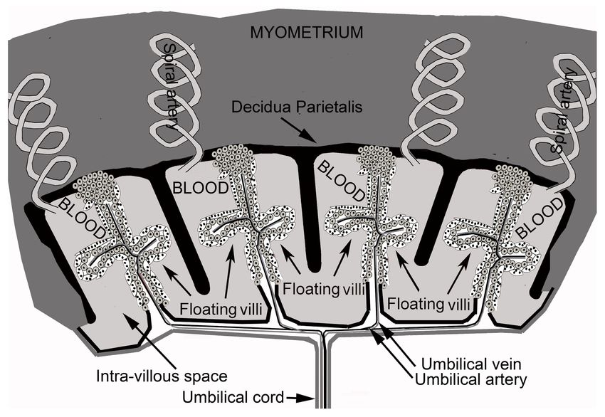
Cole Reproductive Biology and Endocrinology 2012, 10:24                                                                  Page 2 of 18
http://www.rbej.com/content/10/1/24

a molecule containing the name hCG, because it con-                 variants have two very different receptor binding sites.
tains hCG amino acid sequence. I renamed it hypergly-               Here we examine hCG and it variants, the wonders of
cosylated hCG based on its structure.                               today’s science.
   Research by Laub and Jennissen [2] and Lehnert and
Akhurst [3] showed that hCG ß-subunit was part of the               hCG, hyperglycosylated hCG, hemochorial placentation
transforming growth factor ß (TGFß) oncoprotein family              and evolution
of molecules. Lapthorn and collegues determined the 3               A major function of hCG during pregnancy can be
dimension structure of the hormone hCG [4], and                     described as driving hemochorial placentation, or the
showed a 4 peptide cystine knot structure in the ß-sub-             efficient method whereby humans drive nutrient transfer
unit was common to TGFß (Figure 1). It is well estab-               to the fetus. hCG, fetal hCG and hyperglycosylated hCG
lished that the hormone hCG produced in pregnancy                   seeming have many critical roles during pregnancy (See
[5] and the sulfated variant of hCG produced by the                 Section hCG, hyperglycosylated hCG and pregnancy).
pituitary [6], act on an hCG/luteinizing hormone (LH)               Almost every medical text book sold today describe the
receptor to evoke a response. Interestingly, hyperglyco-            sole function of hCG as driving luteal steroidogenesis.
sylated hCG and hCG free ß-subunit have been shown                  This is a very out of date description of hCG, summar-
to be autocrines and to function separately, binding and            izing research in the 1910s, 1920s and 1930s [9-12],
antagonizing a TGFß receptor on the cells that produce              books surely must be more up to date than this period.
these hCG forms [7,8]. So another wonder, hCG and its               Over 100 publications in the 1970s-2010s describe and

 Figure 1 The three dimensional structure of deglycosylated hCG as shown by X-ray defraction [4]. O and N mark O-linked and N-linked
 oligosaccharides and * marks the site of the cystine knot, common to TGFß. Grey is a- and black is ß-subunit.
Cole Reproductive Biology and Endocrinology 2012, 10:24 Page 3 of 18
http://www.rbej.com/content/10/1/24
confirm the many established functions of hCG variants.
Why is everything so out of date? Looking at the section
of this review on hCG function during pregnancy, if
there is only room to describe just one of the many
functions it should be driving hemochorial placentation
as described here, and not just maintaining progesterone
production by the corpus luteum for 3-4 weeks.
It is well established that hyperglycosylated hCG
drives invasion and implantation by placental tropho-
blastic cell deep into the myometrium of the uterus
[13-16]. Hyperglycosylated hCG drives cytotrophoblast
cell growth [8,13,14,16,17], and hCG promotes the
fusion and differentiation of peripheral cytotrophoblast
cells, where the blood supply is, to syncytiotrophoblast
cells [17,18]. Hyperglycosylated hCG and hCG lead the
implantation of placenta tissue into the uterus and the
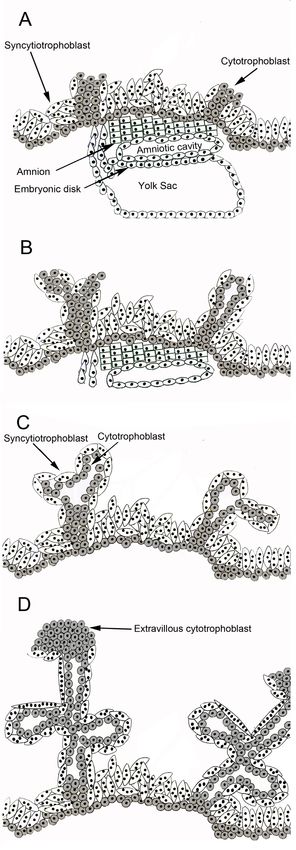
formation of villous trophoblast tissue. As illustrated in
Figure 2 panel A, implanted blastocysts form columns
of cytotrophoblast cells. Columns extend under the
influence of hyperglycosylated hCG. As illustrated in
panels B and C, hCG promotes differentiation of periph-
eral cells to active syncytiotrophoblast cells, closest to
the circulation. Shape of syncytiotrophoblast cells forces
arm formation and folding in developing villi (Figure 2
panels C and D). Taken together this generates villous
trophoblastic tissue (panel D).
While hCG and hyperglycosylated hCG force villous
trophoblast tissue formation, hCG promotes the devel-
opment and growth of uterine spiral arteries [19-26].
Angiogenesis forces the protrusion of arteries to reach
invading villous trophoblast tissue [19-26]. hCG also
promotes the formation of the umbilical circulation in
villous tissue and the formation of the umbilical cord
[27-32]. While there is no clear evidence of how placen-
tal villi, the maternal uterine spiral arteries and fetal
umbilical circulation are tied together to activate hemo-
chorial placentation, all the component of hemochorial
placentation are clearly hCG and hyperglycosylated hCG
controlled. Figure 3 shows a human placenta and active
hemochorial placentation. Histology shows that hemo-
chorial placentation only becomes active by 10 weeks
gestation. As illustrated in Figure 3, maternal blood fills
the decidua parietalis chambers. Nutrients are passaged
accross syncytiotrophoblast cells and into placental villi
and into the developed fetal umbilical circulation.
Hyperglycosylated hCG functions in implantation are
proven [13-16]. That hyperglycosylated hCG drives cyto-
Figure 2 Formation of villous trophoblast. A. Cytotrophoblast
trophoblast growth is shown and confirmed columns in early implanted embryo. B. Extension of columns and
[8,13,14,16,17]. That hCG promotes the fusion of per- differentiation of peripheral cells. B. and C. Folding of extensions
ipheral cytotrophoblast cells to syncytiotrophoblast cells caused by shape of syncytiotrophoblast cells. C and D formation of
is also proven [18]. That hCG drive uterine artery angio- trophoblastic villi. No vascular supply, spiral arteries or fetal
genesis is demonstrated and confirmed multiple times vasculature is shown.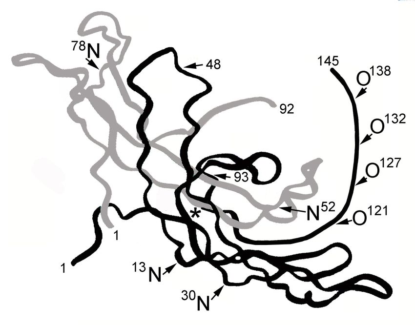
Cole Reproductive Biology and Endocrinology 2012, 10:24                                                                                   Page 4 of 18
http://www.rbej.com/content/10/1/24

 Figure 3 Human placental hemochorial placentation. While hCG and hyperglycosylated hCG force villous trophoblast tissue formation
 [13-17], hCG promotes the development and growth of uterine spiral arteries [19-26]. Angiogenesis forces the protrusion of arteries to reach
 invading villous trophoblast tissue [19-26]. hCG also promotes the formation of the umbilical circulation in villous tissue and the formation of the
 umbilical cord [27-32]. All linked together, villous trophoblast tissue, maternal spiral artery blood and fetal umbilical circulation and you have
 hemochorial placentation, efficient fetal nutrient exchange, as illustrated. In hemochorial placentation, spiral artery bring maternal blood into one
 of 4-7 hemochorial placentation chambers. Blood fills the chamber, nutrients (oxygen/glucse/amino acids) them pass across syncytiotrophoblast
 cells into villous side-arms or floating villi. They are then rapidly absorbed by the umbilical circulation.

[19-26]. Finally that hCG forms the umbilical circulation                    years. Interestingly, chorionic gonadotropin (CG) and
has been demonstrated [27-32]. Putting all these syn-                        hyperglycosylated CG first evolved in this same species.
thetic facts together, putting villous trophoblast tissue                    The logical reason that hemochorial placentation devel-
with maternal spiral arteries with fetal uterine circula-                    oped is the parallel evolution of its driving signal CG
tion and you have hemochorial placentation. Clearly, the                     and hyperglycosylated CG.
combination of hCG and hyperglycosylated hCG drive                             Assuming that CG and hyperglycosylated CG drive
all event leading to hemochorial placentation [14]. This                     hemochorial placentation during pregnancy (see Section
is suggested, however, but it has not been proven. As                        hCG, Hyperglycosylated hCG, Hemochorial Placentation
described in Section hCG, Hyperglycosylated hCG,                             and Evolution) we examine human and primate models
Hemochorial Placentation and Evolution, human hCG is                         (Table 1). Table 1 quotes numerous evolution publica-
dramatically different to primate hCG. Can we prove                          tions [33-44]. As illustrated, prosimian primates, exam-
that hCG and hyperglycosylated hCG drive hemochorial                         ple: lemur, produced LH in pregnancy, biopotency 1X.
placentation in humans. No, the research would be                            This primate used inefficient non-implanting epithelio-
unethical. The only evidence that hCG related molecules                      chorial placentation to manage pregnancy. This primate
are the driving force of hemochorial placentation is the                     had a tiny brain, only 0.07% (1/1428 th) of body weight.
first appearance of invasive hemochorial placentation in                     With more advanced early-simian primates, example:
early simian primates. Primates first evolved 80 million                     old world monkey, evolved CG from a deletion muta-
years ago, early simian primates evolved just 37 million                     tion in LH. CG was produced by fused
Cole Reproductive Biology and Endocrinology 2012, 10:24 Page 5 of 18
http://www.rbej.com/content/10/1/24
Table 1 Parallelisms between placental implantation and hemochorial placentation in primates, sugar structure on CG
or LH, and relative brain masses
Species Placentation Depth of Molecule produce; # oligosaccharides (oligos); pI of Brain mass(% First appearance
characteristics Invasion dimer; circulating 1/2-life; relative biopotency body weight) (million years ago)
Humans Hemochorial 1/3 rd CG; 8 oligos; pI 3.5; 1/2-life 36 h, 109X 2.4% 0.1
myometrium
Advanced Hemochorial 1/10th CG; 6 oligos; pI 4.9; 1/2-life 6 h; 18X 0.74% 20
simian myometrium
primates
Early simian Hemochorial through CG; 5 oligos; pI 6.3; 1/2-life 2.4 h; 7.3X 0.17% 37
primates decidua
Prosimian Epitheliochorial non- LH; 3 oligos; pI 9.0; 1/2-life 0.33 h; 1X 0.07% 55
primate implanting
The circulating 1/2-life of Advanced simian primate and Early simian primate CG was calculated using an equation that consider number of oligosaccharides on
LH and human CG and the known circulating 1/2 life. C1/2 is circulating 1/2-life and number of oligosaccharides is #O, CR = 2.4#O x 1.9. Table summarizes
published data (34-45)
syncytiotrophoblast cells and hyperglycosylated CG was of efficiency. It was this super-hCG and this hemochor-
produced by cytotrophoblast stem cells [7,45]. With the ial placentation that permitted the development of the
evolution of CG and hyperglycosylated CG the early- 2.4% of body mass human brain.
simian primate developed hemochorial placentation The mechanism whereby humans developed ultra-effi-
(Table 1). The initial CG produced by this species was cient placentation has defied evolutionary scientist for
deficient in acidity with only 5 acidic oligosaccharides. It many years [37-39]. This is all explained by the evolu-
has only a 2.4 h calculated circulating 1/2-life (Table 1) tion of CG (biopotency 7, 3X), and the advancing evolu-
or had only 7.3X greater potency than prosimian pri- tion of CG, early-simian primate (biopotency 18X),
mate placental LH. Early simian hyperglycosylated CG advanced simian primate (18X) and humans (109X). In
had only the potency to implant placenta through the the human fetus, developing the large brain is not easy,
thickness of the uterine inner lining or decidua. Hemo- it uses 60% of transferred glucose and oxygen, leaving
chorial placentation in early-simian primate was barely the development of some human organs lacking in
more efficient than placentation in prosimian primates, nutrients [37-39].
leading to the development of a brain 0.17% (1/588 th) of In many respects this review of the role of CG and
body mass (Table 1) [33-44]. hyperglycosylated CG in human evolution does not end
With advancing evolution came advanced simian pri- at this point. Recent research shows that these potent
mates, example: orangutan. Following mutation in the implantation and growth factors are at the root of preg-
CG genes, advanced simian primates produced a more nancy failures in humans (see Section Hyperglycosylated
acidic CG with 6 acidic oligosaccharides. This had a cal- hCG, Failing Pregnancy and Hypertense Pregnancy).
culated circulating 1/2-life of 6 hours (Table 1) or had a Also human malignancies take advantage of these
biopotency of 18X more than prosimian LH. With it super-potent invasion and growth factors in the human
extra biopotency hyperglycosylated CG implanted this genome to stimulate growth and invasion of all
placenta through the decidua and to 10% of the width advanced human malignancies. This is discussed in Sec-
of the uterine wall or myometrial muscle. This species tion Hyperglycosylated hCG, hCG free ß-subunit and
had significantly more potent hemochorial placentation Cancer. CG is a wondrous molecule that seemingly was
leading to the development of a brain of 0.74% of body only generated to advance evolution to humans. Unfor-
mass (Table 1) [33-44]. Humans evolved after many tunately humans have to live with the terrors of this
mutations in the hCG genes. This led to a super acidic wondrous molecule in their genome.
hCG, pI 3.5 with 8 acidic oligosaccharides. This human
hCG is the most acidic glycoprotein with the highest hCG, hyperglycosylated hCG and pregnancy
proportion of sugars occurring in any primate or in hCG is a hormone with multiple functions during preg-
humans. This super-hCG raised the circulating 1/2-life nancy. hCG acts on a joint hCG/LH receptor through a
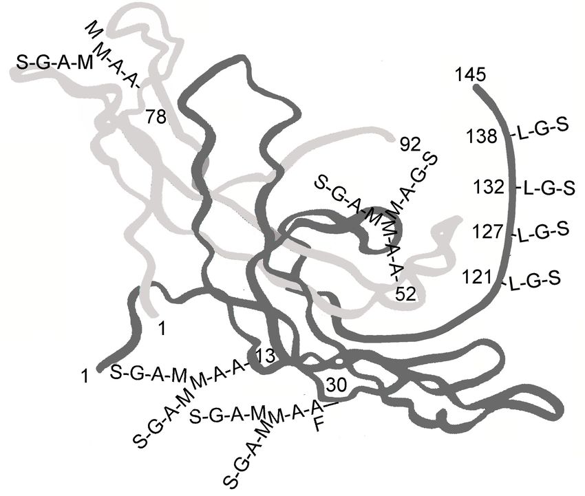
to an incredible 36 hours, or to a biopotency of 109X cyclic AMP intermediate to elicit responses. Figure 4.
over prosimian LH. It is this super hCG and super- presents the representative structure of pregnancy hCG,
hyperglycosylated hCG that drove implantation to as a glycoprotein of molecular weight 37,180. I say repre-
deep as 1/3rd the thickness of the myometrium (Table sentative in that variance in structures is apparent [1].
1), and drove hemochorial placentation to the extreme The a-subunit of hCG comprises 92 amino acids and 2Cole Reproductive Biology and Endocrinology 2012, 10:24 Page 6 of 18 http://www.rbej.com/content/10/1/24 Figure 4 The three dimensional structure of hCG [4]. In order to form crystals hCG was first deglycosylated. hCG and hyperglycosylated hCG have identical amino acid sequence [1], yet vary in function. hCG binds the hCG/LH receptor while hyperglycosylated hCG antagonizes a TGFß receptor [6-8]. As such the glycosylation (all removed) has to make a structural difference to the two hCG forms. This difference is not shown. Oligosaccharide are added to the three dimensional structure assuming the carbohydrate structure observation of Elliott et al. [1]. Oligosaccharides are charged so must project towards the surface of the molecule. L in oligosaccharides is N-acetylgalactosamine, A is N- acetylglucosamine, S is sialic acid or N-acetyl-neuraminic acid, G is galactose, M is mannose and F is fucose. N-linked (Asn-linked) oligosaccharides [46]. The ß-sub- Siemens Immulite series of hCG test. This use two anti- unit comprises 145 amino acids, 2 N-linked (Asn-linked) bodies to the core of ß-subunit and detects most hCG and 4 O-linked (Ser-linked) oligosaccharides [46]. degradation products and variants on a equi-molar basis hCG is measured during pregnancy by either the total [47]. hCG test, which supposedly measured all hCG variants Table 2 shows serum hCG concentration during preg- plus its ß-subunit, or by an intact hCG assay, which nancy as measured by the Siemens Immulite 1000 assay. supposedly measured dimeric molecules only. I say sup- Total hCG concentration rises from pregnancy implan- posedly in that most laboratory total hCG tests sold tation (3rd week of gestation) to a peak at 10 weeks of today, invariably detect hCG, hCG free ß-subunit and gestation. hCG levels rise exponentially during the first other hCG degradation products and variants [47]. It is 7 weeks of pregnancy, increasing approximately 12-fold assumed that a similar variability occurs with intact every week or 1.52-fold every day or 2.3-fold every two hCG assays. I recommend one automated hCG test, the days. Total hCG concentration declines slowly from the
Cole Reproductive Biology and Endocrinology 2012, 10:24                                                                                          Page 7 of 18
http://www.rbej.com/content/10/1/24

10 week hCG peak until a 40 week term (Table 2). hCG                              months of pregnancy is injected into immature female
concentration reach 30% of peak at 15 weeks and 18%                               mice, the ovaries of the mice significantly enlarge and
of peak at 20 weeks, and then hover close to 18% peak                             showed follicular maturation. The test was considered
levels until term [48].                                                           reliable, with an error rate of less than 2%. This test was
   hCG levels vary extraordinarily widely between preg-                           only replaced in 1960 with the induction of the first
nancies [48]. hCG concentration varies among individual                           immunological hCG or pregnancy test, the antibody
and among pregnancies from 0.21 ng/ml to 173 ng/ml                                agglutination test [56] (Table 3). In this test, serum or
in the 4th week of gestation (variation 824X), and varies                         urine was added to a tube and antibody added. When
from 1.86 ng/ml to 1,308 ng/ml in the 5 th week of                                hCG was present antibody-antigen aggregates were
gestation (variation 704X) (Table 2). This variation can                          formed. These generate a cloudy or precipitated
be attributed to poor dating of pregnancy, dating to the                          solution.
first day of the last menstrual period, rather than to the                          In 1967 the hCG radioimmunoassay replaced the
day of true start of pregnancy or implantation. The var-                          agglutination test [57] (Table 3). The radioimmunoassay
iation can also be attributed to varying hCG doubling                             (RIA) was a much more sensitive and quantitative preg-
rate among syncytiotrophoblast cells in individual preg-                          nancy test. In an RIA, a small but known amount of
nancies [48]. Seemingly, hCG levels rise differently                              radio-iodinated hCG competed with the unknown
among different pregnancies. As found, the spare recep-                           serum or urine hCG in binding a limiting quantity of
tor theory explains how pregnancies cope with such                                antibody. The antibody was precipitated and radioactiv-
extreme variations in concentration. Under the spare                              ity measured. The lower the radioactivity the higher the
receptor theory, when a small proportion of receptors is                          unknown concentration of hCG. Unfortunately, due to
activated it may yield similar cellular response to when                          the common a-subunit on hCG and LH, the hCG RIA
all receptors are activated [49-51]. This is due to pla-                          recognized both hCG and LH. In 1972 the hCGß RIA
teaus in receptor G protein and cyclic AMP response                               was introduced [58] (Table 3). This was the first preg-
[49-51]. Also down-regulation [52-54] can explain how                             nancy test detecting only hCG.
a pregnancy accommodates wide variation in hCG con-                                 In 1984 I saw the introduction of a new hCG antibody
centration. A high concentration of hCG, for instance,                            technology, the immunometric assay [59] (Table 3).
may decrease the number of receptor on cells by                                   Simply explained, an antibody to one immunological
degrading the receptor transcript rate.                                           site on hCG (i.e. anti a-subunit) was immobilized on
   hCG assays have evolved through a long history                                 beads or on a tube. Blood or urine was added and this
[55-60] (Table 3). The first pregnancy assay or hCG test                          antibody extracted hCG, the antigen, from the solution.
was the famous Zondek-Aschein Test described in 1930                              An antibody to a second separate site (i.e. anti ß-subu-
[55]. The Aschheim-Zondek test was based upon the                                 nit) was labeled with a radioactivity or other tracer. This
observation that when urine from a female in the early                            antibody, the tracer antibody, was added to the mix, it

Table 2 Concentration of total hCG and hyperglycosylated hCG (hCG-H) in 496 serum samples from 310 women with
term pregnancies measured using the Siemens Immulite 1000 total hCG assay
Gestation age (weeks since start of             N          Median Total        Range Total hCG ng/         Median hCG-H        Range hCG-H ng/ml         hCG-
        menstrual period)                                   hCG ng/ml             ml (variation)              ng/ml                (variation)           H%
  3-weeks (3 weeks 0 days - 6 days)          n = 42       0.26 (16 of 42 <            0.04 - 5.5           0.20(16 of 42 <       0.01 - 6.45 (645X)      87%
                                                             0.1 ng/ml)                                       0.1 ng/ml)
                4 weeks                      n = 42              3.4              0.21 - 173 (824X)               2.5            0.18 - 160 (888X)       51%
                5 weeks                      n = 67              65              1.86 - 1308 (704X)               8.6            0.96 - 698 (731X)       43%
                6-weeks                      n = 29             252               3.80 - 855 (225X)               86             0.76 - 629 (827X)       36%
                7 weeks                      n = 30             3,278             203 - 7,766 (38X)               359              27 - 931 (34X)        16%
                8 weeks                      n = 33             4,331           1,064 - 10,057 (9.4X)             386            67 - 1050 (15.6X)       7.0%
                9 weeks                      n = 24             5,832           1,031 - 11,586 (11.2X)            430            102 - 1158 (11.3X)      5.1%
               10 weeks                      n = 20            10,352           1,952 - 19,958 (10.2X)            521            188 - 1855 (9.9X)       4.3%
             11 - 13 weeks                   n = 41             5,953           1,440 - 15,318 (10.6X)            137             24 - 330 (13.7X)       2.3%
             14 - 17 weeks                   n = 57             2,934            311 - 4,757 (15.2X)              26              6.7 - 129 (19.3X)      1.3%
             18 - 26-weeks                   n = 62             1,931            210 - 6,223 (30.3X)             15.8              5.3 - 95 (17.9X)      0.65%
             27 - 40 weeks                   n = 49             1,911            184 - 8,530 (46.4X)             2.95             0.3 - 12.2 (40.6X)     0.14%
Data from 50 pregnancies that failed due to miscarriage were excluded from this table. Pregnancies which failed to implant in early pregnancy (total hCG < 0.1
ng/ml) are indicated in parenthesis
Cole Reproductive Biology and Endocrinology 2012, 10:24 Page 8 of 18
http://www.rbej.com/content/10/1/24
Table 3 Major discoveries in hCG assays or pregnancy tests 1930-1995 [1-35,61]
Year Description Authors and reference
published
1930 First pregnancy test, the Zondek-Aschein Pregnancy Test Zondek B, Aschein S [55]
1960 First immunological pregnancy test, an antibody Wide L, Gemzell CA [56]
agglutination test
1967 First hCG radioimmunoassay Aono T, Goldstein DP, Taymor ML, Dolch K [57]
1972 Discovery of hCGß radioimmunoassay, assay only detects Vaitukaitis JL, Braunstein GD, Ross GF [58]
hCG
1984 First hCG radio-immunometric assay Armstrong EG, Ehrlich PH, Birken S, Schlatterer JP, Siris E, Hembree WE,
Canfield RE [59]
1995 Automated hCG chemiluminescent-immunometric assay Vankrieken L, Hertogh RE [60]
bound the immobilized antigen to form a sandwich. An though this is the hCG function highlighted in most
immobilized antibody-hCG-tracer antibody-label com- medical text books.
plex was formed. The tracer antibody permitted quanti- Four independent research groups show that hCG
tation of the bound antigen. The following years saw a promotes an anti-macrophage inhibitory factor or a
need to automate and speed up assays, and to develop macrophage migration inhibitory factor. This is a cyto-
new tracers other than radioiodine. Chemiluminescence kine that modulates the immune response during preg-
was discovered, where by a tracer emits light with lim- nancy. This reduces macrophage phagocytosis activity at
ited emission of heat, as the result of a chemical reac- the placenta-uterus interface, preventing destruction of
tion. In 1995 an automated chemiluminescent hCG the foreign fetoplacental tissue by the mothers macro-
assay was introduced [60]. Today, 2011, most laboratory phage system [62-64]. Four other groups have shown
hCG tests are automated and are immunometric assays that hCG may directly suppress any immune action
using the chemiluminescent principal. against the invading foreign tissue by the mother
As described in Section hCG, hyperglycosylated hCG, [65-67,89]. All told, hCG appears to be important in
hemochorial placentation and evolution, hCG drives preventing rejection of fetoplacental tissue during preg-
uterine angiogenesis, umbilical circulation and hemo- nancy [62-67,89]. Most observations suggest that hCG
chorial placentation. As described in Section hCG, has an inhibitory or suppressive function on macro-
hyperglycosylated hCG and pregnancy, a form of hCG phage activity. One group, Wan et al. [64] demonstrated
seemingly drives fetal growth during pregnancy. hCG that chorionic gonadotropin can directly enhance innate
variants do as the title claims, act as a wonder molecule immunity by stimulating macrophage function.
doing just about everything in pregnancy. Functions Multiple groups have found hCG/LH receptor in the
range from controlling uterine, fetal and placental myometrium of the uterus. It has been indicated by two
growth during pregnancy, to protecting pregnancy from groups that uterine growth in line with fetal growth is
myometrial contraction, from immuno-rejection, and controlled by hCG [68,69]. Four other groups have
from macrophage rejection. All the established hCG and shown that hCG relaxes myometrial contractions during
hyperglycosylated hCG functions during pregnancy are pregnancy. hCG acts on a BK-Ca calcium activated
listed in Table 4[6,9-11,13-32,61-91]. channel during pregnancy to relax the myometrium
The original biological activity of hCG was first amd prevent contractions [68,70-72]. hCG levels drop
revealed in the 1920s [9-11]. That hCG takes over from during the final weeks of pregnancy. It has been sug-
LH in promotion of progesterone production by ovarian gested that this drop may be the cause of increased con-
corpus luteal cells in pregnant women was shown in the tractions in the weeks prior to parturition.
nineteen sixties [6,88] (Table 4). As we know today, Four independent reports show that the blastocyst
hCG only promotes progesterone production for 3-4 preimplantation secretes hCG into the uterine space
weeks following pregnancy implantation. This function which is taken up by hCG/LH receptors on the uterine
is active for less than 10% of the length of pregnancy. decidual surface. In response, the decidua is prepared
As shown in Table 2, hCG reaches a peak at 10 weeks for impending implantation [77-80]. These non-vascular
of gestation, or almost one month after progesterone communications by hCG are a critical part of a success-
promotion is complete, then continues to be produced ful pregnancy. Recent studies show the importance of
through the length of pregnancy. Clearly, progesterone hCG preimplantation signaling [81-84]. hCG signaling
production is not the principal function of hCG even directly causes immunotolerance and angiogenesis at theCole Reproductive Biology and Endocrinology 2012, 10:24 Page 9 of 18
http://www.rbej.com/content/10/1/24
Table 4 The biological functions of hCG during pregnancy
Function References
A. hCG
1. Promotion of corpus luteal progesterone production [6,9-11,88]
2. Angiogenesis of uterine vasculature [19-26]
3. Cytotrophoblast differentiation [18,61]
4. Immuno-suppression and blockage of phagocytosis of invading trophoblast cells [62-67,89]
5. Growth of uterus in line with fetal growth [68,69]
6. Quiescence of uterine muscle contraction [68,70-72]
7. Promotion of growth of fetal organs [30-32,73-76]
8. Umbilical cord growth and development [27-32]
9. Blastocysts signals uterine decidua prior to invasion regarding pending implantation [77-80]
10. hCG in sperm and receptors found in fallopian tubes suggesting pre-pregnancy communication [81-86]
11. hCG receptors in hippocampus and brain stem, may cause nausea and vomiting in pregnancy [90,91]
B. Hyperglycosylated hCG
1. Stimulates implantation by invasion of cytotrophoblast cells as occurs at implantation of pregnancy [13-16]
2. Stimulates growth of placenta by promoting growth of cytotrophoblast cells [13,14,16,17]
C. hCG and hyperglycosylated hCG together
1. Drives hemochorial placentation [87]
maternal fetal interface. hCG increases the number of suggesting that placental hCG secretion is directed
uterine natural killer cells that play a key role in the towards the maternal circulation only and is prevented
establishment of pregnancy [81-84]. from entering into fetal circulation [76]. While the hCG
Other new data shows other pre-pregnancy implanta- receptor has been shown in fetal organs, no function
tion function of hCG. Publications from Rao et al. and has been directly demonstrated, just suggested. As such,
by Gawronska et al. [84-86], show the presence of an all the findings regarding the fetus have to be consid-
hCG/LH receptor (shown by presence of mRNA and ered as just suggestions at this time. Unfortunately,
demonstration of receptor action) in human sperm and most animals do not make a form of hCG, making the
in the fallopian tubes. The function of the hCG/LH role of hCG in the fetus difficult to prove.
receptor in the in sperm is unclear. It possibly has some This review claims that there are 5 hCG variants with
relationship with fertility. hCG/LH receptor has recently independent biological activity [44]. There may actually
been demonstrated in adult women’s brain. CNS recep- 6 independent hCG variants when one considers fetal
tors are present in several areas of the brain such as the hCG. It is made by fetal kidneys and liver [73,76] so
hippocampus, hypothalamus and brain stem [90,91]. may not be structurally be similar to syncytiotrophoblast
The finding of an hCG receptor in these parts of the sialylated hCG. Our supplies of fetal hCG may be lim-
brain may explain why hyperemesis gravidarum or nau- ited to umbilical cord hormone. I am not sure that suffi-
sea and vomiting that occurs during normal pregnancy. cient fetal hCG will ever be collected to permit
Exciting new research by multiple research groups is structural analysis. Generally speaking only placental
finding hCG/LH receptors in fetal organs. Goldsmith et and pituitary cells can make glycosylated hCG dimer,
al. [73], have found hCG/LH receptors in the fetal kid- other cells only produce free subunits. I wonder, is fetal
ney and liver. Rao et al. [30-32,74,75], have located hCG a dimer, and normally glycosylated? It is only an
hCG/LH receptors in fetal lungs, liver, kidneys, spleen hCG/LH receptor that has been found in the fetus.
and small and large intestines. Interestingly, this hCG/ Does the fetus produce hyperglycosylated hCG as a
LH receptor is present in the fetal organs but comple- growth promoter, acting on a TGFß receptor?
tely absent in the adult organs. Seemingly, hCG/LH Hyperglycosylated hCG is a super-glycosylated variant
receptors disappear at birth. of hCG. While hCG is made by fused placenta syncytio-
It is suggested that hCG may promote organ growth trophoblast cells, hyperglycosylated hCG is made by
and differentiation in the fetus. The human fetus see- root placental cytotrophoblast cells (13,46). As shown in
mingly produces its own hCG from the fetal kidneys Figure 5 hyperglycosylated hCG is a variant of hCG
and liver [73,76]. The concentrations in fetal circulation with double-size O-linked oligosaccharides and trian-
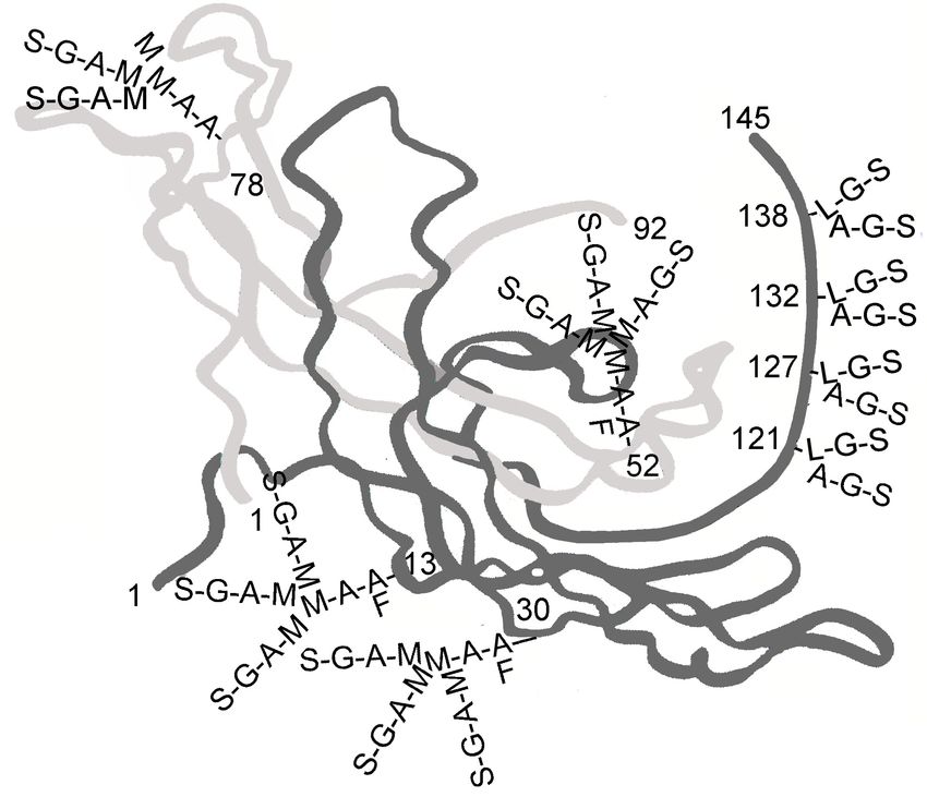
are much lower than maternal concentrations, tennary (vs. biantennary) N-linked oligosaccharides.Cole Reproductive Biology and Endocrinology 2012, 10:24 Page 10 of 18 http://www.rbej.com/content/10/1/24 Figure 5 The three dimensional structure of hyperglycosylated hCG [4]. In order to form crystals hCG was first deglycosylated. hCG and hyperglycosylated hCG have identical amino acid sequence [1], yet vary in function. hCG binds the hCG/LH receptor while hyperglycosylated hCG antagonizes a TGFß receptor [6-8]. As such the glycosylation (all removed) has to make a structural difference to the two hCG forms. This difference is not shown. Oligosaccharide are added to the three dimensional structure assuming the carbohydrate structure observation of Elliott et al. [1]. Oligosaccharides are charged so must project towards the surface of the molecule. L in oligosaccharides is N-acetylgalactosamine, A is N-acetylglucosamine, S is sialic acid or N-acetyl-neuraminic acid, G is galactose, M is mannose and F is fucose. Hyperglycosylated hCG shares all 92 amino acid a-sub- gestation, the proportion hyperglycosylated hCG rapidly unit and a 145 amino acid ß-subunit with hCG. Hyper- declines after this point. The high hyperglycosylated glycosylated hCG is an extreme molecule, molecular hCG in early pregnancy is thought to be the driving sig- weight 42,800, 39% sugar by molecular weight. Hyper- nal of deep pregnancy implantation. Not all commercial glycosylated hCG is the most acidic glycoprotein known laboratory, research, point-of-care and home pregnancy to humans, the peak acidity is pI 3.1. tests detect hyperglycosylated hCG equally with hCG. Table 2 shows the concentration of hyperglycosylated This may make a test inappropriate for early pregnancy hCG in human serum during pregnancy. As shown, detection [47]. In Laurence Cole PhD experience only hyperglycosylated hCG is the principal hCG form pro- the Siemens Immulite series of tests is appropriate for duced in early pregnancy. Hyperglycosylated hCG laboratory tests, only the Quidel and Beckman series of accounts for 87% of total hCG in the 3 rd week of tests are appropriate as Point-of-Care pregnancy tests gestation and 51% of total hCG during the 4th week of and only the Church and Dwight First Response and
Cole Reproductive Biology and Endocrinology 2012, 10:24                                                  Page 11 of 18
http://www.rbej.com/content/10/1/24

Answer Home tests are appropriate at Over-the-Counter       normal term pregnancies (81 and 42 total) produced
pregnancy tests. Hyperglycosylated hCG can be sepa-         greater than 40% hyperglycosylated hCG (% of total
rately detected to total hCG using the antibody B152        hCG) on the day of implantation. In contrast, only 8 of
immunometric test, which only tested hyperglycosylated      36 and 7 of 20 biochemical and spontaneous aborting
hCG [92].                                                   pregnancies produced greater than 40% hyperglycosy-
  Hyperglycosylated hCG and hCG free ß-subunit are          lated hCG on the day of implantation. It is assumed
interchangeable cancer promoters functioning by antag-      that the failures exceeding 40% hyperglycosylated hCG
onizing a TGFß receptor [7,8]. It is thought that hyper-    are the rare genetic abnormalities and that the bulk, 28
glycosylated hCG produced in early pregnancy promotes       of 36 and 13 of 20 pregnancies are pregnancy failures
implantation by a similar mechanism, involving antagon-     due to improper implantation. It is concluded that
ism of a TGFß receptor, blockage of apoptosis, and pro-     hyperglycosylated hCG is an absolute marker of bio-
motion of metalloproteinase and collagenase production      chemical pregnancy and spontaneous abortion, and that
[93-95].                                                    deficiency of hyperglycosylated hCG (< 50% hyperglyco-
  Hyperglycosylated hCG has multiple clear functions in     sylated hCG) is the actual cause of human pregnancy
human pregnancy implantation and promotion of cyto-         failures [15,99].
trophoblast growth (Table 4). Hyperglycosylated hCG is        Unfortunately, to achieve the absolute differentiation
demanded by pregnancy with it unique properties as a        of term pregnancy and failing pregnancy the testing has
TGFß antagonist to drive pregnancy implantation.            to be performed on the day of implantation of preg-
Implantation is driven in a cancer-like manner. In          nancy. Detection of approximately 80% term pregnan-
humans implantation passed through the uterine              cies is possible at later weeks of pregnancy [92,100].
decidua, uterine stroma and connectictive tissue into the     The hyperglycosylated hCG test is licensed to Quest
uterine myometrium or muscle surrounding the uterine        Diagnostics Inc., and can be ordered from any world-
endometrium. Placenta normally implants at 1/3rd the        wide Quest Diagnostics laboratory. It also can be
depth or the myometrium or approximately 40% of the         ordered from the USA hCG Reference Service. The
depth of the uterus. Implantation seemingly uses the        hyperglycosylated hCG test is also valuable as a high
hyperglycosylated hCG-induced cytotrophoblastic metal-      sensitivity marker for Down Syndrome screening
loproteinases and collagenase activities to achieve its     [101,102]. This screening service is currently offered by
goal [94,95]. Cytotrophoblast hyperglycosylated hCG as      Quest Diagnostics Inc.
an autocrine also promotes root placental cytotropho-         As published, The future sees prenatal screening being
blast growth during the course of pregnancy to develop      expanded to pregnancy induced hypertension (PIH) and
and prime growing villous placental tissue (13,14,16,17).   preeclampsia screening, using a hyperglycosylated hCG
                                                            test. A study by Bahado-Singh and colleagues [103] and
Hyperglycosylated hCG, failing pregnancy and hypertense     more recent confirming studies by Brennan and collea-
pregnancy                                                   gues (papers in preparation) shows that hyperglycosy-
As discussed in Section hCG, Hyperglycosylated hCG,         lated hCG measurements may be invaluable for
Hemochorial Placentation and Evolution, and Section         screening women in the first and second trimester of
hCG, Hyperglycosylated hCG and pregnancy, human             pregnancy for these two complication of pregnancy.
hyperglycosylated hCG is the extreme end product of         Today, preeclampsia is the biggest cause of maternal
evolution that drives pregnancy implantation to it          death in pregnancy. Hyperglycosylated hCG promotes
extreme, and drives production of hemochorial placenta-     hemochorial placentation formation [87], preeclampsia
tion to the extreme. Humans having an extreme mole-         and PIH are complication of ineffective hemochorial
cule to drive implantation are faced with the intricacies   placentation, thus it is marked by unduly low hypergly-
and demands of an extreme molecule, so have to face a       cosylated hCG. New hyperglycosylated hCG prenatal
high proportion of rejected pregnancies, this is miscar-    screening tests will identify women at high risk for PIH
riage or spontaneous abortions (17% failure rate) and       and preeclampsia.
biochemical pregnancies (25% failure rate) or 25% +
17% or 42% pregnancy failure rate [15,96]. Simian pri-      Sulfated hCG and the menstrual cycle
mates only have an 8% failure rate since they do not        Sulfated hCG produced by the pituitary is barely detect-
have to cope with human hyperglycosylated hCG.              able during the menstrual cycle. Sulfated hCG parallels
  Scientist show that most biochemical pregnancies and      LH during the menstrual cycle. In a recent study, at the
spontaneous abortion pregnancies, approximately two-        time of the LH peak, hCG level in 277 menstrual cycles
thirds, are due incomplete implantation of pregnancy        averaged 1.54 ± 0.90 mIU/ml [104]. In general, during
[97,98]. As shown by us in two studies examining 62         the menstrual cycle sulfated hCG levels are low,
pregnancies and 127 pregnancies [15,99], all (100%) of      approximately one fiftieth of circulating LH level
Cole Reproductive Biology and Endocrinology 2012, 10:24 Page 12 of 18
http://www.rbej.com/content/10/1/24
[104-106]. While these levels are small, sulfated hCG is Exceptional women can achieve pregnancy up to 60
exactly 50-fold more potent that LH [5]. As such sul- years age. In general, women 40-55 years age are likely
fated hCG may perform a comparable job to LH in pro- to be in perimenopause, and those > 55 years age are in
moting androstenedione production during the follicular menopause [107]. As reported, the range of hCG detec-
phase of the menstrual cycle, a comparable job in pro- tion in non-pregnant menopausal women, > 55 years
moting ovulation and corpus luteal formation. It may age, is < 2 to 13.1 mIU/ml, in non-pregnant menstrual
also perform a comparable job to LH in promoting pro- women 18-40 years age is < 2 to 4.6 mIU/ml, and
gesterone production in the luteal phase of the men- potential perimenopause women is < 2 to 7.7 mIU/ml
strual cycle. All told, one can no longer correctly say [107]. In the USA hCG Reference Service experience,
that LH promotes ovulation, it is LH plus sulfated hCG hCG levels as high as 29 mIU/ml, median 7.2 mIU/ml
[5,104-106]. have been detected in perimenopause and as high as 33
During the menstrual cycle, hypothalamic gonadotro- mIU/ml, median 8.0 mIU/ml are detected in menopause
pin releasing hormone (GnRH) pulses stimulate follicle (Table 5). Higher hCG levels have been recorded, as
stimulating hormone (FSH) ß-subunit and LH ß-subunit high as 39 mIU/ml, in women having oophorectomy
genes (the a-subunit is produced in excess) in pituitary (Table 5).
gonadotrope cells. The problem is that on chromosome The USA hCG Reference Service has examined 88
19 there is a single LH ß-subunit gene located next to 8 women producing pituitary sulfated hCG. This is a list
hCG duplicated ß-subunit genes. GnRH bombards LH of only referred cases and is not a random list of
ß-subunit gene and cannot help bombarding some of woman over 40 years old. Among the 88 cases, sulfated
the adjacent hCG ß-subunit genes leading to pituitary hCG ranged from 1.8 mIU/ml to 39 mIU/ml [106]. In
hCG production. Gonadotrope cells can sulfate glyco- menopausal cases the median level was 8.0 mIU/ml, in
proteins leading to partially sulfated hCG, LH and FSH. perimenopausal cases the median was 7.2 mIU/ml and
In menopause, with the absence of steroid feedback to in induced menopausal cases, women receiving oophor-
the hypothalamus, GnRH pulse become maximal. The ectomy, the median was 6.3 mIU/ml.
result is promotion of vast excesses of LH, hCG and Research by Gronowowski et al. [108], shows that
FSH are produce by gonadotrope cell due to excessive measurement of FSH levels is a powerful predictor of
GnRH pulses. Serum LH increases from, 1-90 mIU/ml pituitary sulfated hCG (FSH > 30 IU/L). The USA hCG
to > 100 mIU/ml in menopause, serum FSH increases Reference Service started using FSH as a confirmation
from 1-29 IU/L to 30-200 IU/L in menopause, and of pituitary sulfated hCG one year ago. We both con-
serum hCG from < 1 - 3 mIU/ml to 2-39 mIU/ml in firm and support the use of FSH testing to affirm the
menopause. All told, pituitary sulfated hCG is very diagnosis of pituitary hCG. Once a woman is diagnosed
detectable in a menopausal woman [104-108]. Perime- as producing pituitary sulfated hCG, what do you do
nopause is the stage prior to menopause marked by oli- next? The only answer is “nothing,” it is normal, it is
gomenorrhea or irregular menstrual periods. Just as natural, you need to completely ignore it. A physician
perimenopause is marked by the start of raised LH and can confirm that we are dealing with pituitary hCG, as
FSH production, it is also marked by detectable hCG described above, by administering a high estrogen oral
levels. contraceptive, to suppress hCG, for 3 weeks.
Table 5 Use of serum free ß-subunit (hCGß plus hyperglycosylated hCGß) and urine ß-core fragment as tumor markers
for detection of malignancies
Malignancy hCGß as a tumor marker ß-core fragment as a tumor marker
Number of Cases Serum hCGß (> 3 pmol/L) Number of Cases Urine ß-core fragment (> 3 pmol/L)
Bladder cancer 170 35% 102 48%
Cervical cancer 60 37%, 410 48%
Colorectal cancer 436 17%
Endometrial cancer 55 33% 157 47%
Lung cancer 243 18% 122 45%
Ovarian cancer 150 38% 207 66%
Pancreatic cancer 29 55%
Vulvar 64 41%
TOTAL 1164 Mean 30% 1027 Mean 48%
All averages are determined by combining total positive cases from multiple reports [109-130,132-138]Cole Reproductive Biology and Endocrinology 2012, 10:24 Page 13 of 18 http://www.rbej.com/content/10/1/24 Hyperglycosylated hCG, hCG free ß-subunit and cancer had no effect [13,14]. When choriocarcinoma cells were My PhD at Medical College of Wisconsin was regarding transplanted into nude mice cancer grew rapidly. Cancer structure of this hCG-variant produced by 2 cervical cell growth and malignancy could be totally suppressed, cancer cell lines, DoT and Caski. As shown, these cell oncostasis, by treating the nude mice with hyperglycosy- lines produce a invariably glycosylated variant of hCG lated hCG antibody [13,14]. Clearly, hyperglycosylated free ß-subunit. Soon after getting my PhD, I went on to hCG was the single driving signal of choriocarcinoma. It a postdoctoral fellowship at University of Michigan in was then shown that ovarian and testicular germ cell Ann Arbor and showed that hCG free ß-subunit was a malignancies took on choriocarcinoma-like cytotropho- tumor marker present in culture fluids, serum and blast morphology, producing high concentrations of urine, and patented hCG free ß-subunit and its variants hyperglycosylated hCG, like choriocarcinoma, and dri- including ß-core fragment as a tumor markers [109]. ven by hyperglycosylated hCG like choriocarcinoma This patent was licensed by Ciba-Corning Laboratories, cells [14]. the predecessor to Quest Diagnostics 1985-1999, While research with hyperglycosylated hCG and chor- Multiple studies in the early 1980s showed that serum iocarcinoma was ongoing in the USA, research in Eur- hCG free ß-subunit and urine ß-core fragment its degra- ope was continuing to investigate hCG free ß-subunit dation product, were tumor marker for all cancers and other malignancies. As found in the 1990s, the (Table 5). As shown and averaged over multiple studies detection of hCG free ß-subunit in serum was a marker [109-125] (Table 5), hCG free ß-subunit and ß-core of poor prognosis of cancer [132]. Later, it was shown fragment marked a proportion of all malignancies. All that hCG free ß-subunit secreted by cancer cells directly told, hCG free ß-subunit marked 30% of 1,164 malig- stimulated cancer cell growth [7,133-138]. hCG free ß- nancies in serum tests, at the extremes, 17% of colorec- subunit blocked apoptosis in cancer cells and enhanced tal cancers, and 38% of ovarian cancers [109-125] (Table growth and malignancy [7,133-138]. In 2000, Stephen 5). All told and averaged, hCG ß-core fragment marked Butler PhD in Europe showed that hCG free ß-subunit 48% of 1,027 malignancies in urine tests, at the produced by bladder cancer cells bound and antago- extremes, 45% of lung cancers, and 66% of ovarian can- nized a TGFß receptor on cancer cells [7]. cers (Table 5) [109-125]. Recently, I examined hyperglycosylated hCG and 2 In the years that followed Acevedo and Krichevsky choriocarcinoma cell lines, and hCG free ß-subunit and [126] and Regelson [127] used cancer membrane flow 2 bladder cancer lines and 2 endometrial cancer cell cytometry methods to show that all advanced cancers lines [8]. I confirmed, hyperglycosylated hCG promoted produced an hCG free ß-subunit variant. An investiga- growth of both choriocarcinoma cell lines and hCG free tion into hCG degradation at that time showed that ß-subunit promoted growth of both bladder and endo- hCG free ß-subunit (circulating 1/2-life 0.72 h) is metrial cancer cell lines [8]. Intriguingly, they were removed from the circulation much more rapidly than interchangeable and could take each other’s roles, hCG (circulating 1/2-life 36 h) [128]. Research showed hyperglycosylated hCG could promote bladder and that hCG free ß-subunit is nicked upon entering the cir- endometrial cancer, and hCG free ß-subunit could pro- culation, and possibly loses its C-terminal peptide mote choriocarcinoma [8]. It was rapidly inferred that [129,130]. As such hCG free ß-subunit had a circulating both antagonize a TGFß receptor making them 1/2-life of just seconds, seconds before it is degraded interchangeable. and excreted in the kidney, or removed by the liver. This research seemingly tied together our knowledge With the miniscule circulating survival time, the claim regarding hCG and cancer, interlinking the general can- that all malignancies produced hCG free ß-subunit, cer stories of Europe and the choriocarcinoma amd while it could only be detected in the serum in just 30% germ cell cancer stories of the USA. The story of hCG of cases made sense. Clearly, most of the hCG free ß- and cancer was very much enhanced and confirmed by subunit produced by cancers was cleared rapidly and ongoing clinical trials with hCG vaccines and advanced was undetectable as a tumor marker. malignancies. Three companies, Celldex, CG Therapeu- Research into choriocarcinoma showed that this can- tics and MCI BioPharma Inc. started in 2000 testing a cer produced hyperglycosylated hCG [1,131]. It was not synthetic hCGß vaccine in treating advanced cancer until the mid-2000s that it was shown that hyperglyco- cases [139-144]. Results were very exciting, with the sylated hCG was a separate and independent molecule finding that hCGß vaccines are considerably extending to hCG acting on cytotrophoblast cells in pregnancy lives of advanced cancer patients. For example, examin- implantation, and in choriocarcinoma cells [13,14]. As ing the clinical trial with hCGß vaccine and colorectal demonstrated, hyperglycosylated hCG directly promoted cancers [141], the average survival of those with optimal choriocarcinoma cytotrophoblast cell growth (in cell cul- antibody response was 45 weeks, compared to just 24 ture) and invasion (in Matrigel chambers). Regular hCG weeks in those without optimal response (p = 0.0003).
Cole Reproductive Biology and Endocrinology 2012, 10:24                                                                             Page 14 of 18
http://www.rbej.com/content/10/1/24

 Figure 6 Proposed pathways of free ß-subunit and hyperglycosylated hCG in advanced cancer cases. As illustrated, free ß-subunit and
 hyperglycosylated hCG are hCG variants with exposed TGFß binding structures [1-4,6], these are autocrines which antagonize the TGFß receptor,
 promoting cell growth and blocking cell apoptosis. As a result of the antagonism, collagenases and metalloproteinases are produced by cells
 [94,95]. As illustrated, cells secrete hCG free ß-subunit or hyperglycosylated hCG. This enters the circulation and rotates around the body. hCG
 free ß-subunit of hyperglycosylated hCG then bind back on a receptor on the cancer cells, a TGFß receptor, and antagonize this receptor. As a
 result cell growth is promoted, and cell apoptosis is blocked. Cell secrete collagenases and metalloproteinases.

This demonstrated that hCG antibodies could poten-                         seemingly modulated completely by hyperglycosylated
tially double longevity. Similar results have been                         hCG, its growth, metastases and grade.
reported with prostate cancer, lung cancer and breast                        The second type of cancer was seeming represented
cancer.                                                                    by all other human malignancies, include lung cancer,
  In conclusion, there are seemingly 2 kinds of cancer as                  breast cancer, leukemia, lymphoma and so on. These
relates to hCG variants. Type 1 is a cancer of hypergly-                   cancers start out as transformed cell driven by an hCG-
cosylated hCG producing cells, choriocarcinoma, gesta-                     independent process. As the cancer progresses and
tional trophoblastic neoplasm, and ovarian and                             becomes advanced it is able to express the hCG ß-subu-
testicular germ cell cancers. This type of cancer pro-                     nit gene and make hCG free ß-subunit. The hCG free
duces hyperglycosylated hCG from the start of malig-                       ß-subunit driven TGFß antagonism mechanism (pro-
nancy. Hyperglycosylated hCG seemingly work through                        posed mechanism, Figure 6) takes over control of the
a mechanism involving antagonism of TGFß (proposed                         cancer, as indicated by the vaccine studies, and has
mechanism, Figure 6) [7,8]. This type of cancer is                         complete control of the advanced disease. Based on the
Cole Reproductive Biology and Endocrinology 2012, 10:24                                                                                          Page 15 of 18
http://www.rbej.com/content/10/1/24

vaccine studies, development of a human high affinity                            7.    Butler SA, Ikram MS, Mathieu S, Iles RK: The increase in bladder carcinoma
                                                                                       cell population induced by the free beta subunit of hCG is a result of an
hCG ß-subunit antibody may be a future answer to                                       anti-apoptosis effect and not cell proliferation. Br J Cancer 2000,
human cancer treatment. Vaccine only works in people                                   82:1553-1556.
with a good functioning immune system. This is where                             8.    Cole LA, Butler SA: Hyperglycosylated hCG, hCGß and Hyperglycosylated
                                                                                       hCGß: Interchangeable Cancer Promoters. Mol Cell Endocrinol 2011.
hCG ß-subunit antibody may shine. I believe that hCG                             9.    Aschner B: Ueber die function der hypophyse. Pflugers Arch Gesamte
ß-subunit antibody based on nude mouse experiments                                     Physiol 1912, 146:1-147.
[13,14], could someday cure Type 1 cancers, and could                            10.   Hirose T: Experimentalle histologische studie zur genese corpus luteum.
                                                                                       Mitt Med Fakultd Univ ZU 1919, 23:63-70.
seemingly offers greatly improved treatment and                                  11.   Aschheim S, Zondek B: Das Hormon des hypophysenvorderlappens.
improved longevity to all Type 2 malignancies.                                         testobjekt zum Nachweis des hormons. Klin Wochenschr 1927, 6:248-252.
                                                                                 12.   Friedman MH, Lapham ME: A simple, rapid procedure for the laboratory
                                                                                       diagnosis of early pregnancies. Am J Obstet Gynecol 1931, 21:405-410.
Conclusions                                                                      13.   Cole LA, Dai D, Butler SA, Leslie KK, Kohorn EI: Gestational trophoblastic
hCG is a wonder of today’s science. It firstly is extreme                              diseases: 1. Pathophysiology of hyperglycosylated hCG-regulated
molecule, including features such as the most acidic                                   neoplasia. Gynecol Oncol 2006, 102:144-149.
                                                                                 14.   Cole LA, Khanlian SA, Kohorn EI: Evolution of the Human Brain, Chorionic
molecule, the most glycosylated molecule and the long-                                 Gonadotropin and Hemochorial Implantation of the Placenta: Insights
est circulating half life. It secondly is unique with multi-                           into Origins of Pregnancy Failures, Preeclampsia and Choriocarcinoma. J
ple variants of hCG having independent functions, and                                  Reprod Med 2008, 53:449-557.
                                                                                 15.   Sasaki Y, Ladner DG, Cole LA: Hyperglycosylated hCG the source of
hCG variants binding 2 separate receptors, hLG/LH                                      pregnancy failures. Fertil Steril 2008, 89:1781-1786.
receptor and TGFß receptor. hCG needs to be consid-                              16.   Guibourdenche J, Handschuh K, Tsatsaris V, Gerbaud MC, Legul F, Muller D,
ered as a placental hormone and autocrine, a pituitary                                 Evain-Brion D, Fournier T: Hyperglycosylated hCG is a marker of early
                                                                                       human trophoblast invasion. J Clin Endocrinol Metab 2010, 95:E240-E244.
hormone and a major cancer promoter.                                             17.   Handschuh K, Guibourdenche J, Tsatsari V, Guesnon M, Laurendeau I, Evain-
                                                                                       Brion D, Fournier T: Human chorionic gonadotropin produced by the
Author’s information                                                                   invasive trophoblast but not the villous trophoblast promotes cell
                                                                                       invasion and is down-regulated by peroxisome proliferator-activated
Laurence A. Cole PhD, The Howard and Friedman Dis-                                     receptor-a. Endocrinology 2007, 148:5011-5019.
tinguished Professor of Obstetrics and Gynecology, USA                           18.   Shi QJ, Lei ZM, Rao CV, Lin J: Novel role of human chorionic
hCG Reference Service, 2412 Calle De Panza NW, Albu-                                   gonadotropin in differentiation of human cytotrophoblasts. Endocrinology
                                                                                       1993, 132:387-395.
querque NM 87104, Phone: 505-263-9635, E-mail: lar-                              19.   Berndt S, Blacher S, d’Hauterive PS, Thiry M, Tsampalas M, Cruz A,
ry@hcglab.com.                                                                         Pequeux C, Lorquet S, Munaut C, Noel A, Foidart JM: Chorionic
                                                                                       gonadotropin stimulation of angiogenesis and pericyte recruitment. J
                                                                                       Clin Endocrinol Metab 2009, 94:4567-4574.
Competing interests                                                              20.   Toth P, Li X, Rao CV, Lincoln SR, Sanfillipino JS, Spinnato JA, Yussman MA:
The author has no conflict of interest in writing this paper, other than being         Expression of functional human chorionic gonadotropin/human
the Director of the USA hCG Reference Service. The author does consult for             luteinizing hormone receptor gene in human uterine arteries. J Clin
Church and Dwight Inc on home pregnancy tests, for Siemens Diagnostics                 Endocrinol Metab 1994, 79:307-315.
Inc. on hCG pregnancy tests, and for Quest Diagnostic Inc. on                    21.   Lei ZM, Reshef E, Rao CV: The expression of human chorionic
hyperglycosylated hCG. This consulting has had no influence in this                    gonadotropin/luteinizing hormone receptors in human endometrial and
research.                                                                              myometrial blood vessels. J Clin Endocrinol Metab 1992, 75:651-659.
                                                                                 22.   Zygmunt M, Herr F, Keller-Schoenwetter S, Kunzi-Rapp K, Munstedt K,
Received: 16 November 2011 Accepted: 28 March 2012                                     Rao CV, Lang U, Preissner KT: Characterization of human chorionic
Published: 28 March 2012                                                               gonadotropin as a novel angiogenic factor. J Clin Endocrinol Metab 2002,
                                                                                       87:5290-5296.
                                                                                 23.   Herr F, Baal N, Reisinger K, Lorenz A, McKinnon T, Preissner KT, Zygmunt M:
References
                                                                                       hCG in the regulation of placental angiogenesis. Results of an in vitro
1. Elliott MM, Kardana A, Lustbader JW, Cole LA: Carbohydrate and Peptide
                                                                                       study. Placenta 2007, 28:(Suppl A):S85-S93.
    structure of the α- and ß-subunits of human chorionic gonadotropin
                                                                                 24.   Zygmunt M, Herr F, Munstedt K, Lang U, Liang OD: Angiogenesis and
    from normal and aberrant pregnancy and choriocarcinoma. Endocrine
                                                                                       vasculogenesis in pregnancy. Eur J Obstet Gynecol Reprod Biol 2003,
    1997, 7:15-32.
                                                                                       110(Suppl 1):S10-S18.
2. Laub M, Jennissen HP: Identification of the anthelix motif in the TGF-ß
                                                                                 25.   Toth P, Lukacs H, Gimes G, Sebestyen A, Pasztor N, Paulin F, Rao CV:
    superfamily by molecular 3D-Rapid Prototyping. Materialwiss Werkst 2003,
                                                                                       Clinical importance of vascular hCG/LH receptors-A review. Reprod Biol
    34:1113-1119.
                                                                                       2001, 1:5-11.
3. Lehnert SA, Akhurst RA: Embryonic expression pattern of TGF beta type-1
                                                                                 26.   Burton GJ, Jauniaux E, Watson A: Maternal arterial connections to the
    RNA suggests both paracrine and autocrine mechanisms of action.
                                                                                       placental intervillous space during the first trimester of human
    Development 1988, 104:263-273.
                                                                                       pregnancy: the Boyd collection revisited. Am J Obstet Gynecol 1999,
4. Lapthorn AJ, Harris DC, Littlejohn A, Lustbader JW, Canfield RE, Machin KJ:
                                                                                       181:718-724.
    Crystal structure of hCG. Nature 1994, 369:455-461.
                                                                                 27.   Rao CV, Li X, Toth P, Lei ZM: Expression of epidermal growth factor,
5. Birken S, Maydelman Y, Gawinowicz MA, Pound A, Liu Y, Hartree AS:
                                                                                       transforming growth factor-alpha and their common receptor genes in
    Isolation and characterization of human pituitary chorionic
                                                                                       human umbilical cords. J Clin Endocrinol Metab 1995, 80:1012-1020.
    gonadotropin. Endocrinology 1996, 137:1402-1411.
                                                                                 28.   Rao CV, Li X, Toth P, Lei ZM, Cook VD: Novel expression of functional
6. Schmitt EJ, Barros CM, Fields PA, Fields MJ, Diaz T, Kluge JM, Thatcher WW:
                                                                                       human chorionic gonadotropin/luteinizing hormone receptor in human
    A cellular and endocrine characterization of the original and induced
                                                                                       umbilical cords. J Clin Endocrinol Metab 1993, 77:1706-1714.
    corpus luteum after administration of a gonadotropin-releasing
                                                                                 29.   Derecka K, Stepien A, Pelliniemi L, Doboszynska T, Gawronska B, Ziecik AJ:
    hormone agonist or human chorionic gonadotropin on day five of the
                                                                                       Evidence for the presence of luteinizing hormone-chorionic
    estrous cycle. J Anim Sci 1996, 74:1915-1929.
You can also read