Converging Light, Energy and Hormonal Signaling Control Meristem Activity, Leaf Initiation
←
→
Page content transcription
If your browser does not render page correctly, please read the page content below
Converging Light, Energy and Hormonal Signaling
Control Meristem Activity, Leaf Initiation, and
Growth1[CC-BY]

Binish Mohammed,a,2 Sara Farahi Bilooei ,a,2 Róbert Dóczi,b Elliot Grove,a Saana Railo,a Klaus Palme,c
Franck Anicet Ditengou ,c,3 László Bögre,a and Enrique López-Juez a,3
a
  School of Biological Sciences, Royal Holloway University of London, Egham, Surrey TW20 0EX, United
Kingdom
b
  Centre for Agricultural Research of the Hungarian Academy of Sciences, H-2462 Martonvasar, Brunszvik u. 2,
Hungary
c
  Institute of Biology II, BIOSS Centre for Biological Signaling Studies, and Centre for Biological Systems
Analysis, University of Freiburg, 79104 Freiburg, Germany
ORCID IDs: 0000-0001-5305-8036 (R.D.); 0000-0002-2728-3835 (K.P.); 0000-0001-5355-0282 (F.A.D.); 0000-0001-7038-604X (L.B.);
0000-0003-4150-6625 (E.L.-J.).

The development of leaf primordia is subject to light control of meristematic activity. Light regulates the expression of thousands
of genes with roles in cell proliferation, organ development, and differentiation of photosynthetic cells. Previous work has
highlighted roles for hormone homeostasis and the energy-dependent Target of Rapamycin (TOR) kinase in meristematic
activity, yet a picture of how these two regulatory mechanisms depend on light perception and interact with each other has yet
to emerge. Their relevance beyond leaf initiation also is unclear. Here, we report the discovery that the dark-arrested
meristematic region of Arabidopsis (Arabidopsis thaliana) experiences a local energy deprivation state and confirm previous
findings that the PIN1 auxin transporter is diffusely localized in the dark. Light triggers a rapid removal of the starvation
state and the establishment of PIN1 polar membrane localization consistent with auxin export, both preceding the induction of
cell cycle- and cytoplasmic growth-associated genes. We demonstrate that shoot meristematic activity can occur in the dark
through the manipulation of auxin and cytokinin activity as well as through the activation of energy signaling, both targets of
photomorphogenesis action, but the organ developmental outcomes differ: while TOR-dependent energy signals alone stimulate
cell proliferation, the development of a normal leaf lamina requires photomorphogenesis-like hormonal responses. We further
show that energy signaling adjusts the extent of cell cycle activity and growth of young leaves non-cellautonomously to available
photosynthates and leads to organs constituted of a greater number of cells developing under higher irradiance. This makes
energy signaling perhaps the most important biomass growth determinant under natural, unstressed conditions.

   Leaves are biological solar panels, the development                        and arrests as the embryo enters dormancy, becoming
of which begins as primordia at the flanks of the shoot                        protected within the seed. Following germination,
apical meristem (Tsukaya, 2005; Kalve et al., 2014a).                         which frequently occurs underground, the develop-
This meristem consists of a pool of stem cells and their                      ment of leaf primordia is arrested in darkness (Chory,
close descendants, is organized during embryogenesis,                         2010). This constitutes part of the skotomorphogenesis
                                                                              developmental program, which helps young seedlings
                                                                              to emerge through the ground, before the photomor-
    1
     Work funded in part by NSF/BBSRC bilateral grant BB/                     phogenesis program commences aboveground. Emer-
M025047 to L.B.                                                               gence into light reinitiates leaf development, including
   2
   3
     These authors contributed equally to the article.                        that of leaf mesophyll cells filled with chloroplasts
     Address correspondence to franck.ditengou@biologie.uni-
                                                                              (Nemhauser and Chory, 2002). In most gymnosperm
freiburg.de and e.lopez@rhul.ac.uk.
   The author responsible for distribution of materials integral to the
                                                                              plants, however, leaves can develop and cells with
findings presented in this article in accordance with the policy de-           chloroplasts can differentiate in the dark, suggesting
scribed in the Instructions for Authors (www.plantphysiol.org) is:            that the skotomorphogenesis program is an evolution-
Enrique López-Juez (e.lopez@rhul.ac.uk).                                      ary innovation to assist seedling establishment (Hills
   B.M. and S.F.B. performed the majority of experiments; R.D., E.G.,         et al., 2015). As a consequence, upon first exposure to
S.R., F.A.D., and E.L.-J. performed essential experiments; B.M., S.F.B.,      light, photosynthesis cannot immediately commence;
R.D., F.A.D., L.B., and E.L.-J. analyzed and discussed data; K.P., L.B.,      instead, photomorphogenesis is activated by informa-
and E.L.-J. supervised work; E.L.-J. wrote the article; all authors con-      tional photoreceptors, most notably the phytochrome
tributed to the final article.                                                 and cryptochrome families (Chory, 2010) that detect the
   [CC-BY]
           Article free via Creative Commons CC-BY 4.0 license.
                                                                              presence, quality, and quantity of light. Accordingly,
   www.plantphysiol.org/cgi/doi/10.1104/pp.17.01730

Plant PhysiologyÒ, February 2018, Vol. 176, pp. 1365–1381, www.plantphysiol.org Ó 2018 The Authors. All Rights Reserved.         1365

                                 Downloaded on February 23, 2021. - Published by https://plantphysiol.org
                                                   Copyright (c) 2017 The Authors.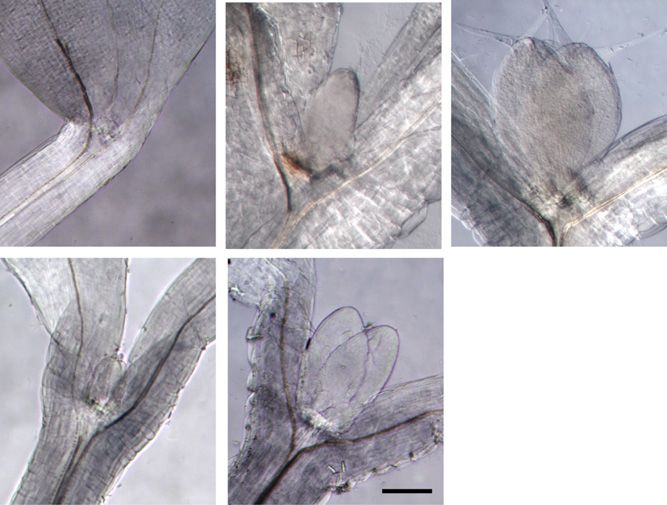
Mohammed et al.

the combined deficiency of phytochromes and crypto-                separate roles in the positioning and early development
chromes prevents leaf initiation in the light (López-Juez         of leaves (Capua and Eshed, 2017).
et al., 2008). Repressors of photomorphogenesis, in-                 An elegant study carried out in tomato (Solanum
cluding DET1 and COP1, target light signaling proteins            lycopersicum) shoot meristems showed that the auxin
for degradation in the dark, as revealed by the fact that         efflux transporter, PIN1, became internalized when
their loss of function leads to constitutive photomor-            light-grown shoot apices were transferred to the dark,
phogenic development (Chory et al., 1994; Lau and                 while in the light, the auxin maxima established by
Deng, 2012).                                                      plasma membrane-localized PIN1 determined the po-
   The response of seedlings to the first light exposure           sitions for cytokinin action to drive leaf initiation
postgermination is so dramatic that it constituted the            (Yoshida et al., 2011). A subsequent study (Pfeiffer et al.,
very first target for large-scale gene expression pro-             2016) demonstrated that sugars acting through the
filing, followed by many subsequent genome-wide                    Target of Rapamycin (TOR) kinase pathway, together
studies (Jiao et al., 2007). However, these studies were          with cytokinin activity, lead to the induction of WUS
of limited use in understanding the initiation of leaves          expression and subsequent meristem activation in the
at the meristem in response to light, since the various           light.
organs show distinct responses to light (e.g. the cot-               Growth is the most resource-consuming process
yledons undergo expansion, while hypocotyls cease to              living organisms undertake, and it is not surprising
elongate). A developmental and transcriptome-wide                 that mechanisms have evolved to sense and interpret
analysis of dissected, etiolated shoot apices when the            the availability of energy and nutrients. Besides their
growth of leaf primordia is initiated upon the first               roles as reduced carbon sources for oxidative phos-
exposure to light addressed this question (López-Juez             phorylation, both Glc and Suc can trigger direct re-
et al., 2008). This analysis revealed a dramatic stimu-           sponses in plants. Exhaustion of reduced carbon has
lation of cell proliferation, peaking between 6 and 24 h          been shown to trigger a common set of genes, named
after light exposure. Gene expression signatures as-              starvation genes, regardless of the means by which the
sociated with cell proliferation and cytoplasmic                  exhaustion takes place (e.g. change of medium com-
growth (protein translation) peaked at 6 h and were               position or loss of available starch in leaves after an
followed by expansion growth-associated signatures,               unexpectedly long night). Starvation genes are turned
including cell wall remodeling and water influx. A                 off when reduced carbon becomes available (Usadel
direct regulation of cell cycle-associated E2F tran-              et al., 2008; Sulpice et al., 2009). The starvation state is
scription factors by photoreceptors, under DET1 and               perceived as a deficiency in the metabolite trehalose-6-
COP1 control, provided a possible mechanism for                   phosphate and acts through the SNF-related protein
meristem activation by light (López-Juez et al., 2008;            kinase, SNRK (Robaglia et al., 2012; Tsai and Gaz-
Berckmans et al., 2011). Furthermore, based on diag-              zarrini, 2014), which negatively regulates TOR, a
nostic gene expression signatures, a transient down-              central kinase that universally mediates resource sig-
regulation of auxin and ethylene signaling at the apex            nals in eukaryotes (Laplante and Sabatini, 2012;
was postulated, one that preceded an up-regulation of             Nukarinen et al., 2016). TOR is a master regulator,
cytokinin responses. The latter coincided with the                controlling a number of growth-signaling cascades,
peak in the cell cycle and ribosome-related gene ex-              which responds to sugar and amino acid availability.
pression activity.                                                One of the fundamental outputs of TOR activity is an
   Hormonal responses are central to leaf initiation.             enhanced ability to manufacture cellular components
Consecutive leaves develop at the flanks of the shoot              through an increase in the cellular translation capacity.
meristem in striking geometric arrangement, known as              TOR also promotes cell proliferation (Xiong et al.,
phyllotaxy, which can be explained by inhibitory fields            2013). In plants, TOR responds both to sugar signals
generated by emerging leaves. Elegant experiments                 (Baena-González et al., 2007; Deprost et al., 2007;
have revealed those fields to be based on the self-                Xiong et al., 2013; Dobrenel et al., 2016a) and to auxin
regulating dynamics of auxin transport (Braybrook                 (Schepetilnikov et al., 2013, 2017). It was shown re-
and Kuhlemeier, 2010). Positions for leaf primordia on            cently that, in shoot meristems, light stimulates the
the epidermis at the peripheral zone of the meristem are          TOR activity via two parallel pathways, through
selected where the auxin concentration is high, and               photosynthates and through light signaling linked to
these points become sinks for further auxin transported           auxin biosynthesis (Li et al., 2017).
from nearby epidermal cells, due to the polar relocali-              In this study, we demonstrate that cell proliferation
zation of the PIN1 auxin transport protein. Cells at this         can be arrested in young primordia by dark exposure,
position then enter rapid cell proliferation, but leaf            or reduced at low light, through a local starvation state
emergence requires a second relocalization of PIN1                in the meristem, and reinitiated by transfer to light,
proteins to export auxin away from the primordium                 which rapidly overcomes such a state. We show that,
into the rib meristem (Reinhardt et al., 2003). These             upon light exposure of dark-arrested leaf primordia,
events constitute the first steps in leaf development.             PIN becomes rapidly polarized and that this precedes
Auxin is further involved in the proliferation of leaf            cell proliferation and growth gene responses. We also
cells and in the differentiation of vasculature (Scarpella        show that shoot meristematic activity can be induced in
et al., 2006, 2010). Thus, auxin plays fundamental and            the dark by exposure to cytokinin, and more efficiently
1366                                                                                                    Plant Physiol. Vol. 176, 2018

                             Downloaded on February 23, 2021. - Published by https://plantphysiol.org
                                               Copyright (c) 2017 The Authors.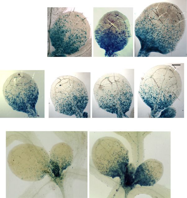
Energy and Hormones Control Leaf Growth in Light

so under reduced auxin sensitivity. It also can occur in
the dark by direct access of the meristem to sugar, in a
TOR-dependent manner (i.e. through the activation of
energy signaling, the second target of photomorpho-
genesis action), but with differing results: energy sig-
nals stimulate cell proliferation, but the development of
a normal leaf lamina requires photomorphogenesis-like
hormonal responses. Lastly, we show that available
photosynthates impact energy signaling and adjust the
extent of cell cycle activity in meristematic cells in a
non-cellautonomous manner, which, under higher ir-
radiance, leads to organs constituted of a greater
number of cells.

RESULTS
The Shoot Meristem and Arrested Primordia of Dark-
Grown Seedlings Experience Local Energy Starvation

   In our earlier analysis of light responses at the shoot               Figure 1. Shoot apices of 3-d-old seedlings exhibit in the dark a star-
apex upon first exposure of dark-grown seedlings, we                      vation response, which disappears within 1 h of light exposure. The
identified nearly 6,000 differentially expressed genes                    expression levels of genes defined as carbon-repressed starvation and
                                                                         carbon-induced feast (Usadel et al., 2008) were plotted using data from
(López-Juez et al., 2008). Among these genes, we
                                                                         a previous microarray experiment (López-Juez et al., 2008). Heat maps
identified one cluster that was composed of hundreds                      represent levels at each time point relative to the average level for the
that responded rapidly, within 1 h, and negatively to                    same gene across all time points (red, above; blue, below). SAp, Shoot
light, exclusively in the shoot apex, not in the coty-                   apex; Cot, cotyledons. The number after each sample type indicates
ledons. Subsequent analysis revealed that this cluster                   hours after light exposure. Color scales are shown above each plot.
was highly enriched in common carbon-repressed
starvation genes, as classified by a previous study
(Usadel et al., 2008). We reexamined the expression                      Light Triggers the Polar Localization of PIN1 to the Plasma
of all such genes in our transcriptome data. The                         Membrane, Allowing Auxin Export That Precedes
resulting expression plot of the complete set of                         Primordia Growth
starvation-defined genes shows a generalized, rapid
down-regulation of transcript levels (Fig. 1). More                         Auxin-responsive genes were shown to be highly
than 50% of starvation genes were expressed 2-fold or                    expressed in the dark-arrested shoot apex, and upon
higher in the dark than after 1 h in the light in the                    light exposure, the expression of these genes was rap-
shoot apex, with more than 20% being 10-fold or                          idly and transiently reduced (López-Juez et al., 2008).
higher. Because the etiolated cotyledons are unlikely                    This could be explained if light activates auxin export
to become photosynthetically competent in the short                      from emerging leaf primordia. To test this hypothesis,
time interval of 1 h, we postulate that the rapid re-                    we examined the localization of the PIN1 protein in the
pression of starvation genes in the shoot apex is a                      arrested meristems of dark-germinated seedlings, be-
consequence of the rapid mobilization of reserves                        fore and after their first exposure to light, using im-
stored in the cotyledons upon light exposure. This is                    munofluorescence labeling. Confirming a previous
in contrast to the growth of the hypocotyl, which                        report (Yoshida et al., 2011), the PIN1 signal was weak,
occurs rapidly at the etiolated stage, demonstrating                     with limited membrane localization, largely diffused
that resources do not limit the growth of another or-                    inside the cells, and difficult to distinguish from back-
gan in the dark. Interestingly, the down-regulation of                   ground in the dark-arrested shoot apex. We found that,
starvation genes in the shoot apex was transient in                      upon light exposure of dark-arrested meristems, the
most cases, expression becoming high again 24 h                          PIN1 localization became polar on the plasma mem-
later. The reason for this is not clear, but it might                    brane within 2 h, in a pattern pointing toward the tips of
represent the fact that the carbon supply could not                      emerging leaf primordia at the epidermal cell layer and
keep up with the rapid growth taking place within the                    away from the tips of primordia in a cell file at the
shoot apex. In contrast, carbon-induced genes, which                     center of the leaf lamina. This pattern was particularly
we refer to as feast genes, exhibited the opposite ex-                   evident 24 h after exposure to light, the position of the
pression pattern, a strong expression between 1 and                      PIN1 signal indicating auxin transport toward the
6 h after light exposure (Fig. 1). We conclude that                      primordia tips and export toward the rib meristem
skotomorphogenesis in the dark imposes a starvation                      (Fig. 2A).
state specifically on cells within the shoot apex and                        Consistent with the diffuse PIN1 localization pattern
that this state is released rapidly upon light exposure.                 in the dark, the DR5:GUS auxin activity reporter
Plant Physiol. Vol. 176, 2018                                                                                                                1367

                                Downloaded on February 23, 2021. - Published by https://plantphysiol.org
                                                  Copyright (c) 2017 The Authors.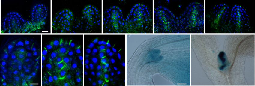
Mohammed et al.

        Figure 2. Light exposure of dark-grown seedlings triggers a rapid PIN1 polarization at the leaf primordia and the establishment of
        localized auxin maxima. A, PIN1 immunofluorescence localization in the first two leaf primordia of wild-type seedlings: PIN1
        (green) and 49,6-diamino-2-phenylindole (DAPI; blue). Seedlings were germinated in the dark for 3 d, then examined immedi-
        ately or after exposure to continuous white light for the times indicated (in hours). B, Enlargements of a primordium tip from the
        PIN1 localization images in A after 0, 2, and 24 h. C, DR5:GUS reporter activity of seedlings in the dark and exposed to white light
        for 24 h. Bars = 10 mm (A), 5 mm (B), and 50 mm (C).

(Ulmasov et al., 1997) revealed a relatively high but                        primordia size was observed in the axr1-12 mutant
delocalized auxin response in dark-grown shoot apices,                       grown in the same conditions (Fig. 3). As expected,
including the meristem and the arrested primordia.                           cytokinin could stimulate wild-type leaf primordia
Given that the GUS protein is stable, we could not                           growth in the dark, but the size of primordia observed
monitor the changes of GUS signal in a similar time                          after the addition of BAP was increased further in the
scale to that used for PIN1 localization. However, 24 h                      axr1 mutant. After 5 d in the dark, the leaf primordium
after transfer to light, the DR5:GUS activity was no                         size of BAP-treated axr1 seedlings reached about one-
longer diffuse and coincided with known, strong auxin                        third that of the wild type in the light in the absence of
maxima at the primordia tips, and a distinct signal ac-                      exogenous hormones. Data obtained from these ex-
companying the differentiation of provascular cells                          periments are consistent with the idea that the removal
emerged in the future mid vein (Fig. 2B).                                    of auxin and the activation of the cytokinin response are
                                                                             required for leaf primordia growth.

Reduction of Auxin Sensitivity Enhances the Ability of
Cytokinin to Induce Leaf Initiation in the Dark                              Active Cell Proliferation in Young Leaf Primordia Can Be
                                                                             Reversibly Arrested in the Dark
   The expression of auxin and cytokinin signature
genes when the dark-arrested meristem was exposed to                            Skotomorphogenesis facilitates seedling establish-
light suggested that, in the dark, auxin might prevent                       ment upon germination in soil, but photoreceptors re-
leaf primordia growth, and this auxin action is rapidly                      main active throughout the life of the plant. We asked
removed upon light exposure, to be replaced by cyto-                         whether the control of leaf development by photo-
kinin to drive growth (López-Juez et al., 2008; Yoshida                      morphogenic pathways remained active after the es-
et al., 2011; Pfeiffer et al., 2016). In agreement, it                       tablishment of leaf primordia, using the well-established
has been shown that an auxin partially insensitive                           CYCLINB1;1:DB-GUS mitotic reporter (Colón-Carmona
mutant (axr1-12; Leyser et al., 1993) and a cytokinin-                       et al., 1999; Donnelly et al., 1999). Seven-day-old,
overproducing one (amp1; Chaudhury et al., 1993) ex-                         light-grown seedlings displayed leaves 1 and 2, which
hibit a deetiolated state in the dark, manifest as                           were about 0.5 mm in length and which exhibited
short hypocotyl and open cotyledons. It also has                             abundant mitotic activity in the proximal region (Fig.
been shown that exposure of wild-type Arabidopsis                            4A; Supplemental Fig. S1). Flow cytometric ploidy
(Arabidopsis thaliana) to the synthetic cytokinin                            analysis of these leaf primordia showed that around
6-benzylaminopurine (BAP) causes leaf initiation in the                      60% of cells had 2N and 40% had 4N nuclear DNA
dark (Chory et al., 1994). We attempted to experimen-                        content (Fig. 4C). Cell cycle analysis of the flow cy-
tally transform the hormonal balance characteristic of                       tometry data revealed that a high proportion of nuclei
dark-arrested meristems (high auxin and low cytokinin                        were undergoing DNA synthesis (Fig. 4B; for extended
activity) into the one normally found after light expo-                      data, see Supplemental Fig. S2A), indicating that these
sure (low auxin and high cytokinin activity) and asked                       cells are very actively proliferating. A further 3 d in the
whether such manipulation would allow leaf initiation                        light led to a pronounced increase in organ size as cells
in the dark. To this end, we exposed the axr1-12 mutant                      exited proliferation and entered cellular expansion.
to BAP on Suc-containing plates in the dark. Without                         Flow cytometry confirmed an increase in the number of
BAP, the leaf primordia remained arrested in the dark                        cells with higher ploidy levels, including cells that en-
in the wild type, while a substantial increase in leaf                       tered endoreduplication (with 8N nuclei; Fig. 4C;
1368                                                                                                                     Plant Physiol. Vol. 176, 2018

                                  Downloaded on February 23, 2021. - Published by https://plantphysiol.org
                                                    Copyright (c) 2017 The Authors.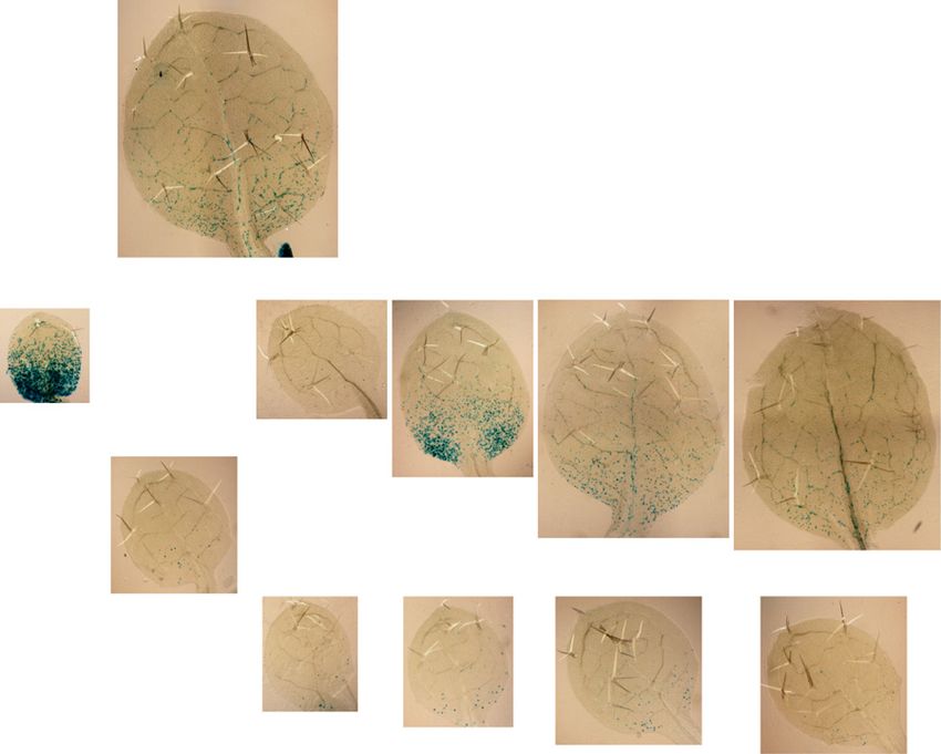
Energy and Hormones Control Leaf Growth in Light

                                                                             endoreduplication-associated         cell      expansion
                                                                             (Supplemental Fig. S2B).
                                                                                The above observations were made on seedlings
                                                                             grown on Suc-containing plates, but similar phenom-
                                                                             ena took place in the absence of exogenous Suc as well.
                                                                             While some aspects of the response, like the increase in
                                                                             the proportion of nuclei in S phase in the light, were not
                                                                             as pronounced (Supplemental Fig. S2), others, like the
                                                                             reinitiation of mitotic events, were even more so
                                                                             (Supplemental Fig. S3). These experiments suggest that
                                                                             prolonged dark exposure of young, developing leaves
                                                                             leads to G1 arrest and block of endoreduplication irre-
                                                                             spective of whether the seedlings are grown on Suc-free
                                                                             or Suc-containing plates. Upon light exposure, the ar-
                                                                             rest in G1 cell cycle phase is reversed and cells rapidly
                                                                             enter into S phase and mitosis.

                                                                             Dark-Arrested and Light-Reactivated Leaf Primordia
                                                                             Exhibit an Arrest/Growth Gene Expression Program

                                                                                We previously observed a program of rapid
                                                                             up-regulation of the expression of growth-related genes
                                                                             at the shoot apex, as leaves initiated development in the
                                                                             light (López-Juez et al., 2008). Having established a
                                                                             system of dark arrest, light reactivation of leaf growth,
                                                                             we made use of it to monitor the expression of genes
                                                                             selected to represent DNA synthesis and mitosis and
                                                                             translation capacity/ribosome buildup (Table I). We
                                                                             assessed whether a comparable gene expression pro-
                                                                             gram to that seen during deetiolation took place during
                                                                             dark arrest and light reactivation of growth in the dis-
                                                                             sected first leaf pair. We performed these experiments
Figure 3. In the axr1-12 mutant, leaves initiate in the dark, this being
enhanced by the addition of cytokinin. A, Seedlings of the Columbia
                                                                             on seedlings grown on Suc-containing medium.
wild type (WT) and axr1-12 were germinated and grown in the dark for            Genes associated with mitosis (CYCB1;1), DNA
5 d, on 1% Suc-containing medium with or without BAP at the con-             synthesis (RNR2A and H2A), and translation (RPS6 and
centrations indicated, or for the wild type in the light for 5 d on medium   EBP1) were all repressed during the 3-d dark period
without BAP. The area of one of the first two leaf primordia is indicated.   and were up-regulated in the first leaf pair within
Error bars represent SE. Asterisks reflect the significance of differences   8 h following reexposure to light; in several cases,
between axr1 and the wild type. B to F, Images of leaf primordia of          up-regulation could be detected already at 3 h after
representative shoot apical regions of seedlings as in A. B to D, The wild   reexposure (Fig. 5).
type. E and F, The axr1-12 mutant. B and E, Dark, no BAP. C and F, Dark,        The originally observed rapid changes in hormonal
10 mM BAP. D, Light. Bar = 200 mm.
                                                                             responses in the shoot apex also took place in the
                                                                             developing leaves: transfer to dark caused a mild
                                                                             elevation of auxin responses, as indicated by
Supplemental Fig. S2B). In contrast, transfer to dark for                    the auxin-responsive AUX1 gene, while light expo-
3 d led to the almost complete losses of mitotic activity,                   sure brought about within 1 h a transient, substantial
organ expansion, and endoreduplication; instead, an                          drop, which preceded a mild up-regulation of
increase in the proportion of 2N nuclei occurred, indi-                      cytokinin-responsive gene expression (ARR5). At
cating a widespread G1 arrest in the dark (Fig. 4;                           later time points during light-reinitiated leaf growth,
Supplemental Fig. S2). Reexposure to light triggered a                       between 3 and 24 h, both the expression of auxin bi-
reinitiation of cell proliferation, as indicated at 12 h by                  osynthesis genes (TAR2 and TAA1) and that of auxin-
the increased mitotic activity (note the GUS mitotic                         responsive AUX1 and HAT2 increased. In contrast,
signal in Fig. 4A), increased number of cells undergoing                     the expression of two genes representing ethylene
DNA synthesis as measured by flow cytometry (note                             response (EIN3 and EBP) was elevated consistently in
the increase of S-phase nuclei in Fig. 4B; Supplemental                      the dark and reduced in the light.
Fig. S2), and an increased percentage of 4N nuclei, in-                         As expected, the expression of starvation genes be-
dicating cells that had passed through DNA synthesis                         came up-regulated in the dark, reflecting the estab-
(Fig. 4C). At the later time point of 48 h, cells with                       lishment of a starvation state, and rapidly dropped
8N nuclei also appeared, indicating the start of                             upon transfer to light, within 1 h (Fig. 5). Since this
Plant Physiol. Vol. 176, 2018                                                                                                      1369

                                  Downloaded on February 23, 2021. - Published by https://plantphysiol.org
                                                    Copyright (c) 2017 The Authors.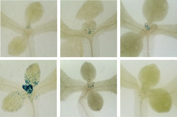
Mohammed et al.

Figure 4. Proliferation activity arrest following
transfer to dark, and reinitiation of mitotic ac-
tivity in the light in proliferation-competent
cells at the leaf base. A, CYCB1;1::DB-GUS-
expressing seedlings were grown for 7 d in
continuous light (7dL), harvested immediately
or transferred to 3 d of continuous light (+3dL)
or continuous dark (+3dD), and the latter were
transferred back to light, after which they were
harvested at the times indicated in hours. A
leaf of the first leaf pair, after visualizing the
GUS reporter, is shown. Blue GUS stain indi-
cates cells undergoing mitosis in an acropetal
gradient. Bar = 500 mm. B, S-phase percentage
of total nuclei determined by flow cytometry
and cell cycle analysis of nuclei from leaf
primordia under the conditions indicated. C,
Percentage of nuclei with different ploidy
levels under the conditions indicated. Error
bars represent SD (n = 3, with each sample
containing a pool of at least five leaves).

happened in spite of the fact that the seedlings were                            exogenous Suc (Supplemental Fig. S4). A notable dif-
grown on Suc-containing plates, the dark-induced ele-                            ference between the experiments on Suc-containing and
vation of transcript levels of starvation genes and their                        Suc-free plates was that, in the latter, cell cycle- and
rapid decrease upon light exposure might be under                                growth-associated genes declined both in the dark
photomorphogenic control in young developing leaves.                             and when seedlings remained in the light. This might
The gene expression changes upon dark arrest and light                           relate to differences in the leaf growth kinetics under
reexposure on plates with Suc also were largely repli-                           these two conditions. However, a clear, further sup-
cated when seedlings were grown in the absence of                                pression during dark acclimation occurs in both

Table I. Genes monitored as representatives of biological growth processes, and products they encode
                      Process                        Gene   Arabidopsis Genome Initiative Code                    Encoded Product
   Cell cycle entry block                     KRP4                     At2g32710                                 Kip-related protein4
   DNA synthesis (S phase)                   RNR2A                     At3g23580                 Ribonucleoside-diphosphate reductase small chain A
                                              H2A                      At1g51060                                      Histone 2A
   Mitosis (M phase)                        CYCB1;1                    At4g37490                                      Cyclin B1;1
   Ribosome biosynthesis                      RPS6                     At4g31700                              40S ribosomal protein S6-1
                                              EBP1                     At3g51800                               ERBB-3 binding protein 1
   Auxin response                            AUX1                      At2g38120                                    Auxin resistant1
                                              IAA1                     At4g14560                            indole-3-acetic acid inducible1
                                              HAT2                     At5g47370                            Homeobox-Leu zipper protein2
   Auxin synthesis                            TAA1                     At1g70560                                Trp aminotransferase1
                                              TAR2                     At4g24670                        Trp aminotransferase-related protein2
   Cytokinin response                         ARR5                     At3g48100                   Arabidopsis two-component response regulator5
   Ethylene response                          EIN3                     At3g20770                                 Ethylene insensitive3
                                               EBP                     At3g16770                     Ethylene-responsive element binding protein
   Starvation of reduced carbon               bZIP1                    At5g49450                                   Basic Leu zipper1
                                              TPS9                     At1g23870                          Trehalose-6-phosphatase/synthase9
   Mesophyll cell (chloroplast) development   GC1                      At2g21280                                  Giant chloroplast1
                                              ARC5                     At3g19720                    Accumulation and replication of chloroplasts5
   Vascular/vein development                 VND6                      At5g62380                                    Vein deficient6
                                             ATHB8                     At4g32880                            Homeobox-Leu zipper protein8

1370                                                                                                                       Plant Physiol. Vol. 176, 2018

                                       Downloaded on February 23, 2021. - Published by https://plantphysiol.org
                                                         Copyright (c) 2017 The Authors.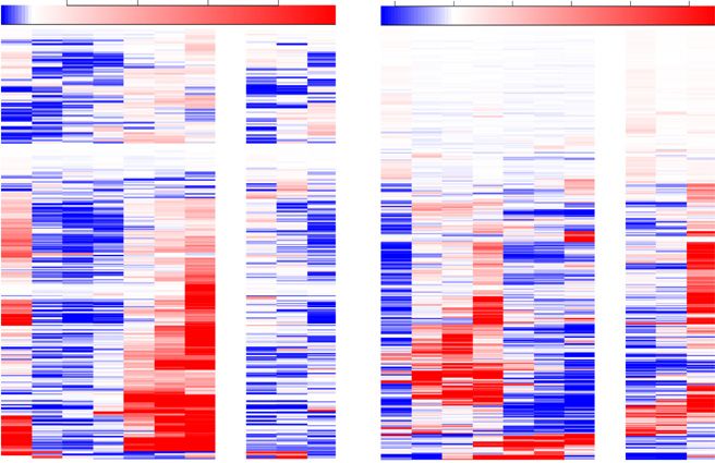
Energy and Hormones Control Leaf Growth in Light

                                                                                                 Figure 5. Expression of signature genes
                                                                                                 during dark arrest and subsequent light ex-
                                                                                                 posure in young leaf primordia. The dark
                                                                                                 arrest blocks the cell proliferation and
                                                                                                 growth genetic program and activates star-
                                                                                                 vation genetic responses. Light reverses
                                                                                                 these and brings about hormonal resetting.
                                                                                                 Wild-type seedlings were gown in light on
                                                                                                 Suc-containing plates, transferred to dark,
                                                                                                 and returned to light under conditions and
                                                                                                 times identical to those for Figure 4 or after
                                                                                                 8 d in continuous light. Seedlings harvested
                                                                                                 at the corresponding times had the primor-
                                                                                                 dia of leaves 1 and 2 dissected, and the
                                                                                                 expression of the genes shown, representing
                                                                                                 the biological process indicated above each
                                                                                                 graph and in Table I, was monitored by
                                                                                                 quantitative real-time PCR. Error bars indi-
                                                                                                 cate SE (between biological replicates).

conditions. These gene expression changes are unlikely                   promoted by reexposure to light (Supplemental Fig.
to be circadian regulated. Although eight out of the                     S5A).
20 selected genes monitored in this study were reported
to exhibit circadian expression, the circadian pattern of
expression of only one (ARR5) coincided with the ob-                     The Starvation/Growth Arrest Gene Expression Program Is
served pattern in our experiment, an elevation at the                    Largely under the Control of the COP1-Dependent
start of light exposure (dawn [Zeitgeber 0 h];                           Photomorphogenic Pathway
Supplemental Table S1). The extended, slightly finer
time course examined for seedlings in the absence of                        To address whether the gene expression program
Suc also showed that the changes occurring did not fit                    upon dark arrest and light reexposure of young de-
an underlying endogenous, circadian control and were                     veloping leaves is imposed by photosynthetic activity
most likely a direct consequence of the light exposure.                  status or light signaling, we performed these experi-
   The reinitiation of leaf development necessitates the                 ments using the cop1-1 mutant. In the dark, this mutant
differentiation of all cell types that, in essence, consist of           maintains active photomorphogenic signaling path-
an epidermis enclosing a combination of photosyn-                        ways, even though photosynthesis is completely ab-
thetic mesophyll and vascular cells. We could indeed                     sent. Transfer of cop1-1 seedlings to dark did not cause a
observe that the dark arrest was accompanied by a re-                    leaf growth arrest, as revealed by the additional area of
duction of the expression of marker genes for early                      white tissue produced in the young leaves during the
chloroplast biogenesis (GC1 and ARC5) and for the in-                    dark exposure, proximal to the green tip developed
itiation of vascular development (VND6 and ATHB8)                        prior to the dark transfer (Fig. 6, inset). We then mon-
and that both types of cellular differentiation were                     itored gene expression signatures associated with
Plant Physiol. Vol. 176, 2018                                                                                                             1371

                                Downloaded on February 23, 2021. - Published by https://plantphysiol.org
                                                  Copyright (c) 2017 The Authors.
Mohammed et al.
Figure 7. Direct Suc access to the meristem reactivates cell prolifera-
tion in the absence of light in a TOR-dependent manner. CYCB1;1::DB-
GUS-expressing seedlings were grown on solid medium plates in light
for 7 d, transferred to Suc-free liquid medium in the dark for 3 d, and
visualized for GUS expression as follows: after subsequent transfer to
Figure 6. The gene expression program change in the light is brought medium containing Suc, or to medium containing Suc plus AZD-8055,
about to a large extent by COP1-dependent photomorphogenesis or to Suc-free medium, for the times indicated. Bar = 500 mm.
pathways. A, Expression in leaf primordia of the genes indicated after
7dL + 3dD is shown, plotted on a log2 scale relative to the levels after
7dL, in the cop1 mutant and its wild type (WT) grown on Suc-containing shown that exposure of the meristem to Suc or Glc can
plates. The inset shows leaf primordia of a 3dD-adapted cop1 seedling.
trigger the further growth of organs in the dark (Roldán
B, Expression of AUX1 after 7dL, + 3dD, and following transfer back to
light (times indicated). Error bars indicate SE.
et al., 1999; Li et al., 2017). We made Suc available to the
shoot apices of seedlings in the dark using the following
strategy: 7-d light-grown seedlings, exhibiting active
growth, hormones (auxin, ethylene, and cytokinin), and meristematic activity, were arrested by transferring to
starvation in cop1-1 mutant seedlings compared with dark in Suc-free liquid culture, and after 3 d, the culture
the wild type upon 3 d in dark (Fig. 6A). Compared medium was replaced, under very dim green safelight,
with the wild type, cell proliferation and growth gene with Suc-containing medium, in which the seedlings
expression signatures were less impacted by the dark continued to grow. Monitoring of the CYCB1;1:DB-
adaptation in cop1-1 (Fig. 6A). The reduced expression GUS reporter demonstrated that the mitotic activity of
of a gene involved in auxin synthesis, as well as the young developing leaves in light (Fig. 4) all but dis-
up-regulation of ethylene action and of the starvation appeared during dark adaptation in the absence of Suc,
response in the dark, were all attenuated in cop1 while exposure to Suc resulted in a reemergence of
(Fig. 6A). The same difference in expression was observed mitotic activity, which was most pronounced after 24 h
for the cell type-specific signature genes (Supplemental (Fig. 7; Supplemental Fig. S6A). The most frequent lo-
Fig. S5B). We then examined the kinetics of the auxin calization of such events was the proximal region of leaf
response by monitoring AUX1 gene expression both primordia (Fig. 7).
during dark arrest and light reactivation. The rapid, We monitored the gene expression program initiated
transient down-regulation of auxin response following by direct exposure of the meristem to Suc in the
reexposure to light was present in the cop1-1 mutant dark (Fig. 8). As expected from the observation of re-
(Fig. 6B). This implies that a COP1-dependent photo- activation of mitotic activity visualized by the CYCB1;1:
morphogenic pathway is responsible for the bulk of the DB-GUS reporter, the cell proliferation- and growth-
gene expression program in the dark. However, the associated gene expression also was strongly stimu-
transient down-regulation of auxin signaling during lated by direct Suc access, with a simultaneous rapid
the dark-to-light transition appears to be independent down-regulation of starvation signature genes (Fig. 8).
of COP1 action. The induction of genes associated with plastid biogen-
esis and vasculature development also exhibited light-
like responses (Supplemental Fig. S5C). Three notable
Direct Suc Access to the Shoot Apex Activates Cell differences, however, could be observed in comparison
Proliferation and the Growth Gene Expression Program in with the response to light. First, the response of growth-
the Dark related genes to direct Suc supply was somewhat
slower than that to light, generally clear after 8 h rather
We have shown that the shoot apex in the dark lo- than 3 h. Second, the rapid, transient down-regulation
cally experiences a starvation state, which is terminated of auxin responses upon the dark-to-light transition
rapidly by light in a way that cannot be explained by was not seen when dark-adapted seedlings were ex-
photosynthetic activity. Intriguing observations have posed to Suc; only a strong increase of such responses
1372 Plant Physiol. Vol. 176, 2018
Downloaded on February 23, 2021. - Published by https://plantphysiol.org
Copyright (c) 2017 The Authors.Energy and Hormones Control Leaf Growth in Light
Figure 8. Direct Suc access activates a prolif-
eration and growth gene expression program.
Wild-type seedlings grown for 7 d in continu-
ous light on solid medium were transferred to
Suc-free liquid medium in darkness for 3 d,
then transferred to Suc-containing medium for
the times indicated. Seedling shoot apices
were dissected, and gene expression was
quantified and displayed as in Figure 5.
was observed, as confirmed by three separate signature initiation. Prolonged growth of shoot apices in contact
genes, suggesting that a rapid activation of auxin export with Suc led to extraordinarily elongated seedlings (Fig.
had not taken place under Suc influence, only the 9E), with unusually long petioles of cotyledons and
up-regulation of auxin synthesis had. Third, ethylene new leaves as well as internodes (Fig. 9, E–H). Elon-
responses, which were rapidly down-regulated by gation of the internodes reflects premature activation
light, were reduced only mildly after Suc exposure in of the rib meristem. Leaf lamina barely developed
the dark (Fig. 8). We conclude that, during leaf devel- (Fig. 9G); however, the transition to flowering occurred
opment, cell proliferation, cytoplasmic growth, aspects (Fig. 9H). Addition of BAP to the medium of seedlings
of plastid biogenesis, and vasculature differentiation all whose shoot meristems were not in contact with Suc
are under Suc control and can occur in the dark. also initiated leaf development, both in the wild type
and in the axr1 mutant background (Fig. 9, I–J). In ad-
dition, we noted in the axr1 mutant occasional tumor-
The Organs Developed by Meristem Activation through like growths on some leaf primordia when exposed to
Direct Access to Sugar Differ in the Dark cytokinin (Fig. 9K). The cop1 mutant also developed leaf
primordia in the dark without direct contact with Suc-
Following an extended 6-d incubation in Suc- containing medium (Fig. 9L). We conclude that sugar
containing liquid medium in the dark, we observed can promote leaf initiation in the dark only through
the appearance of an internode between the youngest direct access to the shoot apex and that the dark arrest
leaf primordia and the point of cotyledon emergence also can be overcome by a light-like shift in hormonal
(Supplemental Fig. S7). To examine this further, we activity or by the removal of COP1, thus activating
administered a prolonged exposure of the meristem to photomorphogenic signaling.
Suc in the dark while avoiding the hypoxia that char- The strategy of enhancing Suc access through the
acteristically occurs in liquid culture, by growing growth of seedlings on Suc-containing vertical plates
seedlings on vertical Suc-containing solid medium, maintains full exposure of the seedlings to ambient air.
where apices of seedlings contact the medium’s surface, This allowed us to also test whether the growth re-
as carried out by Roldán et al. (1999). The shoot apex of sponse of the meristem and young leaf primordia relies
seedlings grown on horizontal, Suc-containing medium on photosynthesis-generated Suc in the light. To this
developed leaves only in the light but was completely end, we designed an experimental setup that depletes
arrested in the dark (Fig. 9, A and B). The meristem of CO2 in air (see “Materials and Methods”; Supplemental
seedlings grown in the dark in liquid medium without Fig. S8). The transfer of seedlings for 3 d into darkness
Suc also was arrested, while if the medium contained on vertical plates without Suc led to the almost com-
Suc, the leaf primordia developed (Fig. 9, C and D). This plete cessation of cell proliferation activity, while on
indicated that direct sugar access is required for leaf Suc-containing vertical plates, with apices being in
Plant Physiol. Vol. 176, 2018 1373
Downloaded on February 23, 2021. - Published by https://plantphysiol.org
Copyright (c) 2017 The Authors.Mohammed et al.
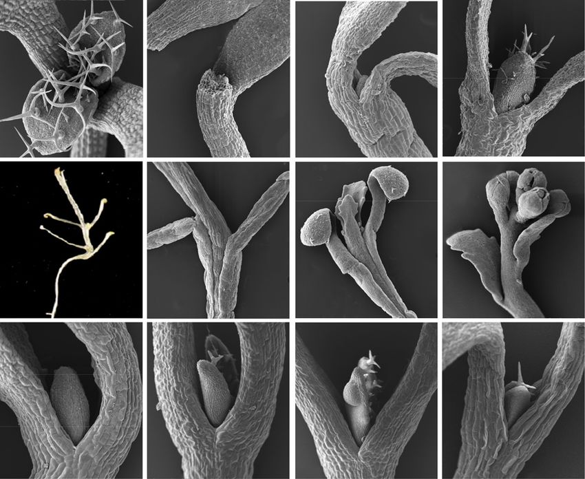
Figure 9. The dark arrest of leaf initiation
can be overcome by direct access to Suc,
change in hormonal response, or by the loss
of COP1. Scanning electron micrographs
show shoot apices of seedlings of the wild
type (A–I), axr1-12 mutant (J and K), and
cop1 mutant (L) genotypes. All seedlings
except that in A were grown in continuous
dark. A, Wild type, continuous light, 7 d,
horizontal Suc-containing plate. B, As in A
but in continuous dark. C, Wild type, 17 d,
Suc-free liquid medium. D, Wild type, 7 d,
Suc-containing liquid medium. E, Wild
type, 28 d, vertical Suc-containing plate. F,
Detail of a seedling equivalent to that in E.
G, Detail of a seedling equivalent to that in
E. H, Detail of a seedling equivalent to that
in E but grown for 42 d. I, Wild type, 7 d,
horizontal Suc-containing plates with
10 mM BAP. J, axr1 mutant, 7 d, horizontal
Suc-containing plates with 2 mM BAP. K, As
in J. L, cop1 mutant, 7 d, horizontal Suc-
containing plates. Arrows in B and C indi-
cate leaf primordia; the arrow in K indicates
a tumor-like growth. Bars = 100 mm (A–D
and I–L), 200 mm (F–H), and 2 mm (E).
contact with the plate, cell proliferation remained ac- phosphorylation, both of which were strongly inhibited
tive. An 8-h light exposure reactivated cell proliferation by AZD-8055 (Dobrenel et al., 2016b; Schepetilnikov
in the shoot meristem of seedlings grown in Suc-free et al., 2017). We carried out treatment with this selective
medium, but this was prevented in CO2-free air, TOR inhibitor, at previously used concentrations, and
where photosynthesis cannot take place (Supplemental observed that it dramatically reduced the mitotic ac-
Fig. S8; for quantitation, see Supplemental Fig. S6B). tivity in young leaf primordia (Fig. 7; Supplemental Fig.
We conclude that the photomorphogenic response S6C). It also reduced the Suc-induced expression of cell
of the meristem and leaf primordia to light requires cycle and cell growth signature genes, confirming that
photosynthesis-generated reduced carbon. these processes are, to a large extent, mediated by the
TOR pathway (Fig. 10; note the log scale). Remarkably,
the up-regulation of two out of three auxin-response
The Growth Response to Suc Is Mediated by the genes also was found to be partially TOR dependent.
TOR Pathway Interestingly, we found genes involved in plastid bio-
genesis to be particularly sensitive to TOR inhibition
Having observed key aspects of the genetic program (Supplemental Fig. S5D). In the dark, the addition of
that are initiated by exposure to Suc, we used a phar- Suc repressed the expression of starvation genes in leaf
macological approach to determine which of those as- primordia, but only to some extent after 24 h (Fig. 10).
pects were TOR dependent. TOR is a structurally and Unexpectedly, the addition of AZD-8055 further re-
functionally conserved protein kinase belonging to the duced their expression, indicating that the sugar re-
PI3K-like protein kinase family (Dobrenel et al., 2016a). pression of starvation genes is modulated, but not
Because of this conservation, highly specific ATP- dependent on TOR signaling. We conclude that Suc
competitive TOR inhibitors developed in animal or access acts on the meristem in a TOR pathway-
yeast cells (Liu et al., 2012), including AZD-8055, have dependent manner, which leads to the bulk of re-
been shown to be effective in plants (Montané and sponses impacting on cell and organ growth.
Menand, 2013; Dong et al., 2015; Kravchenko et al.,
2015; Schepetilnikov et al., 2017). The fact that AtTOR
heterozygous knockout plants are hypersensitive to Light Fluence Rate Increases Lead to an Accelerated
AZD-8055 in terms of root growth (the TOR gene be- Development of Leaves with More Cells
comes haploinsufficient; Montané and Menand, 2013) is
a strong indication that TOR is the genuine target. This One obvious advantage for plants to utilize energy
was experimentally proven by measuring the activity of signaling to determine meristematic activity would be
direct downstream TOR targets, S6 kinase and S6 that it would allow them to adjust organ growth to the
1374 Plant Physiol. Vol. 176, 2018
Downloaded on February 23, 2021. - Published by https://plantphysiol.org
Copyright (c) 2017 The Authors.Energy and Hormones Control Leaf Growth in Light
Figure 10. The gene expression program induced by Suc in the dark is
largely TOR dependent. Expression in the shoot apex and leaf primordia
of the genes indicated is shown following the growth treatment de-
scribed for Figure 7 (7dL in solid medium followed by 3dD in Suc-free
liquid medium), after transfer for a further 24 h to medium containing
Suc with or without AZD-8055, or without Suc. Expression quantitation
by quantitative real-time PCR is displayed as in Figure 6.
Figure 11. During growth in the light, exposure to HL for 8 or 24 h
increases cell proliferation. A, CYCB1;1::DB-GUS-expressing seedlings
constantly changing level of available resources, the were grown for 7dL, transferred to soil, adapted to LL (40 mmol m22 s21)
products of photosynthetic activity. It is known that, until day 11 (see “Materials and Methods”), then harvested immediately
under high irradiance, leaves develop with a multilayer or after transfer to HL (300 mmol m22 s21; top row) or maintained at LL
palisade mesophyll to support photosynthetic perfor- (bottom row) for the times indicated, and visualized for GUS reporter
mance (López-Juez et al., 2007; Kalve et al., 2014b). activity. Leaf 3 is shown. Bar = 200 mm. B, Apical region, displaying
Here, we tested how the mitotic activity becomes primordia of leaves 3 and 4, 8 h after the light transfer, visualized for the
modulated in response to changing light intensity GUS reporter. The arrows indicates mitotic events in the young leaf 2.
throughout the leaf by analyzing the CYCB1;1:DB-GUS
reporter in the palisade layer. We found a rapid in- area in leaves developing in HL (Supplemental Fig.
crease of mitotic activity soon after the transfer from S10B).
low light (LL) to high light (HL; Fig. 11A) as well as an Correspondingly with the immediate increase in cell
increased S-phase proportion measured by flow cy- proliferation activity upon transfer of seedlings from LL
tometry (Supplemental Fig. S9). The mitotic events oc- to HL, the expression of cell cycle and cell growth sig-
curred in the competent, proximal region of young leaf nature genes in young developing leaves of the seedling
primordia (leaf 3 onward), but a few were visible even apex also showed up-regulation (Fig. 12). Notably, the
in leaves 1 and 2 only under HL (Fig. 11B). Cells of auxin-responsive AUX1 expression also increased in
leaves 3 and 4 also entered endoreduplication at an HL, while starvation gene transcript levels decreased,
accelerated rate in HL (Supplemental Fig. S9), as could showing that light quantity sensitively modulates hor-
be expected given the greater extent of cell expansion mone and energy signaling in developing leaves (Fig. 12).
under those conditions. Genes for chloroplast biogenesis and vascular differ-
As a result of an increased mitotic activity, the cell entiation, ARC5 and VND6, respectively, showed a
number across the leaf, as calculated by dividing leaf transient decrease followed by an increase upon HL
area by weighted, average palisade cell areas at proxi- transfer, indicating that the transient burst in cell pro-
mal, middle, and distal regions, over a longer time liferation is accompanied by an early but transient ar-
course, also increased (Supplemental Fig. S10A). The rest in cellular differentiation (Supplemental Fig. S5E).
average size of mesophyll cells was much smaller in the We conclude that, like the dark-to-light transition, a
proximal than in the middle and distal regions, whether change in light intensity rapidly alters the energy,
grown under LL or HL, with size increasing as cell ex- hormonal, cell proliferation, and differentiation pro-
pansion took place. Here, we detected a higher mitotic grams.
activity in leaves at LL compared with those of the same
age at HL, indicating that the entire developmental
program is slowed down and that there is a delayed The Effect of HL on Cell Proliferation in Young Leaves
exit from proliferation to differentiation under LL Is Non-Cellautonomous
(Supplemental Fig. S10A). In agreement, while after 4 d
some mitotic activity remains at the distal region of the If available photosynthates, produced by photosyn-
leaf in LL, mitotic activity had already ceased in this thetically-competent leaves, are indeed the proliferative
Plant Physiol. Vol. 176, 2018 1375
Downloaded on February 23, 2021. - Published by https://plantphysiol.org
Copyright (c) 2017 The Authors.Mohammed et al.
Figure 12. Gene expression changes after
transfer to HL. The expression of signature
genes in the shoot apex and leaf primordia,
following the transfer to HL as described for
Figure 11, is shown. Expression quantitation by
quantitative real-time PCR is displayed as for
Figure 5.
signal in young leaf primordia, one would predict that DISCUSSION
the exposure of mature leaves to HL would be sufficient
How leaves form at the shoot meristem is a central
to stimulate cell proliferation in primordia emerging developmental question. Understanding how light, as a
from the meristem. To test this hypothesis, we allowed natural trigger, brings about the transition from meri-
Arabidopsis rosettes to develop to a larger size and stem arrest to activity, or how light intensity changes
acclimated them to LL. We then used local shading of modulate leaf emergence, can provide fundamental
only the meristematic region, including young pri- clues to this basic biological phenomenon. Taking to-
mordia, or of the entire seedling except that region, gether the results of this and previous studies (López-
during a shift from LL to HL for 8 h (Supplemental Fig. Juez et al., 2008; Yoshida et al., 2011; Pfeiffer et al., 2016;
S11) and monitored the expression of the mitotic re- Li et al., 2017), a picture of how light, hormonal, and
porter. We found an increase in the mitotic activity in energy signaling mechanisms regulate leaf develop-
the young leaf 8 even when it was itself shaded ment emerges.
(remained under LL) and only the mature leaves be- The hormonal switch centers on the biology of auxin.
came exposed to HL. This increase was similar to that Auxin has a complex role in leaf initiation (Braybrook
when the whole plant was uniformly exposed to HL, and Kuhlemeier, 2010; Capua and Eshed, 2017), both
indicating a systemic action of the HL effect from ma- growth promoting and growth inhibiting, but it ap-
ture leaves to very young ones (Supplemental Fig. S11). pears from our data that, in the dark, auxin becomes
We then asked whether the ability to respond to the diffusely localized in the meristem and that this inhibits
HL signal was restricted to a developmental window. the emergence of primordia. One outcome of such ac-
We showed earlier that the first leaf primordia pair of tivity is to prevent the occurrence of auxin maxima,
light-grown seedlings on Suc-containing medium while another may be to prevent cytokinin action. Such
exhibited extensive numbers of cells undergoing mi- an antagonistic action would be consistent with the
totic activity at day 7, a much reduced number if de- unexpected observation of occasional, tumor-like
velopment continued until day 10 in the light, and growths in primordia of the auxin-resistant mutant
almost none if development was arrested for 3 d in the exposed to cytokinin. At least two mechanisms are
dark (Fig. 4). We exposed identically grown, 10-d, known by which this auxin/cytokinin antagonism
constant light-grown seedlings to HL for 48 h. This led could take place: the auxin response factor Monopteros
to a few extra events of mitotic activity (Supplemental inhibits cytokinin signaling (Pacifici et al., 2015; Pfeiffer
Fig. S12), but their number was minimal compared with et al., 2016), and auxin also promotes the expression of
that caused by the light exposure of dark-arrested pri- CKX6, a gene for cytokinin inactivation, in young
mordia of 7-d-old seedlings (Fig. 4). Interestingly, such leaves under simulated shade (Carabelli et al., 2007). A
events at this later stage tended to be associated with close homolog of this gene, CKX5, also is repressed in
provascular or vascular cells throughout the leaf lam- the shoot apex by the first light exposure (López-Juez
ina, not just the proximal region. We conclude that most et al., 2008), and simultaneous inactivation of CKX5 and
leaf primordia cells are competent to respond to light CKX6 enhances the expression of the meristem-
signals with increased mitotic activity only during a organizing WUSCHEL gene (Pfeiffer et al., 2016),
very early developmental window and that, at a later helping to explain, at least in part, the initial meristem-
stage, when most cells have already exited the cell cycle repressive auxin role. Meanwhile, the absence of auxin
during normal development, only vascular cells are maxima prevents the initiation of auxin export, neces-
competent to respond to HL exposure through cell di- sary for leaf initiation (Reinhardt et al., 2003). Indeed,
vision. we observed the simultaneous establishment of polar
1376 Plant Physiol. Vol. 176, 2018
Downloaded on February 23, 2021. - Published by https://plantphysiol.org
Copyright (c) 2017 The Authors.Energy and Hormones Control Leaf Growth in Light
localization of PIN1 toward primordia tip maxima in as do phytochrome mutants of Arabidopsis (Tsukaya,
the epidermis and away in the developing mid vein 2005), and loss of an ethylene-dependent transcription
toward the rib meristem. Once maxima are established, factor gene restored in those pea mutants the wild-type
auxin clearly plays a positive role, needed to direct the leaf phenotype (Weller et al., 2015). Our observations
expansion of primordia and the differentiation of vas- not only confirm a fundamental role for auxin in leaf
culature (Scarpella et al., 2006, 2010). As part of the organ differentiation but also support a role for ethyl-
complex action of auxin, we confirmed that a strong, ene in directing the meristematic cellular activity to-
localized auxin activity occurs at the tips of emerging ward elongating organs, like internodes and petioles in
primordia in the light and that light promotes the ex- the dark, when ethylene response is high, or toward leaf
pression of at least some auxin biosynthesis genes. laminae, with their distinct epidermal and mesophyll
Meanwhile, cytokinin plays an unambiguously posi- cellular makeup in the light, when ethylene responses
tive role, as demonstrated previously (Chory et al., are repressed. Whether this possible ethylene switch of
1994; Yoshida et al., 2011; Pfeiffer et al., 2016), and our the proliferative potential acts solely through auxin
data show that reduced auxin and enhanced cytokinin activities is unknown at present. An elegant genetic
activity not only phenocopy a photomorphogenic state screen recently identified the LEAFLESS tomato gene,
but form an intrinsic part of the endogenous, early deficiency in which results in meristem cells producing
photomorphogenic program under direct light regula- only elongating internodes under auxin action (Capua
tion. Our results further show that their effects interact, and Eshed, 2017). The role of such genes in photomor-
confirming their shared underlying growth output. phogenic leaf initiation also awaits further study. We
A finding, surprising at first, in our experiments was should note, nevertheless, that following a substantially
the fact that energy signaling through direct exposure extended period of dark growth on Suc, after the tran-
of the meristem to Suc is itself capable of promoting at sition to flowering, one could observe comparatively
least some auxin responses, as evidenced by the regu- normal cauline leaves as well as floral buds (Fig. 9H).
lation of signature genes (Fig. 8). This action was, for This could reflect environmental plasticity early in de-
two out of three genes tested, TOR dependent (Fig. 10). velopment, fully subjected to skotomorphogenic or
It has been demonstrated that the TOR kinase, in ad- photomorphogenic regulation, yet enhanced homeo-
dition to mediating cell proliferation and protein syn- stasis of development following the transition to flow-
thesis in response to sugar, also mediates the ering. Whether this in any way relates to ethylene
translational control of expression of several auxin re- signaling, or competence to respond to it, is only a
sponse factors in response to auxin (Schepetilnikov matter of conjecture at present.
et al., 2013). The activation of TOR by auxin occurs Photomorphogenesis acts through a COP1-
through a family of small GTPases (Schepetilnikov dependent pathway. Transcription factors that posi-
et al., 2017). Therefore, this central growth kinase may tively regulate light responses, including hypocotyl
occupy a crucible of growth actions underpinning en- repression, cotyledon unfolding, and the initiation of
ergy and auxin signaling and explain some of their chloroplast biogenesis, are marked by COP1 for prote-
partly shared responses. olysis and are degraded through a proteasome-
Energy signaling plays a central role in the control of dependent activity in the dark (Lau and Deng, 2012).
both cellular growth (Dobrenel et al., 2016a, 2016b) and Although we could observe some degree of response to
cell proliferation (Xiong et al., 2013). It can boost meri- dark adaptation by the cop1 mutant, overall, those re-
stematic activity (Pfeiffer et al., 2016; Li et al., 2017) and, sponses were clearly attenuated. It is a particularly in-
indeed, through direct sugar access to the meristem, triguing aspect of the response to light that it can be
override the dark repression completely. However, on overridden in terms of meristem activation, but not of
its own, it cannot lead to photomorphogenic-like ro- developmental fate, by energy signaling. Light appears
sette leaves. Instead, the meristem overwhelmingly to play what could be described as a gating, or per-
produces petioles and internodes (Fig. 9; Supplemental missive, role toward energy signaling in that the extent
Fig. S7). While such developmental behavior resembles of meristem activity is dependent on seed reserves or,
the phenotype of auxin-overproducing seedlings (Chen later, photosynthates, but only when light is present
et al., 2014), a central key factor may be ethylene, re- does this reduced carbon become accessible to the
sponses to which are strong in the dark and are barely meristem. This light role is dependent on photomor-
affected by Suc exposure. Ethylene signaling is neces- phogenic pathways, as it depends on photoreceptors
sary for hypocotyl hook formation, a component of the (López-Juez et al., 2008) and COP1 (this study). One
skotomorphogenic program (Marín-de la Rosa et al., attractive hypothesis for the mechanism underlying the
2014), and pea (Pisum sativum) phytochrome mutants light-gating phenomenon is that, in a manner analo-
have been shown to exhibit strong ethylene responses gous to auxin export, sugar import into the meristem is
(Foo et al., 2006). Auxin synthesis genes were identified under photoreceptor control in a COP1-dependent
in genetic screens for weak ethylene insensitivity manner. This would explain the dramatic observa-
(Stepanova et al., 2008), because the ethylene actions tions that direct sugar access to the meristem is capable
under observation were mediated by newly synthe- of fully activating the meristem in the dark, which the
sized auxin. Tellingly, pea phytochrome mutants pro- growth of seedlings on Suc-containing solid medium
duced leaves with limited laminae (Weller et al., 2015), alone cannot.
Plant Physiol. Vol. 176, 2018 1377
Downloaded on February 23, 2021. - Published by https://plantphysiol.org
Copyright (c) 2017 The Authors.You can also read